A Comprehensive Guide to LC-MS/MS Method Development for Plasma Analysis: From Fundamentals to Validation
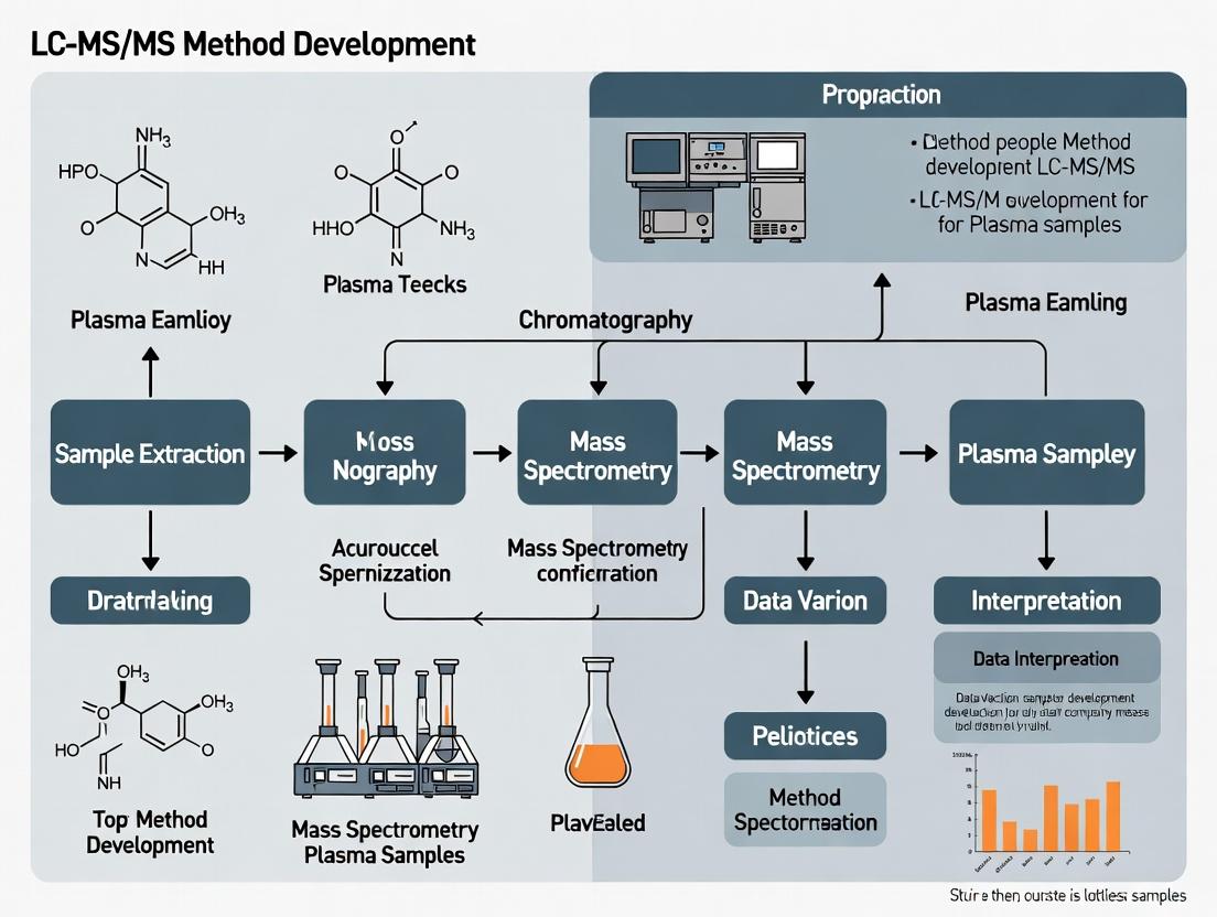
This definitive guide provides a structured, intent-driven roadmap for researchers and bioanalytical scientists developing robust LC-MS/MS methods for plasma samples.
A Comprehensive Guide to LC-MS/MS Method Development for Plasma Analysis: From Fundamentals to Validation
Abstract
This definitive guide provides a structured, intent-driven roadmap for researchers and bioanalytical scientists developing robust LC-MS/MS methods for plasma samples. We cover the essential theoretical foundations of liquid chromatography and tandem mass spectrometry, detail a step-by-step workflow for method creation including sample preparation, chromatography optimization, and mass spectrometer parameter tuning. Critical troubleshooting strategies for common pitfalls like matrix effects and ion suppression are addressed, followed by a comprehensive framework for method validation according to regulatory guidelines (e.g., FDA, EMA) and comparative analysis of different approaches. This guide synthesizes current best practices to empower the development of sensitive, specific, and reproducible assays for pharmacokinetic, metabolomic, and biomarker studies.
LC-MS/MS and Plasma Analysis Fundamentals: Building Your Core Knowledge Base
This chapter establishes the foundational principles of liquid chromatography (LC) separation, a critical first dimension in LC-MS/MS analysis for plasma samples. Effective separation is paramount for reducing ion suppression, isolating analytes from complex matrices, and ensuring accurate quantification in method development.
Core Principles of Chromatographic Separation
Liquid chromatography separates compounds based on their differential distribution between a stationary phase (the column packing) and a mobile phase (the solvent). The separation is governed by the partition coefficient (K), defined as K = Cₛ / Cₘ, where Cₛ is the analyte concentration in the stationary phase and Cₘ is the concentration in the mobile phase.
Key Performance Parameters:
- Retention Factor (k): Measures how long an analyte is retained relative to an unretained compound. k = (tᵣ - t₀) / t₀, where tᵣ is analyte retention time and t₀ is column void time.
- Selectivity (α): The ability to distinguish between two analytes. α = k₂ / k₁ (where k₂ > k₁).
- Theoretical Plates (N): A measure of column efficiency. N = 16 (tᵣ / w)², where w is the peak width at baseline.
- Resolution (Rs): The ultimate measure of separation quality. Rs = 2 (tᵣ₂ - tᵣ₁) / (w₁ + w₂).
Table 1: Quantitative Comparison of Common HPLC Modes for Plasma Analysis
| Mode | Stationary Phase | Mobile Phase | Primary Mechanism | Typical Application in Plasma |
|---|---|---|---|---|
| Reversed-Phase (RPLC) | Hydrophobic (C18, C8) | Polar (Water/Acetonitrile + Modifier) | Hydrophobicity | Small molecules, peptides, most drugs (≥90% of methods). |
| Hydrophilic Interaction (HILIC) | Polar (Silica, Cyano) | Organic-rich (Acetonitrile/Water) | Polarity & Partitioning | Polar metabolites, hydrophilic drugs, glycosylated compounds. |
| Ion Exchange (IEX) | Charged (Quaternary Amine, Sulfonate) | Aqueous Buffer with Salt Gradient | Electrostatic Interaction | Proteins, peptides, nucleotides, charged metabolites. |
| Size Exclusion (SEC) | Porous (Silica, Polymer) | Aqueous Buffer | Molecular Size | Protein aggregation studies, biomolecule purification. |
Detailed Experimental Protocol: Reversed-Phase Method Scouting for Plasma Analytes
This protocol outlines the initial scouting run to determine optimal starting conditions for a new small-molecule analyte in plasma.
Materials & Equipment:
- HPLC system with binary or quaternary pump, autosampler, and column oven.
- MS-compatible columns (e.g., 50-100mm x 2.1mm, 1.7-2.7µm particles): C18, phenyl-hexyl, HILIC.
- Mobile Phase A: 0.1% Formic Acid in Water.
- Mobile Phase B: 0.1% Formic Acid in Acetonitrile.
- Mobile Phase C (for HILIC): 10mM Ammonium Formate, pH 3.0.
- Reconstitution Solvent: 50/50 Water/Acetonitrile.
- Processed plasma sample extract (post-protein precipitation, SPE, or LLE).
Procedure:
- Column Equilibration: Install the first column (e.g., C18). Flush with 20 column volumes of starting mobile phase (e.g., 95% A / 5% B) at the intended flow rate (e.g., 0.4 mL/min).
- Gradient Scouting: Program a generic, wide gradient (e.g., 5% B to 95% B over 5 minutes) with a 2-minute hold and a 2-minute re-equilibration.
- Temperature Scouting: Perform the same gradient at three column oven temperatures: 30°C, 40°C, and 50°C.
- pH Scouting (if needed): Prepare Mobile Phase A at pH 3.0 (formic acid) and pH 6.8 (ammonium acetate). Repeat the gradient with the C18 column.
- Modify Selectivity: Switch to a different column chemistry (e.g., phenyl-hexyl) and repeat steps 1-3.
- HILIC Scouting: Switch to a HILIC column. Equilibrate with 95% Acetonitrile / 5% Buffer C. Run a gradient from 95% to 50% Acetonitrile over 5 minutes.
- Data Analysis: Evaluate chromatograms for peak shape (symmetry factor), intensity (S/N), and retention factor (k). Optimal k is typically between 2 and 10.
Visualization of Method Development Logic
Diagram 1: LC Method Development Decision Workflow (96 chars)
The Scientist's Toolkit: Essential Reagents & Materials for LC Separation of Plasma
Table 2: Key Research Reagent Solutions for Plasma LC-MS/MS
| Item | Function & Rationale |
|---|---|
| C18 Solid-Phase Extraction (SPE) Cartridge | Pre-concentrates analytes and removes phospholipids/salts from plasma, reducing matrix effects and protecting the LC column. |
| Ammonium Acetate / Formate Buffers | MS-compatible volatile buffers for mobile phase pH control; crucial for reproducible retention of ionizable compounds. |
| High-Purity Acetonitrile & Methanol (LC-MS Grade) | Primary organic modifiers; low UV-absorbance and minimal ion suppression background are critical for sensitivity. |
| Formic Acid & Trifluoroacetic Acid (TFA) | Common ion-pairing agents and pH modifiers for reversed-phase LC. TFA provides excellent peak shape for peptides but can suppress ESI. |
| Phospholipid Removal Plate (e.g., HybridSPE) | Specialized sorbent for selective depletion of phospholipids from plasma extracts, a major source of ion suppression. |
| Internal Standard Mix (Stable Isotope Labeled) | Added prior to extraction to correct for variability in recovery, ionization efficiency, and instrument performance. |
| Column Regeneration Solvents | Solutions like water/acetonitrile/isopropanol for cleaning columns contaminated by plasma matrix components. |
Within the framework of LC-MS/MS method development for plasma sample analysis, understanding the operational modes of tandem mass spectrometry is fundamental. This guide delves into the core concepts of Selected Reaction Monitoring (SRM), Multiple Reaction Monitoring (MRM), and the distinct objectives of qualitative versus quantitative analysis, providing the technical foundation for robust bioanalytical method development in drug research and development.
Core Concepts: SRM vs. MRM
SRM and MRM are often used interchangeably in triple quadrupole mass spectrometry, but a subtle distinction exists. Both are highly selective and sensitive quantitative techniques.
- Selected Reaction Monitoring (SRM): Monitors a single, specific precursor ion → product ion transition for a single analyte.
- Multiple Reaction Monitoring (MRM): Monitors multiple SRM transitions concurrently. This can involve several transitions for a single analyte (for confirmation) or transitions for multiple analytes (for multiplexed quantification).
In modern practice, "MRM" is the predominant term, as methods typically monitor several transitions simultaneously. The workflow is identical: Q1 selects a defined precursor ion (e.g., [M+H]⁺), the collision cell (Q2) fragments it, and Q3 selects a specific product ion for detection.
Qualitative vs. Quantitative Analysis Modes
Tandem mass spectrometry operates in distinct modes tailored for identification (qualitative) or measurement (quantitative) purposes.
| Aspect | Qualitative Analysis (e.g., Product Ion Scan, Neutral Loss Scan) | Quantitative Analysis (MRM/SRM) |
|---|---|---|
| Primary Goal | Identify unknown compounds; elucidate structure. | Precisely measure the concentration of known target analytes. |
| Typical Mode | Full scan or scanning modes (Q1 or Q3 scans). | Fixed, non-scanning mode (Q1 and Q3 set to specific m/z). |
| Selectivity | Lower, relies on chromatographic separation and accurate mass. | Very high, from both precursor and product ion selection. |
| Sensitivity | Generally lower due to scanning duty cycle. | Very high due to increased dwell time on specific transitions. |
| Key Output | Mass spectrum for library matching or interpretation. | Chromatographic peak area or height for calibration curves. |
| Application in Plasma | Metabolite identification, biomarker discovery. | Pharmacokinetics (PK), therapeutic drug monitoring (TDM). |
Quantitative Data Comparison: Key Performance Indicators
The following table summarizes typical target performance metrics for a validated quantitative LC-MS/MS (MRM) method for small molecules in plasma, per FDA/EMA bioanalytical guidelines.
| Performance Indicator | Target Acceptance Criteria | Purpose & Rationale |
|---|---|---|
| Accuracy (% Nominal) | ±15% (±20% at LLOQ) | Measures closeness of mean test result to true concentration. |
| Precision (%CV) | ≤15% (≤20% at LLOQ) | Measures repeatability of measurements (within-run & between-run). |
| Lower Limit of Quantification (LLOQ) | Signal-to-Noise ≥ 5, Precision & Accuracy as above | Lowest calibrator that can be measured with acceptable accuracy and precision. |
| Calibration Curve Range | Defined by LLOQ and ULOQ; typically 2-3 orders of magnitude. | The range of reliable response. Must use a weighted regression model (e.g., 1/x²). |
| Carryover | ≤20% of LLOQ area in blank after ULOQ. | Ensures high-concentration samples do not affect subsequent ones. |
| Matrix Effect (IS Normalized) | Mean IS-normalized MF: 85-115%, CV ≤15%. | Assesses ion suppression/enhancement from co-eluting matrix components. |
| Extraction Recovery | Not required to be 100%, but must be consistent and precise. | Efficiency of analyte extraction from the biological matrix. |
Experimental Protocols
Protocol 1: Developing and Optimizing an MRM Method for a New Chemical Entity (NCE) in Plasma
Objective: To establish a sensitive and specific quantitative LC-MS/MS method for an NCE and its internal standard (stable isotope-labeled analog) in human plasma. Materials: See "Scientist's Toolkit" below. Procedure:
- Standard Solution Preparation: Prepare separate 1 mg/mL stock solutions of analyte and internal standard (IS) in appropriate solvent (e.g., DMSO). Combine and serially dilute in methanol-water to create spiking solutions.
- Sample Preparation (Protein Precipitation): Vortex 50 µL of plasma sample. Add 10 µL of IS working solution. Add 200 µL of cold acetonitrile. Vortex vigorously for 1 minute. Centrifuge at 15,000 x g for 10 minutes at 4°C. Transfer 150 µL of supernatant to an autosampler vial for analysis.
- Infusion Optimization (for MS Parameters): Directly infuse a ~100 ng/mL solution of the analyte (neat solvent) into the mass spectrometer via a syringe pump. Using the instrument's automated tuning software, optimize:
- Precursor Ion: Identify the most abundant ion form (e.g., [M+H]⁺, [M-H]⁻).
- Declustering Potential (DP): Optimize for maximum precursor ion intensity.
- Product Ion Scan: Acquire a spectrum to select 2-3 abundant, specific product ions.
- Collision Energy (CE): Optimize for maximum intensity of each selected product ion.
- Cell Exit Potential (CXP): Optimize for transmission of product ions.
- LC-MRM Method Development: Inject standard solutions onto the LC system. Optimize:
- Chromatography: Adjust gradient (e.g., water/acetonitrile with 0.1% formic acid) to achieve symmetric peak shape and retention (typically 1-5 minutes).
- Source/Gas Parameters: Optimize temperature, gas flows, and ion spray voltage for robust ionization in the LC flow stream.
- Method Validation: Perform a full bioanalytical method validation per regulatory guidelines (accuracy, precision, selectivity, sensitivity, matrix effect, stability, etc.).
Protocol 2: Qualitative Screening for Metabolites in Plasma
Objective: To identify potential in vivo metabolites of a drug candidate in preclinical species. Materials: As above, plus metabolite prediction software. Procedure:
- Sample Collection: Collect plasma from dosed animals at multiple time points. Pool samples across time points.
- Sample Preparation (Solid Phase Extraction): Use mixed-mode SPE to broadly capture analyte, metabolites, and related species. Elute with a solvent of increasing strength.
- LC-MS/MS Analysis (Information-Dependent Acquisition -IDA):
- Survey Scan: Use a Q1 full scan (e.g., m/z 100-1000) to detect all ions.
- Triggering Criteria: Set to perform a product ion scan on any ion exceeding an intensity threshold.
- Product Ion Scan: Acquire fragmentation spectra (MS2) of triggered precursors.
- Advanced Scans: Include neutral loss or precursor ion scans if specific metabolic transformations (e.g., glucuronidation) are targeted.
- Data Processing: Use software to identify chromatographic peaks, group related ions (same retention time), and generate MS2 spectra. Compare spectra with the parent drug and use accurate mass data (if using a high-resolution MS) to propose metabolite structures (e.g., +16 Da for oxidation, +176 Da for glucuronidation).
Visualization: Key Workflows and Relationships
Diagram Title: LC-MS/MS Workflow & Mode Selection Logic
Diagram Title: MRM Principle: One Precursor to Many Product Ions
The Scientist's Toolkit: Key Reagent Solutions for Plasma LC-MS/MS
| Item | Function & Rationale |
|---|---|
| Stable Isotope-Labeled Internal Standard (SIL-IS) | Co-elutes with analyte, correcting for losses during prep and ionization variability. Essential for accurate quantification. |
| Acetonitrile & Methanol (LC-MS Grade) | Primary organic solvents for protein precipitation, sample reconstitution, and LC mobile phases. High purity minimizes background noise. |
| Formic Acid / Ammonium Acetate (LC-MS Grade) | Mobile phase additives. Acidic (formic) promotes [M+H]⁺; volatile buffers (ammonium acetate) aid separation for polar compounds. |
| Blank (Control) Plasma Matrix | Human or species-specific. Used to prepare calibration standards and quality controls (QCs). Must be analyte-free. |
| Solid Phase Extraction (SPE) Cartridges | Mixed-mode (C8/SCX) or generic C18. For selective cleanup and concentration of analytes from complex plasma matrix. |
| Phosphate Buffered Saline (PBS) | Used for dilution of samples or preparation of wash buffers in certain extraction protocols. |
Within the thesis "LC-MS/MS Method Development Guide for Plasma Samples Research," the pre-analytical and analytical challenges posed by plasma are foundational. Plasma is not a blank matrix but a complex, variable biological fluid. Its composition, the resulting matrix effects (ME) in LC-MS/MS, and inherent biological variability constitute a triad of interlinked challenges that must be systematically addressed to develop robust, accurate, and precise quantitative methods for drug development and biomarker research.
Composition of Plasma: A Complex Matrix
Human plasma is approximately 90% water, with the remaining 10% comprising a dynamic milieu of salts, lipids, proteins, carbohydrates, hormones, and endogenous metabolites. This composition directly influences sample preparation and analysis.
Table 1: Major Components of Human Plasma and Analytical Implications
| Component | Typical Concentration Range | Primary Analytical Challenge in LC-MS/MS |
|---|---|---|
| Albumin | 35–50 g/L | Non-specific binding of analytes; source of residual matrix effect. |
| Immunoglobulins (IgG) | 8–16 g/L | Contribute to overall protein load. |
| Fibrinogen | 2–4 g/L | Key difference from serum; can clog columns/instrumentation. |
| Lipids (Total) | 4.5–10.0 mmol/L (TG, Chol, PL) | Major cause of ion suppression/enhancement; source of variability. |
| Small Molecules/Electrolytes | (e.g., Na⁺ ~140 mmol/L) | Can influence ionization efficiency. |
Matrix Effects (ME) in LC-MS/MS
ME are the unintended alterations in analyte ionization efficiency caused by co-eluting matrix components. They are the most critical technical challenge in quantitative LC-MS/MS of plasma.
Table 2: Quantification of Matrix Effects in Method Development
| Evaluation Method | Typical Calculation | Acceptability Criterion (Industry Standard) |
|---|---|---|
| Post-column Infusion | Qualitative visualization of ion suppression/enhancement zones. | N/A - Diagnostic tool. |
| Post-extraction Spiking | ME (%) = (Peak Area post-extraction spike / Peak Area neat solution) x 100 | 85–115% is generally acceptable; variability (CV) < 15%. |
| Matrix Factor (MF) | MF = Peak Area in matrix / Peak Area in solvent. Normalized MF = (MF analyte / MF IS) | Normalized MF close to 1.00 with CV < 15%. |
Experimental Protocol: Post-Extraction Spike Method for ME Assessment
Objective: To quantitatively measure ion suppression/enhancement for an analyte in a given LC-MS/MS method. Procedure:
- Prepare six individual lots of control (blank) plasma from different donors (preferably hemolyzed, lipemic, and normal).
- Process each lot through the entire sample preparation protocol (e.g., protein precipitation, SPE, SLE).
- After evaporation and reconstitution in mobile phase, spike a known concentration of the analyte and its internal standard (IS) into the extracted matrix. This is the post-extraction spike sample.
- Prepare equivalent concentration neat solutions of analyte and IS in mobile phase.
- Inject all samples into the LC-MS/MS system.
- Calculate the ME for each lot:
ME (%) = (A_post-extract / A_neat) x 100, where A is the peak area. Calculate the IS-normalized Matrix Factor:MF_norm = (ME_analyte / ME_IS).
Biological Variability
Biological variability refers to the physiologically determined differences in plasma composition between individuals and within an individual over time. It is a key source of imprecision and can confound data interpretation.
Table 3: Sources and Impact of Biological Variability on Plasma Composition
| Source of Variability | Impacted Plasma Components | Consequence for Quantitative Analysis |
|---|---|---|
| Genetics | Enzymes, transporters, baseline protein/lipid levels. | Altered analyte pharmacokinetics; variable baseline ME. |
| Diet | Triglycerides, fatty acids, lipoproteins, glucose. | Major source of lipid-driven ME variability. |
| Age & Sex | Hormones, lipoproteins, albumin. | Different reference ranges; potential for biased results if not stratified. |
| Disease State | Acute-phase proteins (CRP, AAG), lipids, cytokines. | Can dramatically alter protein binding and ME. |
| Circadian Rhythms | Cortisol, melatonin, metabolites. | Intra-individual variability in analyte levels. |
Mitigation Strategies: An Integrated Workflow
Addressing these challenges requires an integrated strategy spanning sample collection, preparation, chromatography, and calibration.
Diagram Title: Integrated Strategy to Mitigate Plasma LC-MS/MS Challenges
Experimental Protocol: HybridSPE-Phospholipid Depletion for ME Reduction
Objective: To selectively remove phospholipids, a major source of ion suppression, from plasma prior to LC-MS/MS analysis. Procedure:
- Conditioning: Piper 200 µL of plasma into a HybridSPE-Phospholipid cartridge (e.g., 96-well plate format).
- Precipitation & Binding: Add 600 µL of 1% formic acid in acetonitrile to the plasma in the well. Vortex mix vigorously. This simultaneously precipitates proteins and acidifies the sample, promoting binding of phospholipids to the zirconia-coated silica sorbent.
- Filtration: Apply vacuum or positive pressure to pass the entire solution through the cartridge. The analytes of interest (many small molecules) pass into the collection plate, while proteins and phospholipids are retained.
- Collection: Collect the eluate in a 96-well collection plate.
- Analysis: Evaporate the eluate under nitrogen at 40°C. Reconstitute in an appropriate mobile phase compatible with the LC-MS/MS method and inject.
The Scientist's Toolkit: Key Research Reagent Solutions
Table 4: Essential Materials for Plasma LC-MS/MS Method Development
| Item | Function | Key Consideration |
|---|---|---|
| K₂EDTA Tubes | Standard anticoagulant for plasma collection. Minimizes metabolic shifts vs. heparin. | Consistent lot-to-lot quality is critical. |
| Stable Isotope-Labeled Internal Standard (SIL-IS) | Co-elutes with analyte, correcting for ME and recovery losses. | Ideal: ¹³C or ¹⁵N labeled; add early in prep. |
| HybridSPE-Phospholipid Plates | Selective removal of phospholipids via zirconia chemistry. | Dramatically reduces late-eluting ME. |
| Supported Liquid Extraction (SLE) Plates | Efficient, consistent liquid-liquid extraction without emulsions. | High recovery for many analytes with clean background. |
| HILIC & Reverse Phase (C18) Columns | Chromatographic separation. HILIC for polar, RPC for non-polar analytes. | Use sub-2µm or core-shell for optimal resolution. |
| Matrix Lots (n≥6 from individuals) | Assessment of ME and variability. | Should include lipemic, hemolyzed, and hyperproteinemic samples. |
| Mass Spectrometer | MRM detection for quantitation. | Source design (e.g., orthogonal spray) can influence ME susceptibility. |
Within the framework of a comprehensive LC-MS/MS method development guide for plasma bioanalysis, defining clear and rigorous analytical goals is the critical first step. These goals establish the performance benchmarks that the method must achieve to generate data fit for its intended purpose in drug development. This technical guide focuses on three interconnected pillars: Sensitivity, defined by the Lower Limit of Quantification (LLOQ); Specificity; and Dynamic Range. Their precise definition dictates experimental design, influences data quality, and ultimately determines the success of pharmacokinetic, toxicokinetic, and biomarker studies.
Core Definitions and Regulatory Context
- Sensitivity (LLOQ): The lowest concentration of an analyte in a sample that can be quantitatively determined with suitable precision and accuracy. The LLOQ is not the limit of detection (LOD), but the lowest point on the calibration curve that meets predefined acceptance criteria (typically ±20% accuracy and 20% CV for bioanalytical methods).
- Specificity: The ability of the method to unequivocally assess the analyte in the presence of other components, such as matrix constituents, metabolites, isomers, or co-administered drugs. In LC-MS/MS, this is achieved through chromatographic separation and selective mass detection.
- Dynamic Range: The interval between the LLOQ and the Upper Limit of Quantification (ULOQ) within which the analytical method provides results with an acceptable level of linearity, precision, and accuracy.
Current regulatory guidance from the FDA (2018) and EMA (2011/2022) emphasizes the need for a well-characterized method whose performance parameters, including these three, are prospectively defined and validated.
Establishing Sensitivity: The LLOQ
The LLOQ is a critical goal that determines the method's utility for detecting drug concentrations at the tail of the elimination phase.
Experimental Protocol for LLOQ Determination
- Preparation: Prepare a minimum of five independent LLOQ samples (at the proposed LLOQ concentration) in the appropriate biological matrix (e.g., plasma).
- Processing & Analysis: Process these samples through the entire analytical method, including sample preparation (e.g., protein precipitation, liquid-liquid extraction, solid-phase extraction) and LC-MS/MS analysis, interleaved with a calibration curve.
- Calculation & Acceptance: For each LLOQ sample, calculate the back-calculated concentration against the calibration curve.
- Acceptance Criteria: The mean accuracy must be within 80-120% of the nominal concentration, and the coefficient of variation (CV) must be ≤20%. At least 80% of the individual LLOQ samples (i.e., 4 out of 5) must meet these criteria.
Key Factors Influencing LLOQ
- Instrumental Sensitivity: MS/MS detector performance, ionization efficiency, and ion source design.
- Sample Cleanup: The efficiency of the sample preparation in removing matrix interferences and reducing ion suppression/enhancement.
- Chromatographic Focus: Peak width and height; sharp, narrow peaks improve signal-to-noise (S/N) ratio.
Ensuring Specificity and Selectivity
Specificity in LC-MS/MS is multi-faceted, addressing interference from the matrix and from structurally related compounds.
Experimental Protocols
A. Assessment of Matrix Interference (Ion Suppression/Enhancement):
- Prepare post-extraction spiked samples at low and high QC concentrations.
- Prepare neat solutions in mobile phase at the same concentrations.
- Compare the analyte response (peak area) of the post-extraction spiked samples to the neat solutions. Calculate the matrix factor (MF).
- Formula: MF = (Peak Area of Post-Extraction Spike) / (Peak Area of Neat Solution)
- Interpretation: MF ≈ 1 indicates minimal matrix effect. Significant deviation from 1 (<0.8 or >1.2) suggests ion suppression or enhancement. The IS-normalized MF (MFanalyte / MFIS) should have a CV ≤15% across lots.
B. Assessment of Interference from Related Substances:
- Independently prepare and analyze samples containing potential interferents: blank matrix from at least 6 different sources, hemolyzed/lipemic matrix, common anticoagulants, known metabolites, and likely co-medications.
- Analyze these samples using the proposed method.
- Acceptance Criteria: The analyte response at its retention time in all blank matrix samples should be ≤20% of the LLOQ response. The IS response should be ≤5% of the average IS response in spiked samples. No interference should be observed for known metabolites/co-medications at expected concentrations.
Specificity Assessment Decision Workflow
Defining the Dynamic Range
The dynamic range should encompass all expected analyte concentrations in study samples without requiring dilution that compromises accuracy.
Experimental Protocol for Calibration Curve and Range Establishment
- Preparation: Prepare a calibration curve consisting of a blank sample (matrix without analyte or IS), a zero sample (matrix with IS only), and a minimum of six non-zero calibrators spanning the anticipated range (e.g., LLOQ, low, mid, high, ULOQ). A quadratic or linear (with 1/x or 1/x² weighting) regression model is typically used.
- Analysis: Analyze the calibration curve in duplicate or singly, interspersed with QC samples.
- Evaluation: The model is accepted if ≥75% of calibrators, including the LLOQ and ULOQ, meet the accuracy criterion (typically 85-115% for non-LLOQ points; 80-120% for LLOQ). The ULOQ is the highest calibrator meeting these criteria.
- Range Confirmation: The defined range (LLOQ to ULOQ) must be validated by analyzing QC samples at LLOQ, Low, Mid, and High concentrations (at least 3 replicates each) with accuracy and precision within ±15% (±20% at LLOQ).
Table 1: Summary of Key Performance Criteria for Analytical Goals
| Parameter | Sub-Parameter | Typical Acceptance Criteria (Small Molecules) | Experimental Evidence Required |
|---|---|---|---|
| Sensitivity | LLOQ Accuracy | 80 - 120% of nominal | Analysis of ≥5 replicates at LLOQ concentration. |
| LLOQ Precision (CV) | ≤ 20% | ||
| Specificity | Matrix Effect (IS-normalized) | CV ≤ 15% | Analysis of post-extraction spikes from ≥6 different matrix lots. |
| Blank/Zero Sample Interference | Analyte response ≤20% of LLOQ; IS response ≤5% | Analysis of blank matrix from ≥6 different sources. | |
| Dynamic Range | Calibrator Accuracy (non-LLOQ) | 85 - 115% of nominal | A minimum of 6 calibration levels analyzed in ≥1 run. ≥75% of calibrators, including LLOQ/ULOQ, must pass. |
| ULOQ Accuracy & Precision | Same as other non-LLOQ calibrators | Established as the highest point on the valid calibration curve. | |
| QC Sample Accuracy & Precision | 85 - 115% (≤20% at LLOQ); CV ≤15% (≤20% at LLOQ) | Analysis of ≥3 replicates at 4 concentrations (LLOQ, Low, Mid, High) across multiple runs. |
Table 2: The Scientist's Toolkit: Essential Reagents and Materials for Method Definition Experiments
| Item | Function / Purpose | Example / Note |
|---|---|---|
| Blank Biological Matrix | The target sample material free of analyte. Used to assess specificity, prepare calibrators, and determine background interference. | Human, rat, or monkey plasma from at least 6 individual donors. Pool after verifying blank status. |
| Analyte Reference Standard | The highly characterized compound of interest with known purity and identity. Used to prepare stock solutions, calibrators, and QCs. | Should be from a certified supplier (e.g., USP, Ph. Eur.) or synthesized to GMP standards. |
| Stable-Labeled Internal Standard (IS) | An isotopically labeled version of the analyte (e.g., ¹³C, ²H). Corrects for variability in sample processing, injection, and ionization efficiency. | Ideally differs by ≥3 Da to avoid cross-talk. Use early in method development. |
| Potential Interferents | Known metabolites, isomers, likely co-administered drugs, and common matrix components (e.g., phospholipids). Used to challenge method specificity. | Stock solutions prepared individually for spiking into test samples. |
| QC Sample Materials | Prepared at LLOQ, Low, Mid, and High concentrations in the relevant matrix. Used to assess accuracy, precision, and define the valid range. | Should be prepared in bulk from a separate weighing of analyte than the calibrators. |
| Sample Preparation Reagents | Solvents, buffers, and materials for extraction (e.g., protein precipitation agents, SPE cartridges, LLE solvents). Critical for achieving LLOQ. | Acetonitrile, methanol, formic acid, ammonium acetate, Oasis HLB or MCX plates, methyl tert-butyl ether. |
Interrelationship of Analytical Goals & Factors
Within the broader framework of developing a robust LC-MS/MS method for plasma bioanalysis, success is predicated on rigorous pre-development planning. This phase systematically evaluates three interdependent pillars: the intrinsic physicochemical and biological properties of the analyte, the constraints and capabilities of the available instrumentation, and the specific regulatory context governing the intended application. Neglecting any one of these considerations can lead to method failure, costly rework, and non-compliance. This guide provides a technical deep dive into each pillar, furnishing researchers and drug development professionals with the structured approach necessary to lay a solid foundation for method development.
Analyte Properties: The Foundational Science
A thorough understanding of the analyte is non-negotiable. Key properties directly dictate choices in sample preparation, chromatography, and mass spectrometry detection.
Physicochemical Properties
These properties influence extraction efficiency, chromatographic retention, and ionization.
Table 1: Key Physicochemical Properties and Their Methodological Impact
| Property | Typical Assessment Method | Impact on LC-MS/MS Method |
|---|---|---|
| pKa | Potentiometric titration, in-silico prediction | Determines charge state; guides choice of mobile phase pH for retention & separation. |
| LogP/D | Shake-flask, HPLC, in-silico prediction | Predicts hydrophobicity; guides choice of extraction solvent (LLE) or SPE sorbent and RP/AP chromatography conditions. |
| Solubility | Kinetic & thermodynamic assays in relevant solvents | Critical for preparing stock & working standard solutions, and for reconstitution post-extraction. |
| Chemical Stability | Forced degradation studies (pH, thermal, oxidative) | Informs handling procedures, stabilizer addition to plasma, and LC solvent compatibility. |
| Protein Binding | Equilibrium dialysis, ultrafiltration | Affects extraction recovery; may require displacement or harsh denaturation for total analyte measurement. |
Experimental Protocol: Rapid Assessment of LogD7.4 via Shake-Flask Method
- Preparation: Create a phosphate buffer (pH 7.4) and presaturated 1-octanol by mutually saturating the two phases overnight.
- Partitioning: Spike the analyte into the presaturated buffer. Combine with an equal volume of presaturated octanol in a glass vial. Shake vigorously for 1 hour at controlled temperature (e.g., 25°C).
- Separation & Analysis: Centrifuge to separate phases. Carefully sample from each phase.
- Quantification: Analyze the concentration in each phase using a UV plate reader or a generic LC-UV method. LogD7.4 = log10([Analyte]octanol / [Analyte]buffer).
- Validation: Ensure mass balance (recovery 85-115%) confirms no adsorption or degradation.
Biological & Pharmacokinetic Context
Understanding the analyte's origin and fate in the biological matrix is crucial.
Table 2: Biological Considerations for Plasma Method Development
| Consideration | Question to Address | Methodological Implication |
|---|---|---|
| Endogenous vs. Xenobiotic | Is the analyte present naturally in plasma? | Requires surrogate matrix or standard addition for calibration for endogenous compounds. |
| Expected Concentration Range | What are the Cmax and trough levels? | Defines required sensitivity (LLOQ) and linear dynamic range of the instrument. |
| Metabolite Profile | Are there known isobaric or interfering metabolites? | Drives need for chromatographic separation from metabolites and investigation of in-source fragmentation. |
| Presence of Prodrug | Is the analyte administered as a prodrug? | May require measurement of both prodrug and active moiety; potential for conversion ex-vivo. |
Diagram Title: Analyte Properties Drive Core LC-MS/MS Method Decisions
Available Equipment: Constraints and Capabilities
Method development must be grounded in the reality of the laboratory's instrumentation.
Table 3: LC-MS/MS System Configuration Assessment
| System Component | Key Specifications to Audit | Impact on Method Performance |
|---|---|---|
| LC System | Pump pressure limits, delay volume, autosampler temperature range, injection volume precision. | Limits column dimensions, flow rates, and gradient speed. Affects carryover and reproducibility. |
| MS Ion Source | Type (e.g., ESI, APCI), available probe geometries, maximum flow rate tolerance. | Defines compatibility with LC flow rate and analyte ionization efficiency. |
| Mass Analyzer | Quadrupole resolution, scan speed, MRM dwell time limits, linear dynamic range. | Determines selectivity, sensitivity, and ability to multiplex transitions. |
| Data System | Software for acquisition, quantitation, and compliance (e.g., 21 CFR Part 11). | Impacts workflow efficiency and regulatory acceptance. |
Experimental Protocol: System Suitability and Capability Test
- Sensitivity Benchmark: Inject a standard of a known compound (e.g., reserpine) at 1 pg/µL. Criteria: S/N > 10:1 for the quantifier transition.
- Chromatographic Integrity: Inject a test mix containing uracil (void time marker) and a series of alkylphenones. Calculate peak asymmetry (should be 0.8-1.2) and plate count (e.g., >10,000 for a 5 cm column).
- Carryover Test: Run a sequence of blank → high concentration standard (upper limit of quantification) → blank. Carryover in the second blank should be <20% of LLOQ.
- Linearity & Dynamic Range: Prepare a series of standards across 4-5 orders of magnitude. Fit a linear (or quadratic) regression; R² should be >0.99.
Regulatory Context: Defining the Rules of the Game
The intended use of the data (research, regulated bioanalysis) dictates the stringency of the development and validation process.
Table 4: Key Regulatory Guidelines for Bioanalytical Method Validation
| Guideline (Agency) | Primary Scope | Critical Pre-Development Considerations |
|---|---|---|
| ICH M10 (ICH) | Bioanalytical method validation for pharmaceuticals in human and animal studies. | Requires stability in matrix, selectivity from endogenous components, and a defined analyte stability in matrix. |
| FDA Bioanalytical Method Validation (FDA) | Supporting data for US regulatory submissions. | Emphasizes use of isotopically labeled internal standards, rigorous matrix effect evaluation, and cross-validation with existing methods. |
| EMA Guideline on Bioanalytical Method Validation (EMA) | Supporting data for EU regulatory submissions. | Similar to FDA, with specific focus on hemolyzed and hyperlipidemic matrix evaluation. |
Diagram Title: Regulatory Path Dictated by Method Purpose
The Scientist's Toolkit: Essential Pre-Development Materials
Table 5: Key Research Reagent Solutions for Pre-Development Assessment
| Item | Function & Rationale |
|---|---|
| Stable Isotope-Labeled Internal Standard (SIL-IS) | Gold standard for correcting for matrix effects and losses during sample preparation; should be added at the earliest possible step. |
| Control (Blank) Plasma from Multiple Sources | At least 6 individual lots (normal), plus lots with hemolysis and hyperlipidemia, to assess selectivity and matrix effects. |
| Analog Internal Standard | Used if SIL-IS is unavailable; must demonstrate extraction and ionization behavior identical to analyte. |
| Matrix Stabilizers (e.g., NaF, esterase inhibitors) | Added immediately upon blood collection to prevent ex-vivo degradation of unstable analytes. |
| SPE Sorbent Test Kit | Contains small cartridges of various chemistries (C18, mixed-mode, HLB) for rapid extraction screening. |
| LC Column Screening Kit | Contains 2-3 cm long columns of different chemistries (C18, phenyl, HILIC) for fast mobile phase and column scouting. |
| Mobile Phase Additives (e.g., formic acid, ammonium acetate, ammonium hydroxide) | For optimizing ionization efficiency and chromatographic peak shape in both positive and negative modes. |
| Carryover Wash Solvents | Strong washes (e.g., high organic, with acid or base) for autosampler needle and injector, identified during pre-dev to mitigate contamination. |
Step-by-Step LC-MS/MS Method Development: A Practical Workflow for Plasma Samples
Within the framework of LC-MS/MS method development for plasma sample analysis, sample preparation is the critical first step to ensure analytical specificity, sensitivity, and reproducibility. Plasma is a complex matrix containing proteins, lipids, salts, and endogenous metabolites that can severely interfere with chromatographic separation and mass spectrometric detection. This guide provides an in-depth technical comparison of three cornerstone techniques: Protein Precipitation (PPT), Liquid-Liquid Extraction (LLE), and Solid-Phase Extraction (SPE), detailing their principles, protocols, and optimal applications in modern bioanalytical workflows.
Protein Precipitation (PPT) is a simplest and fastest method for removing proteins from plasma. It involves adding an organic solvent, acid, or salt to denature and precipitate proteins, which are then separated by centrifugation. It offers high recovery for many analytes but provides limited cleanup, potentially leaving phospholipids and other interferences.
Liquid-Liquid Extraction (LLE) leverages the differential solubility of analytes and matrix components between two immiscible liquids (typically an aqueous sample and an organic solvent). It provides excellent cleanup by removing salts and polar interferences and is highly effective for hydrophobic compounds.
Solid-Phase Extraction (SPE) involves partitioning analytes between a liquid sample (mobile phase) and a solid sorbent (stationary phase). By selectively retaining analytes or impurities, SPE offers the highest degree of cleanup and selectivity. It is the most versatile technique, adaptable via various sorbent chemistries (e.g., reversed-phase, ion-exchange, mixed-mode).
Quantitative Comparison of Techniques The following table summarizes key performance metrics for the three techniques, based on current literature and practical benchmarks.
Table 1: Comparative Overview of PPT, LLE, and SPE
| Parameter | Protein Precipitation (PPT) | Liquid-Liquid Extraction (LLE) | Solid-Phase Extraction (SPE) |
|---|---|---|---|
| Primary Goal | Deproteinization | Broad cleanup & concentration | Selective cleanup & concentration |
| Typical Recovery (%) | 70-100 (analyte-dependent) | 60-95 | 70-100 |
| Cleanup Efficiency | Low (co-precipitates analytes) | Moderate to High | High to Very High |
| Concentration Factor | Low (typically 2-5x) | High (10-100x) | High (10-100x) |
| Throughput (Samples/Day) | High (96-well, >200) | Moderate (50-100) | High (96-well, 100-200) |
| Solvent Consumption | Low (200-400 µL/sample) | High (1-5 mL/sample) | Moderate (1-3 mL/sample) |
| Automation Potential | Excellent (easily automated) | Moderate (phase separation tricky) | Excellent (well-suited) |
| Cost per Sample | Very Low | Low | Moderate to High |
| Best For | High-throughput screens, stable analytes | Non-polar to medium-polar analytes | Complex matrices, low-concentration analytes, ionizable compounds |
Detailed Experimental Protocols
Protein Precipitation (PPT) Protocol
This is a standard protocol for a 96-well plate format, ideal for high-throughput bioanalysis.
Materials: Plasma sample, internal standard (IS) working solution, precipitation solvent (e.g., acetonitrile, methanol, or 2:1 v/v acetonitrile:methanol), vortex mixer, centrifuge, 96-well collection plates.
Procedure:
- Aliquot & Spike: Pipette 50 µL of plasma into a 96-well plate.
- Add IS: Add 10-25 µL of IS working solution in appropriate solvent (e.g., 50/50 methanol/water).
- Precipitate Proteins: Add 200 µL of ice-cold precipitation solvent (e.g., acetonitrile). Seal the plate.
- Mix & Centrifuge: Vortex mix vigorously for 3-5 minutes. Centrifuge at ≥4000 rpm (≈3000-4000 g) for 10-15 minutes at 4°C.
- Collect Supernatant: Transfer 150-200 µL of the clear supernatant to a new collection plate.
- Analysis: Dilute with water or mobile phase if necessary, and inject into the LC-MS/MS system.
Liquid-Liquid Extraction (LLE) Protocol
A typical method for extracting a lipophilic drug from plasma using methyl tert-butyl ether (MTBE).
Materials: Plasma sample, IS working solution, extraction solvent (e.g., MTBE, ethyl acetate, hexane), vortex mixer, centrifuge, evaporation unit (nitrogen evaporator, vacuum concentrator).
Procedure:
- Aliquot & Spike: Transfer 100 µL of plasma to a suitable tube.
- Add IS: Add 20 µL of IS working solution.
- Add Extraction Solvent: Add 1.0 mL of MTBE.
- Extract: Vortex mix for 10 minutes to ensure thorough partitioning.
- Phase Separation: Centrifuge at 4000 g for 5-10 minutes to separate layers.
- Collect Organic Layer: Transfer the upper (organic) layer to a clean tube. For higher recovery, a second extraction of the aqueous layer can be performed and combined.
- Evaporate & Reconstitute: Evaporate the organic extract to dryness under a gentle stream of nitrogen at 30-40°C. Reconstitute the dried extract in 100 µL of an appropriate reconstitution solvent (e.g., initial LC mobile phase).
- Vortex & Inject: Vortex thoroughly to dissolve residues, centrifuge, and inject the supernatant.
Solid-Phase Extraction (SPE) Protocol
A generic protocol for mixed-mode cation exchange (MCX) extraction of a basic analyte.
Materials: Plasma sample, IS working solution, SPE cartridges/plates (e.g., Oasis MCX, 30 mg/well), positive pressure manifold or vacuum system, conditioning solvents (methanol, water), wash solvents (water, 2% formic acid in water), elution solvent (e.g., 5% ammonium hydroxide in methanol).
Procedure:
- Condition: Condition the sorbent with 1 mL of methanol, then 1 mL of water. Do not let the sorbent dry.
- Load: Acidify 100 µL of plasma (spiked with IS) with an equal volume of 2% formic acid in water. Load the sample onto the conditioned cartridge/well.
- Wash: Wash sequentially with 1 mL of 2% formic acid in water (removes interferences), then 1 mL of methanol (removes non-ionizable organics).
- Dry: Apply full vacuum for 5 minutes to dry the sorbent completely.
- Elute: Elute the basic analyte with 1 mL of 5% ammonium hydroxide in methanol. Collect eluate.
- Evaporate & Reconstitute: Evaporate the eluate to dryness under nitrogen. Reconstitute in 100 µL of reconstitution solvent, vortex, centrifuge, and inject.
The Scientist's Toolkit: Key Research Reagent Solutions
Table 2: Essential Materials for Sample Preparation
| Item | Primary Function & Notes |
|---|---|
| Acetonitrile (HPLC/MS Grade) | Primary PPT agent; provides efficient protein denaturation and precipitation with minimal background interference in MS. |
| Methyl tert-butyl ether (MTBE) | Common LLE solvent; low toxicity, good volatility, effective for a wide range of non-polar analytes. |
| Mixed-mode SPE Sorbents (e.g., Oasis MCX/WCX) | Provide dual retention mechanisms (reversed-phase + ion-exchange) for superior selectivity, especially for ionizable analytes. |
| Internal Standard (IS) Solutions | Stable isotope-labeled (SIL) analogs of the analyte are ideal for correcting for losses during sample prep and MS ionization variability. |
| 96-Well Protein Precipitation Plates | Polypropylene plates designed for high-throughput PPT with integrated filter membranes for direct supernatant collection. |
| Positive Pressure Manifold | Provides consistent, low-pressure flow for SPE in 96-well format, improving reproducibility over vacuum manifolds. |
| Phospholipid Removal Plates (e.g., HybridSPE) | Specialized sorbents designed to selectively remove phospholipids, a major source of matrix effects in LC-MS/MS. |
Visualized Workflows
Title: Protein Precipitation (PPT) Basic Workflow
Title: Liquid-Liquid Extraction (LLE) Basic Workflow
Title: Solid-Phase Extraction (SPE) Basic Workflow
Title: Sample Prep Technique Selection Logic
Integration into LC-MS/MS Method Development
The choice of sample preparation technique directly impacts all subsequent stages of method development. PPT is often used for initial method scouting due to its speed. LLE is excellent for eliminating phospholipids and reducing ion suppression. SPE provides the cleanest extracts, crucial for achieving low limits of quantification (LLOQ) and methods requiring high specificity (e.g., in regulated bioanalysis). The optimal technique is selected based on the analyte's physicochemical properties, required sensitivity, matrix complexity, and project throughput needs. A robust LC-MS/MS method for plasma always begins with a sample preparation step that effectively balances recovery, cleanliness, and practicality.
Within the framework of a comprehensive LC-MS/MS method development guide for plasma sample research, chromatography optimization is the cornerstone for achieving reliable, sensitive, and robust analytical results. The selection of the appropriate stationary phase (column), mobile phase composition, and gradient elution profile directly governs the separation efficiency, peak shape, and overall analyte detectability in complex biological matrices. This technical guide provides an in-depth examination of core optimization strategies for reversed-phase (RP) and hydrophilic interaction liquid chromatography (HILIC), with a focus on applications in quantitative bioanalysis of plasma.
Column Chemistry Selection: Reversed-Phase vs. HILIC
The choice between RP and HILIC is primarily dictated by the physicochemical properties of the target analytes (logP, pKa, polarity).
Reversed-Phase (RP) Chromatography: The workhorse for analyzing moderate to non-polar analytes. Separation is based on hydrophobic partitioning between a non-polar stationary phase (typically C18 or C8) and a polar mobile phase (water/organic mixtures).
- Best For: Neutral to non-polar compounds, small molecules, peptides.
- Key Consideration: Requires analytes to be sufficiently retained on the hydrophobic surface. Highly polar compounds may elute with the void volume.
Hydrophilic Interaction Liquid Chromatography (HILIC): Employed for the retention and separation of polar and hydrophilic compounds that are poorly retained in RP. Separation occurs on a polar stationary phase (e.g., bare silica, cyano, amide) using a mobile phase high in organic solvent (typically acetonitrile >70%). A water-enriched layer forms on the stationary phase, and analytes partition between this layer and the bulk eluent.
- Best For: Polar metabolites, carbohydrates, nucleosides, peptides, and charged species.
- Key Consideration: Sensitive to mobile phase buffer concentration and pH; can offer orthogonal selectivity to RP.
Quantitative Comparison of Column Selectivity
Table 1: Comparison of Key Chromatographic Modes for Plasma LC-MS/MS Analysis
| Parameter | Reversed-Phase (C18) | HILIC (e.g., Amide) | Notes for Plasma Analysis |
|---|---|---|---|
| Primary Mechanism | Hydrophobic partitioning | Partitioning & surface adsorption | HILIC often involves ion-exchange secondary interactions. |
| Typical Mobile Phase | Water/Methanol or Acetonitrile + Acid/Volatile Buffer | Acetonitrile/Water (≥70% ACN) + Volatile Buffer (e.g., Ammonium Acetate) | Both require MS-compatible, volatile additives. |
| Typical Start % Organic | Low (5-10%) | High (80-95%) | Gradient elution decreases (RP) or increases (HILIC) aqueous content. |
| Analyte Polarity | Moderate to Non-polar | Polar to Hydrophilic | HILIC complements RP for metabolomics/pharmacokinetics. |
| Retention Order | Polar analytes elute first. | Non-polar analytes elute first. | Orthogonal selectivity can help resolve interferences. |
| MS Signal Response | Can be suppressed in high aqueous initial conditions. | Often enhanced due to high organic content improving desolvation & ionization. | Critical for sensitivity in ESI-MS. |
| Equilibration Time | Moderate (5-10 column volumes) | Longer (10-15+ column volumes) | HILIC requires careful column re-equilibration for reproducibility. |
Decision Workflow for Column and Mobile Phase Selection
Mobile Phase Optimization for LC-MS/MS Compatibility
The mobile phase must facilitate optimal chromatographic separation while maximizing ionization efficiency and minimizing source contamination.
Key Components & Protocols
- Organic Solvent: Acetonitrile (ACN) is preferred over methanol for RP due to lower viscosity and background noise in ESI+. Methanol can offer different selectivity. ACN is essential for HILIC.
- Aqueous Phase: Ultra-pure water (18.2 MΩ·cm).
- Acid/Additives: Formic acid (0.1%) is standard for positive-ion mode. Acetic acid provides weaker ionization suppression. For negative-ion mode, ammonium hydroxide or volatile ammonium acetate/ammonium bicarbonate buffers are used.
- Buffers: Volatile ammonium salts (formate, acetate) at 2-10 mM are used for pH control and to improve peak shape for ionizable compounds, especially in HILIC.
Protocol 3.1: Screening Mobile Phase Additives for Peak Shape and MS Response
- Prepare standard solutions of target analytes (covering a range of pKa values) and internal standards in reconstitution solvent.
- For an initial RP method (e.g., C18, 2.1 x 50 mm, 1.7-1.8 µm), test three mobile phase systems:
- A: 0.1% Formic Acid in Water / 0.1% Formic Acid in ACN.
- B: 10 mM Ammonium Formate, pH 3.0 (aq) / ACN.
- C: 0.1% Acetic Acid in Water / 0.1% Acetic Acid in ACN.
- Inject samples using a shallow, fast gradient (e.g., 5-95% B in 3 min). Use a constant flow rate appropriate for column dimension (e.g., 0.4 mL/min).
- Evaluate chromatograms for peak asymmetry (As), theoretical plates (N), and MS signal intensity (peak area). Select the system offering the best compromise.
Gradient Elution Optimization
Gradient elution is critical for resolving multi-analyte panels from plasma matrix. The goal is to balance resolution, run time, and re-equilibration.
Defining the Gradient Profile
The gradient is defined by initial (%B), final (%B), gradient time (tG), and flow rate (F).
- Scouting Gradient: Start with a broad, linear gradient (e.g., 5% to 95% B in 10 min for RP) to determine the approximate retention window of all analytes.
- Optimization: Adjust gradient steepness (Δ%B / tG) to resolve critical pairs. A shallower gradient improves resolution but increases run time.
- Re-equilibration: Allocate sufficient time (≥5 column volumes for RP, ≥10 for HILIC) at initial conditions for column re-equilibration to ensure retention time reproducibility.
Protocol 4.1: Systematic Gradient Scouting for Plasma Analyte Panels
- Following column and additive selection, prepare a spiked plasma extract containing all target analytes and expected matrix interferences.
- Perform three initial runs with linear gradients of different slopes on the same column:
- Run 1: Fast (5-95% B in 2 min).
- Run 2: Moderate (5-95% B in 5 min).
- Run 3: Slow (5-95% B in 10 min).
- Record retention times (tR) for all peaks of interest.
- Plot tR vs. gradient time for each analyte. Use this data to calculate the optimal gradient slope that provides baseline resolution (Rs > 1.5) for the least resolved pair while minimizing total cycle time.
Table 2: Impact of Gradient Parameters on Method Performance
| Gradient Parameter | Effect on Resolution (Rs) | Effect on Run Time | Effect on Sensitivity (S/N) | Recommendation for Plasma |
|---|---|---|---|---|
| Steepness (Δ%B/min) | ↑ Steepness → ↓ Rs | ↑ Steepness → ↓ Time | Can ↑ or ↓ based on peak focusing | Optimize for critical pair; typical 5-20%/min. |
| Initial %B Hold | Can focus analytes at head of column | ↑ Hold → ↑ Time | Can ↑ S/N by reducing peak width | Useful for very polar analytes (0.5-1 min hold). |
| Gradient Shape (Linear vs. Curved) | Curved can resolve complex mixes | Minimal difference | Minimal difference | Linear is standard; complex gradients for specialty panels. |
| Post-Gradient Equilibration | Critical for Rt reproducibility | Adds to cycle time | Indirect; stable Rt improves integration | RP: 3-5 column volumes; HILIC: 5-10 volumes. |
Gradient Optimization Iterative Workflow
The Scientist's Toolkit: Essential Research Reagent Solutions
Table 3: Key Materials for LC-MS/MS Chromatography Optimization in Plasma Analysis
| Item | Function & Rationale | Example Product/Vendor* |
|---|---|---|
| Hybrid Silica C18 Column (e.g., 2.1 x 50 mm, 1.7-1.8 µm) | High-efficiency, robust RP column for small molecule separation; withstands wide pH range. | Waters ACQUITY UPLC BEH C18, Thermo Accucore C18. |
| HILIC Column (e.g., Amide) | Retains polar analytes; offers orthogonal selectivity to RP. | Waters ACQUITY UPLC BEH Amide, Thermo Accucore HILIC. |
| LC-MS Grade Water | Ultra-pure water minimizes background ions and contaminant interference in sensitive MS detection. | Fisher Chemical LC-MS Grade Water. |
| LC-MS Grade Acetonitrile | High-purity solvent essential for low-noise baselines and consistent ionization efficiency. | Honeywell Burdick & Jackson LC-MS Grade ACN. |
| Ammonium Formate, Optima LC/MS Grade | Volatile buffer salt for pH control in both RP and HILIC without MS source contamination. | Fisher Chemical. |
| Formic Acid, Optima LC/MS Grade | Common acidic mobile phase additive for positive ion mode ESI to promote [M+H]+ formation. | Fisher Chemical. |
| Ammonium Hydroxide, LC-MS Grade | Common basic additive for negative ion mode ESI to promote [M-H]- formation. | Sigma-Aldrich. |
| Stable Isotope Labeled Internal Standards (SIL-IS) | Corrects for matrix effects, recovery variability, and ionization suppression in quantitative plasma assays. | Cayman Chemical, Cerilliant. |
| Protein Precipitation Plates (e.g., 96-well) | High-throughput sample preparation for plasma deproteinization prior to LC-MS/MS injection. | Agilent Captiva ND/Plate, Phenomenex. |
*Vendor examples are indicative; equivalent quality products from other suppliers are suitable.
Optimizing chromatography by strategically selecting between RP and HILIC chemistries, fine-tuning mobile phase additives, and meticulously crafting the gradient elution profile is fundamental to developing a successful LC-MS/MS method for plasma analysis. This process directly addresses the challenges of matrix complexity, enhances sensitivity by improving ionization efficiency, and ensures the specificity required for accurate quantification. The systematic protocols and decision frameworks outlined herein provide a actionable pathway for researchers to build robust, high-performance methods within the broader context of quantitative bioanalytical science.
This guide provides a detailed technical framework for optimizing mass spectrometer parameters, situated within the broader workflow of LC-MS/MS method development for quantitative analysis of drugs and metabolites in human plasma. Proper tuning of the ion source, collision cell, and detector is paramount for achieving the requisite sensitivity, specificity, and robustness in regulated bioanalysis.
The Role of Parameter Tuning in Plasma Analysis
Plasma is a complex matrix containing proteins, lipids, salts, and endogenous metabolites that cause ion suppression or enhancement (matrix effects). Optimal MS parameter tuning mitigates these effects by maximizing analyte signal-to-noise ratio (S/N) and ensuring consistent fragmentation.
Optimizing Ion Source Conditions
Ion source parameters govern the efficiency of converting desolvated analyte molecules into gas-phase ions.
Key Parameters & Their Effects
| Parameter | Typical Range for ESI | Function & Optimization Goal |
|---|---|---|
| Drying Gas Temperature | 250°C - 400°C | Evaporates solvent droplets. Too low reduces sensitivity; too high may degrade thermolabile analytes. |
| Drying Gas Flow | 8 - 12 L/min (N₂) | Assists droplet desolvation. Optimized alongside temperature for peak desolvation efficiency. |
| Nebulizer Pressure/Flow | 30 - 50 psi | Breaks eluent into a fine aerosol. Affects spray stability and initial droplet size. |
| Sheath Gas Temperature/Flow | 300°C - 400°C / 10-12 L/min | Additional heating for enhanced desolvation, often used with higher flow rates. |
| Capillary Voltage (Vcap) | 2.5 - 4.5 kV (positive) | Applies potential to the liquid to induce electrostatic spraying and charging. |
| Nozzle Voltage | 300 - 800 V | Affects ion focusing into the skimmer and can influence in-source fragmentation. |
| Fragmentor Voltage | 100 - 200 V (Agilent) | Voltage between capillary exit and skimmer. Critical for declustering and preventing adduct formation. |
Experimental Protocol: Source Optimization
Objective: To maximize precursor ion signal intensity for the target analyte(s) while minimizing background noise.
- Prepare a neat standard solution of the analyte (e.g., 100 ng/mL in 50/50 methanol/water with 0.1% formic acid).
- Infuse the solution directly into the MS via a syringe pump at a low, constant flow rate (e.g., 5-10 µL/min).
- Set the MS to scan the appropriate m/z range for the precursor ion ([M+H]⁺ or [M-H]⁻).
- Using the instrument's tuning or optimization software, systematically vary one parameter at a time (e.g., Fragmentor voltage from 50V to 250V in 25V steps).
- Record the integrated signal intensity (area or height) for the precursor ion at each step.
- Plot signal intensity vs. parameter value to identify the optimum.
- Repeat for other key parameters (e.g., Gas Temperature, Nebulizer Pressure) using the optimal value from the previous step.
- Finally, confirm optimal settings using a chromatographic run of the neat standard.
Optimizing Collision Energy (CE) for Fragmentation
Collision Energy (CE) in the collision cell (Q2) controls the degree of fragmentation of the precursor ion to produce product ions for MRM transitions.
Quantitative Data on CE Optimization
The optimal CE is compound-dependent and can be predicted from the precursor m/z. Modern software uses linear equations of the form: Optimal CE (V) = Slope * (m/z) + Offset. Empirical determination is critical.
| Compound Class (Precursor m/z) | Typical CE Range (V) | Suggested Slope (V/Da) | Suggested Offset (V) | Primary Optimization Goal |
|---|---|---|---|---|
| Small Molecules (<500 Da) | 10 - 40 | 0.03 - 0.05 | 5 - 15 | Maximize product ion signal for 2-3 transitions. |
| Peptides (500-1500 Da) | 20 - 50 | 0.04 - 0.06 | 5 - 10 | Balance sequence ions (y, b) for identification. |
| Phospholipids / Lipids | 25 - 50 | Varies widely | Varies widely | Promote characteristic head group fragmentation. |
Experimental Protocol: CE Optimization
Objective: To determine the CE that yields the maximum intensity for the selected product ion(s) in MRM mode.
- Using the optimized source conditions, directly infuse or chromatographically introduce a standard of the analyte.
- In the MRM method editor, create a series of experiments for a single precursor → product ion transition.
- Set the CE to vary across a predetermined range (e.g., 5V to 50V in 2V or 5V increments).
- For each CE step, record the peak area or height of the product ion signal.
- Plot product ion intensity vs. Collision Energy. The optimum is typically at the apex of this curve.
- Repeat for all other MRM transitions (quantifier and qualifiers). The optimal CE may differ slightly for each transition.
Optimizing Detector Settings
Detector parameters, primarily the multiplier voltage (or gain), must be set to avoid saturation from high signals while maintaining sensitivity for low-abundance analytes.
Key Detector Parameters
| Parameter | Function & Optimization Consideration |
|---|---|
| Multiplier/Detector Voltage (EMV) | Amplifies the signal from the detector. Increased voltage increases sensitivity but also noise and can lead to saturation. Must be tuned to the linear dynamic range. |
| Dwell Time | Time spent monitoring each MRM transition. Longer dwell times improve S/N but reduce the number of data points across a peak. A minimum of 12-15 points/peak is recommended. |
| Resolution (Q1 & Q3) | Width of the mass filter passband (e.g., 0.7 Da FWHM). Wider settings increase sensitivity but may reduce selectivity. |
Protocol: Avoiding Detector Saturation
Objective: To set the detector gain to ensure the highest calibration standard's signal is within the instrument's linear response range.
- Inject the highest intended calibration standard (e.g., upper limit of quantification, ULOQ).
- Start with the manufacturer's default detector voltage.
- Acquire data for the target MRM transition.
- Inspect the peak shape. A flat-top or "clipped" peak apex indicates detector saturation.
- If saturation occurs, reduce the detector voltage incrementally (e.g., -50 V steps) and re-inject until the peak apex becomes Gaussian.
- Document the final voltage that provides the highest non-saturating signal.
Integrated Optimization Workflow
Diagram Title: LC-MS/MS Parameter Tuning Workflow for Plasma Analysis
The Scientist's Toolkit: Essential Research Reagent Solutions
| Item | Function in Tuning & Plasma Analysis |
|---|---|
| Analyte & Stable-Labeled ISTD Standards | Pure compounds for signal optimization and internal standardization to correct for matrix effects and recovery. |
| Mobile Phase Additives (e.g., Formic Acid, Ammonium Acetate) | Volatile acids or buffers to promote ionization in positive or negative ESI mode. |
| Protein Precipitation Reagents (Acetonitrile, Methanol) | Used for rapid plasma sample cleanup, removing proteins that can foul the ion source. |
| Solid-Phase Extraction (SPE) Kits (C18, Mixed-Mode) | Provide selective cleanup of plasma extracts to reduce phospholipids and other interferents. |
| Phospholipid Removal Plates (e.g., HybridSPE) | Specialized plates for selectively binding phospholipids, a major source of matrix effects. |
| Matrix Effect Test Solutions (Post-Column Infusion Mix) | A mix of analytes infused post-column during a blank matrix injection to visualize ion suppression zones. |
| Tuning/Calibration Solutions (e.g., ESI-L Tuning Mix) | Standard mixtures of known compounds (like polytyrosine) for mass accuracy calibration and performance verification. |
This whitepaper provides an in-depth technical guide for developing a robust Multiple Reaction Monitoring (MRM) assay, framed within a comprehensive LC-MS/MS method development workflow for quantitative analysis of small molecules and peptides in plasma. The selection of optimal precursor/product ion pairs and the subsequent optimization of their transitions are the most critical steps in ensuring assay specificity, sensitivity, and reproducibility for regulated bioanalysis.
Selection of Precursor Ions
The first step involves identifying the most suitable precursor ion (parent ion) for the analyte from the full-scan mass spectrum.
Key Considerations:
- Adduct Formation: For ESI+, [M+H]+ is typically the target. Other common adducts like [M+Na]+ or [M+NH4]+ may be more abundant but are less stable for quantification.
- Isotopic Pattern: The monoisotopic peak is preferred.
- Signal Intensity: Choose the ion with the highest and most consistent signal.
- Chemical Background: Avoid ions that coincide with known background or matrix interferences.
Table 1: Common Precursor Ions Based on Ionization Mode and Analyte Type
| Ionization Mode | Analyte Type | Preferred Precursor Ion | Alternative Ions |
|---|---|---|---|
| ESI+ | Basic, Neutral | [M+H]+ | [M+Na]+, [M+NH4]+ |
| ESI- | Acidic | [M-H]- | [M+Cl]-, [M+FA-H]- |
| APCI+ | Less Polar, Neutral | [M+H]+ | M+• (radical cation) |
| APCI- | Less Polar, Acidic | [M-H]- | M-• (radical anion) |
Selection of Product Ions
Following precursor isolation and fragmentation, product ions are selected from the MS/MS spectrum.
Rules for Optimal Product Ion Selection:
- High Intensity: The product ion should be one of the most abundant fragments.
- High m/z Value: Preferably > m/z 100 to avoid chemical noise. The precursor ion itself can be used if fragmentation is poor (detected as a "transition").
- Structural Specificity: The fragment should be unique to the analyte and originate from a structurally informative cleavage. Avoid non-specific losses (e.g., -H2O, -CO2) as primary quantifiers.
- Stable Isotope Labeled Internal Standard (SIL-IS): Whenever possible, select a product ion that is also produced by the SIL-IS, ensuring co-elution and identical fragmentation behavior.
Table 2: Ranking of Product Ions for a Hypothetical Analyte (MW: 350 Da)
| Product Ion (m/z) | Relative Abundance (%) | Proposed Fragment | Suitability (High/Med/Low) | Rationale |
|---|---|---|---|---|
| 255.1 | 100 | [M+H-C6H8O2]+ | High | High abundance, specific cleavage |
| 188.0 | 85 | [M+H-C9H10O3]+ | High | High abundance, specific |
| 105.0 | 45 | [C7H5O]+ | Medium | Specific but lower m/z |
| 91.1 | 95 | [C7H7]+ | Low | High abundance but non-specific tropylium ion |
| 73.1 | 60 | [M+H-C13H14O4]+ | Medium | Low m/z, potential for background |
Experimental Protocol: MS/MS Spectral Acquisition for Ion Selection
- Sample Preparation: Prepare a standard solution of the analyte (e.g., 1 µg/mL) in a 50:50 mixture of mobile phase A (aqueous) and B (organic).
- LC Conditions: Use a generic gradient (e.g., 5-95% B over 5 min) on a C18 column (50 x 2.1 mm, 1.7 µm) with a flow rate of 0.4 mL/min.
- MS Instrument Setup:
- Ionization: ESI+ or ESI- as appropriate.
- Scan Type: Q1 MS (full scan, m/z 100-1000) to identify precursor ions.
- Followed by Product Ion Scan: Select the precursor ion in Q1, collide in Q2 (collision energy ~20-40 eV, ramped), and scan fragments in Q3.
- Data Analysis: Review the averaged MS/MS spectrum. Identify the 2-3 most intense and structurally specific product ions for subsequent transition optimization.
Diagram Title: Workflow for Selecting Precursor and Product Ions
Optimization of MRM Transitions
Once candidate ion pairs are identified, critical MS parameters must be optimized to maximize the signal for each transition.
Key Parameters to Optimize:
- Declustering Potential (DP): Voltage applied to guide ions into the mass analyzer; optimizes transmission of the precursor.
- Collision Energy (CE): Voltage applied in the collision cell (Q2); critically controls fragmentation efficiency.
- Collision Cell Exit Potential (CXP): Voltage applied to guide product ions into Q3.
Experimental Protocol: Transition Optimization via Direct Infusion
- Setup: Directly infuse a standard solution (100 ng/mL) via syringe pump at 5-10 µL/min.
- Method Creation: Create an MRM method with the candidate precursor/product pairs.
- Parameter Ramping:
- Set DP and CXP to mid-range values initially.
- For each transition, ramp the CE (e.g., from 10 to 50 eV in 5 eV steps).
- Data Analysis: Plot peak area vs. CE for each transition. The CE yielding the maximum intensity is optimal. Repeat process for DP and CXP around the optimal CE.
Table 3: Example Optimization Results for Fictitious Analyte 'X' ([M+H]+ = 401.2)
| Product Ion (m/z) | Optimal DP (V) | Optimal CE (eV) | Optimal CXP (V) | Final S/N Ratio |
|---|---|---|---|---|
| 355.1 (Quantifier) | 85 | 22 | 12 | 1250 |
| 284.0 (Qualifier) | 80 | 28 | 10 | 850 |
| 201.1 (Qualifier) | 90 | 35 | 15 | 620 |
Integration into LC-MS/MS Plasma Method
The optimized MRM transitions are then integrated into a full chromatographic method.
Critical Validation Steps:
- Chromatographic Separation: Ensure baseline separation of analyte from matrix isobars and interferences.
- Matrix Effects: Evaluate signal suppression/enhancement by comparing post-extraction spiked samples with neat standards.
- Specificity: Confirm no interference at the retention time of the analyte in blank matrix from at least 6 different sources.
- Ion Ratio Stability: The ratio of qualifier to quantifier transition intensities must be consistent (<20% variability) across standards and QCs.
Diagram Title: LC-MS/MS MRM Workflow for Plasma Analysis
The Scientist's Toolkit: Research Reagent Solutions
Table 4: Essential Materials for MRM Assay Development
| Item | Function & Importance |
|---|---|
| Stable Isotope Labeled Internal Standard (SIL-IS) | Corrects for variability in sample prep, ionization efficiency, and matrix effects; essential for accurate quantification. |
| Certified Reference Standard | High-purity analyte for preparing calibration standards; ensures method accuracy. |
| Blank Matrix (e.g., Human Plasma, K2EDTA) | For preparing calibration standards and quality controls; must be from the same species as study samples. |
| Solid-Phase Extraction (SPE) Kits (e.g., Mixed-mode, C18) | For selective cleanup and concentration of analytes from plasma, reducing matrix effects. |
| LC-MS Grade Solvents & Additives (Acetonitrile, Methanol, Formic Acid, Ammonium Acetate) | Minimize background noise and maintain consistent ionization. |
| Quality Control Materials (Spiked at LLOQ, Low, Mid, High, ULOQ) | Monitor assay precision, accuracy, and stability during validation and sample analysis. |
| Mass Tuning & Calibration Solutions (e.g., Polypropylene glycol) | Ensure mass accuracy and instrument sensitivity are maintained prior to optimization. |
The meticulous selection of specific precursor/product ion pairs and the systematic optimization of their associated transitions form the non-negotiable foundation of a precise and robust MRM assay. This process, when integrated with appropriate sample preparation and chromatographic separation, enables the development of highly selective and sensitive LC-MS/MS methods capable of meeting the stringent demands of pharmacokinetic, biomarker, and other bioanalytical studies in complex plasma matrices.
Within the comprehensive framework of LC-MS/MS method development for plasma bioanalysis, the selection of an appropriate internal standard (IS) is a critical determinant of analytical accuracy, precision, and reliability. The IS corrects for variability in sample preparation, matrix effects, and instrumental response. This guide provides an in-depth comparison of the two primary IS categories: stable-labeled analogs (SLAs) and structural (or non-labeled) analogs, with a focus on applications in regulated plasma research for drug development.
Core Principles and Comparative Analysis
Stable-Labeled Analogs (SLAs)
SLAs are isotopically labeled versions of the target analyte (e.g., deuterium (^2H), carbon-13 (^{13}C), nitrogen-15 (^{15}N)). Their chemical and physical properties are nearly identical to the native analyte, differing only in mass. They co-elute chromatographically but are distinguished by mass spectrometry.
Structural Analogs
Structural analogs are chemically similar compounds that are not isotopically labeled. They share core functional groups or structural motifs with the analyte but have distinct molecular weights and potentially different chromatographic behavior.
Table 1: Fundamental Comparison of Internal Standard Types
| Characteristic | Stable-Labeled Analog (SLA) | Structural Analog |
|---|---|---|
| Chemical Identity | Virtually identical; isotopologue. | Similar but not identical; homologue or derivative. |
| Chromatographic Retention | Co-elution with analyte. | May elute close to, but not exactly with, the analyte. |
| MS Detection | Distinct mass-to-charge (m/z) ratio. | Distinct m/z ratio. |
| Extraction Recovery | Matches analyte perfectly. | May differ from analyte. |
| Ionization Efficiency (Matrix Effects) | Closely matches analyte. | Can differ significantly. |
| Cost & Availability | High cost, custom synthesis often needed. | Generally lower cost, more readily available. |
| Risk of Cross-Talk/Interference | Low, if label is stable and mass separation sufficient. | Low, if chromatographically resolved. |
| Ideal Application | Regulated bioanalysis (GLP/GCP), definitive quantitative assays. | Early discovery, screening, when SLA is unavailable. |
Quantitative Performance Data
Recent studies and regulatory guidelines consistently demonstrate the superiority of SLAs for definitive quantification.
Table 2: Summary of Method Performance Data from Comparative Studies
| Performance Metric | Method with Stable-Labeled IS | Method with Structural Analog IS | Reference/Context |
|---|---|---|---|
| Accuracy (% Bias) | Typically within ±5% across calibration range. | Often within ±10-15%; can be higher at LLOQ/ULOQ. | EMA/FDA guideline expectations. |
| Precision (% CV) | < 5% (within-run & between-run). | May be > 10-15%, especially at LLOQ. | Inter-laboratory comparison data. |
| Matrix Effect (MF) | Matrix Factor (Analyte) ≈ Matrix Factor (IS). | Significant mismatch common, leading to residual matrix effect. | Post-column infusion experiments. |
| Impact of Hemolyzed/Lipemic Plasma | Minimal, as IS response tracks analyte. | Can be pronounced and uncorrected. | Investigation of abnormal matrices. |
| Cross-Talk/Channel Interference | Negligible with ≥ 3 Da mass separation. | Not applicable if resolved chromatographically. | MRM channel bleed-through assessment. |
Detailed Experimental Protocols
Protocol 1: Assessing IS Compensation for Matrix Effects
Objective: To quantitatively evaluate the ability of an IS to correct for ionization suppression/enhancement.
Materials: Post-column infusion syringe pump, analyte/IS standard solutions, mobile phase, blank plasma extracts from at least 10 individual sources.
Procedure:
- Post-Column Infusion Setup: Continuously infuse a solution of the analyte and the candidate IS (separately or as a mixture) post-column at a constant rate into the MS source.
- LC Injection: Inject a blank, processed plasma sample extract (post-protein precipitation, LLE, or SPE) onto the LC column.
- Data Acquisition: Run a standard gradient. Monitor the MRM transitions for the infused analyte and IS.
- Analysis: Plot the MRM response versus time. A flat line indicates no matrix effect. Any dip (suppression) or peak (enhancement) indicates region of effect. Overlay the traces for analyte and IS.
- Interpretation: For an SLA IS, the suppression/enhancement profiles should superimpose perfectly, demonstrating ideal compensability. For a structural analog, profiles often differ, indicating imperfect correction.
Protocol 2: Determining Extraction Recovery with Different IS Types
Objective: To measure and compare the absolute recovery of the analyte and the IS candidate.
Materials: Spiked plasma samples (low, mid, high QC), appropriate blank matrix, extraction reagents/solids.
Procedure:
- Prepare Three Sets:
- Set A (Pre-Extraction Spiked): Spike analyte and IS into blank plasma before extraction.
- Set B (Post-Extraction Spiked): Spike analyte and IS into the supernatant/eluent of extracted blank plasma.
- Set C (Neat Solution): Prepare analyte and IS in reconstitution solution at equivalent concentrations.
- Process Samples: Extract Set A according to the validated method. Reconstitute all sets (A, B, C) identically.
- LC-MS/MS Analysis: Analyze all samples. Calculate peak areas.
- Calculation:
- Recovery (%) = (Mean Area of Set A / Mean Area of Set B) x 100.
- Calculate separately for analyte and IS.
- Interpretation: An SLA IS will have recovery statistically identical to the analyte (~100% relative recovery). A structural analog may show a different recovery percentage, introducing bias.
Visualizing the Decision Pathway
Internal Standard Selection Decision Pathway (Max 760px)
The Scientist's Toolkit: Key Research Reagent Solutions
Table 3: Essential Materials for Internal Standard Evaluation
| Reagent / Material | Function in IS Assessment |
|---|---|
| Certified Stable-Labeled Analogs ((^{13}C), (^{2}H), (^{15}N)) | Gold-standard IS; provides ideal chromatographic and physicochemical mimicry of the analyte. |
| Structural Analog Library | Collection of related compounds for screening potential non-labeled IS candidates. |
| Charcoal-Stripped / Blank Plasma | Matrix for preparing calibration standards and validating absence of endogenous interference. |
| Individual Donor Plasma Lots (≥10) | For assessing inter-lot variability, matrix effects, and demonstrating IS robustness. |
| Abnormal Plasma Pools (Hemolyzed, Lipemic, Hyperbilirubinemic) | To test IS performance under challenging matrix conditions. |
| Post-Column Infusion Kit (T-union, syringe pump) | Essential hardware for conducting matrix effect experiments via post-column infusion. |
| Solid-Phase Extraction (SPE) Cartridges (e.g., C18, Mixed-Mode) | For evaluating IS behavior during sample cleanup; recovery should match analyte. |
| Liquid-Liquid Extraction (LLE) Solvents (MTBE, Ethyl Acetate, Hexane) | To test partitioning consistency between analyte and IS candidate. |
| LC-MS/MS System with ESI/APCI Source | The analytical platform for separation, detection, and comparison of analyte/IS response. |
For definitive quantitative LC-MS/MS bioanalysis of drugs in plasma, particularly under Good Laboratory Practice (GLP) or Good Clinical Practice (GCP) regulations, stable-labeled analogs are the unequivocal gold standard. They provide unmatched compensation for pre-analytical and analytical variability. Structural analogs can serve as a pragmatic alternative in early research phases but necessitate rigorous validation to demonstrate adequate compensation for matrix effects and recovery. The decision pathway and experimental protocols outlined herein should be integral to any thesis on robust LC-MS/MS method development.
Solving Common LC-MS/MS Pitfalls: Troubleshooting and Advanced Optimization Techniques
Identifying and Mitigating Matrix Effects and Ion Suppression/Enhancement
In the development of robust Liquid Chromatography-Tandem Mass Spectrometry (LC-MS/MS) methods for the quantitative analysis of drugs, metabolites, or biomarkers in plasma, matrix effects (MEs) represent a paramount challenge. Within the comprehensive framework of LC-MS/MS method development for plasma samples, the identification and mitigation of ion suppression or enhancement are non-negotiable steps for ensuring method accuracy, precision, and regulatory compliance. MEs originate from co-eluting matrix components that alter the ionization efficiency of the target analyte in the electrospray ionization (ESI) source, leading to inaccurate quantification.
Matrix effects are predominantly observed in API interfaces like ESI and, to a lesser extent, APCI. The primary mechanism involves competition for charge and droplet space during the solvent evaporation process in the ion source. Common endogenous interferents from plasma include phospholipids, salts, metabolites, and exogenous compounds from sample collection (e.g., heparin) or preparation.
Diagram 1: Mechanism of Ion Suppression in ESI Source
Quantitative Assessment of Matrix Effects
The magnitude of matrix effect is quantitatively expressed as the Matrix Factor (MF). The widely accepted protocol for its determination is as follows:
Experimental Protocol: Post-Extraction Addition Method for MF Calculation
- Prepare Three Sets of Samples:
- Set A (Neat Solution): Analyze the analyte dissolved in pure mobile phase or reconstitution solvent.
- Set B (Post-extraction Spike): Spike the analyte at the same concentration into the extracted blank matrix from at least 6 different individual sources (plasma lots).
- Set C (Control): Spike the analyte into a blank matrix before extraction and process through the entire sample preparation workflow.
- LC-MS/MS Analysis: Analyze all sets and record the peak area responses for the analyte and internal standard (IS).
- Calculation:
- MF (Analyte) = Peak Area (Set B) / Peak Area (Set A)
- MF (IS) = Peak Area of IS (Set B) / Peak Area of IS (Set A)
- Normalized MF = MF (Analyte) / MF (IS)
- An MF = 1 indicates no effect; < 1 indicates suppression; > 1 indicates enhancement.
- Acceptance Criteria: A CV (%) of the normalized MF across different matrix lots ≤ 15% is generally indicative of consistent, well-controlled matrix effects.
Table 1: Interpretation of Matrix Factor (MF) Values
| Normalized MF Value | Interpretation | Impact on Quantification |
|---|---|---|
| 0.80 - 1.20 | Acceptable, minimal effect | Negligible |
| 0.50 - 0.80 or 1.20 - 1.50 | Moderate suppression/enhancement | Requires IS correction; may need mitigation |
| < 0.50 or > 1.50 | Severe suppression/enhancement | Unacceptable; method modification mandatory |
Strategic Mitigation Approaches
A multi-pronged strategy is required to mitigate MEs.
Diagram 2: Integrated Mitigation Strategy Workflow
A. Sample Preparation Optimization The choice of sample clean-up is the first line of defense.
- Protein Precipitation (PPT): Simple but ineffective; transfers all matrix components.
- Liquid-Liquid Extraction (LLE): Excellent for removing phospholipids and polar interferences.
- Solid-Phase Extraction (SPE): Most effective. Selective sorbents (e.g., mixed-mode, phospholipid removal plates) can selectively retain analytes while washing away interferences.
B. Chromatographic Separation Increasing the chromatographic resolution between the analyte and co-eluting matrix components is critical.
- Gradient Elution: A well-optimized gradient can separate analytes from early-eluting phospholipids (typically elute between 1-4 min in reversed-phase).
- Increased Run Time: Allowing more time for separation.
- Column Chemistry: Using alternative stationary phases (e.g., HILIC, phenyl-hexyl) can alter selectivity.
C. Mass Spectrometric and Internal Standard Strategies
- Source Conditions: Optimizing source gas flows, temperatures, and sprayer position can reduce but rarely eliminate MEs.
- Alternative Ionization: Switching from ESI to APCI can change susceptibility, as APCI is less prone to MEs from phospholipids.
- Stable Isotope-Labeled Internal Standard (SIL-IS): The gold standard. A SIL-IS co-elutes with the analyte, experiences nearly identical MEs, and perfectly compensates for them during quantification, provided the IS is added early in the sample prep.
Table 2: Comparison of Mitigation Technique Efficacy
| Technique | Key Principle | Advantages | Limitations |
|---|---|---|---|
| LLE | Differential solubility | Excellent phospholipid removal, clean extracts. | Not universal, may require pH optimization. |
| Selective SPE | Selective retention/washing | High specificity, automated, consistent. | More expensive, requires method development. |
| Chromatography | Temporal separation | Fundamentally addresses the root cause. | Increases run time, may reduce throughput. |
| SIL-IS | Physicochemical mimicry | Compensates for residual effects post-cleanup. | Expensive, not always commercially available. |
The Scientist's Toolkit: Essential Research Reagent Solutions
Table 3: Key Materials for Matrix Effect Studies
| Item | Function & Rationale |
|---|---|
| Blank Plasma Lots (≥6 individual donors) | Assess variability of matrix effects across a biologically relevant population. |
| Hemolyzed & Lipemic Plasma Pools | Challenge the method against extreme matrix variants expected in real samples. |
| Stable Isotope-Labeled Internal Standard (SIL-IS) | The most reliable correction factor for MEs; should be added prior to extraction. |
| Phospholipid Removal SPE Plates (e.g., HybridSPE, Ostro) | Selectively remove phosphatidylcholines and lysophosphatidylcholines, major ESI suppressors. |
| Mixed-Mode (C8/SCX) SPE Sorbents | Provide orthogonal selectivity (reversed-phase + ion-exchange) for cleaner extracts. |
| LC Columns: C18, Phenyl-Hexyl, HILIC | Different selectivities to shift analyte retention away from matrix interference zones. |
| Post-column Infusion Setup | Qualitative tool for visualizing the chromatographic region of ion suppression/enhancement. |
Addressing Carryover, Peak Tailing, and Chromatographic Issues
Thesis Context: This technical guide is a component of a comprehensive LC-MS/MS method development framework for the quantitative analysis of small molecules in plasma samples. Robust chromatographic performance is foundational to achieving reliable, reproducible, and sensitive bioanalytical data in drug development.
Core Chromatographic Issues: Origins and Impacts
Carryover
Carryover refers to the unintended appearance of an analyte signal in a chromatographic run following the injection of a high-concentration sample. In regulated bioanalysis, carryover must typically be ≤20% of the lower limit of quantification (LLOQ) and ≤5% of the internal standard response.
Primary Causes:
- Autosampler Adsorption/Desorption: Analyte adsorbs to injector needle, seal, or sampling syringe.
- Incomplete Elution from Column: High-concentration analyte remains in the column stationary phase or frits.
- System Dead Volume: Pooling in connections, especially with poorly soluble compounds.
Peak Tailing
Peak tailing, measured by the tailing factor (Tf), compromises integration accuracy, reduces sensitivity, and affects resolution. A Tf > 1.5 is generally problematic for quantitative assays.
Primary Causes:
- Secondary Interactions: Unsilized silanol groups on silica-based stationary phases interacting with basic analytes.
- Column Overload: Excessive mass of analyte injected for the column's capacity.
- Incompatible Mobile Phase/Stationary Phase Chemistry.
- Void Formation at Column Inlet.
General Chromatographic Issues
This includes peak splitting, fronting, baseline drift, and retention time shifts, often stemming from method condition mismatches or hardware faults.
Experimental Protocols for Diagnosis and Mitigation
Protocol 2.1: Systematic Carryover Investigation
Objective: Isolate the source of carryover within the LC-MS/MS system.
- Blank Injection Test: Inject a double-blank plasma extract (no analyte/internal standard) immediately after a high-concentration calibration standard (e.g., upper limit of quantification, ULOQ). Observe the MS/MS channel for the analyte.
- Needle Wash Solvent Optimization: Prepare needle wash solutions with varying compositions (e.g., high organic, acidic, or basic modifiers). Test efficacy by injecting ULOQ followed by a blank using each wash solution. The wash should be stronger than the mobile phase.
- Post-Injection Line Purge: Implement and vary the duration of a post-injection purge step if available in the autosampler method.
- Column Bypass Test: Disconnect the column and connect the autosampler outlet directly to the MS. Inject ULOQ followed by blank. Significant signal in the blank indicates carryover from the autosampler or tubing.
- Quantification: Calculate % Carryover = (Peak Area in Blank Post-ULOQ / Peak Area of ULOQ) * 100.
Protocol 2.2: Comprehensive Peak Shape Optimization
Objective: Identify and rectify causes of peak tailing or splitting.
- Mobile Phase pH Scouting: For ionizable analytes, prepare mobile phase buffers at pH values ±2 pH units from the analyte's pKa. Use volatile buffers (ammonium formate/acetate, ammonium bicarbonate). Evaluate peak symmetry and response.
- Stationary Phase Screening: Test columns with different chemistries (e.g., C18, phenyl-hexyl, charged surface hybrid, HILIC) under otherwise identical conditions.
- Additive Screening: Introduce competing bases (e.g., triethylamine, dimethyloctylamine) for basic analytes, or acidic additives for acidic analytes, at low concentrations (e.g., 0.1%) to block silanol sites.
- Injection Solvent Strength Test: Ensure the injection solvent is weaker than the starting mobile phase. Dilute samples with a low-organic aqueous solution and compare peak shape to dilution with the initial mobile phase.
Protocol 2.3: Column Performance Check Protocol
Objective: Diagnose column degradation or hardware issues.
- Inject a known, well-characterized test mix specific to the column chemistry (e.g., for reversed-phase: amitriptyline, propranolol, diphenhydramine, benzene).
- Evaluate asymmetry, plate count, and retention.
- Compare to the chromatogram generated with the same mix on a new, identical column.
- Inspect system pressure trace for irregular fluctuations indicative of a void or clog.
Summarized Data and Solutions
Table 1: Quantitative Impact of Mitigation Strategies on Carryover and Peak Tailing
| Issue | Diagnostic Test | Typical Baseline Value | Mitigation Strategy | Result After Mitigation (Typical) | Key Parameter Measured |
|---|---|---|---|---|---|
| Carryover | Blank after ULOQ | 0.5-2% of ULOQ area | Optimized Needle Wash (e.g., 40/60 Meth/Water w/ 0.1% Formic) | <0.1% of ULOQ area | % Carryover |
| Carryover | Column Bypass Test | High signal in blank | Increased Flush Volume & Strong Wash Solvent | Signal at or near baseline | Peak Area in Blank |
| Peak Tailing | Initial Method | Tf = 2.1 | Mobile Phase pH adjusted to 3.0 (for basic analyte) | Tf = 1.2 | Tailing Factor (Tf) |
| Peak Tailing | Initial Method | Tf = 1.8 | Switched to Charged Surface Hybrid C18 Column | Tf = 1.1 | Tailing Factor (Tf) |
| Retention Time Shift | Consecutive injections | RSD of RT > 1% | Proper column thermostatting (±0.5°C) & buffer prep | RSD of RT < 0.5% | Retention Time (RT) RSD |
Table 2: The Scientist's Toolkit: Key Reagents and Materials
| Item | Function & Rationale |
|---|---|
| Charged Surface Hybrid (CSH) C18 Column | Minimizes secondary silanol interactions for basic analytes, reducing tailing without need for excessive additives. |
| Low Adsorption Autosampler Vials/Liners | Polypropylene vials with polymer inserts reduce surface adsorption of lipophilic or protein-bound analytes. |
| Needle Wash Solvent (e.g., 25:75 IPA:ACN + 0.1% FA) | A strong, partially aqueous wash effectively solubilizes residual analyte from the injection needle and path. |
| High Purity Silanol Blocking Additives (e.g., DMOA) | Competitively binds to residual silanols, improving peak shape for amines at low concentrations compatible with MS. |
| Volatile Buffers (Ammonium Formate/Acetate) | Provides consistent mobile phase pH control for reproducible ionization without causing MS source contamination. |
| Pre-column Filter (0.5µm) or Guard Column | Protects the analytical column from particulates in plasma extracts, prolonging column life and preventing frit blockage. |
| LC-MS Compatible Surfactant (e.g., TFA alternative) | Can improve peak shape for very challenging compounds at low concentrations as a last resort additive. |
Visualized Workflows
Diagnostic Flow for LC-MS/MS Issues
Mechanism of Additive-Based Peak Shape Improvement
Within the framework of a comprehensive LC-MS/MS method development guide for plasma sample research, achieving superior sensitivity and signal-to-noise ratio (S/N) is paramount. Plasma presents a complex matrix laden with salts, phospholipids, and proteins that suppress ionization and contribute to chemical noise. This whitepaper details a systematic, technical approach to enhancing analytical performance, spanning routine source maintenance to the implementation of cutting-edge hardware.
Source Cleaning and Maintenance: The Foundation
Contamination of the ion source is the primary cause of sensitivity loss and noise elevation. A rigorous, scheduled maintenance protocol is non-negotiable.
Experimental Protocol for Source Disassembly and Cleaning:
- Cool Down & Vent: Allow the system to cool and vent according to the manufacturer's instructions.
- Disassembly: Remove the ionization probe, capillary, skimmer cone, and other source components as specified in the vendor's manual.
- Sonication: Place metal components in a beaker with a compatible solvent (e.g., 50:50 methanol:water with 1% formic acid, followed by 50:50 acetonitrile:water). Sonicate for 15-30 minutes.
- Polishing: For cones with stubborn deposits, use a very fine alumina slurry (0.1 µm) on a wet polishing cloth with gentle, circular motions. Rinse thoroughly with water.
- Drying & Reassembly: Dry all parts with a stream of nitrogen or compressed air and reassemble meticulously.
Quantitative Impact of Source Cleaning: Table 1: Signal Recovery Post-Source Maintenance
| Component Cleaned | Typical S/N Improvement | Signal Intensity Recovery |
|---|---|---|
| ESI Sprayer Capillary | 3-5x | 200-400% |
| Skimmer Cone | 2-4x | 150-300% |
| Full Source Assembly | 5-10x | 400-900% |
Advanced Chromatographic Hardware for Plasma Analysis
Reducing extracolumn volume and minimizing dispersion is critical for maintaining narrow peak widths, which directly increases S/N.
Key Hardware Upgrades:
- Narrow-bore Columns: Switching from 4.6 mm to 2.1 mm ID columns increases analyte concentration at the detector, improving sensitivity.
- Reduced Dispersion Systems: Use of 0.12 mm ID peeksil tubing, low-dead-volume mixing tees, and properly sized connection ferrules.
- Heated Electrospray Ionization (HESI) Sources: Enhance desolvation efficiency for higher ion yields, particularly beneficial for high-flow applications.
State-of-the-Art MS Hardware for Ultimate Sensitivity
Recent advancements in instrument design target the fundamental limits of ion transmission and noise reduction.
Experimental Protocol for Evaluating a Novel Ion Source:
- Standard Preparation: Prepare a calibration curve of your target analyte in extracted plasma matrix from 1-1000 pg/mL.
- System Configuration A: Install and tune the standard ESI source. Acquire data for the calibration curve in triplicate.
- System Configuration B: Install the advanced ion source (e.g., a microflow or capillary flow source). Re-tune using the same calibration standard and re-acquire the calibration curve data in triplicate.
- Data Analysis: Compare the slope of the calibration curve, the limit of quantification (LOQ) defined as S/N ≥10, and the peak width at half height for a mid-level standard.
Quantitative Comparison of Advanced Hardware: Table 2: Performance Metrics of Advanced LC-MS/MS Hardware
| Hardware Technology | Principle | Typical Gain in S/N vs. Standard | Best Suited For |
|---|---|---|---|
| Microflow LC (≤50 µL/min) | Reduced droplet size, increased ionization efficiency | 5-20x | Scarce samples, limited sample volume |
| Capillary Flow LC (5-50 µL/min) | Optimal balance of sensitivity and robustness | 3-10x | High-throughput bioanalysis |
| Differential Ion Mobility (FAIMS/SelexION) | Gas-phase separation of isobaric interferences | 2-5x (via noise reduction) | Removing phospholipid matrix noise |
| Next-Gen Ion Optics | Higher transmission efficiency with novel geometries | 2-10x | All applications, especially low-abundance analytes |
The Scientist's Toolkit: Research Reagent Solutions
Table 3: Essential Materials for High-Sensitivity Plasma LC-MS/MS
| Item | Function & Rationale |
|---|---|
| HybridSPE-Phospholipid Plates | Selective precipitation of proteins and removal of >99% of phospholipids, the major source of matrix effect and background noise. |
| Stable Isotope-Labeled Internal Standards (SIL-IS) | Corrects for variability in extraction recovery, ionization suppression, and instrument performance; essential for accurate quantification. |
| Low-Bind Vials and Pipette Tips | Minimizes adsorptive loss of hydrophobic or low-level analytes to container surfaces. |
| LC-MS Grade Solvents & Additives | Minimizes background ions and UV absorbance, reducing chemical noise in sensitive detection windows. |
| High-Purity Nitrogen/Zero Air Generator | Consistent, oil-free gas for nebulization, desolvation, and collision cells ensures stable baseline and prevents contamination. |
Visualizing the Integrated Workflow
Optimized Workflow for Plasma LC-MS/MS
Pillars of S/N Improvement Strategy
Improving sensitivity and S/N in plasma LC-MS/MS is a multifaceted endeavor that must be embedded within the larger method development thesis. It requires a disciplined regimen of source hygiene, strategic investment in low-dispersion chromatographic hardware, and leveraging modern MS technologies designed for superior ion utilization and selectivity. The integration of these elements, as detailed in the protocols and data herein, provides a clear pathway to achieving robust, sensitive, and reliable quantification for demanding bioanalytical applications.
Managing Background Interference and Improving Specificity
In the development of liquid chromatography-tandem mass spectrometry (LC-MS/MS) methods for plasma bioanalysis, managing background interference and achieving high analytical specificity are paramount. Plasma is a complex matrix containing salts, lipids, proteins, and endogenous metabolites that can co-elute with the analyte, causing ion suppression or enhancement, and generating isobaric or isomeric interferences. This in-depth guide, framed within a comprehensive thesis on LC-MS/MS method development for plasma samples, details contemporary strategies to mitigate these challenges, ensuring data integrity for pharmacokinetic, toxicokinetic, and biomarker studies in drug development.
Background interference in plasma LC-MS/MS originates from multiple sources. Understanding and characterizing these is the first step toward effective mitigation.
Table 1: Primary Sources of Background Interference in Plasma LC-MS/MS
| Source Category | Specific Components | Impact on LC-MS/MS Analysis |
|---|---|---|
| Endogenous Matrix | Phospholipids (e.g., LPC, PC, PE), cholesterol esters, triglycerides, bile salts, urea. | Cause significant ion suppression, especially in ESI+. Can foul the ion source and column. |
| Sample Processing | Polymers from plasticware (e.g., PEG, phthalates), surfactants, buffer salts (K+, Na+). | Introduce chemical noise, form adducts ([M+Na]+), and suppress ionization. |
| In Vivo Metabolism | Isobaric metabolites, glucuronides, sulfates, N-oxides. | Produce identical precursor/product ion transitions, leading to false positives. |
| Exogenous | Drugs, dietary components, herbal supplements from study subjects. | Unanticipated interference with analyte of interest. |
Experimental Protocols for Interference Assessment
Protocol 3.1: Post-Column Infusion Experiment for Ion Suppression Mapping
- Objective: To spatially identify regions of ion suppression/enhancement in the chromatographic run.
- Materials: LC-MS/MS system, syringe pump, T-union, analyte standard solution.
- Method:
- Prepare a neat solution of the analyte at a concentration that yields a stable mid-range signal.
- Infuse this solution post-column directly into the MS at a constant flow rate (e.g., 5-10 µL/min) using a syringe pump connected via a T-union.
- Inject a processed blank plasma sample onto the LC system and run the intended gradient.
- Monitor the analyte signal. A dip in the stable signal indicates a region of ion suppression; a peak indicates enhancement.
- Outcome: A chromatogram showing "suppression valleys," typically between 1-3 minutes and around 4-6 minutes for phospholipids in reversed-phase chromatography.
Protocol 3.2: Monitoring Phospholipid Markers
- Objective: To proactively identify the elution profile of major phospholipid classes.
- Materials: LC-MS/MS system in MRM mode.
- Method:
- Identify precursor and product ions for key phospholipids: Lysophosphatidylcholine (LPC 16:0, m/z 496.3 → 184.1), Phosphatidylcholine (PC 34:2, m/z 758.6 → 184.1), Phosphatidylethanolamine (PE 34:1, m/z 716.5 → neutral loss of 141).
- Inject a blank plasma extract and acquire data for these MRM transitions alongside analyte channels.
- Map the elution times of these phospholipid peaks.
- Outcome: Definitive elution windows for phospholipids, enabling chromatographic optimization to shift the analyte away from these regions.
Diagram 1: Workflow for Systematic Interference Investigation
Strategic Approaches to Improve Specificity
Specificity is achieved through orthogonal selectivity at both the chromatography and mass spectrometry levels.
4.1 Chromatographic Optimization
- Stationary Phase Selection: Use charged surface hybrid (CSH) or hydrophilic interaction liquid chromatography (HILIC) phases to alter selectivity and separate phospholipids from analytes.
- Gradient Sculpting: Fine-tune gradient slope and initial organic composition to move the analyte retention time away from phospholipid elution windows identified in Protocol 3.2.
4.2 Advanced Mass Spectrometric Techniques
- Differential Mobility Spectrometry (DMS): A post-ionization, pre-MS separation technique that filters ions based on their mobility in high/low electric fields. Excellent for separating isobaric interferences and reducing chemical noise.
- High-Resolution Mass Spectrometry (HRMS): Using instruments like Q-TOF or Orbitrap to achieve resolving power >30,000 FWHM allows separation based on exact mass, resolving most isobaric interferences.
Table 2: Comparison of Specificity-Enhancing Techniques
| Technique | Key Principle | Best For Mitigating | Approximate Cost Impact | Throughput Impact |
|---|---|---|---|---|
| Optimized RPLC Gradient | Temporal separation | Phospholipids, early eluting interferences | Low | None |
| HILIC Chromatography | Polar stationary phase | Phospholipids, polar analytes | Low | Moderate (longer equilibration) |
| DMS / FAIMS | Gas-phase ion mobility | Isobarics, chemical noise, isomeric metabolites | High | Low |
| HRMS (Q-TOF) | Exact mass measurement | All isobaric interferences | Very High | Low to Moderate |
Diagram 2: Orthogonal Selectivity for Specificity
The Scientist's Toolkit: Key Research Reagent Solutions
Table 3: Essential Materials for Managing Plasma Interference
| Item | Function & Rationale |
|---|---|
| Stable Isotope Labeled Internal Standard (SIL-IS) | Corrects for variability in sample prep recovery and ion suppression. Its identical chemical properties ensure it co-elutes with the analyte, providing a reliable reference. |
| Phospholipid Removal Plates (e.g., HybridSPE, Ostro) | Solid-phase extraction plates with proprietary sorbents designed to selectively bind phospholipids during protein precipitation, dramatically reducing this major interference source. |
| Liquid-Liquid Extraction (LLE) Solvents (MTBE, Hexane-Ethyl Acetate) | Effectively partition analytes away from polar phospholipids and salts into an organic layer, offering clean extracts. |
| Low-Binding Plasticware (Polypropylene) | Minimizes leaching of polymeric interferents (e.g., PEG) and non-specific adsorption of analyte to tube walls. |
| Mass Spectrometry Grade Solvents & Additives | High-purity solvents (ACN, MeOH, Water) and additives (Formic Acid, Ammonium Acetate) reduce baseline chemical noise and source contamination. |
| Characterized Blank Matrix Lots | Multiple lots of control plasma from different sources/donors are essential for interference screening to account for biological variability. |
Integrated Method Development Workflow
A systematic, iterative approach is recommended.
- Initial Screening: Use generic conditions to assess analyte properties.
- Interference Assessment: Execute Protocols 3.1 & 3.2.
- Sample Prep Selection: Choose between protein precipitation (with phospholipid removal), LLE, or SPE based on analyte polarity and required cleanliness.
- Chromatographic Optimization: Sculpt gradient and select column to move analyte away from interference zones.
- MS/MS Optimization: Select multiple MRM transitions (quantifier/qualifier) for added specificity. Consider DMS conditions if available.
- Validation of Specificity: Test against at least 6 individual blank plasma lots, hemolyzed, and hyperlipidemic plasma to ensure no interference at the LLOQ.
Diagram 3: LC-MS/MS Method Dev & Refinement Cycle
Within the comprehensive framework of LC-MS/MS method development for plasma samples, establishing analyte stability is a critical validation parameter. Instability can lead to inaccurate quantification, compromising pharmacokinetic, toxicokinetic, and bioequivalence studies. This guide details the systematic evaluation of stability across three key domains: in the biological matrix (plasma), during sample processing, and in the prepared sample within the analytical instrument's autosampler.
Analyte Stability in Native Plasma
Stability in plasma assesses the compound's resilience to enzymatic degradation, protein binding shifts, and chemical decomposition from the moment of blood draw until processing.
Key Experiments & Protocols
1.1 Bench-Top Stability:
- Protocol: Spiked plasma samples are kept at room temperature (typically ~20-25°C) for a period exceeding the expected sample handling time (e.g., 4, 8, 24 hours). Aliquots are analyzed against a freshly prepared calibration curve.
- Acceptance Criteria: Mean measured concentration within ±15% of the nominal concentration.
1.2 Freeze-Thaw Stability:
- Protocol: Spiked plasma samples undergo multiple freeze (-70°C to -80°C) and thaw (room temperature) cycles (e.g., 3 cycles). After the final thaw, samples are processed and compared to control samples thawed only once.
- Acceptance Criteria: Mean measured concentration within ±15% of the nominal concentration after all cycles.
1.3 Long-Term Stability:
- Protocol: Spiked plasma samples are stored at the intended long-term storage temperature (e.g., -70°C ± 10°C) for a period covering the study sample storage duration. Samples are extracted and analyzed alongside a freshly prepared calibration curve.
- Acceptance Criteria: Mean measured concentration within ±15% of the nominal concentration.
Table 1: Summary of Stability in Plasma Experiments
| Stability Type | Test Condition | Typical Duration | Comparison Standard | Acceptance Criterion |
|---|---|---|---|---|
| Bench-Top | Room Temperature | ~4-24 hours | Time-zero control | ±15% of nominal |
| Freeze-Thaw | 3 Cycles (-80°C to RT) | Cycle duration varies | Cycle 1 control | ±15% of nominal |
| Long-Term | -70°C to -80°C | Covering study duration | Fresh calibration | ±15% of nominal |
Stability During Sample Processing
This evaluates degradation during the extraction procedure itself, which may involve conditions like pH changes, organic solvents, and elevated temperature.
Key Experiment & Protocol
2.1 Processed Sample Stability (or Autosampler Stability Post-Preparation):
- Protocol: Spiked plasma samples are processed (e.g., protein precipitation, liquid-liquid extraction, solid-phase extraction) and the final extract is kept in the autosampler tray or a rack at the designated temperature (e.g., 4-10°C) for the maximum anticipated run time. Re-injection data is compared to the initial injection.
- Acceptance Criteria: Mean measured concentration from re-injections within ±15% of the initial concentration.
Diagram: Stability Assessment Workflow
Title: Phases of Analyte Stability Assessment
Analyte Stability in the Autosampler
Stability of the prepared extract in the injection vial under autosampler conditions ensures reproducibility between the first and last injection of a batch.
Key Experiment & Protocol
3.1 Reinjection Reproducibility:
- Protocol: A single batch of processed samples from spiked plasma is analyzed. The same vials are re-injected after sitting in the autosampler (at a controlled temperature, e.g., 4°C, 10°C) for a period covering the maximum batch runtime (e.g., 24-72 hours). Peak area/response of the analyte and internal standard are compared.
- Acceptance Criteria: Mean concentration from re-injection within ±15% of the initial injection; IS response drift < ±20%.
Table 2: Summary of Post-Processing & Autosampler Stability
| Stability Type | Test Matrix | Test Condition | Key Metric | Acceptance Criterion |
|---|---|---|---|---|
| Processed Sample | Prepared Extract | Autosampler Temp (e.g., 4°C) | Concentration vs. Initial | ±15% of initial |
| Reinjection | Prepared Extract | Autosampler Temp for Batch Duration | Peak Area/IS Response Drift | Conc. ±15%, IS ±20% |
The Scientist's Toolkit: Essential Research Reagent Solutions
Table 3: Key Reagents and Materials for Stability Studies
| Item | Function in Stability Assessment |
|---|---|
| Stabilizing Anticoagulants | EDTA, Citrate, Heparin. Prevent coagulation; choice can affect enzymatic degradation. |
| Enzyme Inhibitors | Esterase inhibitors (e.g., NaF), protease cocktails. Halt specific enzymatic degradation pathways. |
| Antioxidants | Ascorbic acid, Butylated Hydroxytoluene (BHT). Prevent oxidative degradation of susceptible analytes. |
| Stable-Labeled Internal Standard (IS) | Deuterated or 13C-labeled analog of analyte. Compensates for losses during processing and matrix effects; critical for accurate stability assessment. |
| Matrix Storage Tubes | Polypropylene tubes, certified low-adsorption. Minimize analyte loss via adsorption to container walls. |
| Pre-chilled Organic Solvents | Acetonitrile, Methanol (at -20°C). Used in protein precipitation to instantly quench enzymatic activity. |
| pH-Adjustment Solutions | Ammonium acetate/formate buffers, acids/bases. Stabilize pH during extraction to prevent chemical degradation. |
| Autosampler Vials/Inserts | Deactivated glass, polymer vials, low-volume inserts. Minimize adsorption and evaporation in the autosampler. |
A rigorous, multi-stage stability assessment is non-negotiable for a robust LC-MS/MS plasma method. Data from bench-top, freeze-thaw, long-term, processing, and autosampler stability experiments collectively define the handling and storage SOPs for study samples, ensuring the integrity of reported concentrations from collection through final data analysis.
Ensuring Reliability: Method Validation, Regulatory Guidelines, and Comparative Analysis
Within the framework of LC-MS/MS method development for plasma sample analysis, the validation of bioanalytical methods is a critical regulatory requirement. This whitepaper provides an in-depth technical guide to five core validation parameters: Selectivity, Linearity, Accuracy, Precision, and Recovery. These parameters collectively ensure the method is reliable, reproducible, and suitable for generating pharmacokinetic, toxicokinetic, and bioequivalence data in drug development.
Key Validation Parameters: Definitions and Acceptance Criteria
Selectivity/Specificity: The ability of the method to measure the analyte unequivocally in the presence of other components, such as matrix constituents, metabolites, degradation products, or co-administered drugs.
Linearity: The ability of the method to obtain test results that are directly proportional to the concentration of the analyte within a given range.
Accuracy: The closeness of agreement between the value found and a reference value, which is accepted as either a conventional true value or an accepted reference value. It is often expressed as % bias.
Precision: The closeness of agreement between a series of measurements obtained from multiple sampling of the same homogeneous sample under the prescribed conditions. It includes repeatability (intra-day) and intermediate precision (inter-day, inter-analyst, inter-equipment).
Recovery: The efficiency of extraction of the analyte from the biological matrix. It is a measure of the "process efficiency" and is not required to be 100%, but must be consistent, precise, and reproducible.
Table 1: Summary of Key Validation Parameters and Typical Acceptance Criteria
| Parameter | Definition | Typical Acceptance Criteria (Small Molecule LC-MS/MS) |
|---|---|---|
| Selectivity | No interference ≥ 20% of LLOQ for analyte and 5% for IS. | No significant interference (e.g., <20% of analyte LLOQ, <5% of IS response) from ≥6 individual matrix lots. |
| Linearity | Calibration curve fit. | Correlation coefficient (r) ≥ 0.99. Residuals within ±15% (±20% at LLOQ). |
| Accuracy | % Bias from nominal concentration. | Mean within ±15% of nominal (±20% at LLOQ). |
| Precision | % Relative Standard Deviation (RSD). | RSD ≤ 15% (≤20% at LLOQ). |
| Recovery | (Response of extracted spike / Response of post-extraction spike) x 100. | Consistent and reproducible; not necessarily 100%. RSD of recovery typically ≤15%. |
Detailed Experimental Protocols
Protocol for Assessing Selectivity
Objective: To demonstrate that endogenous matrix components do not interfere with the quantification of the analyte or internal standard.
Materials: Blank plasma from at least six individual sources (including hemolyzed and lipemic if possible), QC samples at LLOQ, and zero samples (blank with IS).
Procedure:
- Prepare and analyze blank samples from each of the six individual matrix sources.
- Prepare and analyze zero samples (blank + IS) from each source.
- Prepare and analyze LLOQ samples in each matrix source.
- Chromatographically compare blank, zero, and LLOQ samples.
- Data Analysis: Measure the response at the retention time of the analyte in the blank samples. This response should be less than 20% of the response of the LLOQ sample. The response at the retention time of the IS in blank samples should be less than 5% of the IS response in the zero sample.
Protocol for Establishing Linearity
Objective: To define the calibration range and demonstrate a proportional relationship between concentration and response.
Materials: A minimum of six non-zero calibration standards, prepared in duplicate, spanning the expected range (e.g., LLOQ to ULOQ).
Procedure:
- Prepare calibration standards by spiking analyte and IS into blank matrix.
- Process samples through the entire sample preparation procedure.
- Analyze calibration standards in one batch, typically at the beginning and end of a run.
- Plot peak area ratio (analyte/IS) vs. nominal concentration.
- Data Analysis: Apply a linear (or sometimes quadratic) regression model with a weighting factor (commonly 1/x or 1/x²). The correlation coefficient (r) should be ≥0.99. The back-calculated concentrations of the standards should be within ±15% of nominal (±20% at LLOQ). At least 75% of standards, including the LLOQ and ULOQ, must meet this criterion.
Protocol for Determining Accuracy and Precision
Objective: To evaluate the reliability and reproducibility of the method at relevant concentration levels.
Materials: Quality Control (QC) samples at four levels: LLOQ, Low QC (within 3x LLOQ), Mid QC (~middle of range), and High QC (~75-85% of ULOQ).
Procedure:
- Prepare a minimum of five replicates of each QC level in a single batch for intra-day (repeatability) assessment.
- Analyze the QCs across a minimum of three separate days/runs/batches for inter-day (intermediate precision) assessment.
- Data Analysis:
- Accuracy: Calculate % Bias = [(Mean observed concentration - Nominal concentration) / Nominal concentration] x 100.
- Precision: Calculate % RSD = (Standard Deviation / Mean observed concentration) x 100.
- Acceptance: For each QC level, accuracy (bias) must be within ±15%, and precision (RSD) must be ≤15% (both ±20% at LLOQ).
Table 2: Example Accuracy & Precision Data Summary
| QC Level | Nominal (ng/mL) | Intra-day (n=5) | Inter-day (n=15 over 3 days) | ||||
|---|---|---|---|---|---|---|---|
| Mean (ng/mL) | % Bias | % RSD | Mean (ng/mL) | % Bias | % RSD | ||
| LLOQ | 1.00 | 0.95 | -5.0 | 8.2 | 0.97 | -3.0 | 10.5 |
| Low | 3.00 | 3.12 | +4.0 | 5.1 | 3.09 | +3.0 | 6.8 |
| Mid | 50.00 | 48.75 | -2.5 | 3.4 | 49.50 | -1.0 | 4.2 |
| High | 80.00 | 82.40 | +3.0 | 2.9 | 81.60 | +2.0 | 3.7 |
Protocol for Measuring Recovery
Objective: To evaluate the efficiency and consistency of the sample preparation (extraction) process.
Materials: Three sets of samples at Low, Mid, and High QC concentrations (n=3-5 each). * Set A (Pre-extraction Spike): Analyte spiked into matrix before extraction. * Set B (Post-extraction Spike): Blank matrix extracted, then analyte spiked into the processed extract. * Set C (Neat Solution): Analyte spiked into mobile phase or reconstitution solvent (no matrix).
Procedure:
- Prepare Sets A, B, and C as described.
- Process Set A through the full extraction procedure. Process blank matrix for Set B, then spike.
- Analyze all sets in the same batch.
- Data Analysis:
- Absolute Recovery: (Mean Response of Set A / Mean Response of Set B) x 100.
- Process Efficiency/Ionization Efficiency: (Mean Response of Set A / Mean Response of Set C) x 100. This accounts for both extraction recovery and matrix effects on ionization.
- Recovery need not be 100%, but should be consistent (RSD ≤15%) across concentration levels.
Visualizing the Validation Workflow & Relationships
LC-MS/MS Method Validation Logical Workflow
Analytical Process with Validation Parameter Mapping
The Scientist's Toolkit: Essential Research Reagent Solutions
Table 3: Key Materials for LC-MS/MS Method Validation in Plasma
| Item | Function & Importance |
|---|---|
| Stable Isotope-Labeled Internal Standard (SIL-IS) | Corrects for variability in sample preparation, matrix effects, and ionization efficiency. Crucial for accuracy and precision. |
| Certified Reference Standard (Analyte) | High-purity material of known identity and concentration to prepare calibration standards and QCs. Basis for all quantitative measurements. |
| Blank Control Plasma (from multiple donors) | Matrix for preparing calibration standards, QCs, and assessing selectivity. Must be free of analyte and interfering substances. |
| LC-MS Grade Solvents (Water, Methanol, Acetonitrile) | Minimize background noise and signal suppression/enhancement, ensuring chromatographic reproducibility and detector sensitivity. |
| Ammonium Formate/Acetate & Formic/Acetic Acid | Common volatile buffers and pH modifiers for mobile phases, compatible with MS detection and essential for peak shaping. |
| Solid Phase Extraction (SPE) Cartridges or Protein Precipitation Plates | For sample clean-up and analyte extraction, directly impacting recovery, selectivity, and method robustness. |
| Mass Spectrometry Tuning & Calibration Solutions | To optimize and calibrate the mass spectrometer's performance (sensitivity, resolution, mass accuracy) prior to validation. |
Within the comprehensive framework of LC-MS/MS method development for plasma sample analysis, the reliable determination of the Lower Limit of Quantification (LLOQ) is a critical milestone. The LLOQ represents the lowest analyte concentration that can be quantified with acceptable precision and accuracy, fundamentally defining the sensitivity of a bioanalytical method. This guide details the technical strategies and statistical approaches required to establish the LLOQ with scientific confidence, ensuring robust data for pharmacokinetic, toxicokinetic, and biomarker studies.
Defining LLOQ: Regulatory and Statistical Foundations
The LLOQ is not merely the lowest point on a calibration curve; it is a concentration that must meet predefined analytical performance criteria. According to FDA (2018) and EMA (2011) guidelines, the following criteria are mandated at the LLOQ:
- Accuracy: Mean measured concentration within ±20% of the nominal concentration.
- Precision: Coefficient of variation (CV) ≤20%.
- Signal-to-Noise Ratio (S/N): A minimum S/N of 5:1 is often cited, though precision and accuracy are primary.
- Chromatographic Integrity: The analyte response at LLOQ should be identifiable, discrete, and reproducible.
Experimental Protocol for LLOQ Determination
A systematic, iterative approach is required to establish a reliable LLOQ.
Step 1: Preliminary Estimation
- Prepare a series of calibration standards at descending low concentrations (e.g., 1000, 500, 100, 50, 10, 5, 1 pg/mL).
- Inject each standard (n=3-5 replicates) in an LC-MS/MS batch.
- Plot the response versus concentration. The preliminary LLOQ candidate is the lowest concentration where the detector response is consistently distinguishable from the background and the calibration function remains linear.
Step 2: Formal Assessment of Precision and Accuracy
- Prepare a minimum of five independent LLOQ-level samples from separately spiked stock solutions to account for total variability.
- Analyze these samples across multiple runs (at least three different days/batches).
- Calculate the inter-assay accuracy (% Bias) and precision (% CV).
Step 3: Confirmation with Real Matrix
- Prepare LLOQ samples in at least six different lots of blank plasma to assess matrix effects variability.
- Analyze these samples to confirm that precision and accuracy criteria are met across a biologically relevant matrix population.
Step 4: Establishing Confidence through Inference Using statistical confidence intervals (CI) is superior to point estimates. Calculate the 90% or 95% CI for the mean measured concentration at the LLOQ candidate. The entire CI should fall within the ±20% acceptance limits. This provides a probabilistic guarantee of performance.
Table 1: Inter-Assay Performance at LLOQ Candidate Concentrations (Theoretical Example)
| Nominal Conc. (pg/mL) | Mean Measured Conc. (pg/mL) | Accuracy (% Bias) | Precision (% CV) | 90% CI for Mean (pg/mL) | Meets Criteria? |
|---|---|---|---|---|---|
| 1.0 | 1.15 | +15.0% | 18.5% | [0.92, 1.38] | Yes (CI within ±20%) |
| 0.5 | 0.62 | +24.0% | 22.7% | [0.45, 0.79] | No (Bias >20%, CI exceeds upper limit) |
Table 2: Key Research Reagent Solutions for LLOQ Determination in Plasma
| Item | Function in LLOQ Assessment |
|---|---|
| Analyte-free (Stripped) Plasma | Serves as the definitive blank matrix for preparing calibration standards and QCs. Confirms absence of interference. |
| Stable Isotope-Labeled Internal Standard (SIL-IS) | Corrects for variability in sample preparation, matrix effects, and ionization efficiency. Critical for precision at low levels. |
| Low-Binding Vials/Tubes | Minimizes nonspecific adsorption of analyte at very low concentrations, preventing loss and ensuring accuracy. |
| High-Purity Mobile Phase Additives | Reduces chemical noise, improving S/N ratio. Essential for achieving a clean baseline. |
| Characterized Blank Matrix Lots | Multiple individual or pooled lots of plasma from the target population to assess matrix effect variability at the LLOQ. |
Advanced Considerations for Confident LLOQ
- Ionization Efficiency: Optimize source parameters (e.g., ESI voltage, source temperature) specifically for low-level signals.
- Chromatographic Peak Integrity: Ensure a minimum of 5-6 data points across the peak for reliable integration. Use microflow or nanoscale LC to enhance ionization efficiency.
- Weighting of Calibration Curve: Use a statistically justified weighting factor (e.g., 1/x or 1/x²) to ensure homoscedasticity of residuals across the range, improving accuracy at the lower end.
- Total Error Approach: Consider the sum of absolute %Bias and %CV. A total error ≤30% is a stringent alternative acceptance criterion.
Workflow for LLOQ Determination with Confidence
Factors Contributing to a Confident LLOQ
Determining the LLOQ with confidence is a multi-faceted process that extends beyond a single calibration point. It requires rigorous experimental design, replication across multiple variability sources, and the application of inferential statistics. By integrating these principles into the LC-MS/MS method development workflow for plasma analysis, researchers can establish a foundation of sensitivity that ensures the reliability of all subsequent data, crucial for making pivotal decisions in drug development.
Bench-Top (Short-Term Temperature) Stability
Objective: To evaluate the stability of the analyte(s) in the biological matrix (e.g., plasma) at ambient room temperature for the duration expected during routine sample processing (e.g., weighing, aliquoting, pretreatment). Protocol:
- Prepare at least three replicates of low and high QC samples (e.g., LLOQ, Low QC, Mid QC, High QC).
- Keep these samples on the laboratory bench at room temperature (typically 20-25°C) for the intended processing period (e.g., 4, 8, 24 hours).
- Concurrently, prepare and store fresh QC samples from the same stock at the intended storage temperature (e.g., -70°C).
- After the bench-top period, process and analyze the stability samples alongside the freshly prepared QC samples and a fresh calibration curve.
- Acceptance Criterion: The mean calculated concentration of the stability samples should be within ±15% of the nominal concentration, and precision (%CV) should be ≤15%.
Freeze-Thaw Stability
Objective: To assess the stability of analyte(s) in the matrix through repeated cycles of freezing (at the intended storage temperature) and thawing (typically at room temperature). Protocol:
- Prepare at least three replicates of low and high QC samples.
- Subject the samples to a minimum of three complete freeze-thaw cycles.
- Cycle Definition: Thaw samples unassisted at room temperature (e.g., for 1-2 hours). After complete thawing, refreeze at the specified storage temperature (e.g., -70°C ± 10°C) for a minimum of 12 hours.
- After the final thaw cycle, process and analyze the samples alongside freshly thawed QC samples (control) and a fresh calibration curve.
- Acceptance Criterion: The mean calculated concentration of the stability samples should be within ±15% of the nominal concentration of the control samples.
Long-Term Stability
Objective: To determine the stability of analyte(s) in the biological matrix when stored at the intended storage temperature (e.g., -70°C or -80°C) for the duration matching or exceeding the time between sample collection and final analysis. Protocol:
- Prepare multiple sets of QC samples (low and high) in the relevant matrix.
- Store these samples at the designated long-term storage temperature (e.g., -70°C).
- At predefined intervals (e.g., 1, 3, 6, 9, 12 months), remove a set of stability samples and analyze them alongside freshly prepared QC samples and a fresh calibration curve.
- The storage period should cover the entire timeline of the study from first sample collection to last sample analysis.
- Acceptance Criterion: The mean calculated concentration at each time point should be within ±15% of the nominal concentration.
Processed Sample (Autosampler/Post-Preparative) Stability
Objective: To establish the stability of the processed, ready-to-inject sample when stored in the autosampler under specific conditions (temperature, light) for the expected maximum run time. Protocol:
- Process a batch of QC samples (low and high) through the entire sample preparation procedure (e.g., protein precipitation, extraction, reconstitution).
- Inject these samples immediately to establish the "initial" concentration.
- Store the remaining processed extracts in the autosampler under the conditions of the analytical run (e.g., 4-10°C).
- Re-inject the stored processed samples after a period exceeding the anticipated longest analytical sequence (e.g., 24, 48, or 72 hours later).
- Analyze the aged injections against a fresh calibration curve.
- Acceptance Criterion: The mean calculated concentration of the aged processed samples should be within ±15% of the mean concentration of the initial injections.
The following table summarizes the core parameters and typical acceptance criteria for the four key stability studies in LC-MS/MS method validation for plasma samples.
Table 1: Core Parameters for Stability Studies in Plasma LC-MS/MS Analysis
| Stability Type | Matrix/Condition | Typical Test Conditions | Key Evaluation Metric | Regulatory Acceptance Criteria |
|---|---|---|---|---|
| Bench-Top | Native plasma | Room temp (e.g., 25°C) for 4-24 hrs | % Change vs. fresh control | Mean within ±15% of nominal; %CV ≤15% |
| Freeze-Thaw | Native plasma | ≥3 cycles (e.g., -70°C RT) | % Change vs. unfrozen control | Mean within ±15% of nominal |
| Long-Term | Native plasma | Storage temp (e.g., -70°C) for study duration | % Change vs. nominal over time | Mean at each time point within ±15% of nominal |
| Processed Sample | Processed extract | Autosampler temp (e.g., 4-10°C) for max run time | % Change vs. initial processed sample | Mean within ±15% of initial mean |
The Scientist's Toolkit: Essential Research Reagent Solutions
Table 2: Key Reagents and Materials for LC-MS/MS Stability Studies
| Item | Function/Application |
|---|---|
| Stable Isotope-Labeled Internal Standard (IS) | Corrects for variability in sample preparation, ionization efficiency, and matrix effects; crucial for accurate stability assessment. |
| Control (Blank) Matrix | Drug-free plasma from the same species/type as study samples. Used to prepare calibration standards and QC samples for stability tests. |
| Analyte Stock Solutions | Prepared in appropriate solvent (e.g., methanol, DMSO). Used for spiking into control matrix to create stability QC samples. |
| Protein Precipitation Solvents | Acetonitrile, methanol, often acidified. Used for rapid deproteinization of plasma samples, a common preparation step before analysis. |
| LC-MS/MS Mobile Phase Additives | Formic acid, ammonium acetate/formate, acetic acid. Modulate pH and ionic strength to optimize chromatography, peak shape, and MS sensitivity. |
| Matrix Stabilizers/Chelating Agents | e.g., Sodium fluoride (glycolysis inhibitor), EDTA/ Citrate (anticoagulants and enzyme inhibitors). Preserve sample integrity during collection and initial handling. |
| Low-Binding Microtubes/Plates | Minimize nonspecific adsorption of analytes, especially critical for peptides and hydrophobic compounds, ensuring accurate recovery in stability tests. |
Visualization: Stability Study Workflow and Decision Logic
Diagram Title: Stability Studies Decision Flow in Method Development
Diagram Title: Generic Stability Testing Protocol
Bioanalytical method validation is a cornerstone of drug development, providing the essential data that underpins pharmacokinetic, toxicokinetic, and bioequivalence studies. For methods analyzing drug concentrations in biological matrices like plasma, validation ensures the reliability, reproducibility, and robustness of the generated data. This guide examines the core validation requirements as stipulated by three major regulatory bodies: the U.S. Food and Drug Administration (FDA), the European Medicines Agency (EMA), and the International Council for Harmonisation of Technical Requirements for Pharmaceuticals for Human Use (ICH). The principles discussed herein are integral to the broader thesis of developing and validating robust LC-MS/MS methods for plasma sample analysis in support of clinical and non-clinical studies.
Regulatory Landscape and Harmonization Efforts
The FDA's guidance (May 2018), EMA's guideline (effective 2012), and ICH M10 guideline (adopted June 2022) form the primary regulatory framework. While historically divergent, the ICH M10 guideline represents a significant harmonization effort, aiming to create a unified standard for bioanalytical method validation and study sample analysis.
The following table summarizes the quantitative acceptance criteria for key validation parameters as per the three guidelines.
Table 1: Acceptance Criteria for Key Bioanalytical Method Validation Parameters
| Validation Parameter | FDA Guidance (2018) | EMA Guideline (2012) | ICH M10 Guideline (2022) | Core Objective |
|---|---|---|---|---|
| Accuracy & Precision | Within ±15% (±20% at LLOQ). RE and RSD ≤15% (≤20% at LLOQ). | Within ±15% (±20% at LLOQ). RE and RSD ≤15% (≤20% at LLOQ). | Within ±15% (±20% at LLOQ). RE and RSD ≤15% (≤20% at LLOQ). | Measure closeness to true value and reproducibility. |
| Lower Limit of Quantification (LLOQ) | Signal ≥5x baseline; Accuracy/Precision within ±20%. | Signal ≥5x baseline; Accuracy/Precision within ±20%. | Signal-to-noise ratio ≥5; Accuracy/Precision within ±20%. | Lowest analyte concentration measurable with acceptable accuracy and precision. |
| Calibration Curve | Minimum of 6 non-zero standards. Use simplest adequate model. | Minimum of 6 concentration levels. Back-calculated standards within ±15% (±20% at LLOQ). | Minimum of 6 concentration levels. 75% of standards, including LLOQ and ULOQ, meet ±15% (±20% at LLOQ) criteria. | Establish relationship between response and concentration. |
| Selectivity | No interference ≥20% of LLOQ for analyte and ≥5% for IS. | No interference ≥20% of LLOQ for analyte and ≥5% for IS. | No interference ≥20% of LLOQ for analyte and ≥5% for IS. | Ability to measure analyte uniquely in presence of matrix components. |
| Matrix Effect | Not explicitly required but assessed via post-column infusion or matrix factor. | Must be investigated. Matrix factor 0.8-1.2 with CV ≤15%. | Must be assessed. Matrix factor should be 0.80–1.20 with CV ≤15% for stable isotope-labeled IS; ≤20% for others. | Impact of matrix on ionization efficiency. |
| Carry-over | Should be minimized, ≤20% of LLOQ. | Should be ≤20% of LLOQ. | Should be ≤20% of LLOQ. | Prevents contamination of a sample by a previous one. |
| Dilution Integrity | Accuracy/Precision within ±15% for diluted samples. | Accuracy/Precision within ±15% for diluted samples. | Accuracy/Precision within ±15% for diluted samples. | Ensures accurate quantification of samples diluted beyond ULOQ. |
| Stability | Assess in relevant conditions (bench-top, auto-sampler, freeze-thaw, long-term). | Assess in relevant conditions. Use acceptance criteria of ±15%. | Assess in relevant conditions. Use acceptance criteria of ±15%. | Demonstrates analyte integrity under study storage and handling conditions. |
| Incurred Sample Reanalysis (ISR) | Recommended; ≥10% of samples, 67% within ±20%. | Required; ≥10% of samples, 67% within ±20%. | Required; ≥10% of samples (≥5% for large studies), 67% within ±20%. | Confirms method reproducibility for study samples. |
Detailed Experimental Protocols for Key Validation Experiments
Protocol for Accuracy and Precision (Within- and Between-Run)
Objective: To evaluate the method's closeness to the true value (accuracy) and its repeatability (precision) across multiple runs.
Methodology:
- Prepare Quality Control (QC) samples at four concentration levels: LLOQ, Low QC (≈3x LLOQ), Mid QC (mid-range of standard curve), and High QC (≈75-85% of ULOQ) in the relevant biological matrix (e.g., human plasma).
- Analyze five replicates of each QC level in a single analytical run for within-run (intra-assay) assessment.
- Repeat this process in a minimum of three independent analytical runs on different days to assess between-run (inter-assay) performance.
- For each QC level, calculate:
- Accuracy (% Bias): [(Mean Observed Concentration - Nominal Concentration) / Nominal Concentration] x 100.
- Precision (% CV): (Standard Deviation / Mean Observed Concentration) x 100.
- Acceptance Criteria: Accuracy and precision must be within ±15% of the nominal value, except at the LLOQ, where they must be within ±20%.
Protocol for Selectivity and Specificity
Objective: To demonstrate that the method can unequivocally quantify the analyte in the presence of matrix components, metabolites, and concomitant medications.
Methodology:
- Obtain at least six individual sources of the blank biological matrix (e.g., from six different donors). For anti-coagulated plasma, include samples from different anticoagulants if relevant.
- Process and analyze each blank matrix sample without analyte or internal standard (IS) to check for endogenous interferences.
- Process and analyze each blank matrix sample spiked with the IS only to check for interferences from the IS channel into the analyte channel.
- Process and analyze each blank matrix sample spiked with potential interfering substances (e.g., metabolites, likely co-administered drugs) at their expected highest concentration.
- Process and analyze each blank matrix sample spiked with the analyte at the LLOQ concentration.
- Acceptance Criteria: In blank samples (with/without IS), the response at the analyte retention time should be <20% of the LLOQ response. In blank samples with interferents, the response should be <20% of the LLOQ. In LLOQ samples, the accuracy should be within ±20%.
Protocol for Matrix Effect Assessment
Objective: To evaluate the impact of matrix components on ionization efficiency (ion suppression/enhancement) and ensure consistency across different matrix lots.
Methodology (Post-Extraction Addition / Matrix Factor):
- Prepare Post-extracted Spiked Samples: Extract blank matrix from at least six different sources. After extraction, spike the analyte and IS into the cleaned extract at Low and High QC concentrations.
- Prepare Neat Solutions: Prepare analyte and IS in mobile phase or reconstitution solution at identical concentrations to the post-extracted samples.
- Analyze all samples.
- Calculate the Matrix Factor (MF) for each matrix lot:
MF = Peak response in post-extracted spike / Peak response in neat solution. - Calculate the Internal Standard Normalized MF:
IS-normalized MF = MF (Analyte) / MF (IS). - Calculate the coefficient of variation (%CV) of the IS-normalized MF across the six matrix lots.
- Acceptance Criteria (per ICH M10): The IS-normalized MF should ideally be close to 1.0. The CV of the IS-normalized MF should be ≤15% for a stable isotope-labeled IS or ≤20% for a structural analogue or non-isotopic IS.
Visualizing the Bioanalytical Method Validation Workflow
Diagram 1: Bioanalytical Method Validation Workflow
Diagram 2: Regulatory Guideline Convergence
The Scientist's Toolkit: Key Research Reagent Solutions
Table 2: Essential Materials for LC-MS/MS Bioanalytical Method Validation
| Item | Function & Importance |
|---|---|
| Stable Isotope-Labeled Internal Standard (e.g., ^13C, ^15N, ^2H) | Gold standard for IS. Co-elutes with analyte, compensates for matrix effects and extraction losses, improving accuracy and precision. |
| Certified Reference Standard (Analyte) | High-purity, well-characterized substance used to prepare calibration standards and QCs. Essential for defining the true concentration. |
| Blank Control Matrix | Matrix from untreated subjects (e.g., human plasma, animal plasma). Must be screened for analyte absence. Serves as the foundation for preparing calibration curves and QCs. |
| Appropriate Anticoagulant Tubes | Tubes (e.g., K2EDTA, heparin, citrate) for plasma collection. Validation should match the sample collection conditions of the study. |
| LC-MS/MS Grade Solvents & Reagents | Ultra-pure acetonitrile, methanol, water, and formic acid/ammonium acetate. Minimize background noise, ion suppression, and system contamination. |
| Solid Phase Extraction (SPE) Plates/Cartridges or Liquid-Liquid Extraction (LLE) Tubes | For efficient and reproducible sample clean-up and analyte extraction from plasma, reducing matrix complexity and ion suppression. |
| Low-Binding Microplates & Vials | Prevent adsorption of analyte to plastic surfaces, which is critical for low-concentration analytes and ensures accurate sample transfer. |
| Quality Control Samples (QC Pools) | Prepared in bulk at Low, Mid, and High concentrations from an independent weighing of stock. Used to monitor assay performance during validation and study runs. |
Within the comprehensive framework of a thesis on LC-MS/MS method development for plasma sample research, selecting the optimal development strategy is a foundational decision. This guide provides a comparative evaluation of three dominant paradigms: Traditional Systematic Optimization, Design of Experiments (DoE), and Automated/High-Throughput Screening (HTS). The choice among these strategies impacts development time, resource consumption, robustness, and the comprehensiveness of the final analytical method.
Table 1: Comparative Analysis of Method Development Strategies
| Aspect | Traditional Systematic Optimization | Design of Experiments (DoE) | Automated/High-Throughput Screening (HTS) |
|---|---|---|---|
| Core Principle | One-Factor-At-a-Time (OFAT) variation. | Statistical, multivariate factorial design. | Robotic platforms screen many conditions in parallel. |
| Development Speed | Slow to moderate. Linear time increase with factors. | Fast. Models multiple factors and interactions efficiently. | Very fast initial screening. Parallel processing. |
| Resource Consumption | Low to moderate reagents, high personnel time. | Moderate, optimized use of experiments. | High initial hardware/infrastructure, reduced personnel time per experiment. |
| Statistical Rigor | Low. Cannot detect factor interactions. | High. Quantifies main effects and interactions. | Moderate. Generates large datasets for empirical selection. |
| Optimality of Final Method | May find local optimum, not global. | High probability of identifying robust, global optimum. | High probability of identifying a high-performing condition. |
| Best Application Scenario | Simple methods with 1-2 critical parameters; resource-limited labs. | Complex methods requiring robustness; understanding interactions is key. | Large libraries of compounds; rapid method scouting for new chemical entities. |
| Key Limitation | Inefficient, misses interactions, not statistically defensible. | Steeper learning curve; requires statistical software. | High capital cost; may oversimplify complex optimization. |
Detailed Methodologies & Experimental Protocols
3.1 Protocol: Traditional Systematic Optimization for LC-MS/MS Mobile Phase pH
- Objective: Determine optimal aqueous mobile phase pH for analyte ionization and separation.
- Materials: Plasma samples, analytical column, ammonium formate/formic acid (pH 3.0-3.5), ammonium acetate/acetic acid (pH 4.0-5.0), ammonium bicarbonate/ammonia (pH 8.0-9.0).
- Procedure:
- Prepare buffers at fixed concentration (e.g., 10 mM) across pH range: 3.0, 3.5, 4.0, 4.5, 5.0, 8.0, 9.0.
- Keep all other parameters constant (column temperature, flow rate, organic solvent gradient).
- Inject processed plasma samples at each pH.
- Measure response (peak area, S/N), retention time, and peak shape for each analyte.
- Select pH yielding highest response and adequate separation.
3.2 Protocol: Design of Experiments (DoE) for SPE Optimization
- Objective: Optimize Solid-Phase Extraction (SPE) conditions for maximum recovery and minimal matrix effect.
- Experimental Design: A Central Composite Design (CCD) for three factors.
- Factors & Levels:
- A: Loading pH (Acidic, Neutral)
- B: Wash Solvent Strength (% Methanol in Water: 5%, 10%, 15%)
- C: Elution Solvent Volume (1 mL, 2 mL, 3 mL)
- Procedure:
- Using statistical software (e.g., JMP, MODDE), generate a run table of ~20 experiments.
- Process spiked plasma samples according to each condition.
- Analyze eluates via LC-MS/MS.
- Input recovery (%) and matrix effect (%) as response variables into the software.
- Use response surface modeling to identify the optimal factor combination and visualize interaction effects.
3.3 Protocol: Automated HTS for Column and Solvent Screening
- Objective: Rapidly identify best column/eluent combination for a new chemical entity.
- Materials: Robotic liquid handler, autosampler-coupled column switcher, 96-well plate for samples, library of 5-8 different analytical columns, 3-4 different buffer/organic solvent combinations.
- Procedure:
- Prepare a standard solution of analyte in a 96-well plate.
- Program the automated platform to serially inject the sample onto each column/eluent combination using a generic, fast gradient.
- The system automatically acquires data for all conditions.
- Software ranks conditions based on peak intensity, symmetry, and retention factor.
- Top 2-3 conditions are selected for further, finer optimization.
Strategic Workflow and Decision Pathway
Diagram Title: Decision Pathway for Selecting a Method Development Strategy
The Scientist's Toolkit: Research Reagent Solutions
Table 2: Essential Materials for LC-MS/MS Plasma Method Development
| Item | Function & Rationale |
|---|---|
| Stable Isotope-Labeled Internal Standards (SIL-IS) | Corrects for variability in extraction efficiency, ionization suppression/enhancement, and instrument performance. Essential for quantitative accuracy. |
| Protein Precipitation (PPT) Solvents (e.g., Acetonitrile, Methanol, acidic variants) | Rapid removal of plasma proteins, a simple and fast sample clean-up technique. Often the first step in method scouting. |
| Mixed-Mode SPE Sorbents (e.g., Oasis MCX, WCX) | Provide selective clean-up for ionic analytes, offering superior matrix removal compared to PPT, crucial for sensitive assays. |
| Phosphate/Ammonium Buffer Salts (e.g., Ammonium formate, acetate, bicarbonate) | Volatile buffer components for LC-MS mobile phases. Enable pH adjustment without causing source contamination. |
| Lipid Removal Sorbents (e.g., HybridSPE, Captiva ND Lipids) | Selectively remove phospholipids, a major source of matrix effects and long-term ion source contamination in plasma analysis. |
| Derivatization Reagents (e.g., Dansyl chloride, Girard's Reagent T) | Chemically modify analytes to improve ionization efficiency, chromatographic retention, or selectivity for challenging molecules. |
Conclusion
Successful LC-MS/MS method development for plasma is a meticulous, iterative process that balances foundational science with practical problem-solving. This guide has structured the journey through four critical intents: establishing core knowledge, executing a systematic methodological workflow, proactively troubleshooting issues, and rigorously validating the final assay. The synthesized takeaway is that a robust method is not defined by a single parameter but by the harmonious optimization of sample preparation, chromatography, and mass spectrometry, all validated against stringent criteria. As biomedical research demands analysis of ever-lower analyte concentrations in increasingly complex matrices, the principles outlined here will remain foundational. Future directions point toward greater automation, integration with high-resolution accurate mass (HRAM) platforms for untargeted work, and continued evolution of guidelines to ensure data integrity supports critical decisions in drug development and clinical research.