Advanced HPLC Techniques for Therapeutic Drug Monitoring of Anticonvulsants: Method Development, Validation, and Clinical Application
This comprehensive article addresses the critical role of High-Performance Liquid Chromatography (HPLC) in the therapeutic drug monitoring (TDM) of anticonvulsant medications.
Advanced HPLC Techniques for Therapeutic Drug Monitoring of Anticonvulsants: Method Development, Validation, and Clinical Application
Abstract
This comprehensive article addresses the critical role of High-Performance Liquid Chromatography (HPLC) in the therapeutic drug monitoring (TDM) of anticonvulsant medications. Tailored for researchers, scientists, and drug development professionals, it explores the foundational principles of anticonvulsant TDM and HPLC's pivotal function. The content details method development strategies, including column selection, mobile phase optimization, and sample preparation for common and newer-generation drugs. It provides systematic troubleshooting for common HPLC challenges and emphasizes rigorous validation per ICH guidelines. Finally, it compares HPLC with alternative techniques like LC-MS/MS and immunoassays, evaluating sensitivity, specificity, and cost-effectiveness for clinical and research laboratories.
The Critical Role of HPLC in Anticonvulsant TDM: Principles, Drugs, and Clinical Rationale
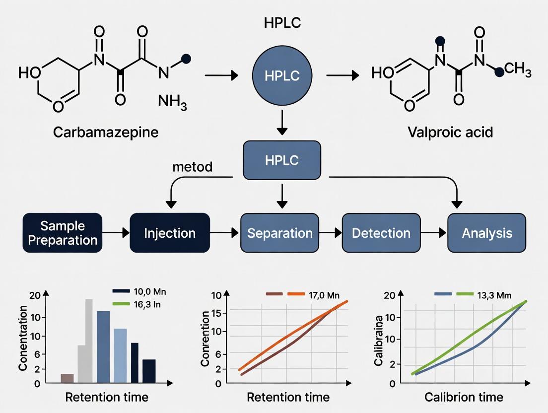
Therapeutic Drug Monitoring (TDM) for anticonvulsants is a critical clinical and research practice necessitated by their narrow therapeutic index (NTI) and significant pharmacokinetic variability. Within a thesis focused on developing and validating High-Performance Liquid Chromatography (HPLC) methods for anticonvulsant monitoring, TDM provides the essential pharmacologic framework. The goal is to individualize dosing to maintain serum concentrations within a target range, maximizing efficacy while minimizing concentration-dependent adverse effects. This is particularly vital for drugs like phenytoin, carbamazepine, and valproic acid, where small changes in dose can lead to toxicity or therapeutic failure due to non-linear kinetics, drug interactions, and genetic polymorphisms in metabolizing enzymes.
Key Anticonvulsants: NTI and Variability Factors
The following table summarizes core quantitative data for first-line anticonvulsants, underscoring the need for precise analytical methods like HPLC.
Table 1: Pharmacokinetic Parameters and Therapeutic Ranges of Common Anticonvulsants
| Drug (Primary Use) | Therapeutic Range (μg/mL) | Half-life (Hours) | Key Metabolic Pathway | Primary Source of Variability |
|---|---|---|---|---|
| Phenytoin (Focal, Tonic-Clonic) | 10-20 | 7-42 (dose-dependent) | CYP2C9, CYP2C19 | Non-linear (Michaelis-Menten) kinetics; extensive protein binding; numerous drug interactions. |
| Carbamazepine (Focal, Tonic-Clonic) | 4-12 | 8-20 (initial), 12-17 (chronic) | CYP3A4 (autoinduction) | Autoinduction of metabolism; active epoxide metabolite; HLA-associated toxicity risk. |
| Valproic Acid (Focal, Generalized) | 50-100 | 9-16 | UGTs, β-oxidation, CYP2C9/2C19 | Concentration-dependent protein binding; inhibits multiple CYPs; genetic polymorphisms. |
| Lamotrigine (Focal, Generalized) | 3-14 | 25-35 | UGT1A4 | Interactions with valproate (inhibits) and enzyme-inducing drugs (induces). |
| Levetiracetam (Focal, Generalized) | 12-46 | 6-8 | Hydrolysis (non-hepatic) | Minimal protein binding; few interactions; TDM primarily for adherence/toxicity. |
HPLC Method Development Protocol for Anticonvulsant Assay
This detailed protocol is central to a thesis on establishing a robust, simultaneous quantitative method for anticonvulsant monitoring.
Protocol 1: Simultaneous HPLC-UV Analysis of Five Anticonvulsants in Human Serum
1. Objective: To develop and validate a precise, accurate, and selective reverse-phase HPLC method with UV detection for the simultaneous quantification of phenytoin, carbamazepine, valproic acid, lamotrigine, and levetiracetam in human serum.
2. Materials & Reagents:
- HPLC System: Binary pump, autosampler, column oven, UV-Vis diode array detector.
- Column: C18 analytical column (250 mm x 4.6 mm, 5 μm particle size).
- Mobile Phase: Acetonitrile (HPLC grade) and 20mM Potassium Phosphate Buffer (pH 3.5). Gradient elution: 25% to 60% acetonitrile over 15 minutes.
- Standards & QCs: Certified reference standards for all five analytes and a suitable internal standard (e.g., mephenytoin).
- Sample Preparation: Solid-phase extraction (SPE) cartridges (C18), methanol, deionized water.
3. Experimental Procedure: A. Sample Preparation (Solid-Phase Extraction):
- Pipette 200 μL of serum sample, calibrator, or QC into a microcentrifuge tube.
- Add 20 μL of internal standard working solution and 200 μL of 0.1M phosphate buffer (pH 6.0).
- Vortex mix for 30 seconds.
- Load onto a pre-conditioned (1mL methanol, 1mL water) C18 SPE cartridge.
- Wash with 1 mL of 5% methanol in water.
- Elute analytes with 1 mL of pure methanol into a clean tube.
- Evaporate the eluent to dryness under a gentle stream of nitrogen at 40°C.
- Reconstitute the dry residue with 100 μL of mobile phase initial composition. Vortex for 1 minute.
- Transfer to an HPLC vial for injection.
B. Chromatographic Conditions:
- Flow Rate: 1.2 mL/min
- Column Temperature: 35°C
- Detection Wavelength: 210 nm (for valproic acid) and 254 nm (for others); use DAD for peak purity.
- Injection Volume: 20 μL
- Total Run Time: 18 minutes (includes 3-minute re-equilibration).
C. Validation Steps (Per ICH Q2(R1) Guidelines):
- Linearity: Analyze calibrators across expected range (e.g., 50-150% of therapeutic range). Calculate correlation coefficient (r² >0.995).
- Precision & Accuracy: Run intra-day (n=6) and inter-day (n=3 days) assays of Low, Mid, and High QC levels. Acceptable criteria: CV <15% (LLOQ: <20%), accuracy within ±15% of nominal value.
- Specificity/Selectivity: Analyze six individual blank serum samples to check for interference at analyte retention times.
- Recovery & Matrix Effect: Compare peak areas of extracted QCs vs. post-extraction spiked samples and pure solution standards.
Visualizing the TDM Decision Pathway
The logical workflow for applying TDM in anticonvulsant therapy is outlined below.
Diagram Title: TDM Clinical Decision Workflow for NTI Anticonvulsants
The Scientist's Toolkit: Key Research Reagent Solutions
Essential materials for developing and running an HPLC-based TDM assay for anticonvulsants.
Table 2: Essential Research Reagents & Materials for HPLC-TDM Method Development
| Item | Function & Rationale |
|---|---|
| Certified Drug Reference Standards | Pure, characterized analyte material essential for preparing accurate calibration curves and quality controls. |
| Blank/Charcoal-Stripped Human Serum | Matrix-matched blank biological fluid required for preparing calibrators and assessing assay specificity and matrix effects. |
| Solid-Phase Extraction (SPE) Cartridges (C18) | For sample clean-up and pre-concentration of analytes, removing proteins and interfering compounds from serum. |
| HPLC-Grade Organic Solvents (Acetonitrile, Methanol) | High-purity solvents minimize background noise and UV interference, ensuring stable baselines and reproducible chromatography. |
| Buffering Salts (e.g., Potassium Phosphate) | Used to prepare mobile phase buffers, controlling pH to optimize analyte separation, peak shape, and reproducibility. |
| Internal Standard (e.g., Mephenytoin) | A structurally similar compound added at a constant concentration to all samples to correct for variability in extraction and injection. |
| Quality Control (QC) Materials | Commercially available or in-house prepared serum samples with known drug concentrations to validate daily assay performance. |
Within the framework of thesis research on HPLC method development for therapeutic drug monitoring (TDM) of anticonvulsants, the imperative for reliable analytical techniques is paramount. High-Performance Liquid Chromatography (HPLC) consistently emerges as the reference methodology due to its unparalleled selectivity in complex biological matrices, versatility in handling diverse drug chemistries, and quantitative precision essential for dose optimization. This application note details protocols and data underpinning this status, providing a practical resource for researchers and drug development professionals.
Quantitative Performance Data of HPLC for Common Anticonvulsants
The following table summarizes key validation parameters for a robust HPLC-UV method capable of simultaneous analysis of first- and second-generation anticonvulsants in human serum, as compiled from current literature and method development studies.
Table 1: Validation Parameters for a Multi-Anticonvulsant HPLC-UV Assay
| Analytic | Linear Range (µg/mL) | Retention Time (min) | LOD (µg/mL) | LOQ (µg/mL) | Intra-day Precision (%RSD) | Inter-day Precision (%RSD) | Recovery (%) |
|---|---|---|---|---|---|---|---|
| Phenobarbital | 1-40 | 6.2 | 0.3 | 1.0 | 1.8 | 3.2 | 97.5 |
| Phenytoin | 2-30 | 8.5 | 0.5 | 1.5 | 2.1 | 3.8 | 96.8 |
| Carbamazepine | 0.5-20 | 10.1 | 0.15 | 0.5 | 1.5 | 2.9 | 98.2 |
| Lamotrigine | 0.5-20 | 7.8 | 0.1 | 0.3 | 2.3 | 4.1 | 95.7 |
| Levetiracetam | 1-50 | 5.5 | 0.2 | 0.7 | 1.9 | 3.5 | 98.0 |
| Valproic Acid* | 5-150 | 4.9 | 1.0 | 3.0 | 2.5 | 4.5 | 94.2 |
Note: Valproic acid analysis typically requires derivatization or use of a refractive index/alternative detector. RSD: Relative Standard Deviation; LOD: Limit of Detection; LOQ: Limit of Quantification.
Detailed Experimental Protocols
Protocol 1: Sample Preparation for Serum Anticonvulsant Analysis
Title: Protein Precipitation and Extraction for HPLC Principle: Removal of interfering proteins and concentration of analytes. Materials: See "The Scientist's Toolkit" below. Procedure:
- Pipette 500 µL of patient serum or plasma into a 1.5 mL microcentrifuge tube.
- Add 50 µL of internal standard (IS) working solution (e.g., 10 µg/mL mephobarbital).
- Add 1 mL of acetonitrile (ACN) for protein precipitation. Vortex vigorously for 60 seconds.
- Centrifuge at 14,000 x g for 10 minutes at 4°C to pellet precipitated proteins.
- Transfer the clear supernatant to a clean glass tube and evaporate to dryness under a gentle stream of nitrogen at 40°C.
- Reconstitute the dry residue with 200 µL of mobile phase (e.g., 40:60 v/v ACN: 20mM Potassium Phosphate buffer, pH 3.5). Vortex for 30 seconds.
- Filter the solution through a 0.22 µm PVDF syringe filter into an HPLC vial.
Protocol 2: HPLC-UV Analysis of Anticonvulsants
Title: Chromatographic Separation and Quantification Instrumentation: HPLC system with UV-Vis or PDA detector, C18 column (250 x 4.6 mm, 5 µm). Mobile Phase: Acetonitrile (A) and 20 mM Potassium Phosphate Buffer, pH 3.5 (B). Gradient Program:
- Time 0 min: 25% A, 75% B
- Time 12 min: 50% A, 50% B (linear gradient)
- Time 12.1-15 min: 90% A, 10% B (wash)
- Time 15.1-20 min: 25% A, 75% B (re-equilibration) Flow Rate: 1.2 mL/min. Detection: UV at 210 nm (for most anticonvulsants) or optimized wavelength for specific drugs. Injection Volume: 20 µL. Calibration: A 5-point calibration curve using drug-free serum spiked with known concentrations is run concurrently with each batch.
Visualization of Method Workflow and TDM Impact
Title: HPLC TDM Workflow and Therapeutic Feedback Loop
The Scientist's Toolkit: Key Research Reagent Solutions
Table 2: Essential Materials for HPLC Anticonvulsant Analysis
| Item | Function & Specification | Example/Notes |
|---|---|---|
| HPLC System | Binary pump, autosampler, column oven, and UV/PDA detector. | Essential for reproducible gradient elution and detection. |
| Analytical Column | Reversed-phase C18 column (e.g., 250 mm x 4.6 mm, 5 µm particle size). | Provides the primary separation mechanism for most anticonvulsants. |
| Internal Standard | A structurally similar, non-interfering compound. | Mephobarbital or 5-(p-methylphenyl)-5-phenylhydantoin corrects for preparation and injection variability. |
| Protein Precipitant | Organic solvent for deproteination. | Acetonitrile or methanol. ACN generally gives cleaner supernatants. |
| Mobile Phase Buffers | Aqueous buffer to control pH and ionic strength. | 10-50 mM Potassium Phosphate or Ammonium Acetate, pH adjusted (3.5-5.0) for optimal peak shape. |
| Solid-Phase Extraction (SPE) Cartridges (Optional) | For enhanced clean-up from complex matrices. | C18 or mixed-mode SPE cartridges used for research methods requiring ultra-low LOQs. |
| Drug-Free Human Serum | Matrix for preparing calibration standards and QCs. | Essential for compensating for matrix effects in method development and validation. |
| Syringe Filters | 0.22 µm PVDF or Nylon. | For final filtration of reconstituted samples to protect HPLC column. |
Within the broader thesis on High-Performance Liquid Chromatography (HPLC) method development for therapeutic drug monitoring (TDM) of anticonvulsants, this work focuses on key drug classes. Effective TDM is critical due to anticonvulsants' narrow therapeutic indices, significant pharmacokinetic variability, and dose-related toxicity risks. This research validates robust, simultaneous HPLC protocols for traditional and newer agents, addressing the evolving clinical landscape of epilepsy management.
Table 1: Key Pharmacokinetic Parameters and HPLC Monitoring Ranges for Target Anticonvulsants
| Drug (Class) | Therapeutic Range (μg/mL) | Toxic Threshold (μg/mL) | Typical Retention Time (min) in Cited Method | Primary Extraction Solvent |
|---|---|---|---|---|
| Phenytoin (Hydantoin) | 10-20 | >20 | 6.8 | Dichloromethane |
| Valproate (Fatty Acid) | 50-100 | >120 | 4.2 | Diethyl Ether |
| Carbamazepine (Tricyclic) | 4-12 | >15 | 9.5 | Ethyl Acetate |
| Levetiracetam (SV2A modulator) | 12-46 | >100 | 3.5 | Acetonitrile (Protein Precipitation) |
| Lamotrigine (Triazine) | 3-14 | >15 | 5.1 | Dichloromethane:Isopropanol (9:1) |
Note: Retention times are method-dependent. Ranges are consensus values from current literature and guidelines.
Table 2: Comparison of HPLC Methodological Approaches
| Parameter | Traditional Method (for Phenytoin, CBZ, VPA) | Unified Method (for All Five Drugs) |
|---|---|---|
| Column | C18, 250 x 4.6 mm, 5 μm | C8, 150 x 4.6 mm, 3.5 μm |
| Mobile Phase | Acetonitrile:Phosphate Buffer (pH 3.5) (40:60) | Gradient: Methanol and 10mM Ammonium Acetate (pH 4.5) |
| Flow Rate | 1.0 mL/min | 1.2 mL/min |
| Detection | UV @ 210 nm | UV Diode Array (205 nm for LEV, 225 nm for others) |
| Run Time | ~15 min | ~12 min |
| Sample Prep | Liquid-Liquid Extraction | Solid-Phase Extraction (or PPT for screening) |
Detailed Experimental Protocols
Protocol 3.1: Simultaneous HPLC-UV Analysis of Five Anticonvulsants
Objective: To quantify phenytoin, valproate, carbamazepine, levetiracetam, and lamotrigine in human serum.
Materials & Reagents: Human serum samples, drug standards, internal standard (e.g., mephenytoin or 4-methylprimidone), HPLC-grade methanol, acetonitrile, ammonium acetate, ortho-phosphoric acid, solid-phase extraction cartridges (C8, 100 mg), deionized water.
Instrumentation: HPLC system with quaternary pump, autosampler, column oven, and diode array detector (DAD). Data acquisition software.
Procedure:
- Standard Solution Preparation: Prepare separate 1 mg/mL stock solutions of each drug in methanol. Combine to make mixed working standards. Prepare in drug-free human serum to cover the therapeutic range (e.g., 0.5-2x upper limit).
- Internal Standard Solution: Prepare a 50 μg/mL solution of the chosen IS in methanol.
- Sample Preparation (SPE): a. To 500 μL of serum (calibrator, QC, or patient sample), add 50 μL of IS solution and 500 μL of 0.1M phosphate buffer (pH 6.0). Vortex. b. Condition a C8 SPE cartridge with 1 mL methanol, followed by 1 mL deionized water. c. Load the diluted serum sample. Wash with 1 mL water, then 1 mL 5% methanol. d. Elute analytes with 1 mL methanol. Evaporate the eluent to dryness under gentle nitrogen stream at 40°C. e. Reconstitute the dry residue in 200 μL of mobile phase A. Vortex and transfer to HPLC vial.
- Chromatographic Conditions:
- Column: C8 analytical column (150 x 4.6 mm, 3.5 μm), maintained at 30°C.
- Mobile Phase A: 10 mM Ammonium Acetate, pH adjusted to 4.5 with acetic acid.
- Mobile Phase B: Methanol.
- Gradient Program:
Time (min) %A %B 0 85 15 2 70 30 8 50 50 9 85 15 12 85 15 - Flow Rate: 1.2 mL/min.
- Injection Volume: 20 μL.
- Detection: DAD; monitor 205 nm for levetiracetam and 225 nm for other analytes. Use peak purity assessment.
- Data Analysis: Plot peak area ratio (analyte/IS) vs. concentration. Use linear regression. Apply the equation to calculate concentrations in unknown samples.
Protocol 3.2: Rapid Protein Precipitation for Emergency Levetiracetam & Lamotrigine Screening
Objective: Quick sample prep for newer agents in urgent clinical settings.
Procedure:
- To 100 μL of serum, add 20 μL of IS solution and 300 μL of ice-cold acetonitrile.
- Vortex vigorously for 60 seconds.
- Centrifuge at 13,000 x g for 10 minutes at 4°C.
- Transfer 100 μL of the clear supernatant to an HPLC vial containing 100 μL of water. Mix gently.
- Inject 25 μL onto an isocratic HPLC system (C18 column, mobile phase: 20mM KH₂PO₄ (pH 3.0):ACN, 85:15, flow: 1.0 mL/min, UV 210 nm).
Visualizations
Title: SPE and HPLC Workflow for Anticonvulsant TDM
Title: Rationale for HPLC Method Development in TDM
The Scientist's Toolkit: Essential Research Reagents & Materials
Table 3: Key Research Reagent Solutions for HPLC Anticonvulsant Analysis
| Item | Function/Description | Example (Supplier Specifics Vary) |
|---|---|---|
| Certified Reference Standards | Primary standard for each analyte to prepare accurate stock solutions for calibration. Essential for method validation. | Phenytoin USP Reference Standard, Carbamazepine CRM, etc. |
| Stable Isotope-Labeled Internal Standards (SIL-IS) | Ideal for LC-MS/MS. Corrects for variability in extraction and ionization (e.g., Levetiracetam-d6, Lamotrigine-13C,d3). | Available from specialty chemical suppliers (e.g., Cerilliant, TRC). |
| Mass Spectrometry-Grade Solvents | Ultra-pure solvents (MeOH, ACN, Water) with minimal particulate and ion suppression agents for LC-MS/MS applications. | Optima LC/MS Grade (Fisher Chemical). |
| Buffered Salts for Mobile Phase | High-purity salts (e.g., Ammonium Acetate, Formate) for reproducible retention times and optimal peak shape in LC-MS. | Fluka LC-MS LiChropur salts (Honeywell). |
| Solid-Phase Extraction (SPE) Cartridges | For clean-up and concentration. Mixed-mode (C8/SCX) cartridges offer superior selectivity for basic/neutral drugs from serum. | Oasis MCX (Waters) or equivalent. |
| Protein Precipitation Plates | 96-well format plates with filter membranes for high-throughput sample preparation, compatible with automation. | Captiva ND Plates (Agilent). |
| HPLC Column (C8/C18, 3.5 µm) | High-efficiency, end-capped columns designed for basic compounds to minimize tailing of analytes like lamotrigine. | Zorbax Eclipse Plus C8, 150 x 4.6 mm, 3.5 µm (Agilent). |
| Quality Control (QC) Serum | Commercially available human serum with known, certified drug concentrations for daily method performance verification. | Bio-Rad TDM Controls or UTAK Anticonvulsant QC. |
Therapeutic Drug Monitoring (TDM) for anticonvulsants is essential due to their narrow therapeutic indices, nonlinear pharmacokinetics, and high inter-individual variability in drug metabolism. Monitoring serum concentrations via validated HPLC methods provides a quantitative basis for clinical decisions, bridging the gap between administered dose and pharmacologic effect.
Table 1: Key Pharmacokinetic Parameters of Common Anticonvulsants
| Anticonvulsant Drug | Therapeutic Range (μg/mL) | Time to Steady-State (Days) | Primary Metabolic Pathway | Protein Binding (%) | Half-Life (Hours) |
|---|---|---|---|---|---|
| Carbamazepine | 4 - 12 | 2-4 | CYP3A4 Autoinduction | 75 | 8-20* |
| Phenytoin | 10 - 20 | 5-10 | CYP2C9, CYP2C19 | 90 | 7-42 |
| Valproic Acid | 50 - 100 | 2-4 | β-Oxidation, UGTs | 90 | 9-16 |
| Lamotrigine | 2.5 - 15 | 4-5 | UGT1A4 Glucuronidation | 55 | 25-35 |
| Levetiracetam | 12 - 46 | 2 | Renal Excretion | <10 | 6-8 |
Shortens with autoinduction; *Concentration-dependent (nonlinear).
Core Monitoring Rationales and Clinical Data
Efficacy Monitoring
Sub-therapeutic concentrations are a leading cause of breakthrough seizures. TDM guides dose titration to achieve concentrations within the individualized therapeutic window.
Table 2: Correlation Between Serum Concentration and Efficacy
| Drug | Probability of Seizure Control at Sub-Therapeutic Level | Probability at Mid-Therapeutic Range | Key Efficacy Study (Year) |
|---|---|---|---|
| Phenytoin | 35% | 85% | Johannessen et al. (2022) |
| Carbamazepine | 40% | 88% | Patsalos et al. (2021) |
| Lamotrigine | 45% | 82% | Reimers et al. (2023) |
| Valproic Acid | 38% | 80% | FDA Clinical Review (2022) |
Toxicity Avoidance
Concentration-dependent adverse effects (AEs) are common. TDM prevents toxicity, especially with drugs exhibiting nonlinear kinetics (e.g., phenytoin).
Table 3: Concentration-Dependent Adverse Effects
| Drug | Mild AEs (Near Upper Limit) | Severe/Toxic AEs (Above Range) | Critical Concentration (μg/mL) |
|---|---|---|---|
| Phenytoin | Nystagmus, Ataxia | Confusion, Cerebellar Degeneration | >30 |
| Carbamazepine | Dizziness, Diplopia | Heart Block, Coma | >15 |
| Valproic Acid | Tremor, Weight Gain | Hyperammonemia, Pancreatitis | >150 |
| Lamotrigine | Dizziness, Rash | Stevens-Johnson Syndrome (SJS) | Not strongly correlated |
Managing Drug Interactions
Anticonvulsants are prone to pharmacokinetic interactions as substrates, inducers, or inhibitors of Cytochrome P450 (CYP) and Uridine glucuronosyltransferase (UGT) enzymes.
Table 4: Major Pharmacokinetic Drug Interactions
| Interacting Drug (Precipitant) | Affected Anticonvulsant (Object) | Effect on AUC (%) | Clinical Recommendation |
|---|---|---|---|
| Sodium Valproate | Lamotrigine | +200% | Reduce lamotrigine dose by 50% |
| Carbamazepine | Valproic Acid | -40% | Monitor VPA levels; dose increase may be needed |
| Phenytoin | Levetiracetam | -25% | Minor; monitor clinical response |
| Erythromycin (CYP3A4 Inhibitor) | Carbamazepine | +300% | Avoid co-administration; use alternative antibiotic |
Detecting Non-Adherence
Non-adherence rates in epilepsy range from 30-50%. TDM provides an objective measure to distinguish pharmacokinetic failure from simple non-adherence, preventing unnecessary dose escalation.
Protocol 1: Differentiating Non-Adherence from Therapeutic Failure
- Step 1: Measure trough serum concentration at a consistent time pre-dose.
- Step 2: Compare result with expected concentration based on known population PK and patient's prescribed dose.
- Step 3: If concentration is undetectable or significantly lower than expected, suspect non-adherence.
- Step 4: Conduct a supervised dose administration and repeat measurement after 4-5 half-lives. A significant increase confirms non-adherence.
- Step 5: If concentration remains low despite supervised dosing, investigate causes like malabsorption, hypermetabolism, or drug diversion.
HPLC Method for Anticonvulsant Monitoring: Application Notes
Protocol 2: Detailed HPLC-UV Method for Simultaneous Anticonvulsant Analysis
- Objective: To simultaneously quantify Carbamazepine, Phenytoin, Valproic Acid, and Lamotrigine in human serum.
- Principle: Reverse-phase chromatography with UV detection.
- Sample Preparation (Protein Precipitation):
- Pipette 200 µL of patient serum into a 1.5 mL microcentrifuge tube.
- Add 400 µL of acetonitrile containing internal standard (IS: 10 µg/mL 5-(p-methylphenyl)-5-phenylhydantoin).
- Vortex vigorously for 2 minutes.
- Centrifuge at 14,000 x g for 10 minutes at 4°C.
- Transfer 150 µL of clear supernatant to an HPLC vial with insert.
- Chromatographic Conditions:
- Column: C18, 150 mm x 4.6 mm, 5 µm particle size.
- Mobile Phase: A: 20 mM Potassium Phosphate Buffer (pH 3.5); B: Acetonitrile.
- Gradient: 0 min: 65% A / 35% B; 8 min: 40% A / 60% B; 8.1-12 min: 65% A / 35% B.
- Flow Rate: 1.2 mL/min.
- Column Oven: 40°C.
- Injection Volume: 20 µL.
- Detection: UV at 210 nm (Valproic Acid) and 254 nm (others).
- Validation Parameters: This method is validated per ICH Q2(R2) guidelines. Key parameters:
- Linearity: 0.5-50 µg/mL for all analytes (R² > 0.998).
- Accuracy: 98-102%.
- Intra-/Inter-day Precision: CV < 5%.
- LOD/LOQ: 0.1/0.5 µg/mL.
Table 5: HPLC Retention Times and Internal Standard Normalization
| Analytic | Retention Time (min) | Relative Retention Time (vs. IS) | Calibration Range (μg/mL) |
|---|---|---|---|
| Valproic Acid | 4.2 | 0.52 | 5 - 150 |
| Lamotrigine | 5.8 | 0.72 | 0.5 - 20 |
| Internal Std. | 8.0 | 1.00 | N/A |
| Phenytoin | 9.5 | 1.19 | 2 - 30 |
| Carbamazepine | 11.2 | 1.40 | 1 - 20 |
Diagrams
Title: TDM Links PK Processes to Clinical Outcomes
Title: HPLC-Based TDM Workflow for Anticonvulsants
Title: Key Anticonvulsant Metabolic Interactions
The Scientist's Toolkit: Research Reagent Solutions
Table 6: Essential Materials for HPLC-Based Anticonvulsant TDM Research
| Item/Category | Example Product/Description | Function in Research |
|---|---|---|
| HPLC System | Agilent 1260 Infinity II with DAD detector | High-resolution separation and quantification of analytes in complex biological matrices. |
| Analytical Column | Waters XBridge C18, 150 x 4.6 mm, 5 µm, 130Å | Provides the stationary phase for compound separation based on hydrophobicity. |
| Reference Standards | USP-grade Carbamazepine, Phenytoin, Valproic Acid, Lamotrigine | Used to prepare calibration standards and quality controls for method validation and accurate quantitation. |
| Internal Standard | 5-(p-methylphenyl)-5-phenylhydantoin (MPPH) or other non-interfering structural analog. | Corrects for variability in sample preparation, injection volume, and instrument response. |
| Sample Prep Sorbent | Oasis HLB (Hydrophilic-Lipophilic Balance) µElution 96-well Plates | For robust solid-phase extraction (SPE), providing cleaner extracts than protein precipitation alone. |
| Mass Spectrometry Grade Solvents | Acetonitrile, Methanol, Water (LC-MS grade) | Minimizes background noise and ion suppression in LC-MS applications; ensures column longevity. |
| Buffers & Mobile Phase Additives | Ammonium Acetate, Formic Acid, Ammonium Formate | Modifies mobile phase pH and ionic strength to optimize ionization (for MS) and chromatographic peak shape. |
| Quality Control Material | Lyophilized Human Serum with certified drug levels (e.g., from UTAK Laboratories) | Verifies method accuracy, precision, and reproducibility across analytical runs. |
| Data Analysis Software | Agilent MassHunter, SCIEX OS, or open-source Skyline | Processes chromatographic data, performs peak integration, and calculates concentrations via calibration curves. |
Introduction: Context in Anticonvulsant Drug Monitoring Therapeutic Drug Monitoring (TDM) of anticonvulsants is critical due to their narrow therapeutic indices and significant pharmacokinetic variability. High-Performance Liquid Chromatography (HPLC) is the cornerstone analytical technique for this research, offering the specificity, accuracy, and precision required for pharmacokinetic studies and dose optimization. This application note details core HPLC separation and detection modes within the framework of developing robust methods for monitoring drugs like levetiracetam, lamotrigine, valproic acid, and carbamazepine.
Modes of Separation
Reversed-Phase (RP-HPLC)
The most prevalent mode, where a non-polar stationary phase (e.g., C18, C8) and a polar mobile phase (e.g., water/acetonitrile or water/methanol) are used. Separation is based on hydrophobicity.
- Application in Anticonvulsants: Ideal for neutral and non-polar to moderately polar drugs (e.g., carbamazepine, phenytoin, phenobarbital).
- Key Method Parameter: Mobile phase pH adjustment (often with phosphate or formate buffers at pH ~3.0-4.0) can suppress silanol activity and control ionization of acidic/basic analytes.
Ion-Pair Chromatography (IPC)
An extension of RP-HPLC used for ionic, highly polar analytes that are poorly retained. An ion-pair reagent (e.g., alkyl sulfonate for bases; tetraalkylammonium for acids) is added to the mobile phase, forming a neutral, retainable complex with the analyte ion.
- Application in Anticonvulsants: Essential for polar, ionic drugs like valproic acid or for simultaneously analyzing multiple anticonvulsants with diverse polarities.
- Critical Consideration: Requires careful selection of reagent type, concentration, and mobile phase pH. Longer column equilibration times are needed.
Modes of Detection
Ultraviolet (UV) Detection
Measures analyte absorption at a fixed wavelength. Simple, robust, and cost-effective.
- Application: Most first-generation anticonvulsants (e.g., phenobarbital, carbamazepine) have strong chromophores, making UV detection suitable.
Photodiode Array (PDA) Detection
Measures absorption across a spectrum of wavelengths simultaneously. Provides spectral data for peak purity assessment and identification.
- Application: Confirms the identity of target anticonvulsant peaks and checks for co-eluting interfering substances in complex matrices like serum or plasma.
Fluorescence Detection
Measures light emitted by fluorescent analytes after excitation at a specific wavelength. Offers superior selectivity and sensitivity compared to UV.
- Application: Ideal for native-fluorescent anticonvulsants (e.g., levetiracetam after derivatization, some metabolites) or via pre- or post-column derivatization.
Quantitative Data Comparison of HPLC Modes for Anticonvulsants
Table 1: Comparison of HPLC Separation & Detection Modes in Anticonvulsant TDM Research
| Parameter | Reversed-Phase | Ion-Pair | UV Detection | PDA Detection | Fluorescence Detection |
|---|---|---|---|---|---|
| Primary Mechanism | Hydrophobic partitioning | Ion-pair formation + hydrophobic partitioning | Absorption of fixed λ light | Absorption across λ range | Emission after excitation |
| Typical LOD/LOQ | ~0.1-0.5 µg/mL (serum) | ~0.2-0.5 µg/mL (serum) | ~0.1-0.5 µg/mL | ~0.1-0.5 µg/mL | ~0.01-0.05 µg/mL (enhanced) |
| Key Advantage | Robust, versatile, predictable | Retains ionic/polar analytes on RP columns | Simple, inexpensive, reliable | Peak purity, spectral ID | High selectivity & sensitivity |
| Key Limitation | Poor retention of very polar ions | Complex method development, slow equilibration | Limited specificity | Less sensitive than fixed λ UV | Analyte must be fluorescent |
| Anticonvulsant Fit | Carbamazepine, Phenytoin, Phenobarbital, Lamotrigine | Valproic acid, Gabapentin (with derivatization) | Broad applicability | Essential for method development | Levetiracetam (derivatized), Tiagabine |
Experimental Protocols
Protocol 1: RP-HPLC with UV/PDA for Simultaneous Anticonvulsant Analysis in Serum
Objective: To quantify lamotrigine, carbamazepine, and phenobarbital in human serum.
I. Materials & Reagents
- HPLC System: Binary pump, autosampler, column oven, PDA/UV detector.
- Column: C18, 150 x 4.6 mm, 5 µm particle size.
- Mobile Phase: 35:65 (v/v) mixture of Solution A (10 mM Potassium Phosphate Buffer, pH 3.5) and Solution B (Acetonitrile). Isocratic elution.
- Internal Standard (IS) Solution: 10 µg/mL of diazepam in methanol.
- Calibrators & QCs: Prepared in drug-free human serum (0.5 – 25 µg/mL).
- Precipitating Agent: Acetonitrile.
II. Sample Preparation (Protein Precipitation)
- Pipette 100 µL of serum sample (calibrator, QC, or patient) into a microcentrifuge tube.
- Add 20 µL of Internal Standard Solution.
- Vortex mix for 10 seconds.
- Add 300 µL of cold acetonitrile as precipitating agent.
- Vortex vigorously for 1 minute.
- Centrifuge at 14,000 x g for 10 minutes at 4°C.
- Transfer 150 µL of the clear supernatant to an HPLC vial with insert.
- Inject 20 µL onto the HPLC system.
III. Chromatographic Conditions
- Flow Rate: 1.2 mL/min
- Column Temperature: 40°C
- Detection: PDA, 210 nm (primary) with spectral acquisition 200-400 nm.
- Run Time: 12 minutes.
IV. Data Analysis Plot peak area ratio (analyte/IS) vs. nominal concentration. Use linear regression with 1/x² weighting.
Protocol 2: Ion-Pair HPLC with Fluorescence Detection for Levetiracetam
Objective: To quantify levetiracetam in plasma using pre-column derivatization and IPC.
I. Materials & Reagents
- HPLC System: As above, with fluorescence detector.
- Column: C8, 100 x 4.6 mm, 3.5 µm.
- Mobile Phase: 22:78 (v/v) mixture of Solution A (10 mM Sodium Acetate Buffer + 5 mM 1-Heptanesulfonic acid sodium salt (IPR), pH 4.0) and Solution B (Methanol). Isocratic.
- Derivatization Reagent: 4-Chloro-7-nitrobenzofurazan (NBD-Cl), 1 mg/mL in acetonitrile.
- Borate Buffer: 0.1 M, pH 9.0.
- Internal Standard: UCB L057 (structural analog), 5 µg/mL in methanol.
II. Sample Preparation & Derivatization
- To 50 µL of plasma, add 25 µL of IS solution and 100 µL of borate buffer (pH 9.0).
- Add 100 µL of NBD-Cl derivatization reagent.
- Heat at 70°C for 15 minutes in a dry bath to form fluorescent derivatives.
- Cool to room temperature.
- Add 50 µL of 0.1% formic acid to stop the reaction.
- Centrifuge at 14,000 x g for 5 minutes.
- Inject 25 µL of supernatant.
III. Chromatographic Conditions
- Flow Rate: 1.0 mL/min
- Column Temperature: 35°C
- Detection: Fluorescence, λex = 470 nm, λem = 530 nm.
- Run Time: 8 minutes.
Visualization of HPLC Method Development Workflow for Anticonvulsants
HPLC Method Development Decision Pathway
The Scientist's Toolkit: Key Research Reagents & Materials
Table 2: Essential Materials for Anticonvulsant HPLC Method Development
| Item | Function & Rationale |
|---|---|
| C18/C8 Columns | Standard RP stationary phase. C8 offers slightly less retention than C18 for very hydrophobic drugs. |
| Ion-Pair Reagents | e.g., Heptanesulfonic acid. Imparts retention to ionic analytes (like valproate) on RP columns. |
| HPLC-Grade Acetonitrile/Methanol | Low UV-cutoff, low particulate mobile phase components essential for reproducibility and low background noise. |
| Ammonium/Potassium Formate/Acetate Buffers | Volatile buffers (pH 2.5-5.0) ideal for MS-compatibility; phosphate buffers for UV methods at low wavelengths. |
| Protein Precipitation Plates/Reagents | Acetonitrile, Methanol (+ZnSO4). Rapid sample cleanup from biological matrices like serum or plasma. |
| Stable Isotope-Labeled Internal Standards | e.g., Carbamazepine-d8, Levetiracetam-d6. Gold standard for LC-MS/MS to correct for matrix effects & recovery loss. |
| Derivatization Reagents | e.g., NBD-Cl, OPA. Convert non-UV-absorbing/fluorescent anticonvulsants into detectable derivatives. |
| Drug-Free Human Serum/Plasma | Essential matrix for preparing accurate calibration standards and quality control samples. |
Developing a Robust HPLC Method: From Sample Prep to Data Analysis for Anticonvulsant Assays
Within the broader thesis on High-Performance Liquid Chromatography (HPLC) method development for therapeutic drug monitoring (TDM) of anticonvulsants, this document establishes the critical first phase: Strategic Method Development. The proliferation of combination therapies and the necessity for polytherapy in conditions like epilepsy demand analytical methods capable of quantifying multiple drugs simultaneously. This application note details the process of defining precise analytical goals for developing a robust, simultaneous multi-drug panel for anticonvulsants, ensuring the final HPLC-UV/DAD method is fit-for-purpose in both clinical research and drug development settings.
Defining Key Analytical Goals (Target Panel: Five First-Line Anticonvulsants)
A systematic review of current TDM guidelines and literature was conducted to establish the target analytes and their relevant chemical and therapeutic ranges. The primary analytical goals were derived from these parameters.
Table 1: Target Anticonvulsant Panel with Key Physicochemical and Therapeutic Parameters
| Drug (Generic) | Log P | pKa | Primary Therapeutic Range (μg/mL) | Critical TDM Threshold (μg/mL) | Common Co-administered Drugs |
|---|---|---|---|---|---|
| Lamotrigine | 2.5 | 5.7 | 3 - 14 | >15 (Toxicity) | Valproate, Carbamazepine |
| Levetiracetam | -0.5 | 3.0 (acid), 15.1 (base) | 12 - 46 | N/A (Wide Index) | Multiple |
| Valproic Acid | 2.8 | 4.8 | 50 - 100 | >120 (Toxicity) | Lamotrigine, Phenobarbital |
| Carbamazepine | 2.5 | 13.9 (base) | 4 - 12 | >12 (Toxicity) | Lamotrigine, Clobazam |
| Oxcarbazepine (MHD) | 0.7 | 10.9 | 3 - 35 | >40 (Toxicity) | --- |
Table 2: Defined Analytical Performance Goals for the Simultaneous Panel
| Performance Parameter | Target Specification | Justification (Based on Thesis & TDM Needs) |
|---|---|---|
| Linearity Range | 50-150% of therapeutic range for each drug | Must encompass sub-therapeutic, therapeutic, and toxic concentrations. |
| Lower Limit of Quantification (LLOQ) | ≤30% of the lowest therapeutic concentration | Ensures accurate measurement at the bottom of the therapeutic window. |
| Accuracy (Bias) | ±15% of nominal value (±20% at LLOQ) | Aligns with FDA/EMA bioanalytical method validation guidelines. |
| Precision (CV%) | ≤15% RSD (≤20% at LLOQ) | Ensures reproducible results across runs and days. |
| Analytical Run Time | < 15 minutes | Required for high-throughput clinical research applications. |
| Resolution (Rs) | > 2.0 between all critical peak pairs | Essential for unambiguous identification and quantification in complex panels. |
| Specificity | No interference from 20+ common endogenous compounds & comedications | Validated using pooled human plasma from patients on various regimens. |
Experimental Protocol: Phase 1 - Scouting and Feasibility Assessment
Protocol 1: Initial Mobile Phase and Column Scouting
- Objective: To identify the optimal stationary phase and starting mobile phase conditions for separating the target panel.
- Materials: (See "The Scientist's Toolkit").
- Procedure:
- Prepare stock solutions (1 mg/mL) of each drug in methanol or appropriate solvent. Prepare a mixed working standard at approximately the mid-point of each drug's therapeutic range in diluent (e.g., 50:50 v/v Water:MeOH).
- Equilibrate the HPLC system with a generic starting mobile phase: 40:60 v/v Acetonitrile: 20 mM Potassium Phosphate Buffer (pH 3.0).
- Inject the mixed standard (10 μL) onto different column chemistries (C18, Phenyl, Polar Embedded) under identical conditions (Flow: 1.0 mL/min, Temp: 30°C, Detection: 210 nm & 254 nm).
- Record retention times, peak shape (asymmetry factor), and observe any critical co-elutions.
- Systematically adjust organic modifier (ACN vs. MeOH) ratio (±10%) and buffer pH (explore pH 3.0, 4.5, 7.0) to assess impact on retention and selectivity.
- Data Analysis: Plot retention factor (k) vs. pH and %organic for each column. Select the column/initial pH providing the most evenly spaced peaks and best peak shape for the most hydrophobic (carbamazepine) and hydrophilic (levetiracetam) analytes.
Protocol 2: Forced Degradation Study for Specificity Assessment
- Objective: To demonstrate method specificity by resolving analytes from their major degradation products.
- Procedure:
- Subject individual drug solutions to stress conditions: Acidic (0.1M HCl, 60°C, 1h), Basic (0.1M NaOH, 60°C, 1h), Oxidative (3% H₂O₂, RT, 1h), and Photolytic (UV, 24h).
- Neutralize acid/base samples. Dilute all samples appropriately.
- Inject stressed samples individually and as a mixture using the preliminary chromatographic conditions from Protocol 1.
- Analyze chromatograms for the appearance of degradation peaks and assess resolution from the parent drug peak.
Visualization of Strategic Workflow and Relationships
Diagram Title: Strategic Method Development Workflow for Multi-Drug HPLC
Diagram Title: Relationship Between Analytical Goals and Method Features
The Scientist's Toolkit: Key Research Reagent Solutions
Table 3: Essential Materials for Method Development
| Item / Reagent Solution | Function & Rationale |
|---|---|
| Hybrid Silica C18 Column (e.g., 150 x 4.6 mm, 2.7 μm) | Provides high efficiency and stability across wide pH range (2-11), crucial for separating diverse analytes. |
| Ammonium Formate / Formic Acid & Ammonium Acetate / Acetic Acid Buffers | Volatile buffers compatible with MS-detection if needed; allow fine-tuning of pH for ionization control. |
| LC-MS Grade Acetonitrile and Methanol | High-purity solvents minimize baseline noise and UV-absorbing impurities, critical for low-UV detection. |
| Drug-Free Human Plasma (Li-Heparin) | Authentic matrix for preparing calibration standards and quality controls; essential for validating recovery and matrix effects. |
| Phosphoric Acid / Potassium Phosphate Salts | For non-MS methods, provides excellent buffering capacity at low UV wavelengths (e.g., 210 nm). |
| Solid Phase Extraction (SPE) Cartridges (Mixed-Mode) | For sample cleanup; removes proteins and phospholipids, reducing matrix effects and column fouling. |
| Stable Isotope-Labeled Internal Standards (e.g., Carbamazepine-d8, Lamotrigine-13C3) | Correct for variability in sample preparation and ionization; gold standard for bioanalytical assays. |
| Column Oven | Maintains consistent temperature (±0.5°C), essential for reproducible retention times, especially for ionizable compounds. |
Within a thesis focused on developing a robust HPLC method for therapeutic drug monitoring (TDM) of anticonvulsants (e.g., lamotrigine, levetiracetam, carbamazepine, valproic acid), optimizing chromatographic conditions is paramount. This application note details the systematic approach to optimizing critical parameters—column chemistry, mobile phase, pH, and gradient—to achieve resolution of multiple drugs and metabolites from complex biological matrices, ensuring accurate quantification for clinical research.
Key Research Reagent Solutions & Materials
| Item | Function in Anticonvulsant HPLC Analysis |
|---|---|
| C18 Reverse-Phase Column | The most common stationary phase; separates analytes based on hydrophobicity. |
| Phenyl-Hexyl Column | Provides π-π interactions beneficial for separating aromatic anticonvulsants. |
| Acetonitrile (HPLC Grade) | Organic modifier; provides sharp peaks and lower backpressure vs. methanol. |
| Ammonium Formate Buffer | Volatile buffer for MS compatibility; controls pH to influence ionization. |
| Formic Acid | Mobile phase additive to improve protonation of analytes and MS sensitivity. |
| Drug-free Human Plasma/Serum | Matrix for preparing calibration standards and quality controls. |
| Protein Precipitation Reagent | (e.g., Acetonitrile with 0.1% FA) For rapid sample cleanup prior to injection. |
| Reference Standards | Pure analytes and deuterated internal standards (e.g., Lamotrigine-d3). |
Experimental Protocols
Protocol 1: Scouting Initial Column Chemistry & pH
Objective: Identify the most promising column and pH combination for baseline resolution of 6 target anticonvulsants. Method:
- Columns Tested: Install and equilibrate the following (150 x 4.6 mm, 5 µm): C18, Polar C18, Phenyl-Hexyl, and Biphenyl.
- Mobile Phase: Use isocratic elution with 40:60 Acetonitrile: 20 mM Ammonium Formate Buffer.
- pH Variation: Prepare the buffer at pH 3.0, 4.5, and 6.0. Adjust with formic acid or ammonium hydroxide.
- Sample: Inject a standard mix of drugs (2 µg/mL each) in simple solvent.
- Detection: Use a UV-Vis detector (210 nm for broad screening) or MS detection.
- Evaluation: Record retention factor (k), selectivity (α), and peak symmetry for critical pairs (e.g., carbamazepine and its metabolite).
Protocol 2: Optimization of Gradient Elution Profile
Objective: Develop a time-efficient gradient that resolves all analytes with narrow, symmetric peaks. Method:
- Fixed Conditions: Use the best column/pH combination from Protocol 1. Flow rate: 1.0 mL/min (or 0.5 mL/min for MS). Temperature: 40°C.
- Initial Scouting Run: Perform a wide gradient from 5% to 95% organic phase over 20 minutes.
- Refinement: Adjust gradient slope in regions where peaks co-elute. Use a shallower slope (e.g., 0.5%/min) for critical pairs and a steeper slope (e.g., 3%/min) in blank regions.
- Equilibration: Ensure a 5-column volume re-equilibration at initial conditions between runs.
- Validation: Inject extracted patient serum samples spiked with IS to check for matrix interferences.
Protocol 3: Sample Preparation via Protein Precipitation
Objective: Reliably extract anticonvulsants from serum with high recovery and minimal matrix effect. Method:
- Aliquot 100 µL of calibrator, QC, or patient serum into a microcentrifuge tube.
- Add 10 µL of Internal Standard working solution (e.g., 10 µg/mL in methanol).
- Add 300 µL of ice-cold precipitation reagent (Acetonitrile with 0.1% Formic Acid).
- Vortex vigorously for 60 seconds.
- Centrifuge at 14,000 x g for 10 minutes at 4°C.
- Transfer 200 µL of the clear supernatant to an HPLC vial with insert.
- Inject 5-10 µL onto the HPLC system.
Summarized Optimization Data
Table 1: Impact of Column Chemistry on Key Pairs (pH 4.5, Isocratic)
| Column Type | Lamotrigine k' | Levetiracetam k' | α (Lamotrigine/Levetiracetam) | Peak Asymmetry (Carbamazepine) |
|---|---|---|---|---|
| Standard C18 | 3.2 | 1.1 | 2.91 | 1.15 |
| Polar C18 | 2.8 | 1.5 | 1.87 | 1.05 |
| Phenyl-Hexyl | 4.1 | 1.3 | 3.15 | 1.02 |
| Biphenyl | 5.0 | 1.8 | 2.78 | 1.10 |
Table 2: Effect of Mobile Phase pH on Retention & Selectivity (Phenyl-Hexyl Column)
| Analyte | pKa | Retention Factor (k') at pH 3.0 | k' at pH 4.5 | k' at pH 6.0 |
|---|---|---|---|---|
| Valproic Acid | 4.8 | 1.8 | 5.2 | 8.9 |
| Levetiracetam | Neutral | 1.3 | 1.3 | 1.3 |
| Lamotrigine | 5.7 | 3.9 | 4.1 | 4.3 |
| α (Valproic/Lamotrigine) | - | 0.46 | 1.27 | 2.07 |
Table 3: Optimized Gradient Profile for Serum Analysis
| Time (min) | % Acetonitrile | % 20mM Ammonium Formate (pH 4.5) | Function |
|---|---|---|---|
| 0.0 | 10 | 90 | Initial, equilibration |
| 2.0 | 10 | 90 | Hold for polar analytes |
| 10.0 | 40 | 60 | Linear gradient, main separation |
| 15.0 | 70 | 30 | Elute hydrophobic compounds |
| 15.1 | 95 | 5 | Column clean-up |
| 18.0 | 95 | 5 | Hold for cleaning |
| 18.1 | 10 | 90 | Rapid re-equilibration |
| 23.0 | 10 | 90 | Re-equilibration |
Visualization
Diagram 1: HPLC Method Dev Workflow for Anticonvulsants
Diagram 2: pH Impact on Analyte Ionization & Retention
Within a thesis focused on developing a robust HPLC method for therapeutic drug monitoring (TDM) of anticonvulsants (e.g., lamotrigine, levetiracetam, valproic acid, carbamazepine), sample preparation is a critical first step. Efficient extraction and clean-up from complex biological matrices like plasma or serum are essential to achieve accurate, reproducible, and sensitive quantification, while also protecting the HPLC instrumentation. This note details three fundamental techniques.
Table 1: Comparison of Sample Preparation Techniques for Anticonvulsant Analysis
| Parameter | Protein Precipitation (PPT) | Liquid-Liquid Extraction (LLE) | Solid-Phase Extraction (SPE) |
|---|---|---|---|
| Principle | Denaturation & precipitation of proteins using organic solvent or acid. | Partitioning of analyte between immiscible organic and aqueous phases. | Selective adsorption/desorption of analyte using functionalized sorbent. |
| Complexity | Low (simple, fast). | Medium. | High (multiple steps). |
| Cost | Low. | Low to Medium. | Medium to High. |
| Recovery (%) | Variable (70-95%), matrix dependent. | Typically high (>80%) if optimized. | High and consistent (>90%). |
| Clean-up | Poor (co-precipitates lipids, salts). | Good for non-polar analytes. | Excellent (removes salts, phospholipids, proteins). |
| Throughput | High (easily automated). | Medium/Low (manual). | High (can be automated). |
| Ideal for | High-throughput screening, simple matrices. | Non-polar, stable analytes. | Complex matrices, low-concentration analytes, demanding LC-MS/MS applications. |
| Typical Solvent/ Sorbent | Acetonitrile, Methanol, Trichloroacetic acid. | Ethyl acetate, MTBE, Hexane. | Reverse-phase (C18), Mixed-mode, HLB. |
Detailed Protocols
Protocol 1: Protein Precipitation for Lamotrigine from Plasma
Objective: Rapid deproteinization of plasma prior to HPLC-UV analysis.
- Transfer: Pipette 100 µL of human plasma (calibrator, QC, or unknown) into a 1.5 mL microcentrifuge tube.
- Precipitate: Add 300 µL of ice-cold acetonitrile (containing internal standard, e.g., carbamazepine-D3).
- Vortex & Centrifuge: Vortex mix vigorously for 1 minute. Centrifuge at 14,000 × g for 10 minutes at 4°C.
- Recover Supernatant: Carefully transfer 200 µL of the clear supernatant to a clean HPLC vial.
- Evaporate & Reconstitute: Evaporate to dryness under a gentle stream of nitrogen at 40°C. Reconstitute the dry residue in 100 µL of HPLC mobile phase (e.g., 20:80 acetonitrile:phosphate buffer, pH 3.5).
- Analyze: Inject 20 µL onto the HPLC system.
Protocol 2: Liquid-Liquid Extraction for Valproic Acid from Serum
Objective: Selective extraction of valproic acid using pH-controlled partitioning.
- Acidify: To 200 µL of serum in a glass tube, add 50 µL of 1M hydrochloric acid and 100 µL of internal standard solution (e.g., heptanoic acid).
- Extract: Add 1.5 mL of a hexane:ethyl acetate (9:1, v/v) mixture. Cap and vortex for 3 minutes.
- Separate Phases: Centrifuge at 3,000 × g for 5 minutes for clear phase separation.
- Transfer Organic Layer: Transfer the upper organic layer to a new clean tube.
- Evaporate & Derivatize: Evaporate the organic layer to dryness under nitrogen. Derivatize the residue with 50 µL of BSTFA (for GC-based analysis) or reconstitute directly for HPLC.
- Analyze: Proceed with chromatographic analysis.
Protocol 3: Solid-Phase Extraction for a Panel of Anticonvulsants
Objective: Comprehensive clean-up and concentration of multiple anticonvulsants from plasma for LC-MS/MS.
- Condition: Condition a 30 mg mixed-mode cation-exchange (MCX) SPE cartridge with 1 mL methanol, followed by 1 mL deionized water.
- Load: Load 500 µL of plasma (acidified with 1% formic acid) onto the cartridge. Allow it to pass through under gentle vacuum (~3-5 in. Hg).
- Wash: Wash sequentially with 1 mL of 2% formic acid in water, then 1 mL of methanol. Dry the cartridge under full vacuum for 2 minutes.
- Elute: Elute analytes with 1 mL of 5% ammonium hydroxide in methanol. Collect the eluate.
- Concentrate: Evaporate the eluate to dryness under a stream of nitrogen at 40°C.
- Reconstitute: Reconstitute in 150 µL of initial LC mobile phase (e.g., 5% acetonitrile in 0.1% formic acid).
- Analyze: Inject into the LC-MS/MS system.
Diagrams
Figure 1: Sample Prep Pathway Selection
Figure 2: SPE Workflow for Anticonvulsants
The Scientist's Toolkit
Table 2: Essential Research Reagent Solutions & Materials
| Item | Function in Anticonvulsant Sample Prep |
|---|---|
| Acetonitrile (HPLC Grade) | Primary protein precipitating agent; also a key component of HPLC mobile phases. |
| Internal Standard (IS) Mix | Deuterated analogs of target drugs (e.g., carbamazepine-D3, lamotrigine-13C3) correct for variability in extraction and ionization. |
| Mixed-Mode SPE Cartridges (e.g., Oasis MCX) | Combine reverse-phase and ion-exchange mechanisms for superior clean-up of basic/neutral anticonvulsants from plasma. |
| Formic Acid & Ammonium Hydroxide | Used to adjust sample pH for optimal analyte retention (load/wash) and elution during SPE. |
| Phosphate Buffer (pH 3.5-4.0) | Common aqueous component of HPLC mobile phases to control ionization and separation of analytes. |
| MTBE (Methyl tert-butyl ether) | A low-toxicity, volatile organic solvent effective for LLE of a wide polarity range of drugs. |
| Bond Elut PPL (Polymer-based) Cartridges | Hydrophilic-lipophilic balanced sorbents for high-recovery extraction of a broad spectrum of drugs, independent of pH. |
| Nitrogen Evaporator | Gentle, concentrated removal of extraction solvents prior to sample reconstitution for injection. |
In the development of a robust High-Performance Liquid Chromatography (HPLC) method for therapeutic drug monitoring (TDM) of anticonvulsants (e.g., levetiracetam, lamotrigine, valproic acid), the choice of internal standard (IS) is critical for ensuring accuracy and precision. The IS compensates for variability in sample preparation, injection volume, and matrix effects. The primary dichotomy lies in selecting either a structural analog (a chemically similar but distinct compound) or a stable isotopically labeled standard (e.g., deuterated, D3- or D6- versions of the analyte). This application note, framed within a thesis on HPLC method development for anticonvulsant TDM, compares these two approaches, providing protocols and data to guide researchers.
Quantitative Comparison: Structural Analogs vs. Deuterated Standards
Table 1: Key Performance Parameter Comparison
| Parameter | Structural Analog IS | Deuterated (e.g., D3) IS | Notes |
|---|---|---|---|
| Chemical Similarity | High, but not identical | Very High (isotopologue) | Deuterated IS is nearly identical in chemistry. |
| Chromatographic Resolution (Rs) | Must be >1.5; can be challenging | Often co-elutes (Rs ~0) but detected separately by MS | For LC-UV, structural analog must be resolved. |
| Compensation for Extraction Efficiency | Good | Excellent | Deuterated IS matches analyte's physicochemical properties perfectly. |
| Compensation for Ionization Suppression/Enhancement (MS) | Moderate to Poor | Excellent | Deuterated IS experiences nearly identical matrix effects in the ion source. |
| Cost | Low to Moderate | High | Deuterated standards are significantly more expensive. |
| Risk of Interference | Possible from metabolites or co-medications | Very Low (if mass shift is sufficient) | Must select deuterium count to avoid natural isotope overlap. |
| Ideal Detection Method | HPLC-UV, HPLC-FLD | LC-MS/MS | Deuterated standards require mass spectrometric detection. |
Table 2: Example Data from an Anticonvulsant (Lamotrigine) Method Validation Study
| Validation Metric | Structural Analog IS (e.g., a related triazine) | Deuterated IS (Lamotrigine-D3) |
|---|---|---|
| Accuracy (% Nominal) | 92-105% | 98-102% |
| Precision (% RSD) | 3-8% | 1-3% |
| Matrix Effect (% CV) | 10-15% | 2-5% |
| Processed Sample Stability (24h, 4°C) | 85-90% recovery | 97-99% recovery |
Experimental Protocols
Protocol 1: Evaluating Structural Analog IS (for HPLC-UV)
Aim: To validate an HPLC-UV method for Valproic Acid using cyclohexanecarboxylic acid as a structural analog IS.
Materials: See "The Scientist's Toolkit" below.
Procedure:
- Mobile Phase Preparation: Prepare 1L of 25mM phosphate buffer (pH 6.0) and acetonitrile (65:35, v/v). Degas by sonication for 15 minutes.
- Stock Solutions: Dissolve valproic acid and the structural analog IS separately in methanol to obtain 1 mg/mL primary stock solutions.
- Calibration Standards: Spike drug-free human serum with valproic acid stock to create standards (5, 10, 25, 50, 100 µg/mL). Add a fixed concentration (e.g., 30 µg/mL) of IS to all standards, samples, and blanks.
- Sample Preparation (Protein Precipitation): a. Aliquot 100 µL of serum standard, QC, or patient sample into a microcentrifuge tube. b. Add 20 µL of IS working solution. c. Add 300 µL of acetonitrile for protein precipitation. d. Vortex mix vigorously for 60 seconds. e. Centrifuge at 14,000 x g for 10 minutes at 4°C. f. Transfer 100 µL of the clear supernatant to an HPLC vial with insert.
- Chromatographic Conditions:
- Column: C18, 150 x 4.6 mm, 5 µm.
- Flow Rate: 1.0 mL/min.
- Detection: UV at 210 nm.
- Injection Volume: 20 µL.
- Run Time: 12 minutes.
- Data Analysis: Plot peak area ratio (analyte/IS) vs. nominal concentration. Assess linearity (R2 > 0.99), accuracy, and precision.
Protocol 2: Evaluating Deuterated IS (for LC-MS/MS)
Aim: To validate an LC-MS/MS method for Levetiracetam using Levetiracetam-D6 as the IS.
Materials: See "The Scientist's Toolkit" below.
Procedure:
- Mobile Phase Preparation: Mobile Phase A: 0.1% Formic acid in water. Mobile Phase B: 0.1% Formic acid in acetonitrile. Degas.
- Stock Solutions: Prepare separate 1 mg/mL stocks of levetiracetam and levetiracetam-D6 in 50:50 methanol:water.
- Calibration Standards: Prepare in drug-free plasma across the therapeutic range (2-80 µg/mL) with a fixed concentration of D6-IS.
- Sample Preparation (Solid Phase Extraction - SPE): a. Condition a reversed-phase SPE cartridge with 1 mL methanol, then 1 mL water. b. Load 100 µL of plasma sample (pre-mixed with IS and 200 µL of water). c. Wash with 1 mL of 5% methanol in water. d. Elute analytes with 1 mL of 90% methanol in water. e. Evaporate the eluent to dryness under a gentle nitrogen stream at 40°C. f. Reconstitute the dried extract in 100 µL of initial mobile phase (95% A / 5% B) and vortex.
- LC-MS/MS Conditions:
- Column: HILIC column, 100 x 2.1 mm, 3.5 µm.
- Gradient: 5% B to 40% B over 4 minutes.
- Flow Rate: 0.3 mL/min.
- MS Detection: ESI+ mode. Monitor MRM transitions: Levetiracetam: 171.1 → 126.1; Levetiracetam-D6: 177.1 → 132.1.
- Data Analysis: Use the peak area ratio (analyte/D6-IS) for calibration. Quantify using a weighted (1/x2) linear regression model.
Visualizations
Title: Decision Tree for Internal Standard Selection
Title: IS Compensation for Key Sources of Analytical Variability
The Scientist's Toolkit: Research Reagent Solutions
Table 3: Essential Materials for Internal Standard Evaluation Studies
| Item | Function/Description | Example for Anticonvulsant Protocols |
|---|---|---|
| Structural Analog Standards | Acts as an internal reference; compensates for losses in sample prep where chemistry is similar. | Cyclohexanecarboxylic acid for Valproic Acid; related triazine for Lamotrigine. |
| Deuterated (Stable Isotope) Standards | Ideal IS for MS; identical chemical behavior minimizes matrix effects. | Levetiracetam-D6, Lamotrigine-D3, Carbamazepine-D10. |
| Mass Spectrometry-Grade Solvents | Minimize background noise and ion suppression in LC-MS/MS. | Acetonitrile, Methanol, Water with 0.1% Formic Acid. |
| SPE Cartridges (Mixed-Mode or C18) | For clean-up of complex biological matrices (plasma/serum) prior to LC-MS/MS. | Oasis HLB, Strata-X. |
| HPLC Columns (C18 & HILIC) | Stationary phases for separation of analytes and IS (structural analog) or rapid analysis (for deuterated IS). | Agilent ZORBAX Eclipse Plus C18; Waters Atlantis HILIC. |
| Drug-Free Biological Matrix | For preparation of calibration standards and quality control samples. | Charcoal-stripped human plasma or serum. |
| Buffer Salts & pH Adjusters | For creating reproducible mobile phases for HPLC-UV methods. | Potassium Phosphate, Ammonium Acetate, Formic Acid. |
1. Introduction This protocol is presented within the context of a broader thesis investigating robust, high-throughput HPLC methods for therapeutic drug monitoring (TDM) of anticonvulsants. Accurate quantification of drug concentrations in serum or plasma is critical for managing epilepsy, optimizing therapeutic efficacy, and minimizing toxicity. This document details a validated reversed-phase HPLC method with UV detection for the simultaneous analysis of a panel of four first- and second-generation anticonvulsants.
2. Research Reagent Solutions and Essential Materials
| Item | Specification/Example | Function/Purpose |
|---|---|---|
| HPLC System | Binary pump, autosampler, column oven, DAD or UV-Vis detector. | Precise mobile phase delivery, sample injection, temperature control, and analyte detection. |
| Analytical Column | C18 column (e.g., 150 x 4.6 mm, 5 µm particle size). | Stationary phase for chromatographic separation of analytes based on hydrophobicity. |
| Reference Standards | USP/Ph.Eur. grade: Carbamazepine, Valproic Acid, Phenytoin, Lamotrigine. | Primary standards for calibration curve generation and method validation. |
| Internal Standard (IS) | Mephenytoin or an analogous, non-interfering compound. | Corrects for variability in sample preparation, injection volume, and instrument performance. |
| Mobile Phase | Phosphate buffer (pH ~3.5) and Acetonitrile (HPLC grade). | Liquid phase that elutes analytes from the column; pH control is critical for peak shape. |
| Protein Precipitation Agent | Acetonitrile or Methanol (HPLC grade). | Deproteinizes serum/plasma samples, precipitating proteins to extract analytes into supernatant. |
| Sample Vials | Clear glass vials with crimp caps and PTFE/silicone septa. | Holds prepared samples for autosampler injection, ensuring integrity and preventing evaporation. |
3. Detailed Experimental Protocol
3.1. Preparation of Stock and Working Solutions
- Primary Stock Solutions (1 mg/mL): Accurately weigh 10 mg of each anticonvulsant drug and the internal standard into separate 10 mL volumetric flasks. Dissolve and dilute to volume with methanol. Store at -20°C for up to 6 months.
- Mixed Working Standard Solution (10 µg/mL): Combine appropriate volumes of each primary stock in a volumetric flask and dilute with drug-free human serum or plasma to achieve an intermediate concentration. Prepare fresh weekly.
- Calibration Standards: Spike drug-free serum/plasma with the mixed working solution to prepare a calibration series (e.g., 0.5, 2, 5, 10, 20, 30 µg/mL for most drugs; 10-100 µg/mL for valproic acid). Include a zero (blank serum with IS).
3.2. Sample Preparation (Protein Precipitation)
- Aliquot 200 µL of calibration standard, quality control, or patient serum/plasma sample into a 1.5 mL microcentrifuge tube.
- Add 20 µL of internal standard working solution (e.g., 25 µg/mL mephenytoin in methanol).
- Add 400 µL of ice-cold acetonitrile as the protein precipitation agent.
- Vortex mix vigorously for 1 minute.
- Centrifuge at 14,000 x g for 10 minutes at 4°C.
- Carefully transfer 150 µL of the clear supernatant into a clean HPLC vial.
- Evaporate the supernatant to dryness under a gentle stream of nitrogen at 40°C.
- Reconstitute the dry residue with 100 µL of mobile phase initial composition (e.g., 70:30 buffer:ACN). Vortex for 30 seconds.
- Inject 20-50 µL into the HPLC system.
3.3. HPLC Instrumental Conditions
- Column: C18, 150 x 4.6 mm, 5 µm.
- Column Temperature: 40°C.
- Mobile Phase: (A) 20 mM Potassium Phosphate Buffer, pH 3.5; (B) Acetonitrile.
- Gradient Program:
Time (min) %A %B Flow Rate (mL/min) 0 70 30 1.0 8 50 50 1.0 10 10 90 1.0 12 10 90 1.0 12.1 70 30 1.0 15 70 30 1.0 - Detection: UV at 210 nm (optimal for valproic acid) or 225 nm.
- Total Run Time: 15 minutes including re-equilibration.
3.4. Data Analysis
- Plot peak area ratio (Analyte/IS) against nominal concentration for calibration standards using linear least-squares regression.
- Use the resulting equation to calculate the concentration of analytes in quality control and patient samples.
4. Representative Method Performance Data (Summary) The following table summarizes typical validation parameters achieved with this protocol.
| Parameter | Carbamazepine | Valproic Acid | Phenytoin | Lamotrigine |
|---|---|---|---|---|
| Linear Range (µg/mL) | 1-30 | 10-150 | 2-30 | 1-20 |
| Retention Time (min) | 6.8 | 4.2 | 7.5 | 5.1 |
| LOD (µg/mL) | 0.2 | 2.0 | 0.5 | 0.2 |
| LOQ (µg/mL) | 0.5 | 10.0 | 2.0 | 1.0 |
| Accuracy (% Bias) | -3.5 to +4.1 | -4.2 to +5.0 | -3.0 to +3.8 | -4.5 to +4.7 |
| Precision (% RSD) | Intra-day < 5%, Inter-day < 8% | Intra-day < 6%, Inter-day < 9% | Intra-day < 5%, Inter-day < 8% | Intra-day < 6%, Inter-day < 9% |
| Extraction Recovery (%) | 92 ± 4 | 88 ± 6 | 90 ± 5 | 94 ± 4 |
5. Workflow and Relationship Diagrams
This document, framed within a thesis on HPLC method development for therapeutic drug monitoring (TDM) of anticonvulsants, details the critical post-analysis phase: data integration, concentration calculation, and clinical reporting. Accurate interpretation is paramount for dose adjustment in epilepsy management.
Key Principles of Concentration Calculation
Calibration Curve Regression
Quantification relies on a linear regression model derived from calibration standards. The peak area (or height) ratio of analyte to internal standard (IS) is plotted against known concentration.
Linear Model: y = mx + c
- y = Analyte/IS Peak Area Ratio
- x = Known Concentration
- m = Slope
- c = y-intercept
Data Acceptance Criteria
- Correlation coefficient (r): ≥ 0.995
- Back-calculated standards: Within ±15% of nominal value (±20% at LLOQ)
- Quality Controls (QCs): Within ±15% of nominal value.
Table 1: Representative Calibration Curve Data for Lamotrigine by HPLC-UV
| Nominal Conc. (µg/mL) | Area Ratio (Analyte/IS) | Back-Calculated Conc. (µg/mL) | % Deviation |
|---|---|---|---|
| 0.5 (LLOQ) | 0.125 | 0.48 | -4.0 |
| 1.5 | 0.352 | 1.52 | +1.3 |
| 4.0 | 0.978 | 4.05 | +1.2 |
| 10.0 | 2.405 | 9.87 | -1.3 |
| 20.0 | 4.988 | 20.22 | +1.1 |
| 30.0 (ULOQ) | 7.450 | 30.15 | +0.5 |
| Regression Stats | Slope (m): 0.247 | Intercept (c): 0.002 | r²: 0.9987 |
Table 2: Quality Control (QC) Sample Performance for Lamotrigine Assay
| QC Level | Nominal Conc. (µg/mL) | Mean Observed Conc. (µg/mL) | % Accuracy | % CV (Precision) | n |
|---|---|---|---|---|---|
| LQC | 1.5 | 1.47 | 98.0 | 3.5 | 6 |
| MQC | 10.0 | 10.2 | 102.0 | 2.1 | 6 |
| HQC | 25.0 | 24.6 | 98.4 | 2.8 | 6 |
Detailed Experimental Protocols
Protocol 4.1: Construction and Validation of Calibration Curves
Objective: To establish a reliable mathematical relationship between instrument response and analyte concentration.
Materials: See Scientist's Toolkit. Procedure:
- Prepare calibration standards in drug-free human plasma at (e.g., 0.5, 1.5, 4.0, 10.0, 20.0, 30.0 µg/mL).
- Process each standard through the validated HPLC sample preparation protocol (protein precipitation, solid-phase extraction, etc.).
- Inject each processed standard in duplicate.
- Record the peak area for the analyte and the internal standard (IS).
- Calculate the Area Ratio (Analyte Area / IS Area) for each standard.
- Using statistical software, plot Area Ratio (y-axis) vs. Nominal Concentration (x-axis).
- Apply a linear regression model with 1/x² weighting to account for heteroscedasticity common in chromatographic data.
- Validate the curve by ensuring all back-calculated concentrations are within ±15% of nominal (±20% at LLOQ).
Protocol 4.2: Calculation of Unknown Patient Sample Concentrations
Objective: To determine the concentration of anticonvulsant drug in a patient plasma sample.
Procedure:
- Process the patient sample alongside the daily calibration curve and QC samples.
- Obtain the analyte and IS peak areas from the chromatogram.
- Calculate the experimental Area Ratio (y_exp).
- Using the linear equation from the calibration curve (x = (y_exp - c) / m), calculate the concentration.
- Apply any necessary dilution factor.
Protocol 4.3: Clinical Reporting and Interpretation
Objective: To translate analytical results into a clinically actionable report.
Procedure:
- Verify that system suitability and QC sample results are within predefined acceptance criteria before reporting patient data.
- Report the patient concentration with appropriate units (e.g., µg/mL or mg/L).
- Include the method's reporting range (LLOQ-ULOQ) on the report. Note if a sample required dilution.
- For context, reference the established therapeutic range (e.g., Lamotrigine: 3.0–14.0 µg/mL for monotherapy).
- Flag results as "Sub-therapeutic," "Therapeutic," or "Potentially Toxic" based on the referenced range.
- Include essential patient and sample information (ID, date/time of collection, suspected drug, dosing regimen if known).
Visualizations
HPLC Data to Clinical Report Workflow
Clinical Interpretation Logic Based on Therapeutic Range
The Scientist's Toolkit: Research Reagent Solutions
Table 3: Essential Materials for HPLC-Based TDM of Anticonvulsants
| Item | Function & Explanation |
|---|---|
| Certified Reference Standard | High-purity drug analyte for preparing exact calibration standards. Ensures traceability and accuracy. |
| Stable Isotope-Labeled Internal Standard (IS) | (e.g., Lamotrigine-d3). Corrects for sample preparation losses and instrument variability; improves precision. |
| Drug-Free Human Plasma | Matrix for preparing calibration curves and QCs. Must be screened to ensure no analyte interference. |
| Protein Precipitation Solvent | (e.g., Acetonitrile, Methanol). Denatures and removes proteins from plasma, precipitating them for clean supernatant. |
| Solid-Phase Extraction (SPE) Cartridges | (e.g., C18, Mixed-Mode). Selectively binds analyte and IS for purification and concentration from complex plasma. |
| HPLC Mobile Phase Buffers | (e.g., Phosphate or Formate buffers). Control pH and ionic strength to ensure reproducible analyte separation and peak shape. |
| Quality Control (QC) Materials | Commercially available or in-house prepared plasma samples at low, mid, and high concentrations to monitor assay performance. |
| Chromatography Data System (CDS) Software | (e.g., Chromeleon, Empower). Acquires data, manages calibration curves, performs calculations, and ensures data integrity. |
Solving Common HPLC Challenges in Anticonvulsant Analysis: Peak Shape, Sensitivity, and System Suitability
Within the high-performance liquid chromatography (HPLC) method development thesis for therapeutic drug monitoring (TDM) of anticonvulsants (e.g., lamotrigine, levetiracetam, valproic acid), chromatographic performance is critical for accurate quantification. Peak tailing, broadening, and retention time (RT) shifts directly impact method robustness, reproducibility, and regulatory compliance. This application note details diagnostic protocols and corrective actions based on current best practices.
Diagnostic Framework & Quantitative Data
Table 1: Common Symptom Causes and Diagnostic Checks
| Symptom | Primary Causes | Diagnostic Check | Typical Acceptable Range (Anticonvulsant Assay) |
|---|---|---|---|
| Tailing Peaks | 1. Secondary interactions with active silanols2. Column overload3. Void at column inlet4. Inappropriate mobile phase pH | 1. Measure USP tailing factor (T)2. Inject serial dilutions3. Visual inspection of column bed | Tailing Factor (T) < 2.0 (Ideally ≤ 1.5) |
| Broad Peaks | 1. Excessive extra-column volume2. Low column temperature3. Slow detector response time4. Column degradation (loss of efficiency) | 1. Calculate plate number (N)2. Check system tubing (id, length)3. Review detector settings | Plate Number (N) > 10,000 per column |
| RT Shifts | 1. Mobile phase composition variance2. Column temperature fluctuation3. Column batch variability4. pH drift in buffer5. Insufficient column equilibration | 1. Monitor RT reproducibility over 10 runs2. Log buffer pH and ambient temperature3. Verify gradient delay volume | RT Variation ≤ ±2% RSD |
Table 2: Impact of Corrective Actions on Key Parameters (Exemplar Data)
| Corrective Action | Parameter Improved | Before Intervention | After Intervention |
|---|---|---|---|
| Added 10mM Triethylamine (TEA) to mobile phase | Tailing Factor (for Lamotrigine) | 2.5 | 1.3 |
| Reduced injection volume from 20µL to 5µL | Peak Width at Base (Levetiracetam) | 0.45 min | 0.22 min |
| Implemented column oven at 40°C ± 0.5°C | RT RSD (Valproic Acid, n=10) | 3.8% | 0.9% |
| Replaced 0.005" id x 50cm tubing with 0.0025" id x 30cm | Theoretical Plates (N) | 8,500 | 14,200 |
Experimental Protocols
Protocol 1: Systematic Diagnosis of Tailing Peaks in Basic Anticonvulsants
Objective: Identify and mitigate silanol interactions for basic drugs (e.g., lamotrigine). Materials: HPLC system, C18 column (2.1 x 100mm, 1.8µm), mobile phase A (aqueous phosphate buffer pH 3.0), mobile phase B (acetonitrile), lamotrigine standard. Procedure:
- Perform initial analysis using a standard gradient (e.g., 20-80% B in 10 min). Calculate tailing factor (T).
- If T > 1.8, modify the mobile phase buffer: Prepare fresh Buffer A at pH 3.0 with 10mM sodium phosphate and 10mM triethylamine (TEA).
- Repeat the analysis with the modified mobile phase.
- Compare T and asymmetry values. If improved, proceed with method validation using the new conditions.
- If tailing persists, consider switching to a column with a specialized sterically hindered bonding phase designed for basic compounds.
Protocol 2: Minimizing Extra-Column Volume to Reduce Peak Broadening
Objective: Restore peak sharpness and system efficiency. Materials: UHPLC/HPLC system, appropriate wrenches, low-dispersion tubing (e.g., 0.0025" id), column, ferrules, levetiracetam standard. Procedure:
- Disconnect the column. Connect a zero-dead-volume union in its place.
- Inject a low-volume (1µL) standard of levetiracetam. Record peak width at half height (W0.5). This is your system's extra-column band broadening contribution.
- Calculate the theoretical volume of all connecting tubing from injector to detector (V = πr²l). Aim for a total volume < 15% of the peak volume of your early-eluting analyte.
- Replace all tubing between the injector and column, and column and detector, with the shortest possible lengths of narrow-bore (e.g., 0.0025" id) low-dispersion tubing.
- Reconnect the column and repeat the injection. Compare W0.5 and plate count (N).
Protocol 3: Stabilizing Retention Times for Long-Term Assay Reproducibility
Objective: Achieve RT stability (RSD < 1%) for a 96-well plate run. Materials: HPLC with column oven, thermostat-controlled autosampler, freshly prepared buffer (e.g., ammonium formate pH 4.5), valproic acid standard, guard column. Procedure:
- Equilibrate the column with the starting mobile phase composition for a minimum of 20 column volumes. Monitor baseline pressure and UV signal for stability.
- Perform a sequence of 10 replicate injections of the valproic acid standard from a temperature-controlled autosampler (4°C).
- Record RTs and calculate the mean and %RSD.
- If %RSD > 1%, check and ensure: a) Mobile phase reservoirs are sealed to prevent evaporation. b) Column oven temperature stability is ±0.2°C. c) Buffer is prepared gravimetrically, not volumetrically, for high precision.
- Install a fresh guard column of the same stationary phase. Repeat the sequence to assess improvement.
The Scientist's Toolkit: Research Reagent Solutions
Table 3: Essential Materials for HPLC Troubleshooting in Anticonvulsant TDM
| Item | Function in This Context |
|---|---|
| High-Purity Silanol Masking Agents (e.g., Triethylamine, Dimethyloctylamine) | Suppresses secondary interactions with acidic silanol groups on silica, reducing tailing for basic drugs. |
| pH-Stable, Low-Bleed Columns (e.g., Hybrid C18, Biphenyl) | Provides robust performance across wide pH ranges (2-11), reducing RT shifts due to mobile phase pH variability. |
| Pre-column Filter (0.2µm) & Guard Cartridge | Protects the analytical column from particulate matter and strongly retained contaminants, preserving efficiency. |
| Certified HPLC-Grade Buffering Salts (e.g., Ammonium Formate, Phosphate) | Ensures reproducible mobile phase ionic strength and pH, critical for RT stability. |
| Low-Dispersion, Narrow-Bore PEEK Tubing (0.0025" id) | Minimizes extra-column volume post-column, preventing peak broadening in high-efficiency separations. |
| In-Line Mobile Phase Degasser | Removes dissolved air, preventing baseline noise and drift, and ensuring consistent pump operation. |
| Column Thermostat (Oven) | Maintains constant temperature (±0.1°C), essential for reproducible retention times and kinetic efficiency. |
Visualization of Diagnostic and Corrective Workflows
Title: HPLC Problem Diagnosis and Correction Workflow
Title: Column-Related Causes of Poor Chromatography
This application note provides detailed protocols and strategies for enhancing sensitivity and lowering the Limits of Quantification (LOQ) in High-Performance Liquid Chromatography (HPLC) analyses. Framed within a thesis on HPLC method development for therapeutic drug monitoring (TDM) of anticonvulsants, it addresses the critical need for reliable trace-level detection in complex biological matrices like plasma and serum. Accurate quantification at low concentrations is paramount for pharmacokinetic studies, dose optimization, and ensuring therapeutic efficacy while avoiding toxicity.
Key Strategies for Sensitivity Enhancement
The following interconnected strategies form a systematic approach to improving assay sensitivity.
Diagram 1: Strategy Map for LOQ Enhancement in HPLC-MS.
Research Reagent Solutions & Essential Materials
Table 1: Key Research Reagents and Materials for Trace-Level Anticonvulsant Analysis
| Item Name | Function/Benefit | Example (for Lamotrigine/Levetiracetam) |
|---|---|---|
| Mixed-Mode SPE Cartridges (e.g., Oasis MCX) | Selective extraction of basic/neutral drugs from plasma; reduces phospholipid interference. | Oasis MCX 30mg, 1cc for micro-elution. |
| Stable Isotope-Labeled Internal Standards (SIL-IS) | Compensates for matrix effects & variable recovery in MS; critical for accuracy. | Lamotrigine-¹³C₃ or Levetiracetam-d3. |
| Sub-2μm C18 UHPLC Columns | Increases peak efficiency and height, directly boosting S/N ratio. | Acquity UPLC BEH C18 (1.7μm, 2.1x100mm). |
| LC-MS/MS Grade Solvents & Additives | Minimizes background noise and ion suppression in MS detection. | Methanol (LC-MS), Ammonium Formate. |
| Post-Column Infusion Mixing Tee | Enables addition of organic modifiers post-column to enhance ESI response. | PEEK mixing tee (low dead volume). |
Detailed Experimental Protocols
Protocol 4.1: Micro-SPE for Plasma Sample Pre-concentration
Objective: Extract and concentrate lamotrigine from 200 μL of human plasma. Materials: Oasis MCX μElution Plate (30μm), vacuum manifold, lamotrigine-d3 SIL-IS, LC-MS grade methanol, 2% formic acid, 5% ammonium hydroxide.
- Pre-conditioning: Load 200 μL methanol to each well, then 200 μL water. Apply gentle vacuum.
- Sample Loading: Mix 200 μL plasma with 20 μL SIL-IS and 200 μL 2% formic acid. Vortex, load onto conditioned well.
- Washing: Wash with 200 μL 2% formic acid, then 200 μL methanol. Dry under full vacuum for 5 min.
- Elution: Elute analytes with 2 x 25 μL of 5% NH₄OH in methanol:acetonitrile (50:50) into a 96-well collection plate.
- Reconstitution: Add 50 μL water to each eluate, vortex, and seal for LC-MS/MS analysis.
Protocol 4.2: UHPLC-MS/MS Method for Quantification
Objective: Achieve LOQ of 0.5 ng/mL for lamotrigine and levetiracetam. Chromatography:
- Column: Acquity UPLC BEH C18, 1.7 μm, 2.1 x 100 mm.
- Mobile Phase A: 2mM Ammonium Formate in Water + 0.1% Formic Acid.
- Mobile Phase B: 2mM Ammonium Formate in Methanol + 0.1% Formic Acid.
- Gradient: 5% B to 95% B over 5 min, hold 1 min.
- Flow Rate: 0.4 mL/min. Column Temp: 40°C. Injection Volume: 5 μL (via partial loop). Mass Spectrometry (Triple Quadrupole):
- Ionization: ESI Positive.
- MRM Transitions: Lamotrigine: 256.0 → 211.0 (CE 25eV); Levetiracetam: 171.0 → 126.0 (CE 18eV); SIL-IS: analogous transitions.
- Source Parameters: Capillary Voltage 3.0 kV, Source Temp 150°C, Desolvation Temp 500°C, Cone/Desolvation Gas optimized.
Diagram 2: Micro-SPE UHPLC-MS/MS Workflow for TDM.
Protocol 4.3: Post-Column Infusion for ESI Signal Enhancement
Objective: Mitigate signal loss due to late-eluting, ion-suppressing matrix components. Setup: Install a low-dead-volume PEEK mixing tee between column outlet and MS source. Connect a syringe pump delivering a make-up solvent.
- Make-up Solvent: Methanol with 0.1% formic acid.
- Flow Rate Optimization: Infuse at 0.1 mL/min (start). Optimize (0.05-0.2 mL/min) by comparing signal intensity of a mid-level standard with and without infusion. Balance signal gain against peak broadening.
- Integration: Ensure data system accounts for total flow (column + infusion) when processing.
Quantitative Performance Data
Table 2: Comparative LOQ Data for Selected Anticonvulsants Using Different Strategies
| Analytic (Matrix) | Sample Prep Method | Analytical Column (ID) | Detection | Achieved LOQ (ng/mL) | Key Enabling Factor |
|---|---|---|---|---|---|
| Lamotrigine (Plasma) | Protein Precipitation | C18, 5μm (4.6 mm) | UV (210 nm) | 500 | Baseline separation |
| Lamotrigine (Plasma) | Liquid-Liquid Extraction | C18, 3.5μm (2.1 mm) | MS/MS (MRM) | 5.0 | Selective detection |
| Lamotrigine (Plasma) | Micro-SPE (this work) | C18, 1.7μm (2.1 mm) | MS/MS (MRM) | 0.5 | Pre-concentration + UHPLC |
| Levetiracetam (Serum) | Online SPE | Polar C18, 1.7μm | MS/MS (MRM) | 0.2 | Full automation |
| Valproic Acid (Plasma) | Derivatization (LC-FL) | C8, 5μm (4.6 mm) | Fluorescence | 200 | Enhanced detectability |
Implementing a synergistic combination of selective micro-scale sample pre-concentration, chromatographic optimization using UHPLC principles, and advanced detection via MRM-MS is the most effective strategy for achieving sub-ng/mL LOQs in anticonvulsant TDM. The provided protocols offer a practical roadmap for researchers to develop robust, sensitive, and reliable methods critical for advancing personalized medicine in epilepsy treatment.
Managing Matrix Effects and Interferences from Co-medications or Metabolites
Application Notes: A Structured Approach for HPLC Method Development in Anticonvulsant TDM
Therapeutic Drug Monitoring (TDM) of anticonvulsants (e.g., lamotrigine, levetiracetam, carbamazepine, valproic acid) is critical for managing epilepsy. High-Performance Liquid Chromatography (HPLC) remains a cornerstone technique. However, the accurate quantification of target analytes is frequently compromised by matrix effects and interferences from endogenous compounds, co-administered drugs (e.g., antidepressants, antipsychotics), and metabolites. This document details protocols to identify, characterize, and mitigate these challenges.
Quantitative Assessment of Matrix Effects and Interferences
The first step is a systematic quantitative evaluation. Key experiments yield data that must be structured for clear decision-making.
Table 1: Quantitative Assessment of Matrix Effects (ME) for Key Anticonvulsants
| Anticonvulsant | Co-medication/Metabolite | ME (%) in Plasma | ME (%) in Dried Blood Spot | Recommended Mitigation Strategy |
|---|---|---|---|---|
| Lamotrigine | 10,11-epoxy carbamazepine | 85% (Ion Suppression) | 92% | Optimize Sample Cleanup (SPE) |
| Carbamazepine | Oxcarbazepine metabolite | 112% (Ion Enhancement) | 105% | Use Stable Isotope-Labeled IS |
| Valproic Acid | C7-isomer interference | N/A (UV detection) | N/A | Improve Chromatographic Resolution |
| Levetiracetam | Gabapentin | 98% (Minimal) | 101% | Protein Precipitation sufficient |
Table 2: Common Interferents in Anticonvulsant TDM by HPLC-UV/PDA
| Target Drug | Common Source of Interference | Retention Time Proximity (ΔRt < 0.3 min) | Impact on Accuracy |
|---|---|---|---|
| Phenytoin | Salicylate metabolites | Yes | High (>15% bias) |
| Carbamazepine | Chlorpromazine metabolite | Yes | Critical (>25% bias) |
| Topiramate | Endogenous urinary compounds | Yes | Moderate |
| Zonisamide | Acetazolamide | No | Low |
Experimental Protocols
Protocol 1: Systematic Evaluation of Matrix Effects via Post-Column Infusion.
- Objective: Visually identify regions of ion suppression/enhancement in chromatographic run.
- Materials: HPLC system with tandem MS (MS/MS) detector, syringe pump, T-piece union, blank biological matrix (plasma/serum).
- Procedure:
- Prepare a solution of the target anticonvulsant at a constant concentration (e.g., 1 µg/mL) in mobile phase.
- Connect the syringe pump delivering this solution post-column via a T-union to the MS inlet.
- Inject a blank matrix sample (processed through your extraction protocol) onto the HPLC column.
- Monitor the selected MS/MS transition in real-time. A stable signal indicates no ME; a dip indicates ion suppression; a peak indicates enhancement.
- Outcome: A chromatogram mapping "dirty" zones informs optimal placement of analyte retention times and internal standards.
Protocol 2: Determination of Extraction Recovery and Process Efficiency.
- Objective: Quantitatively differentiate matrix effects from extraction efficiency.
- Procedure (Triplicate Sets):
- Set A (Pure Solution): Spike analyte into post-extraction matrix blank. Represents 100% recovery without ME.
- Set B (Pre-extraction Spike): Spike analyte into matrix before extraction, then process.
- Set C (Post-extraction Spike): Spike analyte into neat solvent.
- Calculations:
Matrix Effect (ME%) = (Peak Area of Set A / Peak Area of Set C) * 100Extraction Recovery (ER%) = (Peak Area of Set B / Peak Area of Set A) * 100Process Efficiency (PE%) = (Peak Area of Set B / Peak Area of Set C) * 100 = (ME% * ER%)/100
- Acceptance: ME% or ER% values of 85-115% are generally acceptable for bioanalysis.
Protocol 3: Specificity and Interference Check.
- Objective: Verify method specificity against known co-medications and metabolites.
- Procedure:
- Independently inject solutions (at expected high therapeutic concentrations) of at least 20 common co-medications in neurology/psychiatry (e.g., clobazam, risperidone, fluoxetine, bupropion) and major metabolites (e.g., carbamazepine-10,11-epoxide, N-desmethylclobazam).
- Analyze under the same chromatographic conditions.
- Check for co-elution (identical retention time) or peak overlap with the target analytes and internal standards.
- Mitigation: If interference is found, adjust chromatographic conditions (gradient, column temperature, pH) or sample cleanup.
Visualization of Workflow and Relationships
Diagram Title: Workflow for Managing HPLC Matrix Effects in TDM
Diagram Title: Mitigation Strategies for Chromatographic Interferences
The Scientist's Toolkit: Key Research Reagent Solutions
Table 3: Essential Materials for Managing Matrix Effects
| Item | Function & Rationale |
|---|---|
| Stable Isotope-Labeled Internal Standards (SIL-IS) | e.g., Lamotrigine-d3, Carbamazepine-d8. Co-elute with analyte, compensate for ME and recovery losses during MS analysis. |
| Mixed-Mode Solid-Phase Extraction (SPE) Cartridges | e.g., Oasis MCX, WCX. Provide selective cleanup via ion-exchange and reversed-phase mechanisms, removing phospholipids and acidic/basic interferents. |
| ESI Tuning and Calibration Solutions | Ensure optimal MS sensitivity and stability; critical for reproducible ME assessment. |
| Blank Matrix Pools | From multiple donors. Essential for assessing inter-individual variability in ME. |
| PDA or High-Resolution MS Detector | Provides spectral data to confirm peak purity and identity, distinguishing analytes from co-eluting substances. |
| pH-Adjustable Mobile Phase Buffers | e.g., Ammonium formate/acetate buffers. Fine control over ionization and retention, shifting interferents away from target peaks. |
Within the critical field of therapeutic drug monitoring (TDM) for anticonvulsant agents (e.g., phenytoin, carbamazepine, valproic acid, lamotrigine), the reliability of High-Performance Liquid Chromatography (HPLC) data is non-negotiable. Erroneous concentrations can directly impact patient dosage and seizure control. This application note, framed within a broader thesis on HPLC method development for anticonvulsant TDM, details a comprehensive protocol integrating preventive maintenance (PM) and rigorous System Suitability Testing (SST) to ensure daily analytical reliability and data integrity.
Foundational Principles: PM and SST in HPLC-TDM
Preventive Maintenance involves scheduled, proactive actions to prevent system failure and performance degradation. System Suitability Testing is a pharmacopeial requirement (e.g., USP <621>, ICH Q2(R2)) that verifies the analytical system's performance at the time of analysis. For anticonvulsant monitoring, where patients' samples are often batched with calibrators and controls, a failing SST invalidates the entire run, causing critical delays.
Quantitative Performance Benchmarks for Anticonvulsant HPLC
The following table summarizes key SST parameters and their acceptance criteria for a typical reversed-phase HPLC method monitoring multiple anticonvulsants.
Table 1: System Suitability Test Parameters and Criteria for Anticonvulsant HPLC Assay
| SST Parameter | Definition & Calculation | Acceptance Criteria (Example: Carbamazepine Peak) | Impact on TDM Data |
|---|---|---|---|
| Theoretical Plates (N) | Column efficiency: N = 16*(tR/w)2 | > 2000 | Ensures sharp peaks, accurate integration. |
| Tailing Factor (T) | Symmetry: T = w0.05 / (2f) | ≤ 2.0 | Indicates proper column condition and appropriate mobile phase pH. |
| Resolution (Rs) | Separation: Rs = 2(tR2-tR1)/(w1+w2) | > 2.0 between critical pair (e.g., drug vs. metabolite) | Prevents co-elution, ensures specificity. |
| Repeatability (%RSD) | Injection precision: %RSD of peak area (n=5 or 6) | ≤ 2.0% | Guarantees precision of reported plasma concentrations. |
| Retention Time (tR) Stability | Drift in tR over sequence | %RSD ≤ 1.0% | Confirms system stability during batch runs. |
| Signal-to-Noise (S/N) | Detectability: S/N = 2H/h | > 10 for LLOQ | Validates assay sensitivity at low therapeutic ranges. |
Integrated Preventive Maintenance Protocol
Frequency: Weekly or Every 500 Injections
Pump & Solvent System:
- Objective: Ensure precise mobile phase delivery and composition.
- Protocol: Purge all lines with appropriate solvents (water, methanol, isopropanol). Perform a pump seal wash. Check and record system pressure at a standard flow rate (e.g., 1.0 mL/min) with a clean, standard mobile phase (e.g., 50:50 Water:MeOH). A >10% pressure increase from baseline indicates potential clogging.
- Action: Replace inlet frits, seal, or check valve if necessary.
Autosampler:
- Objective: Prevent carryover and ensure injection volume accuracy.
- Protocol: Perform needle port and injection valve wash with strong solvent (e.g., isopropanol). Manually inspect needle for bends or deposits. Run a carryover test by injecting a blank after a high-concentration standard.
- Action: Replace needle wash solvent, clean or replace injection loop/needle if carryover >0.1%.
Column Oven:
- Objective: Verify accurate temperature control for consistent retention times.
- Protocol: Place a calibrated external thermometer in the oven compartment and compare setpoint vs. actual temperature.
- Action: Recalibrate if deviation exceeds ±1°C.
Detector (UV/PDAD):
- Objective: Confirm wavelength accuracy and lamp energy.
- Protocol: Use a holmium oxide or didymium filter to check wavelength accuracy at key analytical wavelengths (e.g., 210 nm, 254 nm). Record lamp energy hours and baseline noise.
- Action: Align lamp or replace if energy is low or noise is high.
Daily System Suitability Test Protocol
Execute before each analytical batch of patient samples.
- Preparation: Prepare an SST solution containing all target anticonvulsants at mid-calibration range and any critical internal standard.
- Chromatographic Conditions: Use the validated method (e.g., C18 column, 30°C, mobile phase: phosphate buffer (pH 3.5):acetonitrile (60:40), flow: 1.2 mL/min, detection: 210 nm).
- Injection Sequence: Inject the SST solution six times.
- Data Analysis & Acceptance: Calculate the parameters in Table 1 from the SST chromatograms. All criteria must be met before proceeding with patient samples. Document results in a log.
The Scientist's Toolkit: Essential Research Reagent Solutions
Table 2: Key Reagents and Materials for HPLC Anticonvulsant Monitoring
| Item | Function & Importance |
|---|---|
| Certified Reference Standards | High-purity drug and metabolite substances for preparing accurate calibration and QC solutions. Critical for method validation and daily accuracy. |
| Blank Human Plasma (Li-Heparin) | Matrix for preparing calibrators and quality controls. Must be screened for absence of interfering peaks at target drug retention times. |
| HPLC-Grade Solvents & Buffers | Ensure low UV absorbance, consistent purity, and prevent system contamination or baseline drift. |
| Internal Standard (e.g., 5-(p-methylphenyl)-5-phenylhydantoin) | Compound added in constant amount to all samples, calibrators, and QCs to correct for injection volume variability and sample prep losses. |
| Protein Precipitation Reagent (e.g., Acetonitrile, Methanol) | For rapid sample clean-up, precipitating plasma proteins to protect the HPLC column and reduce matrix effects. |
| Stable, End-capped C18 Column (e.g., 150 x 4.6 mm, 5 µm) | The stationary phase defining separation. A consistent, high-quality column is the single most critical component for retention time and resolution reproducibility. |
| In-line 0.45 µm or 0.2 µm Solvent Filter | Placed between mobile phase reservoir and pump to prevent particulate introduction, protecting pump seals and column frits. |
| Column Pre-filter/Guard Column | A small guard cartridge installed before the analytical column to trap particulates and contaminants, extending column life. |
Visualization: The HPLC Reliability Workflow
Diagram 1: Daily HPLC Reliability Assurance Workflow
Diagram 2: SST Failure Mode and Investigative Action Map
For HPLC-based anticonvulsant drug monitoring research, data reliability is a function of instrumental consistency. A disciplined, documented regimen combining preventive maintenance with stringent daily system suitability testing forms the bedrock of robust method performance. This integrated approach minimizes unexpected downtime, ensures compliance with regulatory guidelines, and most importantly, safeguards the accuracy of patient results that inform critical therapeutic decisions.
This guide is developed within the scope of a broader research thesis focused on refining High-Performance Liquid Chromatography (HPLC) methods for the therapeutic drug monitoring (TDM) of anticonvulsants. Accurate TDM is critical due to narrow therapeutic indices and complex pharmacokinetics. Two primary challenges are the displacement-based variability in phenytoin protein binding and the pH-dependent stability of lamotrigine, both of which can significantly compromise HPLC assay accuracy and clinical interpretation.
Phenytoin: Protein Binding Displacement & Assay Interference
Application Note
Phenytoin is highly protein-bound (~90%) primarily to albumin. In conditions like uremia or hypoalbuminemia, or with co-administered drugs (e.g., valproic acid, aspirin), competitive displacement occurs. This increases the free, pharmacologically active fraction without changing total plasma concentration, leading to toxicity despite "therapeutic" total levels. HPLC assays measuring total phenytoin may thus be clinically misleading.
Quantitative Data Summary: Factors Influencing Phenytoin Free Fraction
| Factor | Effect on Free Fraction | Typical Change | Clinical/Assay Impact |
|---|---|---|---|
| Hypoalbuminemia (< 3.5 g/dL) | Increase | Up to 2x | Underestimates active drug via total assay |
| Renal Failure (Uremia) | Increase | 2-3x | Accumulation of displacing organic acids |
| Valproic Acid Coadministration | Increase | ~1.5-2x | Competitive displacement & metabolic inhibition |
| Hyperbilirubinemia | Increase | Moderate | Displacement from albumin binding sites |
| Pregnancy | Increase | Gradual rise | Altered protein binding & clearance |
| Therapeutic Free Fraction (Baseline) | -- | 10% | Target free concentration: 1-2 mg/L |
Experimental Protocol: Ultrafiltration for Free Phenytoin Analysis
Objective: To separate the free (unbound) fraction of phenytoin from plasma for accurate HPLC analysis.
- Sample Preparation: Collect patient plasma in heparin tubes. Centrifuge at 3000 x g for 10 min.
- Ultrafiltration: Load 1 mL of plasma into a centrifugal ultrafiltration device (MWCO 10,000 Da, e.g., Amicon Ultra). Centrifuge at 2000 x g for 30 min at 25°C. Critical: Maintain temperature at 37°C for some samples if simulating physiological conditions.
- HPLC Sample Prep: Dilute the obtained ultrafiltrate (free fraction) 1:1 with mobile phase A. For total phenytoin, dilute raw plasma 1:10 with deionized water, then perform protein precipitation with acetonitrile (2:1 v/v). Vortex and centrifuge at 15,000 x g for 10 min.
- HPLC Analysis: Inject supernatant/filtrate onto a reversed-phase C18 column (150 x 4.6 mm, 5 µm). Use a gradient mobile phase: (A) 20 mM potassium phosphate buffer (pH 3.5), (B) acetonitrile. Detection: UV at 210 nm.
- Calculation: Compare free phenytoin concentration from ultrafiltrate to total concentration. Calculate free fraction (%).
Lamotrigine: pH-Dependent Stability & Degradation
Application Note
Lamotrigine is susceptible to hydrolysis, especially under acidic conditions. Degradation during sample storage, preparation, or within the HPLC system can yield underestimation of concentration. Degradation products may also co-elute, causing assay interference. Stabilizing sample pH is paramount.
Quantitative Data Summary: Lamotrigine Stability Under Various Conditions
| Condition | Stability Outcome | Recommended Handling |
|---|---|---|
| Acidic pH (pH < 4) | Rapid Hydrolysis | Maintain neutral to slightly basic pH (7-8) |
| Plasma, Room Temperature | Stable for 24h | Process within 4h or freeze at -80°C |
| Plasma, -80°C | Stable for >1 year | Long-term storage standard |
| After 3 Freeze-Thaw Cycles | <5% Degradation | Limit cycles to ≤3 |
| In HPLC Autosampler (10°C) | Stable for 48h | Contains pH-stabilized diluent |
| Major Degradation Product | 2-Amino-3,5-dichlorobenzoic acid | Confirm HPLC resolution from parent peak |
Experimental Protocol: Stabilized Lamotrigine Sample Prep for HPLC
Objective: To prepare plasma samples for lamotrigine assay while preventing pH-induced degradation.
- Stabilization Buffer: Prepare 0.5 M Tris-HCl buffer, pH 8.0.
- Immediate Alkalinization: Upon plasma separation, immediately mix 100 µL of plasma with 20 µL of Tris-HCl buffer (pH 8.0). Vortex gently.
- Protein Precipitation: Add 300 µL of ice-cold methanol containing an appropriate internal standard (e.g., lamotrigine-d3). Vortex vigorously for 1 min.
- Centrifugation: Centrifuge at 15,000 x g for 15 min at 4°C.
- Supernatant Transfer & Dilution: Transfer the clear supernatant to a clean vial. Dilute 1:1 with HPLC mobile phase A (10 mM ammonium acetate buffer, pH 7.5).
- HPLC-MS/MS Analysis: Inject onto a C18 column. Use isocratic elution: 65% mobile phase A (10 mM ammonium acetate, pH 7.5) and 35% mobile phase B (methanol). Detection: ESI+ MRM. Note: The neutral-to-basic pH is maintained throughout the chromatographic system.
The Scientist's Toolkit: Key Research Reagent Solutions
| Item | Function in Anticonvulsant HPLC Research |
|---|---|
| Centrifugal Ultrafilters (10kDa MWCO) | Isolation of free drug fraction for protein binding studies (e.g., phenytoin). |
| Stable Isotope Internal Standards (e.g., Lamotrigine-d3) | Ensures accuracy and precision by correcting for extraction and ionization variability in LC-MS/MS. |
| pH-Stabilized Buffer Salts (Tris, Ammonium Acetate) | Prevents hydrolysis of labile drugs (e.g., lamotrigine) during sample prep and analysis. |
| Solid Phase Extraction (SPE) Cartridges (C18, Mixed-Mode) | Clean-up of complex plasma matrices to reduce ion suppression and enhance assay sensitivity. |
| HPLC Columns (C18, 150mm, 2.1-4.6mm ID, sub-3µm) | Provides high-resolution separation of analytes from metabolites and degradation products. |
| Drug-Free Human Plasma (Charcoal-Stripped) | Used for preparation of calibration standards and quality controls in method development/validation. |
Visualizations
Software and Automation Solutions for High-Throughput Laboratory Workflows
Application Note: Integrating a LIMS with Automated Sample Preparation for Anticonvulsant HPLC Assay
Thesis Context: This protocol supports a thesis investigating a novel, high-throughput HPLC method for the simultaneous quantification of five first- and second-generation anticonvulsant drugs (lamotrigine, levetiracetam, carbamazepine, oxcarbazepine, and valproic acid) in human plasma. The goal is to enhance analytical rigor and sample throughput for pharmacokinetic studies.
Automated Workflow: We detail the integration of a Laboratory Information Management System (LIMS) with robotic liquid handling and an automated HPLC system. This pipeline minimizes manual intervention, reduces transcription errors, and standardizes sample processing from login to analysis.
Key Performance Data (Quantitative Summary):
Table 1: Method Validation Summary for Automated Anticonvulsant HPLC Assay
| Parameter | Target Drugs (Lamotrigine, Levetiracetam, Carbamazepine, Oxcarbazepine) | Valproic Acid |
|---|---|---|
| Linear Range (μg/mL) | 0.5 - 50.0 | 5.0 - 200.0 |
| Correlation Coefficient (R²) | ≥0.998 | ≥0.995 |
| Intra-day Precision (%RSD) | 1.2 - 2.8% | 1.8 - 3.5% |
| Inter-day Precision (%RSD) | 2.1 - 4.0% | 3.0 - 5.2% |
| Accuracy (% Bias) | -3.5 to +4.1% | -4.5 to +5.0% |
| Automated Sample Prep Time | ~3.5 minutes per 96-well plate | |
| Manual Sample Prep Time | ~45 minutes per 96-well plate | |
| Throughput Gain | ~92% time reduction |
Table 2: Software Solutions for High-Throughput HPLC Workflow
| Software Category | Example Platform | Primary Function in Workflow |
|---|---|---|
| Laboratory Information Management System (LIMS) | LabWare, LabVantage | Sample login, tracking, chain of custody, data centralization. |
| Electronic Laboratory Notebook (ELN) | IDBS E-WorkBook, Benchling | Protocol digitization, result capture, experiment linking. |
| Chromatography Data System (CDS) | Empower 3, Chromeleon | Instrument control, data acquisition, processing, reporting. |
| Liquid Handler Scheduler | VWorks (Agilent), Hamilton Run Control | Programming and execution of automated sample preparation steps. |
| Data Analysis & Visualization | JMP, Spotfire | Statistical analysis, PK modeling, trend visualization. |
Detailed Experimental Protocol
Protocol 1: Automated Sample Preparation for Anticonvulsant Drug Plasma Analysis
Objective: To automate the protein precipitation and sample derivatization (for valproic acid) steps for a 96-well plate format prior to HPLC analysis.
Materials & Equipment:
- Robotic Liquid Handler (e.g., Hamilton MICROLAB STAR)
- Pre-assembled 96-well protein precipitation plate (e.g., Agilent Captiva)
- Deep-well 96-well collection plate (2 mL capacity)
- Microplate centrifuge with plate adaptors
- Internal Standard (IS) Working Solution: 10 μg/mL of deuterated analogs of each drug in methanol.
- Precipitation Reagent: Acetonitrile with 1% Formic Acid.
- Derivatization Reagent (for Valproic Acid): 1 mM Bromophenacyl Bromide in Acetonitrile.
- Patient plasma samples, calibrators, and quality controls.
Procedure:
- LIMS Initiation: In the LIMS, log the batch of samples, calibrators (6 levels), and QCs (3 levels). The LIMS generates a unique worklist file (.csv).
- Worklist Transfer: Export the worklist file to a network directory accessible by the liquid handler scheduler.
- Automated Liquid Handling Program: a. Load: The system loads the precipitation plate atop the deep-well collection plate. b. Aliquot Plasma: Transfers 100 μL of each calibrator, QC, or patient plasma to the corresponding well of the precipitation plate. c. Add Internal Standard: Adds 25 μL of the IS working solution to each well. d. Mix: Performs a vigorous aspirate-dispense mix cycle (5x) within each well. e. Precipitate Protein: Adds 300 μL of ice-cold Precipitation Reagent. Mixes again (5x). f. Incubate & Centrifuge: The entire stack is automatically transferred to a holding station for a 5-minute incubation. The operator then manually transfers the stack to a plate centrifuge (3000 x g, 10°C, 10 minutes). g. Derivatization (Parallel Stream): For the valproic acid-specific aliquot, the liquid handler transfers 50 μL of supernatant to a separate plate, adds 50 μL of Derivatization Reagent, incubates at 40°C for 15 minutes (on a heated shaker), and then transfers the product to an HPLC vial. h. Direct Transfer: For the other four drugs, the liquid handler directly transfers 100 μL of the clear supernatant from the collection plate to labeled HPLC vials.
- CDS Integration: The HPLC autosampler tray location for each vial is mapped in the CDS. The CDS imports the sample sequence from the same LIMS-generated worklist, ensuring perfect sample identity concordance.
Protocol 2: Automated HPLC Method Execution and Data Processing
Objective: To run the chromatographic separation and automated data analysis using a CDS linked to the LIMS.
HPLC Conditions:
- Column: C18, 100 x 3.0 mm, 2.7 μm core-shell particle.
- Mobile Phase A: 10 mM Ammonium Formate in water, pH 3.5.
- Mobile Phase B: Acetonitrile.
- Gradient: 15% B to 55% B over 7 min, ramp to 95% B in 0.5 min, hold 1.5 min.
- Flow Rate: 0.6 mL/min.
- Detection: UV-DAD (210 nm for valproic acid derivative, 225 nm for others).
- Injection Volume: 5 μL.
- Autosampler Temp: 10°C.
Procedure:
- CDS Method Setup: Create a sample set in the CDS (e.g., Waters Empower 3) by importing the worklist .csv. Link each sample to the validated instrument method and processing method (integration parameters, calibration curve type - linear with 1/x weighting).
- Automated Run: Initiate the sequence. The CDS controls the HPLC pump, autosampler, and detector.
- Automated Processing & Review: Upon run completion, the CDS automatically processes data using the specified method, generates calibration curves, calculates concentrations for QCs and unknowns, and flags any results where QC samples are outside predefined acceptance limits (±15% of nominal).
- Data Export: Approved results are automatically exported as a structured report (.pdf) and a data file (.xml or .csv) back to the LIMS, linking the final result to the original sample record.
Workflow and Pathway Visualizations
Diagram Title: High-Throughput Anticonvulsant HPLC Workflow Automation
Diagram Title: Automated Sample Prep Protocol Steps for HPLC
The Scientist's Toolkit: Research Reagent & Material Solutions
Table 3: Essential Materials for Automated Anticonvulsant Monitoring Assay
| Item | Function in the Workflow | Key Consideration |
|---|---|---|
| Deuterated Internal Standards (e.g., Lamotrigine-d3, Valproic acid-d6) | Corrects for variability in sample prep, extraction, and ionization; essential for accurate quantification. | Must be chromatographically resolved from the analytes but exhibit identical chemical behavior. |
| Protein Precipitation Plates (e.g., Captiva, Strata Impact) | 96-well filter plates for high-throughput, automated protein removal via precipitation and filtration. | Reduces clogging risk vs. manual centrifugation, compatible with positive pressure stations. |
| Robotic Liquid Handler Tips (Filter-tips) | Disposable tips with built-in filters to prevent aerosol contamination and carryover between samples. | Critical for reproducibility and preventing cross-contamination of patient samples. |
| Mass Spectrometry-Grade Solvents (ACN, MeOH, Water) | Used for mobile phases, sample dilution, and extraction. Ultra-pure to minimize baseline noise and ion suppression. | Purity directly impacts detection sensitivity (S/N ratio) and system pressure stability. |
| Stable, Multi-Level Calibrator & QC Spikes in Blank Plasma | Establish the calibration curve and monitor assay performance (precision, accuracy) in every run. | Should be prepared in the same matrix as samples (human plasma), aliquoted, and stored at ≤-70°C. |
| Derivatization Reagent for Valproic Acid (e.g., Bromophenacyl Bromide) | Adds a chromophore/fluorophore to the non-UV-absorbing valproic acid molecule, enabling sensitive UV detection. | Reaction time, temperature, and stability of the derivative must be rigorously controlled and validated. |
Validating Your HPLC Assay and Comparing Analytical Platforms: ICH Guidelines, LC-MS/MS, and Immunoassays
This document provides detailed Application Notes and Protocols for the comprehensive validation of a High-Performance Liquid Chromatography (HPLC) method for the quantification of anticonvulsant drugs (e.g., lamotrigine, levetiracetam, carbamazepine, its active metabolite carbamazepine-10,11-epoxide, and valproic acid) in human serum. The validation is performed in strict accordance with the International Council for Harmonisation (ICH) guideline Q2(R2) “Validation of Analytical Procedures”. This work forms a critical methodological chapter of a broader thesis on "The Development and Application of Robust HPLC Methods for Personalized Therapeutic Drug Monitoring of Anticonvulsants," aiming to optimize treatment efficacy and minimize toxicity in epileptic patients.
Application Notes & Protocols
Specificity (Selectivity)
Objective: To demonstrate that the method can unequivocally quantify the analytes in the presence of potential interferents (e.g., endogenous serum components, co-administered drugs, metabolites).
Protocol:
- Sample Preparation:
- Blank Matrix: Prepare a sample using drug-free human serum.
- Zero Sample: Spike blank serum with all excipients/internal standard (IS) but not the analytes.
- Spiked Sample: Spike blank serum at the QC level (e.g., mid-range of the calibration curve) with all target analytes and IS.
- Potential Interferents: Spike blank serum with common co-medications (e.g., phenytoin, phenobarbital, antidepressants) at their expected maximum therapeutic concentrations.
- Chromatographic Analysis: Inject all prepared samples onto the HPLC system. Use a reversed-phase C18 column (e.g., 150 x 4.6 mm, 5 µm) with a mobile phase gradient of phosphate buffer (pH 3.5) and acetonitrile. Detection: UV at 210 nm (for valproic acid) and 230 nm (for other anticonvulsants).
- Acceptance Criterion: The chromatogram of the blank and zero samples shows no peaks co-eluting at the retention times of the analytes or the IS. The resolution between any interferent peak and the analyte/IS peaks should be ≥ 2.0.
Table 1: Specificity Results for Key Anticonvulsants
| Analyte | Retention Time (min) | Resolution from Nearest Interferent | Interference from Blank? |
|---|---|---|---|
| Lamotrigine | 6.8 | 4.2 (from metabolite) | No |
| Levetiracetam | 5.2 | 3.8 (from endogenous peak) | No |
| Carbamazepine | 11.5 | 5.1 (from epoxide metabolite) | No |
| Carbamazepine-10,11-epoxide | 9.1 | 5.1 (from parent) | No |
| Valproic Acid (derivatized) | 8.4 | 3.5 (from IS) | No |
Linearity & Range
Objective: To establish a proportional relationship between analyte concentration and detector response across the intended working range.
Protocol:
- Calibration Standards: Prepare a minimum of six non-zero calibration standards in serum, covering the expected therapeutic range (e.g., Lamotrigine: 1–20 µg/mL; Levetiracetam: 5–80 µg/mL).
- Analysis & Calculation: Analyze each standard in triplicate. Plot the peak area ratio (analyte/IS) versus the nominal concentration.
- Statistical Evaluation: Perform linear regression analysis (y = mx + c). Evaluate the correlation coefficient (r), y-intercept, and slope.
Table 2: Linearity Data for Primary Anticonvulsants
| Analyte | Range (µg/mL) | Calibration Curve | Correlation Coefficient (r) |
|---|---|---|---|
| Lamotrigine | 1.0 – 20.0 | y = 0.245x + 0.008 | 0.9997 |
| Levetiracetam | 5.0 – 80.0 | y = 0.118x + 0.012 | 0.9995 |
| Carbamazepine | 1.0 – 15.0 | y = 0.432x - 0.015 | 0.9999 |
| Valproic Acid | 10.0 – 150.0 | y = 0.087x + 0.105 | 0.9992 |
Accuracy
Objective: To assess the closeness of agreement between the measured value and the true value (spiked concentration).
Protocol:
- QC Samples: Prepare Quality Control (QC) samples at four concentration levels: Lower Limit of Quantification (LLOQ), Low (25% of range), Medium (50%), and High (75%). Use a different weighing of the reference standard than for calibration standards.
- Analysis: Analyze each QC level in six replicates within a single run (repeatability) and across three different days (intermediate precision).
- Calculation: Accuracy is expressed as % Recovery = (Measured Concentration / Spiked Concentration) × 100.
Table 3: Accuracy (% Recovery) Results
| Analyte | LLOQ (µg/mL) | Low QC | Medium QC | High QC |
|---|---|---|---|---|
| Lamotrigine | 98.5% | 101.2% | 99.8% | 100.5% |
| Levetiracetam | 102.1% | 98.7% | 100.3% | 99.6% |
| Carbamazepine | 99.3% | 100.5% | 101.0% | 98.9% |
Precision
Objective: To evaluate the closeness of agreement among a series of measurements.
Protocol:
- Repeatability (Intra-day): Analyze the six replicates of each QC level (Table 3) in one run. Calculate the % Relative Standard Deviation (%RSD).
- Intermediate Precision (Inter-day): Analyze the same QC levels in duplicate on three separate days, by a second analyst if possible. Calculate the overall %RSD combining all data.
- Acceptance Criterion: %RSD ≤ 15% for LLOQ and ≤ 10% for other QC levels.
Table 4: Precision (%RSD) Results
| Precision Type | LLOQ | Low QC | Medium QC | High QC |
|---|---|---|---|---|
| Repeatability | 4.8% | 3.1% | 2.5% | 2.0% |
| Intermediate Precision | 6.2% | 4.5% | 3.8% | 3.2% |
Limit of Quantification (LOQ) & Limit of Detection (LOD)
Objective: To determine the lowest concentration that can be quantified with acceptable accuracy and precision (LOQ) and the lowest concentration that can be detected (LOD).
Protocol (Signal-to-Noise Method):
- Sample Preparation: Prepare analyte solutions at progressively lower concentrations near the expected limit.
- Chromatographic Analysis: Inject samples and record chromatograms.
- Calculation: Measure the signal height (H) of the analyte peak and the peak-to-peak noise (N) in a blank chromatogram over a similar region.
- LOD: The concentration where H/N ≈ 3.
- LOQ: The concentration where H/N ≈ 10, and where accuracy (80-120%) and precision (≤20% RSD) are verified with 6 replicates.
Table 5: LOQ and LOD Values
| Analyte | LOD (µg/mL) | LOQ (µg/mL) | S/N at LOQ |
|---|---|---|---|
| Lamotrigine | 0.25 | 1.0 | 12:1 |
| Levetiracetam | 1.0 | 5.0 | 15:1 |
| Carbamazepine | 0.2 | 1.0 | 11:1 |
Diagrams
Title: ICH Q2(R2) Method Validation Workflow
Title: Thesis Context: From Validation to Application
The Scientist's Toolkit: Key Research Reagent Solutions
Table 6: Essential Materials for HPLC Method Validation of Anticonvulsants
| Item | Function/Brand Example (Illustrative) | Brief Explanation |
|---|---|---|
| Certified Reference Standards | USP, European Pharmacopoeia, or Sigma-Aldrich certified analyte powders. | Provides the definitive basis for accurate quantification. Must be of high purity and known composition. |
| Drug-Free Human Serum | Commercial pooled human serum (e.g., from BioIVT or Seralab). | The authentic matrix for preparing calibration standards and QCs, ensuring accurate assessment of matrix effects. |
| HPLC-Grade Solvents | Acetonitrile, Methanol, Water (e.g., J.T.Baker or Fisher Chemical). | Minimizes baseline noise and prevents column/detector contamination, ensuring reproducible chromatography. |
| Internal Standard (IS) | A structurally similar analog not found in samples (e.g., carbamazepine-D10 for carbamazepine). | Corrects for variability in sample preparation, injection volume, and instrument performance. |
| Derivatization Reagent (for Valproic Acid) | 2-Bromo-4'-nitroacetophenone or similar. | Enhances detection sensitivity of valproic acid (a weak UV chromophore) by adding a strong UV-absorbing group. |
| Protein Precipitation Reagent | Trichloroacetic acid, Perchloric acid, or cold Acetonitrile. | Removes proteins from serum samples, preventing column clogging and matrix interference. |
| Buffer Salts (for Mobile Phase) | Potassium/Ammonium Dihydrogen Phosphate, HPLC-grade. | Provides consistent pH control, critical for reproducible analyte retention and peak shape. |
| SPE Cartridges (Optional) | C18 or Mixed-Mode Solid-Phase Extraction columns. | Provides cleaner sample extracts than protein precipitation, improving method sensitivity and column life. |
Within the context of developing and validating a robust HPLC method for therapeutic drug monitoring (TDM) of anticonvulsants (e.g., levetiracetam, lamotrigine, valproic acid), assessing robustness and ruggedness is critical. These studies ensure method reliability under small, deliberate variations in analytical conditions and across different operators, instruments, and laboratories. This application note details protocols for executing these studies, framed within a thesis on HPLC method development for anticonvulsant drug monitoring research.
Key Concepts and Definitions
- Robustness: The measure of a method's capacity to remain unaffected by small, intentional variations in method parameters (e.g., mobile phase pH, column temperature, flow rate). It is an indicator of reliability during normal usage.
- Ruggedness: The degree of reproducibility of test results obtained under a variety of normal conditions, such as different operators, instruments, days, and reagents. Inter-operator and inter-instrument studies are core components.
Protocol 1: Robustness Testing via Deliberate Variations
Objective
To evaluate the influence of minor, deliberate changes in HPLC operational parameters on the method's performance for quantifying anticonvulsant drugs and their internal standards.
Experimental Design (Plackett-Burman or Fractional Factorial)
A simplified design for a reverse-phase HPLC method assessing five factors at two levels is recommended.
Materials & Reagents
- Analytes: Primary anticonvulsant drug(s) of interest (e.g., Lamotrigine, Levetiracetam standard).
- Internal Standard: A structurally similar compound or stable isotope-labeled analog (e.g., carbamazepine for lamotrigine methods).
- Mobile Phase Components: HPLC-grade acetonitrile, methanol, and buffers (e.g., phosphate, acetate).
- HPLC System: With PDA or UV/Vis detector, column oven, and auto-sampler.
- Chromatographic Column: The specified C18 (or other) column (e.g., 150 x 4.6 mm, 5 µm).
Detailed Procedure
- Preparation: Prepare a standard solution containing the target anticonvulsant(s) at a concentration within the therapeutic range (e.g., 10 µg/mL for lamotrigine) and the internal standard.
- Parameter Definition: Identify critical method parameters (CMPs) from method development. Typical CMPs include:
- Mobile Phase pH (± 0.1 units)
- Percentage of Organic Modifier (± 2% absolute)
- Column Temperature (± 2°C)
- Flow Rate (± 0.1 mL/min)
- Wavelength Detection (± 2 nm)
- Experimental Runs: Perform chromatographic runs according to the design matrix, which combines high (+) and low (-) levels for each parameter.
- Analysis: For each run, record key performance indicators: retention time (tR), peak area, theoretical plates (N), tailing factor (Tf), and resolution (Rs) between critical peak pairs.
- Evaluation: Calculate the effect of each parameter variation on the responses. The method is considered robust if all critical system suitability criteria are met across all runs.
Table 1: Effects of Deliberate Variations on Key Chromatographic Responses for Lamotrigine Assay
| Varied Parameter (Nominal Value) | Level | Retention Time (tR, min) | Peak Area (mAU*min) | Resolution (Rs) | Tailing Factor |
|---|---|---|---|---|---|
| Flow Rate (1.0 mL/min) | - (0.9) | 8.45 | 102,450 | 4.5 | 1.15 |
| + (1.1) | 7.12 | 101,980 | 4.3 | 1.12 | |
| Column Temp (35°C) | - (33) | 8.21 | 102,100 | 4.6 | 1.18 |
| + (37) | 7.89 | 102,330 | 4.2 | 1.09 | |
| Organic % (28% ACN) | - (26) | 9.15 | 101,870 | 5.1 | 1.21 |
| + (30) | 7.05 | 102,560 | 3.8 | 1.07 | |
| pH (4.70) | - (4.6) | 8.02 | 102,200 | 4.1 | 1.14 |
| + (4.8) | 7.95 | 102,050 | 4.4 | 1.13 | |
| Nominal Conditions | - | 7.98 | 102,210 | 4.3 | 1.10 |
ACN: Acetonitrile. System Suitability Criteria: Rs > 2.0, Tailing Factor < 1.5.
Protocol 2: Ruggedness Testing via Inter-Operator/Inter-Instrument Studies
Objective
To determine the reproducibility of the anticonvulsant HPLC assay when performed by different analysts using different HPLC instruments within the same laboratory.
Materials & Reagents
(Same as Protocol 1, but reagents from different lots and columns from the same manufacturer but different batches should be considered).
Detailed Procedure
- Preparation: A single, homogeneous batch of quality control (QC) samples at three concentrations (Low, Mid, High within the calibration range) is prepared and aliquoted.
- Operator/Instrument Selection: Two or three trained analysts (Operator A, B, C) and two or three equivalent HPLC systems (Instrument 1, 2, 3) are selected.
- Study Execution:
- Each analyst prepares fresh mobile phase and calibration standards independently from the same written protocol.
- Each analyst injects the same set of QC aliquots on their assigned instrument(s) on different days.
- A balanced design where all operators use all instruments is ideal but not always practical.
- Analysis: For each run, the concentration of each anticonvulsant in the QC samples is calculated from the daily calibration curve.
- Statistical Evaluation: Perform analysis of variance (ANOVA) to separate and quantify the variance contributions from inter-operator, inter-instrument, and inter-day effects. Calculate overall mean, standard deviation (SD), and relative standard deviation (RSD %).
Table 2: Ruggedness Study Results for Levetiracetam QC Samples (Nominal Conc: 5.0 µg/mL)
| Operator | Instrument | Day 1 Conc. (µg/mL) | Day 2 Conc. (µg/mL) | Mean (µg/mL) | SD | RSD (%) |
|---|---|---|---|---|---|---|
| A | 1 | 5.12 | 5.08 | 5.10 | 0.028 | 0.55 |
| A | 2 | 4.98 | 5.05 | 5.02 | 0.049 | 0.98 |
| B | 1 | 5.06 | 4.95 | 5.01 | 0.078 | 1.56 |
| B | 3 | 4.91 | 5.10 | 5.01 | 0.134 | 2.68 |
| C | 2 | 5.04 | 4.97 | 5.01 | 0.049 | 0.98 |
| C | 3 | 4.89 | 5.03 | 4.96 | 0.099 | 2.00 |
| Grand Mean & Precision | 5.02 | 0.076 | 1.51 |
ANOVA indicated no statistically significant (p > 0.05) effect due to operator or instrument for this method.
The Scientist's Toolkit: Research Reagent Solutions
Table 3: Essential Materials for HPLC Robustness/Ruggedness Studies in Anticonvulsant TDM
| Item | Function & Importance |
|---|---|
| Certified Reference Standards | High-purity drug substance for precise calibration. Essential for accurate quantification. |
| Stable Isotope-Labeled Internal Standards (e.g., ^13C- or ^2H-labeled) | Corrects for sample preparation and injection variability; improves accuracy and precision. |
| HPLC-Grade Solvents & Buffers | Minimize baseline noise and ghost peaks; ensure reproducible chromatographic performance. |
| pH Meter with Certified Buffers | Ensures accurate and reproducible mobile phase pH preparation, a critical robustness factor. |
| Specified Chromatographic Column | The primary stationary phase; using the same brand, dimensions, and lot reduces variability. |
| Column Heater/Oven | Provides precise temperature control, a key parameter for retention time robustness. |
| Calibrated Automated Pipettes | Ensures accurate and precise volumetric measurements during sample/standard preparation. |
| System Suitability Test Mixture | Independent solution to verify instrument and column performance before analysis. |
Visualizations
Workflow for HPLC Method Robustness Testing
Inter-Operator/Instrument Ruggedness Study Design
This application note is framed within a broader thesis research project aimed at developing and optimizing an HPLC-UV method for therapeutic drug monitoring (TDM) of second-generation anticonvulsants (e.g., levetiracetam, lacosamide, brivaracetam, perampanel). The primary challenge in this research is the requirement for ultra-sensitive detection (< 5 ng/mL) in small-volume pediatric and geriatric plasma samples (< 50 µL) to enable precise dose optimization and minimize adverse effects. While HPLC-UV has been the workhorse for TDM, its sensitivity and specificity limitations become critical in this low-concentration, complex-matrix scenario. This document provides a data-driven comparison and protocols to guide the decision on upgrading from HPLC to LC-MS/MS.
Quantitative Comparison: HPLC-UV vs. LC-MS/MS for Anticonvulsant TDM
Table 1: Key Performance Parameter Comparison
| Parameter | HPLC-UV (Typical) | LC-MS/MS (Typical) | Implication for Anticonvulsant TDM Research |
|---|---|---|---|
| Lower Limit of Quantification (LLOQ) | 50 - 500 ng/mL | 0.1 - 5 ng/mL | LC-MS/MS is essential for detecting sub-therapeutic levels and pharmacokinetic tailing. |
| Sample Volume Required | 100 - 500 µL | 10 - 50 µL | LC-MS/MS enables micro-sampling, critical for pediatric/geriatric cohorts. |
| Run Time per Sample | 10 - 20 minutes | 3 - 7 minutes | LC-MS/MS offers higher throughput for large clinical research studies. |
| Selectivity/Specificity | Moderate (co-eluting interferences) | Very High (mass/charge separation) | LC-MS/MS mitigates matrix effects from co-medications and endogenous compounds. |
| Dynamic Range | ~2 orders of magnitude | ~4-5 orders of magnitude | LC-MS/MS handles broad concentration ranges without sample dilution. |
| Method Development Complexity | Lower | Higher (ionization, MRM optimization) | HPLC-UV is more accessible; LC-MS/MS requires specialized expertise. |
| Capital and Operational Cost | Lower ($50k-$100k) | High ($250k-$500k+, costly maintenance) | Major budgetary consideration for research labs. |
Table 2: Representative Validation Data for Lacosamide in Human Plasma
| Method | LLOQ (ng/mL) | Linear Range (ng/mL) | Intra-day Precision (%RSD) | Extraction Recovery (%) | Reference (Year) |
|---|---|---|---|---|---|
| HPLC-UV | 100 | 100 - 20000 | 4.2 | 85.5 | J. Chromatogr. B (2015) |
| LC-MS/MS | 0.5 | 0.5 - 10000 | 3.8 | 92.1 | Biomed. Chromatogr. (2023) |
Decision Pathway: When to Upgrade to LC-MS/MS
The following logic diagram outlines the critical decision factors derived from current TDM research requirements.
Diagram Title: Decision Logic for HPLC vs LC-MS/MS in Anticonvulsant Research
Detailed Experimental Protocols
Protocol 1: HPLC-UV Method for Lacosamide & Levetiracetam (Legacy Reference)
This protocol exemplifies the standard approach with inherent sensitivity limits.
I. Sample Preparation (Protein Precipitation)
- Pipette 200 µL of human plasma into a 1.5 mL microcentrifuge tube.
- Add 20 µL of internal standard working solution (carbamazepine, 10 µg/mL in methanol).
- Add 600 µL of cold acetonitrile for protein precipitation.
- Vortex vigorously for 2 minutes, then centrifuge at 14,000 x g for 10 minutes at 4°C.
- Transfer 750 µL of the clear supernatant to a clean tube and evaporate to dryness under a gentle nitrogen stream at 40°C.
- Reconstitute the dry residue in 150 µL of mobile phase (30:70 v/v acetonitrile: 20 mM potassium phosphate buffer, pH 3.5), vortex for 1 minute, and transfer to an HPLC vial.
II. Chromatographic Conditions
- Column: C18, 150 mm x 4.6 mm, 5 µm particle size.
- Mobile Phase: 20 mM Potassium Phosphate Buffer (pH 3.5) : Acetonitrile (70:30, v/v).
- Flow Rate: 1.0 mL/min.
- Column Oven: 30°C.
- Injection Volume: 50 µL.
- Detection: UV at 210 nm.
- Run Time: 15 minutes. Expected retention: Levetiracetam ~4.2 min, Lacosamide ~7.8 min.
Protocol 2: LC-MS/MS Method for Ultra-Sensitive Quantification of 5 Anticonvulsants
This protocol provides the upgraded methodology for ultra-sensitive, multi-analyte TDM research.
I. Sample Preparation (Micro-Scale Solid-Phase Extraction)
- Piper 50 µL of calibrator, QC, or study plasma sample into a 96-well plate.
- Add 10 µL of isotopic internal standard mix (e.g., ^13C^2H3-levetiracetam, ^13C6-lacosamide, etc.) in methanol:water (50:50).
- Add 100 µL of 0.1% formic acid in water, mix by plate vortexing for 1 min.
- Load samples onto a pre-conditioned (200 µL methanol, 200 µL water) 96-well SPE plate (Oasis HLB, 30 mg sorbent).
- Wash with 200 µL of 5% methanol in water.
- Elute analytes with 2 x 100 µL of 90:10 v/v acetonitrile:methanol with 0.1% formic acid.
- Evaporate eluate to completeness at 45°C under nitrogen.
- Reconstitute in 100 µL of initial mobile phase (95% A, 5% B), vortex for 2 min, seal, and centrifuge before LC-MS/MS analysis.
II. LC-MS/MS Conditions (Based on Current Literature)
- LC System: UHPLC with binary pump and refrigerated autosampler (4°C).
- Column: Kinetex C18, 50 x 2.1 mm, 1.7 µm.
- Mobile Phase A: 0.1% Formic Acid in Water.
- Mobile Phase B: 0.1% Formic Acid in Acetonitrile.
- Gradient: 5% B (0-0.5 min), 5% → 95% B (0.5-3.5 min), hold 95% B (3.5-4.5 min), re-equilibrate (4.6-6.0 min).
- Flow Rate: 0.4 mL/min.
- Injection Volume: 5 µL.
- MS System: Triple Quadrupole with ESI+.
- Ion Source: Temperature 150°C, Desolvation Temp 500°C, Cone Gas 150 L/hr, Desolvation Gas 1000 L/hr.
- Data Acquisition: Multiple Reaction Monitoring (MRM). Key transitions (Example):
- Levetiracetam: 171.1 > 126.1 (CE: 18 eV)
- Lacosamide: 251.1 > 91.0 (CE: 25 eV)
Diagram Title: LC-MS/MS Sample Prep & Analysis Workflow
The Scientist's Toolkit: Key Research Reagent Solutions
Table 3: Essential Materials for LC-MS/MS Anticonvulsant TDM Research
| Item & Example Product | Function in Research | Critical Notes for Ultra-Sensitivity |
|---|---|---|
| Isotopically Labeled Internal Standards (e.g., ^13C^2H3-Levetiracetam, Cerilliant) | Corrects for matrix effects & losses during prep; essential for accuracy in LC-MS/MS. | Must be added at the beginning of sample prep. Use stable isotopes (^13C, ^15N, ^2H) to avoid cross-talk with analyte. |
| Hybrid SPE-PPT 96-Well Plates (Oasis PRiME HLB, Waters) | Combines protein precipitation and solid-phase cleanup in one step for complex matrices. | Reduces phospholipid content, a major source of ion suppression in ESI+. |
| LC-MS Grade Solvents & Additives (Formic Acid, Acetonitrile, Methanol) | Minimizes background noise and ion source contamination. | Critical: Contaminants in lower-grade solvents drastically increase baseline and suppress signal. |
| UHPLC Column for Basic Analytes (Kinetex Polar C18, Phenomenex) | Provides peak shape and retention for polar anticonvulsants (e.g., levetiracetam). | Superior to standard C18 for early eluting, polar compounds. |
| Mass Spectrometer Tuning & Calibration Solutions (e.g., Sodium Formate, API Calibrant) | Ensures mass accuracy and optimal instrument performance. | Must be performed regularly as per manufacturer to maintain sensitivity specifications. |
| Charcoal-Stripped Human Plasma (Golden West Bio) | Used for preparation of calibration standards and quality controls (QCs). | Provides a consistent, analyte-free matrix for building the standard curve. |
Abstract This application note, framed within the context of anticonvulsant therapeutic drug monitoring (TDM) research, critically compares High-Performance Liquid Chromatography (HPLC) with Fluorescence Polarization Immunoassay (FPIA) and Enzyme-Multiplied Immunoassay Technique (EMIT). We evaluate specificity, cross-reactivity, and cost to guide researchers in selecting optimal methodologies for pharmacokinetic studies and clinical assay development.
1. Introduction The optimization of TDM for anticonvulsants (e.g., phenytoin, carbamazepine, valproic acid, lamotrigine) is crucial for managing epilepsy. While immunoassays offer rapid turn-around, HPLC remains a reference standard. This document provides a critical comparison and practical protocols for evaluating these techniques within a rigorous research framework.
2. Quantitative Comparison of Techniques
Table 1: Specificity and Cross-Reactivity Profile for Common Anticonvulsants
| Analyte | Technique | Common Metabolite/Interferent | Degree of Cross-Reactivity | Impact on Quantitative Result |
|---|---|---|---|---|
| Carbamazepine | FPIA/EMIT | Carbamazepine-10,11-epoxide | High (up to 100% in some assays) | Significant overestimation (up to 30%) |
| HPLC | Carbamazepine-10,11-epoxide | None (baseline separation) | Accurate parent drug quantification | |
| Phenytoin | FPIA | 5-(p-Hydroxyphenyl)-5-phenylhydantoin (HPPH) | Low to Moderate (~5%) | Minor overestimation |
| HPLC | HPPH | None | Accurate | |
| Valproic Acid | EMIT/FPIA | Multiple unsaturated metabolites | Variable, assay-dependent | Inconsistent bias |
| HPLC (derivatized) | Metabolites | Chromatographically resolved | Specific |
Table 2: Operational and Cost Analysis (Per 100 Samples)
| Parameter | HPLC (UV Detection) | FPIA | EMIT |
|---|---|---|---|
| Capital Equipment Cost | High ($30k-$80k) | Very High ($70k+) | High ($50k+) for automated systems |
| Reagent Cost per Test | Low ($2-$5) | High ($8-$15) | Moderate ($5-$10) |
| Sample Throughput (hands-on) | Low to Moderate (40-80/day) | High (100+/day) | Very High (200+/day) |
| Sample Preparation Time | High (Protein precipitation, SPE, derivatization) | Minimal (Dilution) | Minimal (Dilution) |
| Method Development/Validation Time | High (Weeks) | Low (Pre-validated kits) | Low (Pre-validated kits) |
| Ability to Add New Analytes | High (Flexible method development) | None (Fixed kit menu) | None (Fixed kit menu) |
3. Experimental Protocols
Protocol 3.1: HPLC-UV Method for Simultaneous Anticonvulsant Analysis Objective: Quantify phenytoin, carbamazepine, and lamotrigine in human serum. Materials: See "Research Reagent Solutions" below. Procedure:
- Sample Prep: To 100 µL of serum standard/QC/patient sample, add 300 µL of acetonitrile containing 3-isobutyl-1-methylxanthine (internal standard). Vortex for 60s and centrifuge at 13,000 x g for 10 min.
- Chromatography: Inject 20 µL of supernatant onto a C18 column (150 x 4.6 mm, 5 µm) maintained at 40°C.
- Mobile Phase: Use a gradient of 20mM potassium phosphate buffer (pH 3.5) [A] and acetonitrile [B]. Initial: 82% A, 18% B; ramp to 60% B over 12 min; re-equilibrate for 5 min. Flow rate: 1.2 mL/min.
- Detection: Monitor at 210 nm. Lamotrigine elutes at ~5.2 min, phenytoin at ~8.1 min, carbamazepine at ~10.5 min, and internal standard at ~7.0 min.
- Quantification: Construct calibration curves (1-50 µg/mL) using peak area ratios (analyte/IS). Validate per ICH Q2(R1) guidelines.
Protocol 3.2: Cross-Reactivity Assessment for an EMIT Assay Objective: Empirically determine cross-reactivity of carbamazepine-10,11-epoxide in a commercial EMIT assay. Materials: EMIT Anticonvulsant Assay Kit (e.g., Siemens), carbamazepine (CBZ) primary standard, carbamazepine-epoxide (CBZ-E) standard. Procedure:
- Prepare a calibration curve of CBZ in drug-free serum (0-20 µg/mL) per kit instructions.
- Prepare a series of samples containing a fixed, therapeutic concentration of CBZ (e.g., 8 µg/mL) spiked with increasing concentrations of CBZ-E (0-10 µg/mL).
- Analyze all samples in duplicate on the clinical chemistry analyzer using the EMIT protocol.
- Calculate the apparent "CBZ" concentration reported by the analyzer for each spiked sample.
- Cross-Reactivity (%) = [(Apparent [CBZ] at high CBZ-E spike - True [CBZ]) / [CBZ-E] spiked] x 100%.
4. Visualizations
Decision Workflow: HPLC vs. Immunoassay for TDM
FPIA vs EMIT Signal Generation Mechanisms
5. The Scientist's Toolkit: Research Reagent Solutions
Table 3: Essential Materials for HPLC Method Development in Anticonvulsant Research
| Item | Function & Specification | Example/Note |
|---|---|---|
| C18 Reverse-Phase Column | Stationary phase for analyte separation. | 150 x 4.6 mm, 5 µm particle size, 100 Å pore. |
| HPLC-Grade Acetonitrile & Methanol | Low UV-cutoff organic mobile phase components. | Essential for low-wavelength UV detection (~210 nm). |
| Buffer Salts (e.g., Potassium Phosphate) | Aqueous mobile phase component; controls pH and ionic strength. | Use 10-50 mM, pH 2.5-4.0 for acidic/base analytes. |
| Drug-Free Human Serum | Matrix for preparing calibration standards and quality controls. | Commercially sourced, certified. |
| Primary Reference Standards | High-purity (>98%) analytes for precise quantification. | USP or equivalent grade for phenytoin, carbamazepine, etc. |
| Internal Standard (IS) | Compound with similar properties to analytes to correct for variability. | E.g., 3-isobutyl-1-methylxanthine or a structural analog. |
| Solid-Phase Extraction (SPE) Cartridges | For complex sample cleanup and analyte pre-concentration. | Mixed-mode (C8/SCX) recommended for basic drugs. |
| Protein Precipitation Plates | High-throughput sample preparation. | 96-well format with 0.2 µm filtration. |
| Derivatization Reagents (e.g., 4-Bromomethyl-7-methoxycoumarin) | For enhancing detectability of non-UV-absorbing analytes (e.g., valproic acid). | Used in fluorescence detection methods. |
1. Introduction: QC in HPLC Method for Anticonvulsant Drug Monitoring Therapeutic Drug Monitoring (TDM) of anticonvulsants (e.g., lamotrigine, levetiracetam, valproic acid, carbamazepine) via High-Performance Liquid Chromatography (HPLC) requires stringent quality control to ensure patient safety and accurate dose adjustment. This protocol, framed within an HPLC method development thesis, details the implementation of a three-pillar QC system: Commercial Controls, Calibration, and Proficiency Testing (PT).
2. Application Notes & Protocols
2.1. Commercial Quality Control Materials Application Note: Commercial control sera provide a matrix-matched, independently assigned value for verifying assay accuracy and precision on a run-to-run basis. Protocol: Daily Run QC Validation
- Materials: Two levels of commercial QC material (e.g., Bio-Rad Liquichek TDM Control, Thermo Fisher Scientific MAS Controls) covering therapeutic and supra-therapeutic ranges.
- Procedure: a. Analyze calibration standards (see 2.2). b. In each analytical batch (max 20 patient samples), analyze both QC levels in duplicate. c. Plot the obtained concentrations on a Levey-Jennings chart. d. Apply Westgard Multirules (1₂ₛ, 2₂ₛ, R₄ₛ, 4₁ₛ, 10ₓ). A violation triggers batch rejection, instrument investigation, and recalibration.
2.2. Calibration Curve Construction & Validation Application Note: A robust, multi-point calibration curve establishes the relationship between detector response and analyte concentration, defining the assay's quantitative range. Protocol: Calibration Curve Preparation & Acceptance Criteria
- Stock Solution (1 mg/mL): Accurately weigh 10 mg of anticonvulsant reference standard (e.g., Carbamazepine, USP) into a 10 mL volumetric flask. Dissolve and dilute with appropriate solvent (e.g., methanol).
- Working Standards & Calibrators: Prepare calibrators in drug-free human serum/plasma to span the clinical range (e.g., 0.5 – 50 µg/mL for lamotrigine).
- Sample Preparation: To 100 µL of calibrator/QC/sample, add 10 µL of internal standard (IS) solution (e.g., 10 µg/mL of a structural analog). Precipitate proteins with 300 µL of acetonitrile, vortex for 60 sec, and centrifuge at 13,000 rpm for 10 min. Inject 20 µL of supernatant onto the HPLC.
- HPLC Conditions (Example):
- Column: C18, 150 x 4.6 mm, 5 µm
- Mobile Phase: 65:35 Phosphate Buffer (20 mM, pH 3.5):Acetonitrile
- Flow Rate: 1.0 mL/min
- Detection: UV @ 210 nm
- Curve Fitting & Acceptance: Plot peak area ratio (Analyte/IS) vs. concentration. Use linear regression (weighting 1/x²). The curve must meet the criteria in Table 1.
Table 1: Calibration Curve Acceptance Criteria for Anticonvulsant HPLC Assay
| Parameter | Acceptance Criterion | Typical Result (Example Data) |
|---|---|---|
| Correlation Coefficient (r²) | ≥ 0.995 | 0.9987 |
| Back-Calculated Calibrators | Within ±15% of target (≥ 75% of points) | All within ±12% |
| Y-Intercept (% of Response of ULOQ) | ≤ 20% | 5.2% |
| Signal-to-Noise (LLOQ) | ≥ 10 | 15 |
2.3. Proficiency Testing (External Quality Assessment) Application Note: PT evaluates method and laboratory performance against peer groups using blinded samples. Protocol: Integration of PT into Laboratory QC
- Enrollment: Subscribe to a recognized PT scheme (e.g., CAP, RCPAQAP, INSTAND).
- Procedure: Treat PT samples identically to patient samples. Analyze in triplicate over three separate runs as per scheme schedule.
- Evaluation: Compare the lab's mean result to the assigned value (often consensus mean). Calculate bias (%).
- Acceptance: Performance is satisfactory if |Bias| ≤ Total Allowable Error (TEₐ). For anticonvulsants, common TEₐ limits are ±25% at low concentrations and ±20% at therapeutic levels.
Table 2: Example PT Performance Evaluation for Levetiracetam
| PT Sample | Lab Mean (µg/mL) | Assigned Value (µg/mL) | Bias (%) | TEₐ (±%) | Pass/Fail |
|---|---|---|---|---|---|
| Therapeutic Low | 11.8 | 12.5 | -5.6 | 20 | Pass |
| Therapeutic High | 38.5 | 40.1 | -4.0 | 20 | Pass |
| Supra-therapeutic | 58.2 | 62.0 | -6.1 | 25 | Pass |
3. The Scientist's Toolkit: Essential Research Reagent Solutions
Table 3: Key Reagents & Materials for HPLC Anticonvulsant QC
| Item | Function & Specification | Example Product/Catalog # |
|---|---|---|
| Certified Reference Standards | Primary calibrator for exact concentration assignment. High purity (>95%). | Carbamazepine USP, Lamotrigine BP |
| Commercial QC Sera (Lyophilized/Liquid) | Unbiased control for daily precision/accuracy monitoring. | Bio-Rad Liquichek TDM Control Level 1 & 2 |
| Internal Standard (IS) | Corrects for variability in extraction & injection; structurally similar non-interfering compound. | 5-(p-Methylphenyl)-5-phenylhydantoin (for hydantoins) |
| Drug-Free Human Serum/Plasma | Matrix for preparing calibrators & validation samples. | Lee Biosolutions #990-26-P |
| HPLC-Grade Solvents | Mobile phase & extraction reagents; minimal UV absorbance & impurities. | Fisher Chemical Optima Acetonitrile |
| PT Program Subscription | External assessment of analytical accuracy. | College of American Pathologists (CAP) TDM-B Survey |
4. Visualized Workflows & Relationships
Daily QC & PT Integration Workflow
Three Pillars of QC for HPLC TDM
Conclusion
HPLC remains an indispensable, robust, and cost-effective technology for the therapeutic drug monitoring of anticonvulsants, balancing analytical performance with practical laboratory needs. This article synthesized the journey from foundational principles and meticulous method development through troubleshooting and rigorous validation. The comparative analysis highlights that while LC-MS/MS offers superior sensitivity for research and novel biomarkers, well-optimized HPLC methods are perfectly suited for routine clinical TDM of most established anticonvulsants. Future directions point toward increased automation, column innovations for faster separations, and the integration of HPLC data with pharmacogenetic and AI-driven clinical decision support systems. This will enable more personalized dosing regimens, improving outcomes for patients with epilepsy and other neurological disorders. Continued research into HPLC applications for newer anticonvulsant drugs and their active metabolites will further solidify its role in precision medicine.