PBPK Modeling for Organ Impairment: Advancing Inclusive and Ethical Clinical Trials
This article provides a comprehensive guide for researchers and drug development professionals on the application of Physiologically-Based Pharmacokinetic (PBPK) modeling for patients with organ impairment in clinical trials.
PBPK Modeling for Organ Impairment: Advancing Inclusive and Ethical Clinical Trials
Abstract
This article provides a comprehensive guide for researchers and drug development professionals on the application of Physiologically-Based Pharmacokinetic (PBPK) modeling for patients with organ impairment in clinical trials. We explore the foundational rationale for using PBPK to overcome ethical and logistical challenges in this vulnerable population, detail the methodological framework for model development and application, address common troubleshooting and optimization strategies, and examine validation approaches and comparative analyses with traditional methods. The content synthesizes current regulatory perspectives and best practices to enable more inclusive trial design, improved dose selection, and enhanced drug labeling for patients with renal or hepatic dysfunction.
Why PBPK is a Game-Changer for Organ Impairment Studies: Foundations and Rationale
The Ethical and Practical Hurdles of Clinical Trials in Organ Impairment
1. Introduction and Thesis Context Within the broader thesis on advancing PBPK (Physiologically-Based Pharmacokinetic) modeling for patients with organ impairment (OI), the primary ethical and practical hurdles in clinical trial conduct are explored. PBPK modeling is posited as a tool to mitigate these hurdles by optimizing trial design, reducing unnecessary patient risk, and extrapolating from existing data, thereby addressing the core challenges of inclusivity, safety, and feasibility in this vulnerable population.
2. Key Quantitative Hurdles: Prevalence and Exclusion Clinical trial exclusion of organ impairment patients is widespread, creating evidence gaps. Recent analyses quantify this issue.
Table 1: Prevalence of Organ Impairment and Clinical Trial Exclusion Rates
| Organ System | Estimated Prevalence in General Population (Adults) | Typical Exclusion Rate in Phase III Trials | Primary Ethical Concern |
|---|---|---|---|
| Renal Impairment (CKD Stage 3-5) | ~8% (US) | 50-75% | Denies access to novel therapies for a common comorbidity. |
| Hepatic Impairment (Child-Pugh B/C) | 1-2% (Cirrhosis) | >80% | Creates significant uncertainty for dosing in a high-risk population. |
| Cardiac Impairment (HF, reduced ejection fraction) | ~2% (US) | 60-85% | Excludes patients likely to use the drug post-approval, safety data lacking. |
| Multi-Organ Impairment | Increasing with aging population | ~90%+ | Real-world patient heterogeneity is not represented in trial data. |
3. Ethical Hurdles and Framework
- Principle of Justice: Systematic exclusion violates equitable access to the benefits of research.
- Informed Consent: Cognitive dysfunction (e.g., hepatic encephalopathy) can impair decision-making capacity, requiring robust assessment protocols.
- Risk-Benefit Assessment: Weighing potential toxicity against the prospect of direct therapeutic benefit is complex. Therapeutic misconception is a heightened risk.
- Vulnerability: Increased susceptibility to coercion or exploitation necessitates additional safeguards.
4. Practical Hurdles and Mitigation Strategies via PBPK
Table 2: Practical Hurdles and PBPK-Informed Mitigation Protocols
| Hurdle Category | Specific Challenge | PBPK-Informed Mitigation Strategy | Proposed Experimental Protocol |
|---|---|---|---|
| Patient Recruitment | Limited eligible population, stringent criteria. | Use PBPK to refine eligibility (e.g., simulate PK in mild-moderate OI to include them safely). | Protocol 1: PBPK-Simulated Dose-Finding for Mild OI. 1. Develop a validated PBPK model using data from healthy volunteer and severe OI studies. 2. Simulate exposure for mild-moderate OI patients across a range of doses. 3. Identify the dose predicted to match safe exposure in non-impaired subjects. 4. Propose this dose for a small, targeted PK study in the mild OI cohort. |
| Safety & PK Variability | Altered drug clearance leading to toxicity or lack of efficacy. | A priori PBPK simulations to guide initial dose selection and intensive sampling schedules. | Protocol 2: Optimized Sparse Sampling for OI Trials. 1. Use the PBPK model to perform virtual trials (n=1000) in the OI population. 2. Identify the time windows where PK variability is most informative for estimating key parameters (e.g., AUC, Cmax). 3. Design a sparse sampling scheme (2-4 time points) targeting these critical windows to reduce patient burden. |
| Trial Design | Difficulty conducting parallel, controlled studies. | Support the use of adaptive or staggered trial designs and justify extrapolation. | Protocol 3: PBPK-Justified Extrapolation from Renal to Hepatic Impairment. 1. For a drug primarily renally excreted, conduct a standard renal impairment study. 2. Develop a PBPK model incorporating renal and hepatic physiology. 3. Validate the model's ability to predict hepatic PK using in vitro metabolic data. 4. Use the validated model to simulate PK in hepatic impairment, potentially obviating a separate study if no significant change is predicted. |
| Polypharmacy | High medication burden complicating PK and safety. | Incorporate competitive inhibition/induction mechanisms into the PBPK model to assess DDI risks. | Protocol 4: Assessing DDI Risk in OI Polypharmacy. 1. Identify the 3-5 most common concomitant medications in the target OI population. 2. Populate the PBPK model with in vitro inhibition/induction parameters (Ki, EC50) for these drugs. 3. Simulate the investigational drug's exposure with and without the concomitant medications. 4. Flag combinations predicted to cause >2-fold exposure change for targeted monitoring or exclusion. |
5. Visualization: Integrating PBPK into the OI Trial Workflow
Title: PBPK Model-Informed Strategy for Organ Impairment Trials
6. The Scientist's Toolkit: Key Research Reagent Solutions
Table 3: Essential Materials for PBPK and OI Clinical Research
| Item/Category | Function in OI Research | Example/Specification |
|---|---|---|
| Human Hepatocytes (Cryopreserved) | To assess drug metabolism and enzyme inhibition/induction potential, critical for hepatic impairment modeling. | Donor-specific lots (healthy & impaired), high viability (>80%). |
| Recombinant CYP Isozymes | Quantify the contribution of specific cytochrome P450 enzymes to drug clearance. | Human, expressed in baculovirus-insect cell system (e.g., CYP3A4, 2D6). |
| Plasma Protein Solutions | Determine fraction unbound (fu) in plasma; altered in hepatic/renal disease. | Human serum albumin (HSA), alpha-1-acid glycoprotein (AAG), at physiological concentrations. |
| Transporter-Expressing Cell Lines (e.g., OATP1B1, OCT2, MDR1) | Characterize uptake/efflux transport, often impaired in OI. | Stably transfected mammalian cell lines (HEK293, MDCK). |
| Physiological Simulation Software | Platform for building, validating, and simulating PBPK models. | GastroPlus, Simcyp Simulator, PK-Sim. |
| Validated Bioanalytical Assay Kits (LC-MS/MS preferred) | Quantify drug and metabolite concentrations in complex biological matrices from OI patients. | Kit includes stable-labeled internal standards, optimized for low sample volumes. |
| Cognitive Assessment Tools (e.g., MoCA, bCAP) | Ethically assess informed consent capacity in patients with potential encephalopathy. | Brief, validated instruments sensitive to mild cognitive impairment. |
Physiologically Based Pharmacokinetic (PBPK) modeling is a mathematical framework that integrates physiological, biochemical, physicochemical, and drug-specific information to predict the absorption, distribution, metabolism, and excretion (ADME) of compounds in vivo. Its core principle is the mechanistic representation of the body as interconnected compartments corresponding to real organs and tissues, linked by the circulatory system. This approach is uniquely relevant to altered physiology—such as in organ impairment—as it allows for the explicit modification of system parameters (e.g., organ blood flow, enzyme expression, glomerular filtration rate) to simulate disease states and predict their impact on drug exposure, thereby de-risking clinical trials in vulnerable populations.
The following table summarizes the key system- and drug-related parameters that constitute a PBPK model.
Table 1: Core PBPK Model Parameters
| Parameter Category | Specific Parameters | Typical Values (Healthy 70kg Adult) | Typical Alteration in Hepatic Impairment (e.g., Child-Pugh B) |
|---|---|---|---|
| Physiological System | Cardiac Output (L/h) | 360 | Unchanged or Slightly Decreased |
| Hepatic Blood Flow (L/h) | 81 | Decreased by 20-50% | |
| Renal Blood Flow (L/h) | 114 | Decreased (correlates with GFR) | |
| Glomerular Filtration Rate, GFR (mL/min) | 120 | Decreased (Staging: Mild >90, Mod 60-89, Severe <30) | |
| Hematocrit | 0.45 | May be decreased | |
| Tissue Composition | Organ Volumes (L): Liver, Kidneys, Muscle, Adipose | Liver: 1.8, Kidneys: 0.31, Muscle: 29, Adipose: 14.5 | Ascites increases body water; Muscle may decrease |
| Biochemical | Hepatic CYP3A4 Abundance (pmol/mg protein) | 80-150 | Decreased by 20-70% |
| Serum Albumin (g/L) | 45 | Decreased (e.g., 30) | |
| Drug-Specific | Lipophilicity (Log P) | Compound-specific | Unchanged |
| Fraction Unbound in Plasma (fu) | Compound-specific | May increase with hypoalbuminemia | |
| Intrinsic Clearance (CLint) | Determined in vitro | Intrinsic capacity may be reduced |
Application Notes: PBPK in Altered Physiology
A PBPK model's predictive power in organ impairment hinges on the quality of the physiological and biochemical data integrated. For hepatic impairment, critical modifications include reductions in hepatic blood flow, functional hepatocyte mass, and enzyme/transporter abundances. For renal impairment, reductions in GFR, renal blood flow, and active secretion capacity are key. The model workflow involves:
- Model Development & Verification: Building and validating a robust model using data from healthy populations and in vitro systems.
- System Parameter Alteration: Replacing healthy physiology parameters with values representative of the impaired population (see Table 1).
- Simulation & Prediction: Running simulations to predict PK in the impaired population.
- Informed Study Design: Using predictions to optimize clinical trial design (e.g., dose selection, sampling schedule) for organ impairment studies.
Experimental Protocols
Protocol 1:In VitroHepatocyte Incubation for Intrinsic Clearance (CLint) Determination
This protocol is essential for obtaining a key drug-specific parameter for PBPK models, especially for assessing metabolism changes in hepatic impairment.
1. Objective: To determine the intrinsic metabolic clearance of a test compound using cryopreserved human hepatocytes from healthy and hepatically impaired donors.
2. Materials (Research Reagent Solutions):
| Item | Function |
|---|---|
| Cryopreserved Human Hepatocytes (Healthy & CP-B) | Primary cells expressing relevant metabolic enzymes. Impaired lot reflects disease state metabolism. |
| Hepatocyte Thawing/Plating Medium | Provides nutrients and supplements for cell viability post-thaw. |
| Williams' E Medium (Incubation Medium) | Serum-free medium for compound incubation. |
| Test Compound (1 mM stock in DMSO) | Substrate for metabolic reactions. |
| Substrate Depletion Method Kit | Provides reagents for quantifying parent compound over time. |
| Liquid Chromatography-Tandem Mass Spectrometry (LC-MS/MS) System | Analytical platform for quantifying compound concentration. |
3. Methodology:
- Thawing & Viability Check: Rapidly thaw hepatocyte vials (healthy and CP-B lots) and determine viability via trypan blue exclusion. Proceed only if viability >80%.
- Suspension Preparation: Adjust cell density to 1.0 x 10^6 viable cells/mL in pre-warmed incubation medium.
- Incubation Setup: Pre-incubate cell suspensions at 37°C for 10 min. In a 96-well plate, mix 180 µL of cell suspension with 20 µL of test compound (final concentration 1 µM, DMSO ≤0.1%). Include control wells with heat-inactivated cells.
- Sampling: At time points (0, 5, 15, 30, 45, 60 min), transfer 50 µL aliquots to acetonitrile-containing plates to stop the reaction. Centrifuge and collect supernatant for analysis.
- Analysis: Quantify parent compound concentration in each sample using validated LC-MS/MS methods.
- Data Analysis: Plot natural log of remaining compound concentration vs. time. The slope (k, min⁻¹) is used to calculate CLint (µL/min/million cells): CLint = k * (incubation volume / number of cells). Compare CLint between healthy and CP-B hepatocytes.
Protocol 2: Plasma Protein Binding Determination via Rapid Equilibrium Dialysis (RED) for Altered Physiology Studies
Determines the fraction unbound (fu), a critical parameter that can change in disease states like hepatic or renal impairment.
1. Objective: To measure the fraction of drug unbound to plasma proteins using plasma from healthy and organ-impaired subjects.
2. Materials:
- RED device (e.g., 96-well plate format) and membranes.
- Pooled human plasma (healthy and disease-state, e.g., hypoalbuminemic).
- Phosphate Buffered Saline (PBS), pH 7.4.
- Test compound.
- LC-MS/MS system.
3. Methodology:
- Preparation: Spike test compound into plasma (final concentration therapeutic range). Load 150 µL of spiked plasma into the donor chamber.
- Equilibration: Add 350 µL PBS to the receiver chamber. Seal plate and incubate at 37°C for 4-6 hrs with gentle agitation.
- Sampling: Post-incubation, aliquot equal volumes from donor (plasma) and receiver (PBS) chambers.
- Matrix Matching: To account for matrix effects, add PBS to the plasma aliquot and plasma to the PBS aliquot to achieve identical matrices.
- Analysis: Quantify drug concentrations in both matrices via LC-MS/MS.
- Calculation: fu = (Concentration in Receiver Chamber / Concentration in Donor Chamber) * Dilution Factor. Compare fu between healthy and impaired plasma.
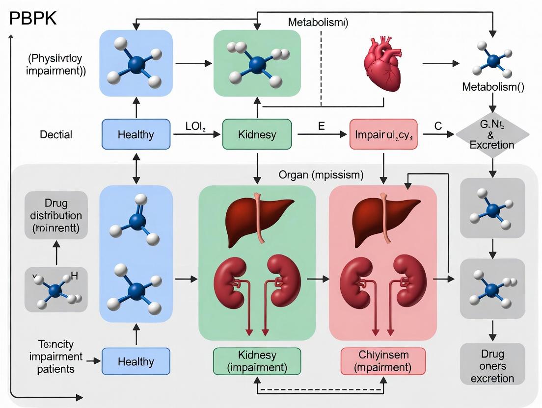
Model Development and Application Workflow
PBPK Workflow for Organ Impairment
Key Signaling Pathways in Drug Disposition
PBPK models integrate knowledge of pathways governing drug ADME. The liver is a key site for metabolism.
Hepatic Drug Uptake, Metabolism, and Efflux
Within the broader thesis on applying Physiologically-Based Pharmacokinetic (PBPK) modeling to optimize clinical trials for patients with organ impairment, regulatory guidance is a primary catalyst. Both the U.S. Food and Drug Administration (FDA) and the European Medicines Agency (EMA) have issued explicit guidelines encouraging the submission of PBPK analyses to support drug development and regulatory reviews, particularly for special populations like those with hepatic or renal impairment.
Table 1: Current FDA and EMA Guidelines on PBPK Submissions
| Agency | Guideline Title | Key PBPK Encouragement & Focus Areas | Reference Code | Year |
|---|---|---|---|---|
| FDA | Physiologically Based Pharmacokinetic Analyses — Format and Content | Explicit guidance on the format and content for submitting PBPK reports to support INDs, NDAs, ANDAs, and BLAs. Encourages use for drug-drug interaction (DDI), pediatrics, organ impairment. | Guidance for Industry | 2023 (Revised) |
| EMA | Guideline on the qualification and reporting of physiologically based pharmacokinetic (PBPK) modelling and simulation | Detailed framework for PBPK model qualification and reporting. Encourages use in DDI, pediatrics, organ impairment, and biopharmaceutics. | CHMP/458101/2016 | 2021 (Updated) |
| FDA | Pharmacokinetics in Patients with Impaired Renal Function—Study Design, Data Analysis, and Impact on Dosing and Labeling | Recommends PBPK as an alternative or supplement to dedicated clinical studies in renal impairment. | Guidance for Industry | 2020 |
| EMA | Guideline on the evaluation of the pharmacokinetics of medicinal products in patients with decreased renal function | Suggests PBPK modeling can be used to inform on dosing adjustments in renal impairment. | EMA/CHMP/83874/2014 | 2016 |
| FDA & EMA | Questions and Answers on modeling and simulation in pharmacokinetics (EMA); Various PBPK-focused webpages (FDA) | Provide practical Q&A on application, verification, and submission requirements for PBPK models. | - | Continuously Updated |
Quantitative Data from Recent Submissions (2018-2023): Table 2: PBPK Submission Trends to FDA (Adapted from Public Data)
| Application Area | Percentage of Submissions Containing PBPK (Approx.) | Primary Regulatory Purpose |
|---|---|---|
| Drug-Drug Interactions (DDI) | ~70% | To support waiver of dedicated clinical DDI studies or inform labeling. |
| Pediatric Extrapolation | ~50% | To inform first-in-pediatric doses and study design. |
| Hepatic/Renal Impairment | ~35% and increasing | To support dose recommendations, often to waive or supplement clinical studies. |
| Formulation/Biowaiver | ~25% | To support bioequivalence assessments. |
Application Notes: PBPK for Organ Impairment within Regulatory Framework
Application Note 1: Regulatory Strategy for Hepatic Impairment Studies
- Objective: To justify waiver of a dedicated clinical hepatic impairment study per FDA/EMA guidance.
- PBPK Approach: Develop and qualify a full PBPK model using data from healthy volunteers and in vitro systems. Extrapolate to virtual populations representing varying degrees of hepatic impairment (Child-Pugh A, B, C) by modifying system parameters (e.g., hepatic blood flow, enzyme/transporter activity, plasma protein levels).
- Regulatory Deliverable: A comprehensive report demonstrating model qualification and simulation outcomes (e.g., AUC, Cmax ratios vs. healthy). The submission must clearly show that the predicted exposure changes are minimal (<2-fold) or confidently predictable, thereby supporting a waiver and specific label language.
Application Note 2: Optimizing Renal Impairment Trial Design
- Objective: To optimize the design of a required renal impairment study (e.g., select most informative severity groups).
- PBPK Approach: Develop a PBPK model incorporating renal clearance mechanisms. Simulate pharmacokinetics (PK) across a continuum of renal function (eGFR from 90 to <15 mL/min). Identify the "breakpoint" of eGFR where PK changes become clinically significant.
- Regulatory Deliverable: Simulations submitted in the clinical trial protocol to justify the selected patient cohorts (e.g., severe and mild impairment, omitting moderate) and proposed dosing, making the trial more efficient and ethical.
Detailed Experimental Protocols
Protocol: PBPK Model Development for a Renally Eliminated Drug
Title: In Vitro to In Vivo Extrapolation (IVIVE) and PBPK Model Building for Renal Impairment Predictions.
I. Objectives:
- To develop a mechanistic PBPK model for Drug X, primarily cleared renally.
- To qualify the model against clinical PK data in healthy volunteers.
- To simulate exposure in virtual patients with varying degrees of renal impairment.
II. Materials & Software (The Scientist's Toolkit): Table 3: Essential Research Reagents & Solutions for PBPK Modeling
| Item/Category | Function in Protocol | Example/Notes |
|---|---|---|
| In Vitro Assay Systems | Determine fundamental drug parameters. | Human hepatocytes (metabolism); Transfected cell lines (transporter kinetics); Human plasma (protein binding). |
| Specific Chemical Inhibitors | Characterize enzymatic/transporter pathways. | Ketoconazole (CYP3A4); Rifampin (OATP1B1); Cimetidine (MATEs). |
| Reference Compounds | Validate assay performance. | Metoprolol (CYP2D6 probe); Digoxin (P-gp probe). |
| PBPK Software Platform | Integrate data, build model, run simulations. | Commercial (e.g., GastroPlus, Simcyp Simulator, PK-Sim) or open-source. |
| Clinical PK Datasets | For model qualification and verification. | Historical or proprietary data from Phase I studies in healthy subjects. |
| Physiological Database | Provide system parameters for virtual populations. | Built into software (e.g., age, weight, organ volumes, blood flows, enzyme abundances). |
| Virtual Population Libraries | Generate representative cohorts for simulation. | Simcyp's "Renal Impairment" population; FDA's "Virtual Population". |
III. Methodology: Step 1: Data Collation (Input Parameterization)
- Collect in vitro data: LogP, pKa, solubility, permeability, fraction unbound in plasma (fu), blood-to-plasma ratio.
- Determine renal clearance mechanism: Conduct in vitro transporter assays (e.g., using HEK293 cells expressing OAT1, OAT3, OCT2, MATE1/2K) to obtain Km and Vmax.
- If metabolism is involved, obtain intrinsic clearance (CLint) from human liver microsome or hepatocyte assays.
Step 2: IVIVE and Base Model Building
- Use PBPK software to scale in vitro clearance data to in vivo organ clearance (IVIVE).
- Develop a minimal PBPK model (e.g., whole-body) incorporating absorption, distribution, and clearance pathways.
- Enter all collected physicochemical and in vitro parameters.
Step 3: Model Qualification (Healthy Volunteers)
- Simulate single and multiple-dose trials in a virtual healthy population (n=100, 10 trials) matching the demographics of the clinical studies.
- Compare simulated plasma concentration-time profiles and key PK parameters (AUC, Cmax, t1/2) to observed clinical data.
- Apply diagnostic criteria (e.g., visual predictive checks, fold-error of <1.5 - 2 for AUC/Cmax) to accept the model.
Step 4: Extrapolation to Renal Impairment
- Within the software, select or create a "Renal Impairment" virtual population. This modifies system parameters: reduced glomerular filtration rate (GFR), altered renal blood flow, potential changes in fu (due to albuminuria), and possible changes in non-renal clearance (per guidance).
- Run simulations in virtual populations stratified by eGFR (e.g., >60, 30-59, 15-29, <15 mL/min/1.73m²).
- Output predicted exposure (AUC) ratios (impaired/healthy) for each group.
Step 5: Sensitivity Analysis
- Perform sensitivity analysis on key uncertain parameters (e.g., fractional contribution of a specific transporter to renal secretion, fu) to assess their impact on the final exposure predictions in impaired populations.
Protocol: Executing a Virtual Hepatic Impairment Study
Title: PBPK Simulation to Support a Waiver for a Clinical Hepatic Impairment Study.
I. Objectives:
- To predict the pharmacokinetics of Drug Y (metabolized by CYP3A4) in patients with hepatic impairment.
- To assess if dedicated clinical study can be waived per regulatory criteria.
II. Methodology: Step 1: Robust Model Qualification
- Qualify a PBPK model for Drug Y using data from healthy volunteers and potentially from drug-drug interaction studies with CYP3A4 inhibitors/inducers. This is critical to establish confidence in the model's metabolic component.
Step 2: Virtual Population Generation
- Utilize built-in virtual populations (e.g., Simcyp's Cirrhosis Population). These populations incorporate disease-specific changes: reduced hepatic CYP enzyme abundance and activity (Child-Pugh specific), reduced hepatic blood flow, increased shunting, and altered plasma protein levels.
Step 3: Simulation & Output Analysis
- Simulate the proposed clinical dosing regimen in virtual healthy, Child-Pugh A, B, and C populations (n=100 per group, 10 trials).
- Generate primary outputs: AUC ratio (impaired/healthy) and Cmax ratio.
Step 4: Waiver Justification Assessment
- Apply regulatory logic: If the predicted increase in systemic exposure (AUC) is less than 2-fold in all impairment categories, and the model is well-qualified, a strong case for a waiver exists.
- If predictions show >2-fold increase, the PBPK results can still inform the design of a necessary clinical study (e.g., focus on the most severe impairment group).
Visualizations
Regulatory Decision Flow for Organ Impairment
PBPK Workflow for Regulatory Submission
Application Notes: Integrating PBPK in Organ Impairment (OI) Studies
Physiologically-based pharmacokinetic (PBPK) modeling is a mechanistic computational framework that integrates physiological, physicochemical, and biochemical parameters to predict drug pharmacokinetics (PK). Within the thesis context of optimizing clinical trials for organ impairment patients, PBPK serves as a pivotal tool for ethical and efficient drug development.
Reducing Clinical Burden: Traditional dedicated hepatic or renal impairment studies require recruitment of vulnerable patients, posing ethical challenges and often delaying development. PBPK modeling, when adequately verified, can simulate PK in these populations, potentially reducing or replacing the need for some clinical studies. Regulatory agencies like the FDA and EMA now accept PBPK to support dose recommendations for OI patients, thereby minimizing their direct participation in trials.
Informing Trial Design: For trials where OI patients must be enrolled, PBPK models inform optimal trial design. They can predict the degree of PK alteration, helping to determine necessary sample sizes, appropriate dosing regimens, and optimal blood sampling schedules. This leads to more robust, "right-sized" trials with a higher probability of conclusive outcomes.
Extrapolation: A verified PBPK model allows for extrapolation beyond studied conditions. It can predict PK in untested severities of organ impairment (e.g., Child-Pugh C from B), in multi-organ dysfunction, or when impairment is complicated by drug-drug interactions. This extrapolation capability is central to the thesis of broadly applying PBPK to support label claims across the impairment spectrum.
| Drug/Therapeutic Area | Organ Impairment | PBPK Application | Regulatory Outcome | Reference (Public Source) |
|---|---|---|---|---|
| Novel Oral Anticoagulant | Hepatic (Child-Pugh A-C) | Replace dedicated hepatic study; dose recommendation | Accepted by EMA (CHMP) | EMA Assessment Report (2023) |
| Oncology (Small Molecule) | Renal (eGFR 15-89 mL/min) | Inform dosing in Phase Ib trial; support label | FDA Clinical Pharmacology Review (2022) | FDA Drugs@FDA |
| Metabolic Disease Drug | Hepatic & Renal (Mild/Moderate) | Simulate PK for dual impairment; trial waiver | Internal company white paper, cited in FDA guidance update (2024) | FDA PBPK Guidance Update |
| Antibiotic | Renal (Severe Impairment) | Optimize sparse sampling design for confirmatory study | Protocol agreed via EMA Qualification Advice | EMA Qualification of Novel Methodologies (2023) |
Experimental Protocols for PBPK Model Development & Verification
Protocol 2.1:In VitrotoIn Vivo(IVIVE) Parameterization for a Hepatically Cleared Drug
Objective: To develop a drug-specific PBPK model by parameterizing hepatic clearance via IVIVE. Materials: See "Scientist's Toolkit" (Section 4). Methodology:
- Determine Fraction Unbound in Plasma ((fu)): Using equilibrium dialysis against human plasma (n=3 donors). Incubate drug at therapeutic concentration for 6 hours at 37°C. Calculate (fu).
- Determine Microsomal Intrinsic Clearance ((CL{int,mic})): Incubate drug (1 µM) with human liver microsomes (0.5 mg/mL) and NADPH. Withdraw aliquots over 60 minutes. Determine substrate depletion half-life. Scale (CL{int,mic}) to whole liver using scaling factors (45 mg microsomal protein/g liver, 21.4 g liver/kg body weight).
- Account for Non-Microsomal Metabolism: If relevant, conduct similar assays with hepatocytes or cytosolic fractions.
- Incorporate into PBPK Software: Input (fu), scaled (CL{int}), and physicochemical properties (logP, pKa, molecular weight) into a platform (e.g., GastroPlus, Simcyp, PK-Sim).
- Verify with Clinical PK in Healthy Volunteers: Simulate a single-dose PK study in a virtual healthy population (n=10 trials, 100 subjects/trial). Optimize model within 2-fold of observed AUC and Cmax.
Protocol 2.2: Verification of a PBPK Model in a Renal Impairment Population
Objective: To verify the predictive performance of a PBPK model for a renally excreted drug across varying degrees of renal impairment. Methodology:
- Base Model Development: Establish and verify a drug model in healthy volunteers as per Protocol 2.1, including precise characterization of renal clearance (active secretion, glomerular filtration).
- Population Model Construction: Within the PBPK platform, select the "Renal Impairment" population module. This module typically scales down renal blood flow, glomerular filtration rate (eGFR), and function of transporters based on published physiological correlations.
- Define Virtual Cohorts: Create virtual cohorts matching the demographics and renal function (e.g., mild: eGFR 60-89, moderate: 30-59, severe: 15-29 mL/min) of a published clinical study.
- Simulation and Comparison: Simulate the PK profile (e.g., after a single dose) for each virtual cohort. Extract predicted AUC, Cmax, and other relevant PK parameters.
- Validation Criterion: Compare predictions to observed clinical data. A model is considered verified if the predicted/observed ratios for AUC and Cmax in each impairment group fall within the 0.8 – 1.25 range (or 2-fold for exploratory stages).
Visualizations
PBPK Model Development and Extrapolation Workflow
PBPK's Role in Trial Design Decision Logic
The Scientist's Toolkit: Key Research Reagent Solutions
| Item Name / Solution | Supplier Examples | Function in PBPK for OI |
|---|---|---|
| Human Liver Microsomes (HLM) | Corning, XenoTech, BioIVT | Contain CYP450 enzymes; used to measure metabolic intrinsic clearance (IVIVE). |
| Cryopreserved Human Hepatocytes | Lonza, BioIVT, CellzDirect | Intact cellular system to study hepatic metabolism, transport, and potential toxicity. |
| Human Kidney Microsomes & Cells | XenoTech, Sekisui | Study renal metabolic and transporter pathways (e.g., UGTs, OCT2, MATE). |
| Pooled Human Plasma (from various OI conditions) | BioIVT, Sera Labs | Determine plasma protein binding (fu) in disease states, a critical parameter for PK. |
| Recombinant Human CYP & Transporter Enzymes | Sigma-Aldrich, Thermo Fisher | Identify specific enzymes involved in drug clearance. |
| Equilibrium Dialysis Devices | HTDialysis, Thermo Fisher (Pierce) | Gold-standard method for measuring fraction unbound (fu) in plasma or tissue homogenates. |
| PBPK Modeling Software (Simcyp Simulator, GastroPlus, PK-Sim) | Certara, Simulations Plus, Bayer | Integrated platforms containing population databases and OI modules for model building and simulation. |
| Systems Biology Databases (PK-Sim Ontogeny Database, IIV Database) | Open Systems Pharmacology, PubChem, DrugBank | Provide physiological, ontogeny, and variability data for system parameters in models. |
Physiologically Based Pharmacokinetic (PBPK) modeling is a critical tool for predicting drug pharmacokinetics (PK) in special populations, particularly patients with hepatic or renal impairment. Its value lies in its ability to integrate physiological, physicochemical, and biochemical parameters to mechanistically simulate drug absorption, distribution, metabolism, and excretion (ADME). Within the context of organ impairment, PBPK modeling can optimize clinical trial design, inform dosing recommendations, and potentially replace dedicated clinical studies, thereby accelerating drug development and enhancing patient safety.
Decision Tree: When to Apply PBPK Modeling for Organ Impairment
A structured decision tree helps identify scenarios where PBPK modeling offers the highest value.
Decision tree for applying PBPK in organ impairment.
Key Use Cases and Application Notes
Use Case 1: Hepatic Impairment (HI) Study Waiver
Application Note: PBPK can support a request to regulatory authorities (e.g., FDA, EMA) to waive a dedicated HI clinical study. Success depends on demonstrating model credibility and accurate prediction of exposure changes.
Key Quantitative Data Summary: Table 1: Example PBPK Predictions vs. Observations for Drugs in HI
| Drug (Metabolism Pathway) | Child-Pugh Class | Predicted AUC Ratio (HI/Normal) | Observed AUC Ratio (HI/Normal) | Prediction Success | Regulatory Outcome |
|---|---|---|---|---|---|
| Drug A (CYP3A4 substrate) | Moderate (B) | 2.5 | 2.7 | Within 1.25-fold | Study Waiver Granted |
| Drug B (CYP2C8 substrate) | Severe (C) | 3.8 | 3.2 | Within 1.5-fold | Study Recommended |
| Drug C (UGT substrate) | Mild (A) | 1.3 | 1.2 | Within 1.25-fold | Study Waiver Granted |
Use Case 2: Dose Recommendation for Renal Impairment (RI)
Application Note: PBPK models incorporating glomerular filtration rate (GFR) and transporter changes can predict PK across all RI stages, enabling precise dosing guidance without extensive trials in each subpopulation.
Use Case 3: Predicting Complex Drug-Disease Interactions
Application Note: For drugs where organ impairment alters non-elimination pathways (e.g., plasma protein binding, tissue distribution, transit times), PBPK's holistic physiology is uniquely valuable.
Experimental Protocols for Model Building and Verification
Protocol 1: Developing a PBPK Model for Hepatic Impairment
Objective: To build and qualify a PBPK model for a CYP3A4-metabolized drug to predict PK in patients with varying degrees of hepatic impairment.
Detailed Methodology:
- System Characterization: Populate the simulator (e.g., GastroPlus, Simcyp, PK-Sim) with a "healthy" population physiology.
- Drug Parameterization:
- Obtain in vitro data: lipophilicity (Log P), pKa, blood-to-plasma ratio, fraction unbound (fu), and CYP3A4 intrinsic clearance (CLint) from human liver microsomes.
- Optimize system-dependent parameters (e.g., absorption rate constant) by fitting the model to observed PK data from healthy volunteer studies.
- HI Physiology Incorporation: Modify the virtual population parameters for HI as per published guidance:
- Reduce hepatic CYP enzyme abundance (e.g., 30% for CP-B, 50% for CP-C).
- Reduce hepatic blood flow.
- Adjust plasma protein levels (e.g., albumin).
- Model Qualification: Predict PK in HI (CP-B) and compare to any available clinical data using fold-error analysis. Qualify if predictions are within 2-fold of observations.
- Simulation & Extrapolation: Simulate PK in severe HI (CP-C) and propose dosing recommendations.
Protocol 2: Validating a Renal Impairment PBPK Model Using Renal Transporter Data
Objective: To validate a PBPK model for a renally secreted drug (substrate of OAT1/3) in RI populations.
Detailed Methodology:
- Base Model Development: Develop a model incorporating active secretion via OAT1/3, parameterized using in vitro transporter data (Vmax, Km) from transfected cell lines.
- RI Physiology Implementation: Scale renal transporter activity in correlation with measured GFR or CKD stage, based on published in vitro-in vivo extrapolation (IVIVE) relationships.
- Prospective Validation: Use the model to predict PK data from a published clinical RI study not used in model development.
- Sensitivity Analysis: Perform sensitivity analysis on key parameters (GFR, transporter activity, fu) to identify the dominant drivers of exposure change in RI.
The Scientist's Toolkit: Key Research Reagent Solutions
Table 2: Essential Materials for PBPK-Focused Organ Impairment Research
| Item/Category | Example Specifics | Function in PBPK Workflow |
|---|---|---|
| In Vitro ADME Assay Kits | Hepatocyte stability kits (e.g., from BioIVT, Corning), Caco-2 permeability kits. | Generates crucial input parameters (CLint, permeability) for the model. |
| Transfected Cell Systems | HEK293 cells overexpressing human OAT1, OATP1B1, OCT2, etc. | Quantifies transporter-mediated uptake kinetics for renal/hepatic drugs. |
| Human Biomatrices | Plasma from healthy and organ-impaired donors, human liver microsomes (HLM) from HI donors. | Measures disease-specific binding (fu) and enzyme activity for model refinement. |
| PBPK Software Platform | Simcyp Simulator, GastroPlus, PK-Sim/Open Systems Pharmacology. | Provides the physiological framework and algorithms to integrate data and run simulations. |
| Clinical PK Datasets | Public (e.g., NIH ClinicalTrials.gov) or internal data from healthy volunteer and organ impairment studies. | Used for model calibration, verification, and assessing predictive performance. |
Visualization of PBPK Modeling Workflow for Organ Impairment
PBPK workflow for organ impairment studies.
Building and Applying PBPK Models for Hepatic and Renal Impairment: A Step-by-Step Guide
Application Notes
The successful development and application of Physiologically-Based Pharmacokinetic (PBPK) models for predicting drug exposure in organ impairment (OI) patients rely on the rigorous integration of three distinct data domains. These models are pivotal within clinical trial research for dose adjustment justification and informing regulatory submissions. The following notes detail the critical parameters within each domain.
System-Specific Parameters
These are the physiological parameters of the virtual population. For OI populations, these must be carefully altered to reflect the pathophysiology of the impaired organ(s).
- Hepatic Impairment: Changes include reduced hepatic blood flow, functional liver volume, and levels of cytochrome P450 (CYP) enzymes and transporters. Child-Pugh or NCI-ODWG classifications are used to stratify severity.
- Renal Impairment: Changes include reduced glomerular filtration rate (GFR), renal blood flow, and expression of renal transporters (e.g., OAT, OCT). CKD-EPI or MDRD equations estimate GFR for stratification.
Drug-Specific Parameters
These are the compound-specific physicochemical and pharmacokinetic properties.
- Fundamental Properties: Molecular weight, logP, pKa, blood-to-plasma ratio, and fraction unbound in plasma (fu).
- Disposition Parameters: Permeability, tissue-plasma partition coefficients (Kp), and elimination pathways (e.g., fraction metabolized by specific CYP enzymes, fraction excreted unchanged in urine).
- In Vitro Parameters: Michaelis-Menten constants (Vmax, Km) for enzymes, transporter kinetics (Jmax, Kt), and inhibition/induction constants (Ki, EC50).
Trial-Specific Parameters
These define the clinical scenario being simulated.
- Dosing Regimen: Route, dose, frequency, and duration.
- Population Characteristics: Demographic distributions (age, weight, BMI, sex), genotype prevalences for polymorphic enzymes, and the proportion of subjects in each OI severity class.
- Co-medications: For assessing drug-drug interactions (DDIs) in a polypharmacy-prone OI population.
Table 1: Core Data Requirements for OI PBPK Modeling
| Domain | Parameter Category | Example Parameters | OI-Specific Considerations |
|---|---|---|---|
| System | Physiology | Organ volumes, blood flows, hematocrit, GFR | Modify based on OI severity (e.g., -40% liver volume in Child-Pugh C). |
| System | Enzyme/Transporter Abundance | CYP3A4, UGT1A1, OATP1B1, P-gp levels | Quantify reduction in impaired organ (e.g., ~50% OATP1B1 in cirrhosis). |
| Drug | Physicochemical | logP, pKa, B/P ratio, fu | fu may change in OI due to altered plasma protein levels (e.g., albumin). |
| Drug | Metabolism/Transport | fmCYP2C9, CLint, Kp, Kt, Jmax | Key target for system parameter modulation. Verify in vitro assay conditions. |
| Drug | Inhibition/Induction | Ki, IC50, EC50, kinact | Critical for DDI risk assessment in polypharmacy OI trials. |
| Trial | Design | Dose, route, formulation, sampling schedule | Reflect planned or historic trial protocol. |
| Trial | Population | Age range, BMI, OI stratification criteria | Define virtual cohort matching eligibility criteria. |
| Trial | Co-medications | Drug, dose, timing | Common medications in OI population (e.g., diuretics, analgesics). |
Experimental Protocols
Protocol 1: Determination of Fraction Unbound (fu) in Plasma from Hepatically Impaired Patients
Objective: To measure the fraction unbound of a drug in plasma from healthy volunteers and patients with varying degrees of hepatic impairment, accounting for potential changes in protein concentrations.
Materials:
- Test compound (stable label recommended)
- Pooled human plasma (healthy control)
- Pooled plasma from patients classified as Child-Pugh A, B, and C
- Rapid Equilibrium Dialysis (RED) device with inserts (e.g., 8 kDa MWCO)
- PBS (pH 7.4)
- LC-MS/MS system
Procedure:
- Prepare a stock solution of the test compound in DMSO and subsequently spike into each plasma type (healthy, CP-A, CP-B, CP-C) to a final therapeutic concentration (e.g., 1 µM). Keep DMSO concentration ≤0.5%.
- Load 200 µL of spiked plasma into the donor chamber of the RED insert.
- Load 350 µL of PBS (pH 7.4) into the receiver chamber.
- Assemble the plate and incubate at 37°C with gentle agitation (e.g., 300 rpm) for 6 hours (pre-validate time to equilibrium).
- Post-incubation, aliquot equal volumes (e.g., 50 µL) from donor (plasma) and receiver (buffer) chambers.
- Quench samples with an equal volume of acetonitrile containing an internal standard. Vortex and centrifuge.
- Analyze supernatant via validated LC-MS/MS to determine compound concentrations in donor [D] and receiver [R] chambers.
- Calculate fu:
fu = [R] / [D]. Correct for volume differences if necessary.
Data Integration: The disease-specific fu values are used as direct inputs for the PBPK model to adjust plasma protein binding.
Protocol 2: In Vitro Assessment of Hepatocyte Clearance in Disease-Mimicking Conditions
Objective: To obtain intrinsic clearance (CLint) data in hepatocytes under conditions mimicking the uremic milieu of renal impairment.
Materials:
- Cryopreserved human hepatocytes (pooled)
- Uremic human serum (pooled from CKD patients) or synthetic uremic toxin cocktail (e.g., containing indoxyl sulfate, p-cresol sulfate at pathological concentrations)
- Williams' Medium E
- Test compound
- Collagen-coated incubation plates
- LC-MS/MS system
Procedure:
- Preparation of Media: Prepare two incubation media: (A) Standard Williams' Medium E with 2% healthy human serum. (B) Disease-mimicking media: Williams' Medium E with 2% uremic human serum or supplemented with a defined mixture of uremic toxins at clinically relevant concentrations.
- Hepatocyte Thawing & Plating: Thaw cryopreserved hepatocytes and suspend in both media types. Determine viability (trypan blue exclusion). Plate cells in collagen-coated 24-well plates at a density of 0.5 x 10^6 viable cells/well in their respective media.
- Dosing & Incubation: Pre-incubate plates for 30 min at 37°C, 5% CO2. Add test compound (final concentration << Km, typically 1 µM) to triplicate wells for each media condition. Include control wells (no cells) for each media to account for non-specific loss.
- Sampling: At predetermined time points (e.g., 0, 15, 30, 60, 90, 120 min), remove 50 µL of supernatant from each well and transfer to a stop solution (acetonitrile with IS) on a pre-chilled plate.
- Analysis: Centrifuge stopped samples and analyze supernatant via LC-MS/MS to determine parent compound depletion over time.
- Calculation: Plot natural log of remaining compound concentration versus time. The slope (k) from the linear phase is used to calculate in vitro CLint,hep:
CLint,hep = k * (Volume of incubation / Number of viable cells). Compare CLint between standard and disease-mimicking media.
Data Integration: The relative change in CLint informs the scaling factor for metabolic/transport processes in the renal impairment PBPK model.
Visualizations
PBPK Model Data Integration Flow
OI PBPK Model Development Workflow
The Scientist's Toolkit
Table 2: Key Research Reagent Solutions for OI PBPK Data Generation
| Item | Function in OI Context |
|---|---|
| Disease-State Human Biomatrices (e.g., Hepatically or Renally Impaired Plasma/Serum) | Essential for measuring disease-altered binding (fu) and creating disease-mimicking in vitro incubation media. |
| Cryopreserved Hepatocytes from Organ-Impaired Donors (if available) | The gold standard for directly assessing metabolic capacity in disease state. Often scarce; disease-mimicking media are a practical alternative. |
| Synthetic Uremic Toxin Cocktail (Indoxyl sulfate, p-cresol sulfate, etc.) | Allows controlled in vitro simulation of the renal impairment milieu to study its impact on hepatic/renal transporters and enzymes. |
| Rapid Equilibrium Dialysis (RED) Device | High-throughput method for reliable determination of fraction unbound (fu) in plasma, critical for accurate PK prediction. |
| LC-MS/MS System with High Sensitivity | Required for quantifying low drug concentrations in complex biomatrices, especially from low-volume in vitro assays and clinical microsampling. |
| PBPK Software Platform (e.g., GastroPlus, Simcyp, PK-Sim) | Integrates system, drug, and trial data to build, validate, and simulate virtual populations and trials. |
| Curated Physiological Databases (e.g., IMI REDI, PK-Sim Ontology) | Provide quantitative, peer-reviewed system parameters for healthy and disease populations, ensuring model reliability. |
Within the broader thesis on refining PBPK modeling for special populations in clinical trials, hepatic impairment (HI) presents a critical challenge. Accurate prediction of drug exposure in HI patients is essential for dose adjustment and regulatory approval. This application note details a systematic PBPK framework for HI, integrating quantitative changes in key hepatic physiological parameters: metabolic enzyme activity, hepatic blood flow, and plasma protein binding. The protocols herein are designed for researchers to develop, qualify, and apply HI PBPK models to inform trial design and labeling.
The severity of hepatic impairment, commonly classified by Child-Pugh (CP) score, systematically alters drug disposition parameters. The following table summarizes literature-derived quantitative changes.
Table 1: Quantitative Changes in Key Hepatic Parameters by Child-Pugh Class
| Parameter | Child-Pugh A (Mild) | Child-Pugh B (Moderate) | Child-Pugh C (Severe) | Data Source & Notes |
|---|---|---|---|---|
| Hepatic Blood Flow | ~80-90% of normal | ~70-80% of normal | ~60-70% of normal | Based on indocyanine green clearance studies. |
| Cytochrome P450 (CYP) Activity | Highly variable; general trends shown. | |||
| - CYP1A2 | ↓ 30% | ↓ 50% | ↓ 70% | Correlation with prothrombin time. |
| - CYP2C9 | ↓ 20% | ↓ 40% | ↓ 60% | |
| - CYP2C19 | ↓ 20% | ↓ 40% | ↓ 60% | |
| - CYP2D6 | ↓ 10% | ↓ 30% | ↓ 50% | Preserved longer than other CYPs. |
| - CYP3A4 | ↓ 30% | ↓ 50% | ↓ 70% | Correlates with erythromycin breath test. |
| UDP-Glucuronosyltransferase (UGT) Activity | ↓ 0-20% | ↓ 20-50% | ↓ 50-70% | Substrate-dependent (e.g., bilirubin, AZT). |
| Albumin Concentration | 3.5-4.0 g/dL | 2.8-3.5 g/dL | <2.8 g/dL | Direct measure from CP score. |
| α1-Acid Glycoprotein (AAG) | Variable (↑ or ↓) | Variable (↑ or ↓) | Variable (↑ or ↓) | Inflammatory responses can increase AAG. |
| Liver Volume | ~90% of normal | ~80% of normal | ~70% of normal | Imaging-based assessments. |
| Hepatocyte Mass / Function | ~70% of normal | ~50% of normal | ~30% of normal | Functional estimate based on galactose elimination. |
Experimental Protocols for Parameter Estimation
Protocol 3.1: In Vitro Determination of Fraction Unbound in HI Plasma (fu,p) Objective: To measure the drug-specific fraction unbound in plasma from HI patients for incorporation into PBPK models. Materials: See Scientist's Toolkit. Method:
- Plasma Pool Preparation: Obtain human plasma from healthy volunteers and HI patients (CP A, B, C). Pool samples within each group (n≥10 donors) to minimize inter-individual variability.
- Equilibrium Dialysis: a. Assemble Teflon equilibrium dialysis cells separated by a semi-permeable membrane (MW cutoff > 10x drug molecular weight). b. Add 1 mL of patient plasma (spiked with drug at therapeutic concentration) to the donor chamber and 1 mL of isotonic phosphate buffer (pH 7.4) to the receiver chamber. c. Incubate at 37°C with gentle agitation for 4-8 hours (validate time to reach equilibrium).
- Sample Analysis: a. Post-incubation, collect aliquots from both chambers. b. Quantify drug concentrations in plasma ([C]plasma) and buffer ([C]buffer) using a validated LC-MS/MS method. c. Account for volume shift due to osmotic pressure using published corrections.
- Calculation: fu,p = [C]buffer, corrected / [C]plasma, corrected. Report mean ± SD for each CP class.
Protocol 3.2: Retrograde Drug-Drug Interaction (DDI) Study to Scale CYP Activity Objective: To estimate in vivo CYP activity in HI populations using a clinical DDI study design. Method:
- Study Design: A fixed-sequence, open-label study in HI patients (stratified by CP class) and matched healthy volunteers.
- Probe Dosing: a. Day 1 (Baseline): Administer a selective CYP probe cocktail (e.g., caffeine [CYP1A2], warfarin [CYP2C9], omeprazole [CYP2C19], dextromethorphan [CYP2D6], midazolam [CYP3A4]) at microdoses. b. Collect serial blood samples over 24-48 hours for each probe.
- Pharmacokinetic Analysis: Perform non-compartmental analysis (NCA) for each probe to determine clearance (CL).
- Activity Scaling: For each CYP, calculate the relative activity in HI: RACYP,HI = CLprobe, HI / CLprobe, Healthy. Incorporate RA as a scalar on intrinsic clearance (CLint) in the PBPK model.
PBPK Model Building and Verification Workflow
Diagram 1: HI PBPK Model Development and Qualification Workflow (98 chars)
Signaling Pathways: Impact of Liver Disease on Drug Disposition
Diagram 2: Pathophysiological Drivers of Altered PK in HI (99 chars)
The Scientist's Toolkit: Key Research Reagent Solutions
Table 2: Essential Materials for HI PBPK Research
| Item | Function/Application | Example/Supplier Note |
|---|---|---|
| Matched HI & Healthy Human Plasma | For in vitro protein binding (fu) studies. | Commercially available from biorepositories (e.g., BioIVT, Seralab). Must be characterized by CP score. |
| Human Hepatocytes (from HI donors) | For assessing metabolic activity (CLint) in vitro. | Limited availability. Consider cryopreserved pools from vendors like Lonza or Corning. |
| CYP-Specific Probe Substrates & Inhibitors | For in vitro enzyme phenotyping and in vivo DDI studies. | Use selective probes (e.g., Bupropion [CYP2B6], Phenacetin [CYP1A2]). Available from Sigma-Aldrich, Tocris. |
| PBPK Modeling Software | Platform for building, simulating, and populating models. | Commercial (GastroPlus, Simcyp, PK-Sim) or open-source (R/mrgsolve, Pumas). |
| Clinical PK Data in HI Populations | For model verification and qualification. | Extracted from literature, regulatory filings (e.g., FDA EDR), or internal trials. |
| Equilibrium Dialysis Device | Gold-standard for measuring plasma protein binding. | HTD96b dialysis cells (HTDialysis) or RED devices (Thermo Fisher). |
| LC-MS/MS System | For sensitive and specific quantification of drugs and metabolites in biological matrices. | Essential for generating in vitro and in vivo PK data. |
Application Notes for PBPK Model Development
Within the broader thesis of developing robust PBPK models for organ impairment populations, renal dysfunction presents a critical challenge. Accurate prediction of pharmacokinetics in renal impairment (RI) requires mechanistic integration of altered glomerular filtration, tubular secretion/reabsorption, and fluid balance dynamics. These models are essential for optimizing trial design and dose adjustment strategies without exposing vulnerable patients to unnecessary risk.
Core Quantitative Parameters for Renal Impairment Stratification
The table below summarizes key quantitative parameters that must be adjusted in a RI-PBPK model, stratified by Kidney Disease: Improving Global Outcomes (KDIGO) stages.
Table 1: Key Physiological Adjustments by CKD Stage for PBPK Modeling
| Parameter | CKD Stage G1 (Normal, ≥90) | CKD Stage G2 (Mild, 60-89) | CKD Stage G3a (Mild-Mod, 45-59) | CKD Stage G3b (Mod-Severe, 30-44) | CKD Stage G4 (Severe, 15-29) | CKD Stage G5 (Kidney Failure, <15) | Source/Justification |
|---|---|---|---|---|---|---|---|
| Measured GFR (mL/min/1.73m²) | ≥90 | 60-89 | 45-59 | 30-44 | 15-29 | <15 | KDIGO 2012 Classification |
| Renal Plasma Flow (RPF) Adjustment Factor | 1.00 | 0.85-0.95 | 0.70-0.80 | 0.55-0.65 | 0.40-0.50 | 0.20-0.30 | Deduced from nephron loss & vascular changes. |
| Hematocrit Adjustment | Baseline | Baseline to -5% | -5% to -10% | -10% to -15% | -15% to -25% | -25% to -35% | Correlates with declining erythropoietin production. |
| Albumin Concentration (g/dL) | 4.0-4.5 | 3.8-4.3 | 3.5-4.0 | 3.2-3.7 | 2.9-3.4 | 2.5-3.2 | Increased capillary permeability & inflammation. |
| Fractional Fluid Volume Increase | 0% | 0-2% | 2-5% | 5-8% | 8-12% | 12-20% | Due to impaired sodium/water excretion. |
| Tubular Secretory Capacity (Relative) | 1.00 | 0.80 | 0.65 | 0.50 | 0.35 | 0.20 | Non-linear decline steeper than GFR for many transporters. |
Integrating Secretory Pathways: OATs, OCTs, and MATEs
Active secretion primarily occurs via transporters in the proximal tubule. Their activity does not decline linearly with GFR and must be modeled independently.
Table 2: Key Renal Transporters and Impact of Uremic Toxins
| Transporter | Gene | Location | Substrate Examples | Impact in RI (Activity/Expression) | Notable Inhibitory Uremic Toxins |
|---|---|---|---|---|---|
| OAT1 | SLC22A6 | Basolateral | Methotrexate, β-lactams, antivirals | Downregulated (≤50% in severe RI) | p-Cresol sulfate, Indoxyl sulfate |
| OAT3 | SLC22A8 | Basolateral | Furosemide, Cimetidine | Downregulated | 3-Carboxy-4-methyl-5-propyl-2-furanpropanoate (CMPF) |
| OCT2 | SLC22A2 | Basolateral | Metformin, Cisplatin | Variably downregulated; competitive inhibition key | Dimethylarginines, Guanidinosuccinate |
| MATE1 | SLC47A1 | Apical (Canalicular) | Metformin, Cimetidine | Potentially downregulated; critical for efflux | Elevated intracellular pH may affect function. |
| MATE2-K | SLC47A2 | Apical (Brush Border) | Metformin | Data limited; assumed parallel decline with GFR. | -- |
| P-gp | ABCB1 | Apical | Digoxin, Tacrolimus | Conflicting data; may be induced or unchanged. | -- |
Diagram Title: Renal Drug Handling: Filtration, Secretion & Uremic Inhibition
Experimental Protocols
Protocol 1:In VitroTransporter Inhibition Assay Using Uremic Serum
Objective: To quantify the inhibitory potential of serum from RI patients on key renal transporters (OAT1, OAT3, OCT2) for parameterizing PBPK models.
Materials:
- HEK293 cells stably expressing human OAT1, OAT3, OCT2, and Mock-transfected controls.
- Serum pools from healthy volunteers and patients stratified by CKD stage (G3a, G4, G5).
- Radio-labeled or fluorescent probe substrates (e.g., [³H]p-aminohippurate for OATs, [¹⁴C]Metformin for OCT2).
- Uptake buffer (HBSS with HEPES, pH 7.4).
Procedure:
- Serum Preparation: Thaw serum pools on ice. Dialyze (10 kDa MWCO) against uptake buffer to remove endogenous small molecule substrates. Filter sterilize (0.22 µm).
- Cell Preparation: Seed cells in 24-well poly-D-lysine coated plates 48h prior to assay at 200,000 cells/well. Ensure confluency >90%.
- Uptake Assay: a. Pre-incubate cells with serum-supplemented buffer (10% v/v dialyzed serum from each pool) or control buffer for 15 min at 37°C. b. Aspirate and add uptake buffer containing probe substrate (typical Km concentration) and the same 10% serum condition. Incubate for a predetermined linear time (e.g., 2-5 min). c. Terminate uptake by rapid washing with ice-cold buffer. d. Lyse cells with 0.1% Triton X-100. Analyze lysate via liquid scintillation counting or fluorescence.
- Data Analysis: Subtract non-specific uptake (Mock cells). Calculate uptake velocity (pmol/min/mg protein) for each serum condition. Express as % of activity in healthy serum control.
The Scientist's Toolkit: Key Research Reagent Solutions
| Item | Function in Protocol |
|---|---|
| Stable Transporter-Expressing Cell Lines (e.g., HEK293-OAT1) | Provides a consistent, high-expression system for isolating specific human transporter activity. |
| CKD Patient Serum Pools (Stratified) | Source of pathophysiological uremic toxins to test clinical, non-specific inhibition. |
| 10 kDa MWCO Dialysis Cassette | Removes endogenous transporter substrates from serum while retaining larger protein-bound and proteinaceous inhibitors. |
| Radio-labeled Probe Substrates (e.g., [³H]PAH) | Allows sensitive, specific, and quantitative measurement of low-level transporter-mediated uptake. |
| Poly-D-Lysine Coated Plates | Enhances cell adhesion, especially for transporter assays requiring vigorous washing steps. |
Protocol 2: Determining Fraction Reabsorbed via Urinary Recovery in Preclinical RI Models
Objective: To empirically determine the fraction of filtered drug reabsorbed in the tubule in a controlled rat model of renal impairment for model verification.
Materials:
- Male Sprague-Dawley rats with Adenine-induced chronic kidney disease (CKD) and sham controls.
- Test compound solution for IV bolus.
- Metabolism cages for precise urine collection.
- Surgical materials for cannulation (jugular vein, urinary bladder).
- LC-MS/MS system for bioanalysis.
Procedure:
- Model Induction & Validation: Administer 0.75% w/w adenine mixed in powdered feed to rats for 4 weeks to induce tubulointerstitial fibrosis and CKD. Validate via plasma creatinine and BUN.
- Surgical Preparation: Implant jugular vein and urinary bladder catheters under anesthesia 24h prior to experiment. Connect bladder catheter to a refrigerated fraction collector.
- Dosing and Sampling: a. Administer IV bolus of test compound. b. Collect serial blood samples via jugular catheter over the compound's elimination phase. c. Collect total urine continuously in timed intervals (e.g., 0-2, 2-6, 6-24h) post-dose, keeping samples on ice or at 4°C.
- Bioanalysis & Calculation: Quantify compound concentration in plasma and urine volumes. a. Calculate total amount excreted unchanged in urine (Ae). b. Calculate total filtered load: ∫GFR(t) * Cp(t) dt, where GFR is measured via FITC-inulin clearance in a separate cohort. c. Fraction Reabsorbed (Fr) = 1 - (Ae / Filtered Load). d. Compare Fr between CKD and sham groups.
Diagram Title: PBPK Model Workflow for Renal Impairment
Application Notes
Within the thesis context of PBPK modeling for organ impairment (OI) patients in clinical trials, the selection of a robust software platform is critical. These tools enable the simulation of altered physiology and pharmacokinetics (PK) to inform trial design, dose adjustment, and regulatory submissions. The following application notes detail the use of three leading platforms for OI research.
- GastroPlus (Simulations Plus): Its strength lies in a detailed mechanistic absorption and compartmental absorption & transit (ACAT) model, integrated with PBPK. For hepatic impairment, the software's Population Estimates for Age-Related Physiology (PEAR) module can be used to generate virtual populations with age- and disease-related physiological changes. Key applications include predicting the impact of reduced hepatic enzyme activity, altered plasma protein binding, and portal blood flow on drug exposure.
- Simcyp Simulator (Certara): A leader in population-based PBPK, its Simcyp Disease module provides extensively verified physiological, biochemical, and anatomical parameters for virtual populations with hepatic or renal impairment. It is particularly powerful for simulating the complex interplay of enzyme/transporter changes and endogenous biomarkers (e.g., serum creatinine). It facilitates the assessment of inter-individual variability in PK within OI populations.
- PK-Sim (Open Systems Pharmacology): As part of the open-source, modular Open Systems Pharmacology suite, PK-Sim offers high flexibility for modeling disease states. OI models are built by modifying system parameters (e.g., organ volumes, blood flows, enzyme expression levels) within its detailed ontologies. Its integration with MoBi allows for complex customizations of disease progression models, making it suitable for mechanistic research into severe or multi-organ dysfunction.
Table 1: Quantitative Comparison of Key Platform Features for Organ Impairment Modeling
| Feature | GastroPlus (v9.8.2) | Simcyp Simulator (v21) | PK-Sim (v11) |
|---|---|---|---|
| Pre-defined OI Populations | Hepatic (Child-Pugh A-C), Renal (various eGFR) via PEAR | Extensive Hepatic & Renal populations (Child-Pugh, NIDDK, CKD stages) | Custom-built; extensive library of physiological parameters for modification |
| Key System Parameters for OI | Blood flows, enzyme abundances, plasma protein levels, hematocrit | Tissue volumes/flows, enzyme/transporter abundances, glomerular filtration rate (GFR), organic anion/cation transport | All system parameters are freely adjustable via built-in ontologies or custom equations |
| Typical Output Metrics | Plasma concentration-time profiles, AUC, Cmax, tissue concentrations | Plasma/ tissue PK, population variability statistics (CV%), DDI risk in OI | Concentration-time profiles in any compartment, enzyme/transporter occupancy |
| Regulatory Submission Use | Widely used in IND/NDA filings | Industry standard for regulatory PBPK (FDA, EMA) | Used in academic and industry submissions; open model transparency |
| Core Modeling Approach | Mechanistic absorption-linked PBPK | Population-based PBPK | Whole-body, physiology-based PK/PD |
Experimental Protocols
Protocol 1: Simulating a Hepatic Impairment Trial using the Simcyp Simulator Objective: To predict the change in exposure (AUC) of a primarily hepatically cleared Drug X in patients with moderate hepatic impairment (Child-Pugh B) compared to healthy volunteers.
- Compound File Development: Input Drug X's in vitro data (logP, pKa, B:P ratio, fu) and PK parameters (CL, Vss) obtained from healthy volunteer studies. Define clearance mechanisms (e.g., CYP3A4 metabolism, hepatic uptake).
- Virtual Trial Design:
- Healthy Population: Select "Sim-Healthy Volunteer" population, n=100 (10 trials x 10 subjects), age range matching clinical data.
- Hepatic Impairment Population: Select "Child-Pugh B (Moderate)" population from the Simcyp Disease module. Match age, sex, and genotype distributions to the healthy cohort.
- Simulation Execution: Administer the intended clinical dose (e.g., oral, 100 mg, once daily) to both virtual populations. Set simulation duration to cover 5 half-lives.
- Output Analysis: Extract individual and mean plasma concentration-time profiles. Calculate geometric mean ratios (GMR) and 90% confidence intervals for AUC0-inf and Cmax (HI/Healthy). Assess if the GMR falls within the typical bioequivalence range (0.80-1.25) to evaluate the need for dose adjustment.
Protocol 2: Building a Renal Impairment Model in PK-Sim Objective: To develop a PBPK model for Drug Y (renally cleared) and simulate its PK across chronic kidney disease (CKD) stages.
- Base Model Development: Create a PBPK model for Drug Y in a healthy individual. Enter physicochemical and in vitro data. Calibrate the model by optimizing renal clearance parameters (e.g., glomerular filtration fraction, tubular secretion rate) to match observed healthy volunteer PK data.
- Disease Parameterization: For each CKD stage (e.g., Stage 2: eGFR 60-89; Stage 5: eGFR <15), create a new individual in PK-Sim.
- Modify the glomerular filtration rate (GFR) to the stage-specific value.
- Adjust relevant physiological parameters: reduce kidney volume, reduce renal blood flow, alter plasma protein levels (e.g., albumin), and adjust hematocrit as per literature values.
- Scenario Simulation: Run simulations for the healthy individual and each CKD stage individual after a single intravenous dose of Drug Y.
- Validation & Prediction: Compare simulated exposure in CKD Stage 4-5 against any available clinical data for validation. Predict the AUC increase in Stage 5 and recommend a dosing regimen (e.g., dose reduction, interval extension) to match healthy exposure.
Diagram: PBPK Workflow for Organ Impairment
The Scientist's Toolkit: Essential Research Reagents & Materials
| Item | Function in PBPK Modeling for Organ Impairment |
|---|---|
| High-Quality In Vitro Assay Data | Determines fundamental drug parameters: fraction unbound (fu), intrinsic clearance (CLint), permeability, and transporter kinetics. Critical for accurate base model building. |
| Clinical PK Data from Healthy Volunteers | Used for initial model calibration and verification before extrapolation to disease states. |
| Validated In Silico Prediction Tools | Used to estimate missing physicochemical (e.g., pKa, logP) or ADME properties when experimental data is scarce. |
| Organ Impairment Population Libraries | Pre-validated virtual patient cohorts (within software) representing specific disease severities (e.g., Child-Pugh Class). Essential for efficient, standardized simulations. |
| Physiological Parameters Database | A curated resource (e.g., literature, ICRP publications) of disease-specific changes in organ size, blood flow, enzyme abundance, and protein levels. Required for custom population building. |
| Scripting Interface (e.g., R, MATLAB) | For advanced platform automation, custom statistical analysis of virtual trial outputs, and creation of bespoke visualizations. |
Application Notes: PBPK-Guided Drug Development in Organ Impairment
Physiologically-based pharmacokinetic (PBPK) modeling serves as a critical tool for informing clinical trial design and dosing recommendations for patients with hepatic or renal impairment. Its integration into regulatory submissions is now commonplace, as outlined in recent FDA and EMA guidances (2022-2024). The core application lies in simulating the pharmacokinetic (PK) impact of altered physiology—such as reduced metabolic enzyme activity, blood flow, or plasma protein binding—to optimize trial protocols without exposing vulnerable patients to unnecessary risk.
Key Quantitative Insights from Recent Literature (2020-2024): Table 1: Summary of PBPK Modeling Impact on Trial Design for Organ Impairment
| Drug Class | Organ Impairment | Simulation Outcome | Regulatory Impact & Trial Strategy |
|---|---|---|---|
| CYP3A4 Substrates | Moderate Hepatic (Child-Pugh B) | Predicted AUC increase of 150-250% | Justified reduced dosing arm; informed staggered enrollment (healthy vs. impaired). |
| Renally Excreted (>30%) | Severe Renal (eGFR <30 mL/min) | Predicted AUC increase of ≥200% | Supported dose adjustment recommendation; replaced dedicated clinical study with simulation. |
| Low-Extraction Ratio Drugs | Hepatic Impairment | Minimal change in systemic exposure predicted (<50% AUC change). | Waiver for dedicated hepatic impairment study granted by regulatory agency. |
| Prodrugs (Hepatic Activation) | Hepatic Impairment | Predicted 50-70% reduction in active metabolite formation. | Informed critical PK endpoints for a small, confirmatory PK study (n=8 per group). |
Experimental Protocols for PBPK Model Development and Verification
Protocol 1: In Vitro to In Vivo (IVIVE) Parameterization for Organ Impairment PBPK
Objective: To develop and parameterize a compound-specific PBPK model using in vitro data, then scale to populations with organ impairment. Materials: See Scientist's Toolkit below. Methodology:
- Compound Input Parameterization:
- Determine physicochemical properties (logP, pKa, B/P ratio) experimentally.
- Measure in vitro metabolic stability in human hepatocytes or microsomes from healthy donors to derive CLint.
- Assess plasma protein binding (fu) across a range of concentrations using equilibrium dialysis.
- Determine permeability and transport kinetics (e.g., using Caco-2 or transfected cell lines) if relevant.
- System Model Configuration:
- Select a robust population-based PBPK platform (e.g., Simcyp, GastroPlus, PK-Sim).
- Import the "Healthy Volunteer" population.
- Input compound parameters. Verify the model by simulating clinical PK data from Phase I studies in healthy subjects (e.g., single ascending dose). Optimize only parameters within a priori defined bounds (typically 2-fold of in vitro value).
- Organ Impairment Population Scaling:
- Activate the built-in "Renal Impairment" or "Hepatic Impairment" population module.
- For hepatic impairment, the model algorithmically reduces hepatic blood flow, CYP enzyme abundances (based on Child-Pugh score or NCI organ dysfunction working group criteria), and alters plasma protein levels (albumin).
- For renal impairment, the model adjusts glomerular filtration rate (GFR) and actively secreted drug clearance based on reported eGFR or creatinine clearance bins.
- Simulation and Output:
- Simulate typical clinical trial scenarios (e.g., n=10 virtual subjects per trial, 100 trials) for the target organ impairment severity.
- Output key PK metrics: AUC, Cmax, Tmax, and trough concentrations.
- Compare simulated exposure ratios (Impaired/Healthy) against predefined clinically relevant thresholds (e.g., 2-fold).
Protocol 2: Prospective PBPK-Based Clinical Study Design for Confirmatory Evaluation
Objective: To design a minimal, efficient clinical study to verify PBPK predictions in an organ impairment population. Methodology:
- Simulation-Informed Design:
- Use the verified PBPK model from Protocol 1 to simulate the anticipated PK variability in the impairment population.
- Perform virtual power analyses to determine the sample size required to detect the predicted exposure change with ≥80% power.
- Simulate various sampling schedules to identify a sparse, yet informative, PK sampling scheme (e.g., 4-6 time points post-dose).
- Finalized Clinical Protocol Parameters:
- Sample Size: Typically 6-8 patients per impairment severity group (e.g., Child-Pugh A, B, C), as informed by simulation.
- Control Arm: Use historical healthy volunteer PK data from Phase I as the comparator, rather than a concurrent healthy control arm.
- Dosing: Administer a single, therapeutic dose. The dose may be reduced from the standard if simulations indicate a significant (>2-fold) exposure increase.
- PK Sampling: Implement the sparse sampling schedule identified by simulation.
- Endpoints: Primary endpoint: Geometric mean ratio of AUC0-∞ (Impaired/Healthy reference). Secondary endpoints: Cmax ratio, safety, and tolerability.
Visualizations
Diagram 1: PBPK Workflow for Organ Impairment
Diagram 2: Key Physiological Changes in Hepatic Impairment Model
The Scientist's Toolkit: Key Research Reagent Solutions
Table 2: Essential Materials for PBPK Modeling in Organ Impairment Research
| Item / Reagent | Function in PBPK Workflow |
|---|---|
| Human Hepatocytes (Pooled & Single Donor) | In vitro assessment of intrinsic metabolic clearance (CLint) and enzyme phenotyping. Single-donor from impaired organs can inform variability. |
| Human Liver Microsomes/S9 Fractions | Cost-effective system for measuring metabolic stability and reaction phenotyping. |
| Transfected Cell Systems (e.g., OATP-HEK293) | To quantify kinetics of transporter-mediated hepatic uptake, a critical parameter for some drugs. |
| Equilibrium Dialysis Device | Gold-standard method for determining fraction unbound in plasma (fu), critical for accurate distribution predictions. |
| PBPK Software (e.g., Simcyp Simulator, GastroPlus) | Industry-standard platforms containing validated population libraries for healthy, renal, and hepatic impairment. |
| Clinical PK Datasets (Phase I) | Essential for verifying the base model in healthy volunteers before extrapolation to special populations. |
Overcoming PBPK Challenges: Troubleshooting and Model Optimization Strategies
Application Notes & Protocols: A PBPK Framework for Organ Impairment
1.0 Thesis Context: Advancing Clinical Trial Design for Organ Impairment This document provides application notes and experimental protocols within the broader thesis that physiologically-based pharmacokinetic (PBPK) modeling is indispensable for optimizing clinical trial design and dose selection for patients with hepatic or renal impairment. The strategic application of PBPK can de-risk trials, support regulatory waivers, and ensure patient safety, contingent upon rigorous avoidance of common pitfalls.
2.0 Pitfall 1: Data Gaps in Special Populations
- Challenge: Critical physiological, biochemical, and drug-specific data for organ impairment populations are often sparse or inconsistent, leading to high model uncertainty.
- Protocol 2.1: Systematic Literature Mining & Data Collation
- Objective: To construct a standardized database of system-dependent parameters for hepatic and renal impairment (Child-Pugh A-C; CKD stages 3-5).
- Methodology:
- Define search strings (e.g., "hepatic impairment plasma volume", "renal fibrosis CYP3A4 expression", "organ impairment plasma protein binding").
- Query biomedical databases (PubMed, Embase) and regulatory agency repositories (FDA, EMA) for the last 10 years.
- Apply inclusion/exclusion criteria: human studies, clear severity stratification, quantitative parameter reporting.
- Extract data into a pre-defined template. Record mean/median, variability measure (SD, range), sample size, and severity classification.
- Perform quality scoring based on study design and analytical methods.
- Data Summary Table: Common Data Gaps & Representative Values
| Parameter | Healthy Population (Typical) | Hepatic Impairment (Child-Pugh C) | Renal Impairment (CKD Stage 5) | Key Data Source Gap |
|---|---|---|---|---|
| Hepatic CYP3A4 Abundance | 137 pmol/mg protein (CV 30%) | Estimated 50-70% reduction | Largely unaffected | Quantitative proteomics in explant livers |
| Renal GFR (mL/min) | 120 | Unchanged* | <15 | Drug-specific transport changes |
| Plasma Albumin (g/L) | 40-50 | 25-35 | 30-40 | Disease-specific binding affinity changes |
| Hematocrit | 0.40-0.50 | May be reduced | Often significantly reduced | Impact on blood-to-plasma ratio data |
| Biliary Clearance | Drug-dependent | Severely impaired | Unchanged | Qualitative/quantitative in vivo data |
*Note: GFR in hepatic impairment is variable; may be reduced in hepatorenal syndrome.
3.0 Pitfall 2: Parameter Sensitivity & Uncertainty
- Challenge: Model predictions are disproportionately influenced by a subset of parameters. Unquantified uncertainty propagates, misleading decision-making.
- Protocol 3.1: Global Sensitivity Analysis (GSA) & Uncertainty Quantification
- Objective: To identify and rank influential parameters and quantify prediction confidence intervals.
- Methodology:
- Define model output of interest (e.g., AUC in Child-Pugh C).
- Specify all input parameters (e.g., enzyme abundances, fu, CLrenal) with their plausible ranges (PDFs) based on Protocol 2.1.
- Employ a GSA method (e.g., Sobol's variance-based method or Morris screening) using software (e.g., R
sensitivity, MATLAB, Simulx). - Generate thousands of virtual patients across the parameter space.
- Calculate sensitivity indices (Total-order indices). Rank parameters.
- Perform uncertainty propagation: plot prediction intervals (e.g., 5th-95th percentiles) against observed data.
- Visualization: Parameter Sensitivity Workflow
Diagram Title: Workflow for Global Sensitivity & Uncertainty Analysis
4.0 Pitfall 3: Model Misspecification
- Challenge: Incorrect model structure (e.g., missing a key elimination pathway, wrong driver of DDI) invalidates predictions regardless of parameter tuning.
- Protocol 4.1: Mechanism-Driven Model Qualification
- Objective: To test and verify the structural adequacy of the PBPK model for the intended organ impairment context.
- Methodology:
- Hypothesize Pathways: Map all known ADME pathways for the drug. For organ impairment, explicitly hypothesize which are altered (e.g., hepatic uptake via OATP1B1, renal secretion via OCT2/MATEs).
- Design Crucial Experiments: Identify in vitro or in vivo data that can falsify the structural hypothesis.
- Stepwise Verification: a. Calibrate model using in vitro data (e.g., metabolic stability, transporter kinetics). b. Predict healthy volunteer PK without fitting to clinical PK data. Compare to observed. c. Key Step: Predict organ impairment PK using only physiology-driven parameter changes from Protocol 2.1 (e.g., scaling hepatic clearance using CP-specific liver volume and enzyme abundance). Do not fit drug-specific parameters to impairment data.
- Accept/Reject Structure: If predictions fall within 2-fold of observed data, structure is qualified. If not, revisit the hypothesized ADME pathway map.
- Visualization: Model Qualification Logic
Diagram Title: Logic for PBPK Model Qualification
5.0 The Scientist's Toolkit: Research Reagent Solutions
| Item / Solution | Function in PBPK for Organ Impairment |
|---|---|
| PBPK Software (e.g., GastroPlus, Simcyp, PK-Sim) | Platform for building, simulating, and validating mechanistic models incorporating disease physiology. |
Global SA/UA Tools (e.g., R sensitivity, Simulx) |
To perform variance-based sensitivity analysis and Monte Carlo uncertainty propagation. |
| System Parameter Database (e.g., PK-Sim Ontogeny Database, Simcyp Population Library) | Provides curated, age-dependent physiological parameters for healthy and disease populations. |
| In Vitro Transporter Assay Kits (e.g., SOLVO, Corning) | To quantify drug-specific kinetic parameters (Km, Vmax) for key uptake/efflux transporters affected in disease. |
| Human Biomatrix Banks (e.g., Hepatic/Renal Impairment Plasma, Tissue) | To measure critical drug-specific parameters (e.g., fraction unbound, metabolic stability) in matrices from impaired organs. |
| Systemic Literature Review Tools (e.g., DistillerSR, Rayyan) | To manage and streamline the data collation process from heterogeneous sources (Protocol 2.1). |
| Model Qualification Framework (e.g., FDA's Best Practices, EMA's Guideline) | Provides regulatory-grade criteria for assessing model credibility and domain of applicability. |
Within physiologically based pharmacokinetic (PBPK) modeling for organ impairment (OI) populations, sensitivity analysis (SA) is a fundamental tool for quantifying the influence of physiological and biochemical parameters on model outputs, such as drug exposure (AUC, Cmax). Identifying critical parameters streamlines model development, informs clinical trial design for vulnerable populations, and supports regulatory submissions by justifying model assumptions.
For OI patients, key physiological parameters (e.g., hepatic blood flow, glomerular filtration rate, plasma protein levels, enzyme activity) are often altered. SA systematically perturbs these inputs to rank their impact, ensuring the PBPK model is robust and focused on the most sensitive, disease-altered processes. This is critical for predicting dose adjustments and avoiding adverse events in clinical trials.
Key Physiological Parameters and Quantitative Data
Based on current literature and regulatory guidance, the following parameters are frequently prioritized in SA for OI PBPK models. The table summarizes typical baseline values and plausible ranges for perturbation.
Table 1: Key Physiological Parameters for SA in Organ Impairment PBPK Models
| Parameter | Typical Baseline (Healthy) | Perturbation Range for SA (± %) | Primary Organ Impairment Relevance | Impact on PK (Example) |
|---|---|---|---|---|
| Hepatic Blood Flow (Qh) | 90 L/h | 20-50% | Hepatic | High for high-extraction ratio drugs |
| Glomerular Filtration Rate (GFR) | 120 mL/min | 30-80% | Renal | Critical for renally excreted drugs |
| Cytochrome P450 Enzyme Activity (e.g., CYP3A4) | 100% (Relative) | 50-80% | Hepatic, Intestinal | Major for metabolized drugs |
| Fraction Unbound in Plasma (fu) | Compound-specific | 10-40% | Hepatic, Renal | Impacts clearance and distribution |
| Cardiac Output (CO) | 350 L/h | 15-30% | Cardiac, Multi-organ | Affects perfusion-limited distribution |
| Hematocrit (HCT) | 0.45 L/L | 15-25% | Renal, Hepatic | Influences blood-to-plasma ratio |
| Intestinal Transit Time | Compound-specific | 30-60% | Hepatic (cirrhosis) | Affects absorption profile |
Detailed Experimental Protocols for Sensitivity Analysis
Protocol 3.1: Local (One-at-a-Time) Sensitivity Analysis
Objective: To assess the individual effect of a single parameter on a PK endpoint while keeping all others constant. Materials: PBPK software (e.g., GastroPlus, Simcyp, PK-Sim), compound file with established base model. Procedure:
- Define Output: Select key PK output (e.g., AUC, Cmax).
- Select Parameters: Choose parameters from Table 1 relevant to the drug's ADME and the OI condition.
- Set Variation Range: Perturb each parameter individually (e.g., -50%, -20%, +20%, +50% from baseline).
- Run Simulations: Execute the model for each parameter value.
- Calculate Sensitivity Coefficient (SC): SC = (% Change in Output) / (% Change in Input). |SC| > 0.5 is often considered sensitive.
- Rank Parameters: Order parameters by the absolute magnitude of SC.
Protocol 3.2: Global Sensitivity Analysis (Morris Method Screening)
Objective: To explore the entire parameter space and identify critical parameters, including interaction effects.
Materials: PBPK software with SA toolkit, R/Python with sensitivity package.
Procedure:
- Parameter Space: Define plausible min/max bounds for each parameter based on OI pathophysiology.
- Elementary Effects (EE) Design: Use the Morris method to generate a trajectory through parameter space. Each parameter is varied in discrete steps across its range.
- Run Ensemble Simulation: Execute the PBPK model for each set of parameters in the trajectory.
- Compute Statistics: For each parameter, calculate the mean of the absolute EE (μ*) which indicates overall influence, and the standard deviation (σ) of the EE which indicates nonlinearity/interactions.
- Visualization & Identification: Plot μ* vs. σ. Parameters with high μ* are deemed critical.
Visualization: Workflow and Pathway Diagrams
Title: Sensitivity Analysis Workflow for OI PBPK Models
Title: Parameter-Outcome Links in Hepatic Impairment
The Scientist's Toolkit: Research Reagent Solutions
Table 2: Essential Tools for Conducting Sensitivity Analysis in PBPK Modeling
| Tool/Reagent Category | Specific Example/Software | Function in SA |
|---|---|---|
| PBPK Simulation Platforms | Simcyp Simulator, GastroPlus, PK-Sim/Open Systems Pharmacology Suite | Core environment for building OI PBPK models and running perturbation simulations. |
| SA & Statistical Software | R (with sensitivity, ggplot2 packages), Python (SALib, NumPy, Matplotlib), MATLAB |
Design global SA studies, compute sensitivity indices, and visualize results (e.g., tornado plots, scatterplots). |
| Parameter Databases | PK-Sim Ontogeny Database, ICRP Publications, National Health and Nutrition Examination Survey (NHANES) | Provide population-specific baseline values and variance for physiological parameters (e.g., organ weights, blood flows). |
| Clinical Data Sources | Organ Impairment Clinical Trial Literature, Liver-Kidney Biomarker Studies (e.g., Child-Pugh Score, eGFR equations) | Inform realistic perturbation ranges for parameters based on observed clinical measurements in disease populations. |
| High-Performance Computing (HPC) | Local compute clusters, Cloud computing services (AWS, GCP) | Facilitate the thousands of model runs required for robust global SA, which is computationally intensive. |
Within the broader thesis on PBPK modeling for organ impairment patients, the co-existence of dysfunction in multiple organs (e.g., renal and hepatic) alongside comorbidities like heart failure or diabetes presents the ultimate validation challenge. Traditional single-organ impairment models fail to capture the nonlinear pharmacokinetic (PK) shifts arising from interdependent clearance pathways, altered distribution volumes, and pharmacodynamic (PD) sensitivity. This document provides application notes and protocols for extending PBPK frameworks to these complex, physiologically plausible scenarios.
Quantitative Data on Pathophysiological Alterations in Multi-Organ Impairment
The following tables summarize key physiological and biochemical changes that must be parameterized within a PBPK model.
Table 1: Representative Hemodynamic and Plasma Protein Changes in Common Comorbidity Patterns
| Comorbidity Pattern | Cardiac Output (% Change vs. Healthy) | Hepatic Blood Flow (% Change) | Renal Plasma Flow (% Change) | Serum Albumin (g/L) | α1-Acid Glycoprotein (% Change) |
|---|---|---|---|---|---|
| Moderate Hepatic + Moderate Renal Impairment | -10 to +5 | -30 to -50 | -50 to -70 | 28-35 | +50 to +100 |
| Severe Heart Failure (CHF) + Renal Impairment | -20 to -30 | -20 to -40 | -40 to -60 | 30-38 | +20 to +50 |
| Obesity (BMI >35) + NAFLD | +10 to +30 | Normal to +20 | +20 to +50 | Normal | Normal |
| Advanced Liver Cirrhosis + Hepatorenal Syndrome | -15 to +5 (Hyperdynamic) | -60 to -80 | -70 to -90 | <28 | Variable |
Data synthesized from recent clinical studies (2021-2024) on integrated pathophysiology.
Table 2: Impact on Major Drug-Metabolizing Enzymes and Transporters
| Organ System Impairment | CYP3A4 Activity | CYP2C9 Activity | CYP2D6 Activity | UGT1A1 Activity | P-gp Expression (Intestinal/Hepatic) | OATP1B1/1B3 Activity |
|---|---|---|---|---|---|---|
| Moderate Renal (eGFR 30-59) | to ↓ 20% | ↓ 20-30% | to ↑ | ↓ (Uremic inhibition) | ↓ (Uremic inhibition) | |
| Moderate Hepatic (Child-Pugh B) | ↓ 30-50% | ↓ 30-50% | ↓ 20-30% | ↓ 40-60% | ↓ (Hepatic) | ↓ 50-70% |
| Combined Hepatic & Renal | ↓ 40-70% | ↓ 50-80% | ↓ 20-40% | ↓ 50-70% | ↓↓ | ↓↓ >70% |
= minimal change; ↓/↑ indicate direction of change. Based on recent *in vitro and clinical PK probe studies.*
Experimental Protocols for Model Input and Verification
Protocol 3.1:In VitroAssessment of Transporter Inhibition in Uremic Plasma
Objective: To quantify the inhibitory potential of plasma from patients with multi-organ impairment on key hepatic uptake transporters (e.g., OATP1B1) for parameterizing in vitro to in vivo extrapolation (IVIVE).
Materials: HEK293 cells stably expressing OATP1B1. [³H]-Estradiol-17β-glucuronide (E17βG). Pooled human plasma (healthy control). Uremic plasma pools (from patients with combined hepatic/renal impairment, with documented creatinine, bilirubin, and uremic toxin levels). Hanks' Balanced Salt Solution (HBSS, pH 7.4).
Methodology:
- Plasma Preparation: Thaw pooled plasma samples at 4°C. Centrifuge at 3000xg for 10 minutes to remove precipitates. Prepare uptake buffer containing 5% (v/v) test plasma in HBSS.
- Cell Culture & Uptake Assay: Seed HEK293-OATP1B1 cells in 24-well plates. At confluence, wash cells twice with pre-warmed HBSS.
- Inhibition Assay: Incubate cells with uptake buffer containing 1 μM [³H]-E17βG and 5% test plasma (uremic or control) for 5 minutes at 37°C. Include wells with 100 μM cyclosporine A as a positive inhibition control.
- Termination & Analysis: Terminate uptake by adding ice-cold HBSS. Lyse cells with 0.1% Triton X-100. Measure radioactivity via liquid scintillation counting. Normalize protein content via BCA assay.
- Data Analysis: Calculate % remaining OATP1B1 activity relative to healthy plasma control. Fit data to determine the unbound inhibitory fraction (fu,inc) for PBPK input.
Protocol 3.2: Pilot PK Study Design for Model Verification
Objective: To obtain sparse PK data for a model compound (e.g., a dual CYP3A/UGT substrate with renal excretion) in a small cohort of patients with defined multi-organ impairment for PBPK model verification.
Study Design: Open-label, parallel-group, single-dose study. Cohorts: (n=6 per cohort) 1) Healthy matched controls; 2) Moderate hepatic impairment (Child-Pugh B); 3) Moderate renal impairment (eGFR 30-59); 4) Combined moderate hepatic & renal impairment. Dosing: Single oral dose of probe drug (e.g., midazolam + furosemide combination). Sampling: Sparse sampling (4-6 time points up to 48h) tailored per individual using optimal design (D-optimal) principles derived from the prior PBPK simulation. Bioanalysis: LC-MS/MS for parent drug and major metabolites. Parameter Estimation: PopPK analysis to derive CL/F, Vd/F, and compare with PBPK predictions. Key verification metric: prediction error for AUC and Cmax within ±30%.
The Scientist's Toolkit: Research Reagent Solutions
Table 3: Essential Materials for PBPK Model Development in Multi-Organ Impairment
| Item | Function & Application |
|---|---|
| Human Hepatocytes (Suspension & Sandwich-Cultured) | Gold standard for assessing intrinsic hepatic clearance and biliary excretion; crucial for studying impairment due to cirrhosis or NASH. |
| Transporter-Expressing Cell Lines (HEK293, MDCKII) | Stable cell lines expressing OATP1B1, OATP1B3, OATs, OCTs, P-gp, BCRP to quantify transporter inhibition by uremic/toxic solutes. |
| Characterized Human Plasma Pools | Plasma from well-phenotyped patients (specific organ impairment scores, comorbidities) for in vitro plasma protein binding and inhibition studies. |
| Physiologically Relevant In Vitro Media (e.g., SIF, FaSSIF) | Media simulating intestinal fluid in disease states (altered pH, bile salt composition) to predict dissolution and absorption. |
| Commercial PBPK Software (GastroPlus, Simcyp, PK-Sim) | Platforms with pre-built population libraries for organ impairment; allow incorporation of custom disease progression algorithms. |
| Probe Drug Cocktails (e.g., Basel, Cooperstown) | Validated combinations of low-dose CYP/transporter substrates to phenotype patients in verification studies efficiently. |
| Biobanked Human Tissue (Diseased Liver/Kidney) | Microsomes, cytosolic fractions, or tissue slices from impaired organs to measure enzyme/transporter abundance via proteomics. |
Visualizing Pathways and Workflows
Title: PBPK Workflow for Multi-Organ Impairment
Title: Drug Disposition in Multi-Organ Impairment
Within the thesis on PBPK modeling for organ impairment patients in clinical trials, a robust and efficient workflow for model qualification and refinement is paramount. This application note details protocols for systematically developing, qualifying, and iteratively refining PBPK models to predict drug pharmacokinetics in patients with hepatic or renal impairment, thereby supporting regulatory submissions and dose adjustment recommendations.
Core Workflow Protocol
A standardized, iterative four-phase workflow is recommended for PBPK model development in organ impairment.
PBPK Workflow for Organ Impairment
Detailed Phase Protocols & Data Presentation
Phase 1: Base Model Development in Healthy Population
Objective: Develop a robust PBPK model using in vitro and healthy volunteer data.
Protocol:
- Data Collation: Gather physicochemical (logP, pKa), in vitro ADME (CLint, fu, B/P ratio), and human PK data after single and multiple doses from healthy subjects.
- Model Building (Software: GastroPlus, Simcyp, PK-Sim):
- Implement a full-PBPK distribution model.
- Incorporate mechanistic absorption (ACAT) and elimination models.
- Populate system parameters with the software's default healthy population.
- Sensitivity Analysis: Perform local sensitivity analysis on key parameters (e.g., CLint, fu, Ka) to identify influential factors.
- Visual Predictive Check (VPC): Compare simulated PK profiles (n=100 trials) against observed healthy volunteer data.
Table 1: Example Base Model Performance Metrics (Theoretical Drug X)
| PK Parameter | Observed Geometric Mean (CV%) | Predicted Geometric Mean (Predicted/Observed Ratio) | Acceptance Criteria Met? (0.8-1.25) |
|---|---|---|---|
| Cmax (ng/mL) | 125.5 (25%) | 118.2 (0.94) | Yes |
| AUCinf (h*ng/mL) | 1020.0 (30%) | 1095.0 (1.07) | Yes |
| t1/2 (h) | 12.5 (15%) | 13.1 (1.05) | Yes |
Phase 2: Initial Model Qualification
Objective: Qualify the base model against clinical data not used for development.
Protocol:
- Blinded Prediction: Use the finalized base model to predict (not fit) PK in a distinct healthy cohort (e.g., different dose, elderly subjects).
- Qualification Metric: Calculate the fold error of predicted vs. observed AUC and Cmax. Use goodness-of-fit plots.
- Decision Gate: If ≥75% of PK parameters fall within a 2-fold error range, proceed to Phase 3.
Phase 3: Refinement for Organ Impairment
Objective: Adapt and qualify the model for hepatic or renal impairment populations.
Protocol for Hepatic Impairment (HI):
- System Parameter Adjustment: Replace the healthy virtual population with a HI-specific population (e.g., Simcyp's Cirrhosis Population). This modifies organ volumes, blood flows, and importantly, hepatic enzyme abundances (e.g., CYP reductions).
- Drug Parameter Adjustment: Adjust fraction unbound (fu) based on observed changes in albumin concentration. Consider modulating intrinsic clearance (CLint) if evidence of disease-specific inhibition/induction exists.
- Qualification: Simulate PK in virtual HI populations (Child-Pugh A, B, C) and compare against observed HI study data.
Table 2: Key System Parameters Altered for Hepatic Impairment PBPK
| Parameter | Change in Moderate HI (vs. Healthy) | Physiological Basis | Impact on Drug PK |
|---|---|---|---|
| Hepatic CYP3A4 Abundance | ↓ 40-50% | Reduced synthetic function | ↑ AUC for CYP3A4 substrates |
| Hepatic Blood Flow | ↓ 20-30% | Portal hypertension, shunts | Variable impact on clearance |
| Serum Albumin | ↓ 30-40% | Reduced synthesis | ↑ fu for highly bound drugs |
| Haematocrit | ↓ 10-15% | Chronic anaemia | Altered blood-to-plasma ratio |
HI Impact on Key PBPK Parameters
Phase 4: Final Model Application & Iterative Refinement
Objective: Apply the qualified organ impairment model for simulation and decision-making.
Protocol:
- Scenario Simulation: Run "what-if" simulations to predict exposure in untested scenarios (e.g., severe HI with concomitant inhibitors, novel dosing regimens).
- Dose Recommendation: Simulate various doses to identify one that matches healthy exposure, informing label recommendations.
- Iterative Loop: If new clinical data (e.g., from Phase III) becomes available, return to Phase 3. Compare predictions vs. observations, refine parameters if needed (e.g., refine CLint scaling factor), and re-qualify.
The Scientist's Toolkit: Key Research Reagent Solutions
Table 3: Essential Tools for PBPK Workflow in Organ Impairment
| Item/Category | Function in Workflow | Example/Note |
|---|---|---|
| PBPK Software Platform | Core engine for building, simulating, and refining models. | Simcyp Simulator, GastroPlus, PK-Sim/Open Systems Pharmacology. |
| In Vitro ADME Assay Kits | Generate critical input parameters (fu, CLint). | Hepatocyte incubation kits (e.g., Corning Hepatocyte Stability), plasma protein binding assays (HTDialysis, RED). |
| Virtual Population Libraries | Provide system parameters for healthy and diseased populations. | Simcyp's Population Libraries (Renal Impairment, Cirrhosis), PK-Sim's COPD. |
| Clinical PK Database | Source for observed data for model building and qualification. | Internal clinical study reports, published literature, DrugBank. |
| Statistical & Scripting Tools | Automate sensitivity analyses, result aggregation, and plotting. | R (ggplot2, mrgsolve), Python (NumPy, SciPy), MATLAB. |
| Visual Predictive Check (VPC) Template | Standardized graphical method to assess model performance. | Custom R/Python scripts or software-integrated VPC tools. |
Application Notes: PBPK in Organ Impairment
The application of PBPK modeling in organ impairment populations is critical for optimizing clinical trial design and supporting regulatory submissions. These models integrate physiological, biochemical, and drug-specific parameters to predict pharmacokinetic (PK) alterations.
Table 1: Comparative Outcomes of PBPK Modeling in Organ Impairment
| Case Study Drug Class | Primary Organ Impairment | Model Prediction vs. Observed PK | Outcome & Key Learning |
|---|---|---|---|
| Rivaroxaban (FXa Inhibitor) | Renal Impairment (RI) | Predicted AUC increase of 1.5-fold in severe RI vs. normal. Observed increase: 1.6-fold. | Success: Model supported dose adjustment recommendations without a dedicated RI study. |
| Mavoglurant (mGluR5 Antagonist) | Hepatic Impairment (HI) | Predicted 2-fold ↑ in AUC in moderate HI. Observed >4-fold ↑. | Failure: Underprediction due to unaccounted for inhibition of metabolizing enzymes (CYP2C8, UGTs). |
| Pexidartinib (CSF1R Inhibitor) | Hepatic Impairment | Predicted 2.5 to 4.1-fold ↑ in AUC across HI. Observed 2.8 to 4.5-fold ↑. | Success: Model accurately informed contraindication in moderate/severe HI. |
| Methotrexate | Renal Impairment | Standard model failed to predict prolonged exposure. | Adaptive Success: Model refined with dynamic transporter expression (OAT1/3, MRP2) linked to eGFR. |
Experimental Protocols
Protocol 1: In Vitro to In Vivo Extrapolation (IVIVE) for Enzyme/Transporter Activity Scaling Objective: To quantify changes in enzyme/transporter activity from organ impairment biomarker data. Materials: Human liver/renal microsomes (healthy & impaired), specific probe substrates, LC-MS/MS system. Procedure:
- Obtain in vitro intrinsic clearance (CLint) data for the drug using microsomes from healthy tissues.
- Correlate biomarker levels (e.g., albumin, bilirubin, creatinine clearance) with in vitro CYP or transporter activity from impaired tissue samples.
- Establish a quantitative relationship (scaling factor) between biomarker severity and CLint reduction.
- Integrate the scaled CLint into the PBPK software (e.g., GastroPlus, Simcyp, PK-Sim).
- Validate the scaled model against sparse clinical PK data from early-phase trials in impaired populations.
Protocol 2: Prospective PBPK Model Validation for Dose Recommendation Objective: To prospectively validate a PBPK model and justify a modified dosing regimen. Materials: Populated PBPK model, virtual patient populations (healthy, mild, moderate, severe impairment), clinical trial simulation software. Procedure:
- Develop a base PBPK model using in vitro and in silico parameters, validated against healthy volunteer PK.
- Introduce mechanistic alterations: e.g., reduce hepatic blood flow and enzyme activity for HI; reduce glomerular filtration rate and renal transporter function for RI.
- Simulate a virtual trial (n≥100 per group) matching the demographics of the intended clinical study.
- Predict exposure (AUC, Cmax) differences across impairment groups. Propose dosing regimens to achieve exposure matching (e.g., ≤25% difference) to normal function.
- Submit model simulations and proposed dosing to regulators as part of trial protocol design. Conduct the clinical study and compare observed PK with predictions.
Visualizations
Title: PBPK Pathways Altered in Hepatic & Renal Impairment
Title: Iterative PBPK Model Refinement Protocol
The Scientist's Toolkit: PBPK Research Reagent Solutions
| Item | Function in PBPK for Organ Impairment |
|---|---|
| Human Biomimetic In Vitro Systems (e.g., HepatoPac, co-cultures) | Provides sustained metabolic and transporter activity for assessing drug disposition in a controlled system mimicking impaired physiology. |
| Specific Chemical/Probe Inhibitors (e.g., Ketoconazole for CYP3A4, Rifampicin for OATP1B) | Used in in vitro assays to identify and quantify the contribution of specific enzymes/transporters to a drug's clearance. |
| LC-MS/MS System | Essential for quantifying drug and metabolite concentrations in complex biological matrices from in vitro assays and clinical samples. |
| PBPK Simulation Software (e.g., Simcyp Simulator, GastroPlus, PK-Sim) | Platform for integrating physiological, drug, and population data to build, simulate, and validate mechanistic models. |
| Virtual Population Databases (e.g., Simcyp's RI, HI populations) | Contain demographic, physiological, and biochemical parameters defining virtual patients with varying degrees of organ impairment. |
| Biomarker Assay Kits (e.g., for ALT, Albumin, Creatinine, Cystatin C) | Used to characterize the severity of organ impairment in both clinical samples and to parameterize virtual populations. |
Validating PBPK Predictions and Comparing to Traditional Pharmacokinetic Methods
Within the broader thesis on developing and applying Physiologically-Based Pharmacokinetic (PBPK) models to optimize clinical trials for patients with organ impairment (OI), rigorous validation is paramount. Given the ethical and practical challenges of conducting trials in these vulnerable populations, a robust PBPK model can serve as a critical tool for dose selection and trial design. This document outlines structured application notes and protocols for three fundamental validation paradigms, ensuring model credibility for regulatory and clinical decision-making.
Internal Validation: Assessing Model Self-Consistency
Internal validation ensures the mathematical and numerical integrity of the model structure and its implemented parameters.
Protocol 1.1: Sensitivity Analysis (Local)
- Objective: To identify model input parameters (e.g., organ blood flows, enzyme activity in OI, fraction unbound) that have the greatest influence on key outputs (AUC, Cmax).
- Methodology:
- Define a base simulation using mean or typical physiological parameters for the OI population (e.g., reduced glomerular filtration rate (GFR) for renal impairment).
- Select parameters (
P_i) for analysis (e.g., hepatic CYP3A4 activity, renal clearance, plasma protein binding). - Vary each
P_iindividually by a defined fractional change (typically ±10% or ±50% from the baseline value), while holding all other parameters constant. - Run simulations for each variation and record the change in pharmacokinetic (PK) outputs (
O_j). - Calculate the normalized sensitivity coefficient (NSC):
NSC = (ΔO_j / O_j_base) / (ΔP_i / P_i_base).
- Data Presentation:
Internal Validation Workflow: Local Sensitivity Analysis
The Scientist's Toolkit: Internal Validation
| Item | Function in PBPK Context |
|---|---|
| PBPK Software Platform (e.g., GastroPlus, Simcyp, PK-Sim) | Provides the numerical solver, physiological databases, and framework for building, varying, and simulating the model. |
| Scripting Interface (e.g., R, Python, MATLAB) | Enables automation of repetitive tasks like parameter perturbation, batch simulation, and calculation of sensitivity indices. |
| ODE Solver (Integrator) | Core computational engine for solving the system of ordinary differential equations representing drug ADME processes. |
| Parameter Sampling Tool | For advanced internal validation (e.g., Monte Carlo analysis), tools to sample parameters from defined statistical distributions. |
External Validation: Benchmarking Against Independent Data
External validation evaluates the model's predictive performance by comparing its simulations against observed clinical data not used during model development.
Protocol 2.1: Predictive Check Using Clinical PK Data from OI Populations
- Objective: To quantify the model's ability to predict PK profiles in a specific organ impairment cohort.
- Methodology:
- Develop and internally validate the PBPK model using a training dataset (e.g., PK data from healthy volunteers and mild OI patients).
- Identify an external dataset from a published clinical study in the target OI population (e.g., moderate/severe hepatic impairment).
- Input the precise study characteristics (demographics, dosage regimen, OI severity metrics) into the validated model.
- Execute the simulation and generate predicted PK profiles and parameters (e.g., AUC, Cmax, trough concentrations).
- Conduct a quantitative comparison between predictions and observations.
- Data Presentation:
External Validation Workflow: Predictive Check
Prospective Validation: Informing Clinical Trial Design
Prospective validation represents the highest standard, where model predictions explicitly guide the design of a new clinical study, and the study outcome is used to confirm the prediction.
Protocol 3.1: Prospective PBPK-Guided Dose Recommendation for a Hepatic Impairment Trial
- Objective: To use a qualified PBPK model to predict an appropriate dose for a Phase I trial in patients with hepatic impairment, then validate the prediction with trial results.
- Methodology:
- Qualification: Ensure model has passed internal and external validation for related compounds and populations.
- Simulation Scenario: Simulate the planned trial protocol (multiple dose levels) in virtual populations representing healthy volunteers and various Child-Pugh classes (A, B, C).
- Target Metrics: Define a target exposure range (e.g., AUC within 80-125% of healthy exposure) to guide dose selection.
- Dose Recommendation: Propose a specific dose for each hepatic function group that is predicted to achieve the target exposure.
- Clinical Trial Execution: Conduct the actual clinical trial using the model-recommended doses.
- Prospective Comparison: Compare the observed trial PK data with the model's a priori predictions and predefined success criteria.
Table 3: Prospective PBPK Predictions for a Hepatic Impairment Trial Design
| Virtual Population | Model-Predicted AUC at Standard Dose (mg·h/L) | Predicted Fold-Change vs. Healthy | Model-Recommended Dose | Predicted AUC at Recommended Dose (mg·h/L) |
|---|---|---|---|---|
| Healthy (HV) | 100 [Reference] | 1.0 | 100 mg | 100 |
| Child-Pugh A (Mild) | 135 | 1.35 | 75 mg | 101 |
| Child-Pugh B (Moderate) | 210 | 2.10 | 50 mg | 105 |
| Child-Pugh C (Severe) | 320 | 3.20 | 30 mg | 96 |
Prospective Validation & Trial Design Workflow
Application Notes
The Imperative for PBPK in Organ Impairment
Within the broader thesis that PBPK modeling is essential for ethical and efficient clinical trials in organ impairment populations, benchmarking predictive accuracy is a critical validation step. Application notes from regulatory agencies and industry consortia highlight a structured approach: using rich clinical data from mild-to-moderate impairment studies to validate and then prospectively predict pharmacokinetics (PK) in severe impairment or untested scenarios.
Key Performance Metrics
The accuracy of PBPK predictions is typically benchmarked using fold-error analysis comparing predicted vs. observed PK parameters (AUC, C~max~). Successful models generally demonstrate a high percentage of predictions within a predefined acceptance criterion (e.g., 0.5-2.0 fold error).
Table 1: Benchmarking Accuracy of PBPK Predictions in Hepatic Impairment
| Drug Class (Example) | Number of Compounds Analyzed | % Predictions within 0.5-2.0 Fold Error (AUC) | Key Model Refinement for Accuracy |
|---|---|---|---|
| CYP3A4 Substrates | 15 | 87% | Incorporation of Child-Pugh score-dependent changes in CYP3A4 activity and hepatic blood flow. |
| Drugs with High Hepatic Extraction | 8 | 75% | Accurate scaling of sinusoidal transporter activity (OATP1B1/1B3) and biliary clearance. |
| Low Extraction, Albumin-Bound Drugs | 10 | 92% | Implementation of disease-driven changes in plasma protein levels and reduced synthesis. |
Table 2: Benchmarking in Renal Impairment
| Elimination Pathway | % Predictions within 0.8-1.25 Fold Error (AUC~ss~) | Critical System Parameter Adjustments |
|---|---|---|
| Primarily Glomerular Filtration | 95% | GFR (via CKD-EPI equation) scaled to fraction of renal function. |
| Active Tubular Secretion | 78% | Adjustment of renal transporter activity (OAT, OCT, MATE) based on residual function. |
| Mixed Elimination | 85% | Combined adjustment of GFR and non-renal clearance pathways (e.g., hepatic). |
Strategic Applications
- Dose Selection: Validated models support dose recommendation for impaired populations without dedicated trials.
- Trial Waiver Justification: Provides scientific rationale for regulatory waivers of clinical studies in severe organ impairment.
- Risk Assessment: Identifies drugs with high risk of altered exposure, prioritizing clinical evaluation.
Experimental Protocols
Protocol 1: Retrospective Validation of a Hepatic Impairment PBPK Model
Objective: To validate a developed PBPK model by retrospectively predicting PK in mild and moderate hepatic impairment and comparing predictions to observed clinical data.
Materials & Workflow:
- Model Development: Develop a robust PBPK model for the drug in healthy subjects using prior IV and PO clinical data. Define system parameters (blood flows, organ sizes) using a healthy population library.
- Data Curation: Gather published clinical PK data from hepatic impairment studies (Child-Pugh A & B). Record dose, regimen, demographics, and observed AUC and C~max~.
- System Parameter Adjustment: Create virtual hepatic impairment populations.
- Scale hepatic CYP enzyme activity based on Child-Pugh score (e.g., CP-A: 70% of healthy; CP-B: 50%).
- Reduce hepatic blood flow proportionally.
- Adjust plasma protein levels (albumin, AAG).
- Simulation: Run simulations using the virtual impairment populations and the study's dosing regimen.
- Benchmarking: Calculate predicted/observed ratios for AUC and C~max~. Apply pre-defined success criteria (e.g., ≥70% of predictions within 0.5-2.0 fold).
Diagram 1: Retrospective PBPK Model Validation Workflow
Protocol 2: Prospective Prediction for Severe Renal Impairment
Objective: To prospectively predict steady-state exposure in severe renal impairment (eGFR <30 mL/min) using a model validated in mild-moderate impairment.
Materials & Workflow:
- Model Validation: Ensure the base model is validated against observed PK data from a study in mild-moderate renal impairment (CKD Stage 2 & 3).
- Define Severe Population: Generate a virtual population representing severe renal impairment (CKD Stage 4/5).
- Set GFR to 15-29 mL/min for Stage 4, <15 for Stage 5.
- For renally secreted drugs, scale relevant renal transporter activities (e.g., OAT1/3, OCT2) in correlation with residual renal function.
- Consider compensatory changes in non-renal clearance (scientific judgment/literature-based).
- Dosing Simulation: Simulate the proposed clinical dose regimen in the severe virtual population.
- Exposure Prediction & Dosing Guidance: Predict AUC~ss~ and C~max,ss~. Compare to healthy exposure to calculate the predicted increase. Recommend dose adjustment if exposure exceeds therapeutic window.
- Prospective Comparison: If a clinical study is later conducted, compare predictions to observed data to complete the benchmarking cycle.
Diagram 2: Prospective Prediction for Severe Renal Impairment
The Scientist's Toolkit: Key Research Reagent Solutions
Table 3: Essential Tools for PBPK Model Benchmarking in Organ Impairment
| Item/Category | Function in Benchmarking | Example/Specification |
|---|---|---|
| PBPK Software Platform | Core engine for building models, simulating virtual populations, and running sensitivity analyses. | Simcyp Simulator, GastroPlus, PK-Sim. |
| Virtual Population Libraries | Provide pre-defined, physiologically characterized virtual subjects (healthy and organ-impaired). | Simcyp's Renal Impairment, Hepatic Impairment, and Population-based ADAM libraries. |
| Clinical PK Datasets | Gold-standard observed data for model validation and benchmarking accuracy. | Internal Phase I study reports, published literature from journals like CPT:PSP. |
| System Parameters Database | Quantified changes in enzyme/transporter activity, organ size, and blood flow in disease states. | Literature-derived scalars (e.g., CYP3A4 activity in Child-Pugh B). |
| Statistical Analysis Scripts (R/Python) | Automate calculation of performance metrics (fold-error, geometric mean, confidence intervals). | Custom scripts for generating prediction-accuracy plots and summary tables. |
| Regulatory Guidance Documents | Inform acceptance criteria and model development expectations. | FDA's "PBPK Analyses—Content and Format" guidance, EMA reflection papers. |
Application Notes
The integration of special populations, particularly patients with hepatic or renal impairment (HI/RI), into clinical development is an ethical and regulatory imperative. Traditional approaches to dose selection for these populations rely on two primary methods: (1) empirical allometric scaling from healthy volunteer PK data, and (2) conducting dedicated, standalone PK studies in the impaired population. Physiologically-based pharmacokinetic (PBPK) modeling offers a third, mechanistic paradigm. The following notes detail their comparative application within a thesis on optimizing clinical trials for organ impairment.
- Dedicated PK Studies: The current regulatory gold standard. These are controlled clinical trials that directly measure PK parameters in carefully recruited HI/RI patients versus matched healthy controls. They provide definitive data but are costly, time-consuming, logistically challenging, and raise ethical concerns about exposing vulnerable patients to experimental drugs without clear dose rationale.
- Allometric Scaling: A simplified, empirical method often used for first-in-human dose projection or to support waiver requests for dedicated studies. It scales PK parameters (like clearance) from healthy subjects to impaired patients using body weight and fixed exponents, sometimes incorporating in vitro data (e.g., fraction metabolized by a specific enzyme). Its major limitation is its empirical nature, failing to capture complex, non-linear changes in physiology (e.g., compensatory metabolic pathways, altered plasma protein binding, transporter function).
- PBPK Modeling: A systems-based approach that integrates drug-specific properties (e.g., logP, pKa, in vitro metabolism/transporter kinetics) with a virtual population representation of human physiology. For organ impairment, "systems" parameters (e.g., hepatic blood flow, enzyme/transporter abundances, glomerular filtration rate, hematocrit) are modified according to disease severity (Child-Pugh score, CKD stage). PBPK allows for the prospective simulation of PK in virtual impairment populations, enabling hypothesis testing, dose optimization, and potentially reducing or refining the need for dedicated studies.
Table 1: Comparative Analysis of Methods for PK Assessment in Organ Impairment
| Feature | Dedicated PK Study | Allometric Scaling | PBPK Modeling |
|---|---|---|---|
| Primary Basis | Empirical, direct measurement | Empirical, mathematical scaling | Mechanistic, physiology-based |
| Time Required | 12-24 months (planning to report) | Days to weeks | Weeks to months (model development & verification) |
| Typical Cost | Very High (>$1M) | Low | Moderate (increasing with complexity) |
| Population Specificity | High (actual patients studied) | Low to Moderate | High (virtual populations tunable to severity) |
| Mechanistic Insight | Low (descriptive outputs only) | Very Low | High (identifies key drivers of PK change) |
| Regulatory Acceptance | High (definitive evidence) | Moderate (supportive evidence) | High (for specific contexts-of-use, per FDA/EMA guidelines) |
| Key Limitation | Ethical/logistical burden, lack of generalizability | Oversimplification of complex physiology | Quality of predictions dependent on model verification & input data |
| Best Application | Definitive label recommendations; drugs with narrow therapeutic index. | Early-stage planning; supportive evidence for waivers in low-risk scenarios. | Prospective dose rationale; informing & optimizing dedicated study design; extrapolation to untested severities. |
Experimental Protocols
Protocol 1: Conducting a Dedicated Pharmacokinetic Study in Hepatic Impairment
1. Objective: To characterize the single-dose pharmacokinetics of [Drug X] in subjects with varying degrees of hepatic impairment compared to healthy matched controls.
2. Study Design: Open-label, parallel-group, single-dose study.
3. Subject Population:
- Group 1 (Severe HI): n=8, Child-Pugh Class C (score 10-15).
- Group 2 (Moderate HI): n=8, Child-Pugh Class B (score 7-9).
- Group 3 (Mild HI): n=8, Child-Pugh Class A (score 5-6).
- Group 4 (Healthy): n=8, matched to impairment groups by age, sex, and BMI.
- Key Exclusion: Fluctuating or rapidly deteriorating hepatic function; significant renal impairment; use of strong enzyme inducers/inhibitors.
4. Dosing & Procedures:
- A single oral dose of [Drug X] is administered in the fasted state.
- Serial blood samples (e.g., 3-4 mL into K2EDTA tubes) are collected pre-dose and at: 0.5, 1, 2, 4, 6, 8, 12, 24, 48, 72, and 96 hours post-dose.
- Samples are centrifuged (1500 x g, 10 min, 4°C), plasma aliquoted, and stored at ≤-70°C until LC-MS/MS analysis.
- Clinical laboratory tests (albumin, bilirubin, INR, creatinine) and vital signs are monitored.
5. Bioanalysis & PK Analysis:
- Plasma concentrations of [Drug X] and its major metabolites are quantified using a validated LC-MS/MS method.
- Non-compartmental analysis (NCA) is performed using Phoenix WinNonlin to determine: AUC0-inf, Cmax, Tmax, t1/2, CL/F, and Vz/F.
- Statistical comparison (ANOVA) of dose-normalized PK parameters between groups.
Protocol 2: Developing & Verifying a PBPK Model for Renal Impairment Extrapolation
1. Objective: To develop a verified PBPK model to simulate the PK of [Drug Y] (renally cleared) across all stages of chronic kidney disease (CKD).
2. Model Building (Software: e.g., GastroPlus, Simcyp, PK-Sim):
- Drug File Creation: Input physicochemical (MW, logP, pKa), binding (fup), and permeability data.
- Clearance Mechanism: Incorporate in vitro data defining the fraction excreted unchanged in urine (fe) and any active secretory transport (CLsec) from transfected cell assays.
- Base Model Verification: Simulate Phase I single/multiple-dose trials in healthy volunteers. Optimize within 2-fold of observed data for AUC and Cmax.
3. Incorporation of Renal Impairment Physiology:
- Select a virtual population simulator with CKD populations.
- For each CKD stage (1-5), the software modifies: Glomerular Filtration Rate (GFR), renal blood flow, hematocrit, plasma protein concentrations (albumin, alpha-1-acid glycoprotein), and potentially gastrointestinal pH/motility.
- The drug's renal clearance (CLR = GFR * fu + CLsec) is automatically scaled by these system parameters.
4. Model Verification & Simulation:
- Verification: Simulate any available PK study in mild/moderate RI patients. Qualify model if predictions fall within pre-defined acceptance criteria (e.g., prediction error < 30%).
- Prospective Simulation: Simulate virtual trials (n=1000) for each CKD stage using the verified model. Output predicted exposure (AUC, Cmax) distributions.
- Dose Recommendation: Based on simulated exposure margins relative to safety/efficacy targets, propose dose adjustments (e.g., no change, reduced dose, or extended interval) for each CKD stage.
Visualizations
Title: Decision Pathway for Organ Impairment Dosing
Title: PBPK Model Application Workflow for HI/Ren
The Scientist's Toolkit: Key Reagent Solutions for PBPK Modeling
| Item | Function in PBPK Context |
|---|---|
| Transfected Cell Systems (e.g., HEK293, MDCKII overexpressing OATP1B1, P-gp) | To determine in vitro kinetic parameters (Km, Vmax) for drug transport, essential for modeling transporter-mediated clearance and DDIs. |
| Human Liver Microsomes (HLM) & Hepatocytes | To quantify metabolic stability, identify major CYP isoforms involved via reaction phenotyping, and obtain intrinsic clearance (CLint) values. |
| Recombinant CYP Enzymes | To determine enzyme-specific kinetic parameters for metabolic pathways and assess inhibition potential. |
| Plasma Protein Binding Assay Kits (e.g., Rapid Equilibrium Dialysis) | To measure fraction unbound in plasma (fup), a critical parameter for scaling hepatic clearance and understanding free drug exposure. |
| Caco-2 Cell Monolayers | To assess intestinal permeability and potential for efflux transporter interactions (e.g., P-gp), informing oral absorption modeling. |
| PBPK Software Platform (e.g., Simcyp Simulator, GastroPlus, PK-Sim) | Integrative software that houses compound files, physiological population databases, and algorithms to execute mechanistic simulations. |
| Clinical PK Datasets (Healthy & Impaired) | Critical for model verification and qualification. Serves as the benchmark for evaluating model predictive performance. |
Within the broader thesis on the application of Physiologically-Based Pharmacokinetic (PBPK) modeling to optimize clinical trials in patients with organ impairment (OI), regulatory acceptance is a critical milestone. This review analyzes successfully submitted and endorsed PBPK cases, focusing on their application to support dosing recommendations in hepatic and renal impairment. The convergence of regulatory guidance from the FDA and EMA with advanced modeling practices has established a pathway for using PBPK to potentially waive dedicated OI clinical studies.
Analysis of Submitted and Endorsed PBPK Cases
Recent reviews and regulatory documents indicate a growing track record of PBPK submissions for organ impairment. The success hinges on robust model validation and addressing specific regulatory concerns.
Table 1: Summary of Regulatory PBPK Submission Outcomes for Organ Impairment
| Application Area | Typical Regulatory Request | Key Success Factors | Example Outcome |
|---|---|---|---|
| Hepatic Impairment | Waiver for Child-Pugh B/C study | Validation against CP-A & published OI data; sensitivity analysis on critical parameters (e.g., CYP activity, hepatic blood flow). | Endorsed waiver for a drug primarily metabolized by CYP3A4, with simulation showing ≤ 2-fold exposure change in CP-B. |
| Renal Impairment | Dose adjustment recommendation | Integration of measured renal function impact on non-renal clearance; incorporation of dialysis. | Approved label with reduced dosing in severe renal impairment based on PBPK-predicted exposure. |
| Drug-Drug Interactions (DDI) in OI | Risk assessment in polymedicated OI populations | Complex model incorporating dual OI and DDI pathways (e.g., CYP inhibition in cirrhotic liver). | Accepted rationale for no additional study in OI patients on common co-medications. |
Detailed Application Notes
Note 1: Framework for a Successful OI PBPK Submission The regulatory acceptance framework is built on a "Learn-Confirm-Apply" paradigm. First, a base model is learned and validated using data from healthy volunteers and in vitro systems. It is then confirmed against any available clinical PK data in mild organ impairment (e.g., Child-Pugh A). Finally, the verified model is applied to simulate PK in moderate-to-severe impairment (e.g., Child-Pugh B/C or severe renal impairment) to inform dosing.
Note 2: Addressing Key Regulatory Questions Regulators focus on model credibility. Key questions include:
- How well does the model recover observed PK in healthy and mild OI populations?
- Are the system (physiology) and drug parameters for severe OI populations sufficiently verified and justified (e.g., using published probe studies)?
- Has uncertainty and sensitivity been adequately quantified around critical assumptions?
Experimental Protocols
Protocol 1: Developing a PBPK Model for Hepatic Impairment Dosing Recommendations
Objective: To develop and qualify a PBPK model for a novel hepatically cleared drug to support a waiver for a dedicated Child-Pugh C clinical study.
Workflow:
- Base Model Development: Develop a full-PBPK model using in vitro ADME data (permeability, metabolic stability, plasma protein binding) and physicochemical properties (logP, pKa).
- Healthy Volunteer Validation: Calibrate and verify the model using Phase I single- and multiple-ascending dose PK data in healthy subjects.
- System Parameter Scaling: Incorporate verified physiological changes for hepatic impairment (e.g., from the literature: reduced hepatic CYP activity, increased plasma volume, reduced albumin, altered hepatic blood flow) for Child-Pugh A, B, and C populations.
- Model Qualification: Qualify the OI model by simulating PK in Child-Pugh A (and B, if data exists) and comparing predictions to observed clinical data. Apply predefined success criteria (e.g., prediction within 1.5-fold of observed AUC and Cmax).
- Simulation & Sensitivity Analysis: Simulate PK in Child-Pugh C population. Conduct global sensitivity analysis to identify parameters (e.g., fraction unbound, intrinsic clearance) driving exposure variability.
- Risk-Benefit Assessment: Compare simulated exposure in Child-Pugh C to the established therapeutic window. Provide dosing recommendations.
The Scientist's Toolkit: Key Research Reagent Solutions for PBPK Modeling
| Item / Reagent | Function in PBPK Workflow |
|---|---|
| Human Hepatocytes / Microsomes | To measure in vitro intrinsic clearance for hepatic metabolic pathways. |
| Transfected Cell Systems (e.g., OATP, P-gp) | To determine kinetic parameters (Km, Vmax) for transporter-mediated uptake/efflux. |
| Plasma Protein Binding Assays | To measure fraction unbound in plasma, critical for predicting clearance and distribution. |
| PBPK Software Platform (e.g., GastroPlus, Simcyp, PK-Sim) | Integrated platform for model building, population simulation, and virtual trial execution. |
| Clinical PK Dataset | Observed data from healthy volunteer and mild OI studies for model verification. |
Protocol 2: Protocol for PBPK-Based Renal Impairment Assessment
Objective: To use PBPK modeling to predict the pharmacokinetics of a renally cleared drug in patients with severe renal impairment (eGFR <30 mL/min) and on hemodialysis.
Workflow:
- Base Model & Renal Component: Develop a PBPK model incorporating glomerular filtration (GFR) for the parent drug and any active metabolites.
- Parameterization of Renal Impairment: Scale system parameters based on eGFR: reduced renal clearance, potential changes in non-renal clearance (via established correlations), alterations in plasma protein binding (uremia), and increased extracellular fluid volume.
- Dialysis Module Implementation: Integrate a hemodialysis module, specifying dialyzer characteristics (blood flow, dialysate flow, clearance) and treatment schedule.
- Verification: Verify the model using PK data from subjects with mild or moderate renal impairment.
- Final Simulation: Simulate exposure at steady-state in virtual populations with severe renal impairment, both on and off dialysis. Propose dose adjustment or timing relative to dialysis.
Visualizations
PBPK Model Development for Organ Impairment Workflow
Data Integration for OI PBPK Predictions
Application Note: PBPK Model Development for Hepatic Impairment
Objective: To develop and qualify a PBPK model for a novel small molecule oncology drug (Drug X) to predict pharmacokinetic (PK) alterations in patients with varying degrees of hepatic impairment (HI), thereby informing clinical trial design and dose adjustment.
Background: Hepatic impairment can alter drug metabolism and excretion, posing risks for toxicity or under-dosing. A "Model-Informed" approach uses prior in vitro and in vivo data to simulate clinical scenarios, reducing the need for dedicated HI trials.
Key Data Inputs & Model Parameters: Data were gathered from recent literature and internal studies (2023-2024) on Drug X and relevant system parameters.
Table 1: Physicochemical and *In Vitro Parameters for Drug X*
| Parameter | Value | Source/Assay |
|---|---|---|
| Molecular Weight | 450.2 g/mol | - |
| LogP | 3.2 | Shake-flask method |
| fu (plasma) | 0.15 | Equilibrium dialysis |
| B:P Ratio | 0.8 | In vitro blood cell partitioning |
| CLint, liver | 18 µL/min/10^6 cells | Human hepatocytes |
| CYP3A4 Contribution (%) | 85% | Chemical inhibition/CYP phenotyping |
| Vss (Predicted) | 2.1 L/kg | Mechanistic tissue composition model |
Table 2: System Parameters for Hepatic Impairment
| Child-Pugh Class | Hepatic CYP3A4 Activity (% of Normal) | Hepatic Blood Flow (% of Normal) | Albumin (g/dL) |
|---|---|---|---|
| A (Mild) | 75% | 90% | 3.5 |
| B (Moderate) | 50% | 75% | 2.8 |
| C (Severe) | 25% | 60% | 2.2 |
Model Simulation & Results: A whole-body PBPK model was built in a commercial software platform (e.g., GastroPlus, Simcyp). The model was verified against observed PK data from Phase I trials in healthy volunteers. Simulations were then performed for virtual populations (n=1000) with mild, moderate, and severe HI.
Table 3: Simulated Exposure (AUC0-∞) of Drug X in Hepatic Impairment
| Population | Simulated AUC0-∞ (ng·h/mL) | Ratio vs. Normal | Predicted Dose Adjustment |
|---|---|---|---|
| Normal Hepatic Function | 3200 [2800-3650] | 1.0 | Reference (100 mg) |
| Child-Pugh A (Mild) | 4100 [3500-4800] | 1.28 | Reduce to 75 mg |
| Child-Pugh B (Moderate) | 6050 [5100-7200] | 1.89 | Reduce to 50 mg |
| Child-Pugh C (Severe) | 9100 [7500-11000] | 2.84 | Reduce to 35 mg |
Conclusion: The PBPK model predicts a significant increase in Drug X exposure with worsening hepatic function. These results support a model-informed dose reduction strategy for HI patients entering Phase III trials, pending confirmation with sparse PK sampling.
Protocol: Prospective Evaluation of a PBPK Model in a Renal Impairment Trial
Title: Protocol for Sparse PK Sampling in a Phase IIb Trial of Drug Y to Validate a Prior PBPK Model in Patients with Renal Impairment (RI).
Purpose: To prospectively validate a PBPK model for Drug Y in patients with moderate and severe renal impairment (eGFR 15-59 mL/min) enrolled in a Phase IIb study.
Experimental Design: An open-label, parallel-group, pharmacokinetic substudy.
Methods:
Subject Stratification:
- Enroll at least 8 subjects per renal function group: Normal (eGFR ≥90), Moderate RI (eGFR 30-59), Severe RI (eGFR 15-29).
- All subjects receive the standard dose of Drug Y (established in Phase II).
Blood Sample Collection (Sparse Sampling):
- Predose (0 h), and post-dose at 1, 4, 8, and 24 hours. A total of 5 samples per subject.
- Samples are collected into K2EDTA tubes, centrifuged (1500g, 10 min, 4°C), and plasma is stored at -80°C until analysis.
Bioanalytical Method:
- Quantify Drug Y and its major (potentially renally cleared) metabolite using a validated LC-MS/MS method.
- Use stable isotope-labeled internal standards. Calibration range: 1-1000 ng/mL.
PK Analysis & Model Validation:
- Perform non-compartmental analysis (NCA) to determine observed AUC, Cmax, and t1/2 for each group.
- Compare observed PK parameters with the prior PBPK model predictions.
- Validation criterion: The observed geometric mean for each parameter must fall within the 90% prediction interval of the simulation.
Model Refinement:
- If predictions are outside the acceptance interval, refine the model (e.g., adjust renal clearance or transporter parameters) using the new data.
- The refined model will be used to simulate exposure for end-stage renal disease (ESRD) patients to guide dosing without a dedicated trial.
Visualizations
Diagram 1: PBPK Modeling Workflow for Organ Impairment
Diagram 2: Key Mechanisms Altered in Hepatic Impairment
The Scientist's Toolkit: Key Research Reagent Solutions
Table 4: Essential Materials for PBPK Modeling in Organ Impairment
| Item / Solution | Function in Research | Example Vendor/Product |
|---|---|---|
| Cryopreserved Human Hepatocytes | Determine intrinsic metabolic clearance (CLint) for the liver model. | Thermo Fisher Scientific (Gibco), BioIVT |
| Transfected Cell Systems (OATP1B1/1B3, OCT2, etc.) | Assess transport kinetics (Km, Vmax) for hepatic/renal uptake. | Corning (Gentest), Solvo Biotechnology |
| Human Liver & Kidney Microsomes/Cytosol | Identify metabolic pathways and contribution of specific enzymes. | XenoTech, Tebu-Bio |
| Human Plasma (Normal & Disease-State) | Measure drug protein binding (fu) in normal and impaired conditions. | BioIVT, SeraCare |
| PBPK Modeling Software | Platform to integrate data, build models, and simulate populations. | Certara (Simcyp), Simulations Plus (GastroPlus) |
| LC-MS/MS System | Gold standard for quantifying drug and metabolite concentrations in biological matrices. | Sciex, Waters, Agilent |
| Virtual Population Libraries | Simulated patient demographics with disease-specific physiological changes. | Built into software (e.g., Sim-NML, Sim-Renal) |
Conclusion
PBPK modeling represents a paradigm shift in the clinical development of drugs for patients with organ impairment, moving from high-burden, often exclusionary trials to a more predictive, ethical, and efficient model-informed approach. By establishing a strong physiological foundation, methodologically applying models to specific impairment scenarios, proactively troubleshooting, and rigorously validating predictions, researchers can significantly de-risk development and ensure safer dosing for these vulnerable populations. The future points toward greater regulatory reliance on these models, increased integration with emerging biomarkers and real-world data, and their pivotal role in designing truly inclusive clinical trials that better represent real-world patient populations. This evolution promises not only streamlined drug development but also more equitable access to effective therapies.