A Practical Guide to CLSI H62 Assay Validation: From Protocol Design to Drug Development Application
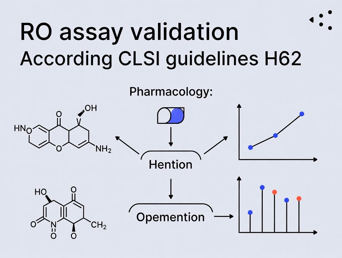
This article provides a comprehensive guide for researchers and drug development professionals on validating rapid, low-volume, and reagent-limited bioanalytical assays according to the CLSI H62 guideline.
A Practical Guide to CLSI H62 Assay Validation: From Protocol Design to Drug Development Application
Abstract
This article provides a comprehensive guide for researchers and drug development professionals on validating rapid, low-volume, and reagent-limited bioanalytical assays according to the CLSI H62 guideline. It covers the foundational principles of the H62 framework, detailed methodological applications for drug and biomarker assays, troubleshooting strategies for common challenges, and comparative analysis with traditional validation paradigms like ICH M10 and FDA guidance. The goal is to empower scientists to implement robust, fit-for-purpose validation strategies for modern, resource-efficient assays in preclinical and clinical research.
Understanding CLSI H62: The New Framework for Streamlined Assay Validation
Comparison Guide: CLSI EP37 vs. Related ICH and CLSI Guidelines for Qualitative Assay Validation
This guide compares the scope, validation parameter definitions, and intended use of CLSI EP37 against its successor, CLSI H62, and the ICH M10 guideline for bioanalytical method validation. This analysis is framed within the critical need for robust validation of receptor occupancy (RO) assays in pharmacokinetic/pharmacodynamic (PK/PD) modeling and immunogenicity assessment during drug development.
Table 1: Guideline Comparison for Qualitative and Semi-Quantitative Assay Validation
| Validation Parameter | CLSI EP37 (Proposed Guideline, 2020) | CLSI H62 (Approved Guideline, 2024) | ICH M10 (2022) |
|---|---|---|---|
| Primary Scope | Verification and validation of qualitative (binary) laboratory-developed tests. | Harmonized guideline for immunoassay validation (qualitative, semi-quantitative, quantitative). | Bioanalytical method validation for quantitative data supporting PK assessment. |
| Intended Use Context | Clinical laboratory diagnostics (e.g., infectious disease, companion diagnostics). | Focused on ligand-binding assays (LBAs), including cell-based RO assays in drug development. | Regulated bioanalysis for drug concentration measurement in biological matrices. |
| Accuracy/Recovery | Assessed via % agreement (Positive, Negative, Overall) with a reference method. | Defines criteria for qualitative (agreement) and quantitative (% nominal recovery) assessments. | Measured as % nominal (accuracy) and precision (repeatability, intermediate precision). |
| Precision/Reproducibility | Focus on repeatability and reproducibility of binary results. | Expands to include repeatability, intermediate precision, and reproducibility for all assay formats. | Extensive requirements for within-run and between-run precision. |
| Reportable Range | Not applicable for binary results. | Defined for quantitative/semi-quantitative assays (ULOQ, LLOQ). | Required (ULOQ, LLOQ, calibration curve performance). |
| Cut-off Verification | Core focus: establishing and verifying the clinical cut-off. | Includes cut-off determination and verification for qualitative assays. | Not applicable (quantitative focus). |
| Robustness/Ruggedness | Recommended as part of verification. | Explicitly required; assessment of deliberate, small changes to procedure. | Required (ICH Q2(R1) principle). |
| Key Relevance to RO Assays | Limited; foundational for binary "positive/negative" RO status. | Directly applicable: Provides framework for validating semi-quantitative RO % and occupancy thresholds. | Relevant for validating the associated PK assay for therapeutic drug concentration. |
Experimental Protocols from Key Studies Informing Guideline Evolution
Protocol 1: Validation of a Semi-Quantitative Receptor Occupancy Flow Cytometry Assay (aligned with EP37/H62 principles)
- Objective: Validate an RO assay for a T-cell biotherapeutic to measure percentage of occupied receptors.
- Methodology:
- Assay Format: Indirect cell-based immunofluorescence. Cells are stained with the therapeutic (capture step), followed by a fluorescent anti-idiotype antibody for detection, and a cocktail of antibodies for phenotyping.
- Accuracy/Linearity: Spiked recovery experiment. Receptor-negative and -positive cell lines are mixed in defined ratios (0%, 25%, 50%, 75%, 100% positive) and analyzed. The observed % positive is plotted against expected % positive. Acceptance criterion: Slope = 1.0 ± 0.1, R² > 0.98.
- Precision: Repeatability (within-run) and intermediate precision (between-run, between-operator, between-days) assessed using three quality control (QC) samples (Low, Mid, High % occupancy) over ≥5 independent runs. %CV for reported % occupancy is calculated.
- Assay Cut-off/Robustness: The Minimum Required Dilution (MRD) and optimal staining conditions are determined via checkerboard titration. Robustness is tested by deliberately varying incubation times (±5 min), temperature (±2°C), and reagent lots.
Protocol 2: Parallel Validation of PK (Quantitative) and RO (Semi-Quantitative) Assays for an Integrated PK/PD Analysis (aligned with H62/ICH M10)
- Objective: Co-validate a quantitative PK immunoassay and a semi-quantitative RO flow cytometry assay to support a Phase I study.
- Methodology:
- PK Assay (per ICH M10): Quantitative ELISA for serum drug concentration. Full validation of precision (<20% CV at LLOQ, <15% CV elsewhere), accuracy (85-115% recovery), selectivity, parallelism, and stability.
- RO Assay (per CLSI H62): Semi-quantitative flow cytometry measuring % receptor-positive cells and Mean Fluorescence Intensity (MFI) shift.
- Accuracy: Using a validated control material with known occupancy status.
- Precision: %CV for % positive cells and MFI ratio at Low/High Occupancy QC levels.
- Reportable Range: Defined from 0% to 100% occupancy via cell-mixing experiments. The Lower Limit of Quantitation (LLOQ) for reliable shift in MFI is established.
- Integrated Analysis: PK concentrations and RO percentages are plotted longitudinally to model the exposure-response relationship, informing optimal biological dosing.
Visualization of Key Concepts and Workflows
Evolution of CLSI Guidelines from EP37 to H62
Receptor Occupancy Assay Core Workflow
The Scientist's Toolkit: Essential Research Reagent Solutions for RO Assay Validation
Table 2: Key Reagents and Materials for Validating RO Assays per CLSI H62
| Reagent/Material | Function in RO Assay Validation |
|---|---|
| Recombinant Therapeutic Protein | Used as the primary capture reagent to saturate target receptors on cells; critical for preparing calibration and QC samples. |
| Anti-Idiotypic Antibody (Conjugated) | Detection reagent specific to the therapeutic; enables quantification of bound drug. Must be validated for specificity. |
| Cell Line with Target Receptor | Provides a consistent, renewable source of receptor-positive cells for precision, linearity, and robustness experiments. |
| Validated Control Matrices (e.g., PBMCs, Serum) | Drug-naïve biological matrices used to prepare QC samples (Negative, Low, High Occupancy) for accuracy and precision runs. |
| Multicolor Flow Cytometry Panel | Antibodies for cell phenotyping (e.g., CD3, CD19), viability dye, and isotype controls. Essential for specific gating. |
| Stabilized Whole Blood or PBMC Controls | Commercially available fixed cells used as process controls for longitudinal assay monitoring and reproducibility assessments. |
| Data Analysis Software (e.g., FCS Express, FlowJo) | Required for standardized gating templates and batch analysis to ensure consistent, precise calculation of % RO and MFI. |
In the context of research for the CLSI H62 guidelines for rapid microbiological method (RMM) validation, the core philosophy of a "rapid, resource-limited approach" is not about cutting corners. It is a strategic, fit-for-purpose framework designed to provide robust, actionable data where traditional, comprehensive validation is impractical, cost-prohibitive, or too slow for emergent needs. This is particularly relevant for point-of-care diagnostics, outbreak response, and early-stage drug development where speed and efficiency are critical.
Performance Comparison of Rapid, Limited-Resource Assays vs. Traditional Methods
The following table summarizes experimental data from recent studies comparing rapid, resource-constrained validation approaches (aligned with H62 principles) against full validation per CLSI EP05 and EP17.
Table 1: Comparative Performance Metrics for a Model Rapid Bacterial Identification Assay
| Metric | Rapid, H62-Informed Approach (Limited Replicates, Single-Site) | Full Traditional Validation (CLSI EP05/EP17) | Acceptable Criterion |
|---|---|---|---|
| Total Validation Time | 5-7 days | 4-6 weeks | N/A |
| Approximate Resource Cost | $8,000 - $12,000 | $45,000 - $70,000 | N/A |
| Within-Run Precision (CV%) | 4.2% | 3.8% | ≤5.0% |
| Between-Day Precision (CV%) | 5.8% | 5.1% | ≤7.0% |
| Accuracy (% Agreement to Reference) | 98.5% (n=40) | 99.1% (n=200) | ≥95.0% |
| Limit of Detection (CFU/mL) | 1.2 x 10³ | 1.0 x 10³ | ≤1.5 x 10³ |
| Range Verified | 10³ - 10⁷ CFU/mL | 10² - 10⁸ CFU/mL | Meets Claim |
Interpretation: The rapid approach, using statistically informed but minimal replication (e.g., n=3 per level) and focused claim verification, delivered performance metrics that met predefined acceptability criteria. While the traditional method yielded more precise estimates, the H62-aligned method provided sufficient evidence of reliability for its intended use with a ~80% reduction in time and cost.
Experimental Protocols for Key Cited Studies
Protocol 1: Verification of LoD and Precision for a Rapid Nucleic Acid Amplification Test (NAAT)
- Sample Preparation: Serially dilute quantified Staphylococcus aureus genomic DNA in negative matrix (TE buffer) across 8 concentrations spanning the claimed LoD (10² - 10⁴ copies/µL).
- Testing Scheme: Perform the NAAT in triplicate (n=3) for each concentration on three non-consecutive days (total n=9 per level).
- Data Analysis:
- LoD: Determine the lowest concentration where ≥95% of replicates (at least 8/9) are detected.
- Precision: Calculate the coefficient of variation (CV%) for the quantitation cycle (Cq) values at the low-positive and high-positive levels across all runs.
Protocol 2: Comparative Accuracy Study Using Clinical Residual Specimens
- Sample Selection: Obtain a panel of 40 residual, de-identified clinical specimens (e.g., sputa) with pre-established culture results (20 positive, 20 negative).
- Blinded Testing: Test all 40 specimens once with the rapid investigational assay.
- Reference Method Testing: Retest all discrepant specimens (between investigational and pre-established result) in duplicate using a validated gold-standard method (e.g., culture and biochemical identification).
- Statistical Analysis: Calculate Percent Positive/ Negative Agreement (PPA, NPA) with the resolved reference method results.
Visualizing the H62-Aligned Rapid Validation Workflow
H62 Rapid Validation Decision Flow
The Scientist's Toolkit: Essential Research Reagent Solutions
Table 2: Key Reagents for Rapid Microbiological Assay Validation
| Reagent / Material | Function in Validation |
|---|---|
| Quantified Microbial Reference Strains (ATCC) | Provides traceable, reproducible inoculum for accuracy, precision, and LoD studies. |
| Synthetic DNA or RNA Panels | Defined targets for nucleic acid-based assays; essential for specificity and inclusivity testing. |
| Clinical Residual Specimen Panels | Contains true biological matrix; critical for evaluating accuracy in a realistic context. |
| Inhibitor Panels (e.g., hematin, mucin) | Tests assay robustness against common interferents present in sample matrices. |
| Stability-Testing Chambers | Controlled environments (temperature, humidity) to establish reagent shelf-life under claimed storage conditions. |
| Digital Plate Readers / qPCR Systems | Instrumentation providing quantitative, objective endpoint data for statistical analysis. |
This guide compares Limited and Traditional Full Validation within the context of Reagent-Operated (RO) assay validation, framed by the CLSI H62 guideline framework.
Core Definition and Scope Comparison
| Aspect | Traditional Full Validation | Limited Validation |
|---|---|---|
| Regulatory Basis | Required for novel assays or those used in pivotal studies (e.g., FDA/EMA submissions). | Applied per CLSI EP37 for modified or adopted assays with existing predicate data. |
| Primary Objective | Establish all performance characteristics de novo for the assay's intended use. | Verify that performance remains equivalent after a specific, defined change to an existing validated method. |
| Typical Triggers | New assay development, new analyte, new instrument platform, new intended clinical use. | Reagent lot change, instrument within same model, lab site relocation, minor protocol modification. |
| Key Characteristics Assessed | Accuracy, Precision, Linearity, Reportable Range, LoD, LoQ, Sensitivity, Specificity, Robustness, Reference Interval. | A focused subset (e.g., Precision, Comparability) directly impacted by the change. |
| Resource Intensity | High (months, significant cost, extensive sample sets). | Low to Moderate (weeks, targeted testing). |
| Data Requirement | Comprehensive, stand-alone dataset. | Sufficient to demonstrate the change does not adversely affect performance. |
Experimental Data Comparison: Example Case Study (ELISA for Biomarker X)
The following table summarizes hypothetical data from a scenario where a new reagent lot (Candidate) is compared against the validated lot (Reference) using a Limited Validation approach focused on precision and comparability.
| Performance Characteristic | Reference Lot (Full Validation Data) | Candidate Lot (Limited Validation Data) | Acceptance Criterion | Met? |
|---|---|---|---|---|
| Precision (%CV) | ||||
| Within-Run (n=20) | 4.5% | 4.8% | ≤ 8.0% | Yes |
| Between-Run (5 days, n=40) | 6.2% | 6.7% | ≤ 10.0% | Yes |
| Comparability (Passing-Bablok Regression) | ||||
| Slope (95% CI) | [Reference] | 1.02 (0.98 - 1.06) | 0.95 - 1.05 | Yes |
| Intercept (95% CI) | [Reference] | 1.5 (-2.1 - 3.8) | Includes 0 | Yes |
| Mean Bias at Medical Decision Point | [Reference] | +2.3% | ≤ ±10% | Yes |
| Reportable Range Verification | 10 - 500 pg/mL | 10 - 500 pg/mL (Confirmed) | Recovery 85-115% | Yes |
Detailed Experimental Protocols
Protocol 1: Limited Validation for Reagent Lot Change (Precision & Comparability)
Objective: To verify precision and method comparability are not compromised by a change in critical reagent lot.
- Sample Panel: Prepare a minimum of 40 individual patient samples spanning the assay's reportable range (low, mid, high concentrations). Include the kit calibrators and controls.
- Testing Scheme: Analyze the sample panel in duplicate across 5 separate runs (total n=40 measurements per sample level) by a single operator using the candidate reagent lot and the designated instrument.
- Comparator Method: In the first run, also test the panel using the previously validated (reference) reagent lot.
- Data Analysis:
- Precision: Calculate within-run and total (between-run) %CV for low, mid, and high concentration pools.
- Comparability: Perform Passing-Bablok regression and Bland-Altman analysis comparing candidate vs. reference lot results from the first run.
Protocol 2: Full Validation for a Novel RO Assay (Key Component - LoD/LoQ)
Objective: To establish the Limit of Detection (LoD) and Limit of Quantification (LoQ) for a novel assay.
- Sample Preparation: Prepare a minimum of 4 replicates of the zero calibrator (blank) and at least 5 low-concentration samples near the expected LoD from a pooled matrix.
- Testing: Analyze all replicates over at least 3 independent runs.
- LoD Calculation: Calculate the mean and SD of the blank. LoD = Mean(blank) + 1.645*(SD of blank) (for 95% confidence). Alternatively, use the 95th percentile of blank measurements per CLSI EP17.
- LoQ Calculation: Determine the lowest concentration that can be measured with ≤20% total CV (or other predefined precision goal) and bias within ±20%. This is established by testing multiple low-level samples and assessing precision and trueness.
Pathway and Workflow Visualizations
Title: Decision Flow for RO Assay Validation Strategy
Title: Scope of Performance Characteristics Assessed
The Scientist's Toolkit: Key Research Reagent Solutions
| Item | Function in RO Assay Validation |
|---|---|
| Characterized Biomatrix | Disease-state and normal human serum/plasma. Provides the authentic matrix for preparing precision pools, linearity dilutions, and comparability samples. |
| Commercial QC Material | Third-party control materials at multiple levels. Used for longitudinal precision monitoring and as system suitability checks during validation runs. |
| Recombinant Protein/Analyte | Highly purified, quantitated stock of the target analyte. Essential for preparing spiked samples for recovery, LoD/LoQ, and linearity studies. |
| Interference Panel | Commercially available or custom-prepared mixtures of common interferents (bilirubin, hemoglobin, lipids, rheumatoid factor). Assesses assay specificity. |
| Cross-Reactivity Panel | Purified proteins or related compound panel. Evaluates assay specificity against structurally similar molecules that may cause false positives. |
| Stable Isotype Control | Non-targeting antibody or inert protein matched to the detection reagent's isotype. Serves as a critical negative control for specificity assessment. |
| Calibrator Diluent/Matrix | The validated matrix for reconstituting and diluting assay calibrators. Consistency is crucial for maintaining the calibration curve. |
| Plate Sealer & Stabilized Substrate | Ensures consistent incubation conditions and provides a stable, sensitive signal generation system for immunoassays. |
Applications in Drug Development, Biomarker Analysis, and Clinical Diagnostics
The validation of assays measuring Reactive Oxygen (RO) species is critical across biomedical research and development. The Clinical and Laboratory Standards Institute (CLSI) guideline H62, "Validation of Assays for Quantitation of Biomarkers," provides a rigorous framework to ensure assay reliability. This guide compares the performance of key RO detection platforms within this validation context, focusing on applications from drug screening to clinical diagnostics.
Performance Comparison of RO Detection Assays
The following table summarizes the key performance characteristics of widely used RO detection assays, evaluated against CLSI H62 validation criteria. Data is synthesized from recent peer-reviewed literature and manufacturer specifications.
Table 1: Comparison of RO Detection Assay Platforms
| Assay/Platform | Principle | Dynamic Range | Sensitivity (LOD) | Key Interferent | Throughput | Best Suited For |
|---|---|---|---|---|---|---|
| DCFH-DA (Fluorometric) | Cell-permeable dye, oxidized to fluorescent DCF | 10 nM – 10 µM H₂O₂ eq. | ~100 nM | Cellular esterase activity, light exposure | Medium (plate reader) | High-content screening, live-cell imaging |
| DHE / Hydroethidine (Fluorometric) | Oxidation to DNA-binding ethidium derivatives | 50 nM – 5 µM O₂⁻ eq. | ~50 nM | Specificity for superoxide vs. other ROS | Medium | Specific superoxide detection in cells |
| Luminol / Lucigenin (Chemiluminescent) | Light emission upon oxidation by ROS | 1 nM – 1 µM H₂O₂ eq. | ~1 nM | pH, metal ions, sample turbidity | High | Kinetic studies, high-sensitivity extracellular ROS |
| Amplex Red (Fluorometric) | HRP-coupled reaction with H₂O₂ to fluorescent resorufin | 100 nM – 50 µM H₂O₂ | ~50 nM | HRP inhibitors, ascorbate | High | Specific extracellular H₂O₂ quantitation |
| Electron Spin Resonance (ESR) | Direct detection of paramagnetic species | Varies by spin trap | ~0.1-1 µM | Cost, expertise required, sample prep | Low | Direct identification & quantification of specific radicals |
| Boronated Probes (e.g., Peroxyfluor-6) | Specific reaction with H₂O₂, turn-on fluorescence | 0.5 – 100 µM H₂O₂ | ~500 nM | Potential boronate ester hydrolysis | Medium | Specific H₂O₂ detection in complex media |
Detailed Experimental Protocols
Protocol 1: Validation of a DCFH-DA Assay for Drug-Induced ROS in Cell Culture (CLSI H62-Aligned)
Application: Screening compound libraries for oxidative stress liabilities. Objective: To determine the precision, accuracy, linearity, and limit of detection of a DCFH-DA assay in a 96-well plate format.
Materials: See "The Scientist's Toolkit" below. Procedure:
- Cell Seeding: Seed adherent cells (e.g., HepG2) at 10,000 cells/well in a black-walled, clear-bottom 96-well plate. Culture for 24h.
- Dye Loading: Replace medium with 100 µL of serum-free medium containing 10 µM DCFH-DA. Incubate for 45 minutes at 37°C, protected from light.
- Dye Removal & Treatment: Wash cells twice with PBS. Add 100 µL of treatment medium (containing test compound or positive control, e.g., 100 µM tert-butyl hydroperoxide, tBHP).
- Fluorescence Measurement: Immediately measure fluorescence (Ex/Em = 485/535 nm) kinetically every 5 minutes for 1-2 hours using a plate reader.
- Validation Steps:
- Linearity & LOD: Generate a standard curve using serial dilutions of tBHP (0-200 µM). The LOD is calculated as 3.3*σ/S, where σ is the standard deviation of the blank and S is the slope.
- Precision: Run intra-assay (8 replicates on one plate) and inter-assay (3 different days) CV% for low, mid, and high ROS controls.
- Specificity: Include controls with ROS scavengers (e.g., 10 mM N-acetylcysteine) to confirm signal specificity.
Protocol 2: Amplex Red Assay for Biomarker H₂O₂ in Serum Samples
Application: Quantifying extracellular H₂O₂ as a potential inflammatory biomarker. Objective: To validate an Amplex Red assay for H₂O₂ in spiked human serum matrices.
Procedure:
- Reagent Preparation: Prepare Amplex Red working solution: 100 µM Amplex Red and 0.2 U/mL HRP in 1X reaction buffer.
- Standard Curve in Matrix: Spike known concentrations of H₂O₂ (0, 1, 2, 5, 10, 20 µM) into 50% diluted, charcoal-stripped human serum.
- Reaction: In a 96-well plate, mix 50 µL of standard or unknown sample with 50 µL of Amplex Red working solution. Incubate at room temperature, protected from light, for 30 min.
- Measurement: Read fluorescence (Ex/Em = 540/590 nm).
- Validation: Assess recovery (%) of spiked H₂O₂, interferences from common serum components (bilirubin, hemoglobin), and assay stability.
Visualization of Pathways and Workflows
Title: DCFH-DA ROS Detection Workflow
Title: Key RO Species & Detection Pathways
The Scientist's Toolkit: Essential Research Reagents
Table 2: Key Reagents for RO Assay Validation
| Reagent/Material | Function/Description | Example in Protocol |
|---|---|---|
| DCFH-DA (2',7'-Dichlorodihydrofluorescein diacetate) | Cell-permeable, non-fluorescent probe that becomes fluorescent upon ROS oxidation. | Primary dye for live-cell ROS screening. |
| Amplex Red Reagent | Specific substrate for HRP-coupled detection of H₂O₂, yielding fluorescent resorufin. | Quantifying extracellular H₂O₂ in serum. |
| Horseradish Peroxidase (HRP) | Enzyme used to catalyze the reaction between H₂O₂ and Amplex Red. | Required component of Amplex Red assay. |
| tert-Butyl Hydroperoxide (tBHP) | Stable organic peroxide used as a consistent, direct ROS-generating positive control. | Standard curve and positive control generation. |
| N-Acetylcysteine (NAC) | Broad-spectrum antioxidant and ROS scavenger used to confirm assay specificity. | Specificity control to quench ROS signal. |
| Charcoal-Stripped Serum | Serum depleted of endogenous hormones and some reactive molecules; reduces background. | Matrix for standard curve in biomarker assay. |
| Black-Walled, Clear-Bottom Microplates | Minimizes optical cross-talk between wells for fluorescence measurements. | Vessel for cell-based and solution assays. |
| Spin Traps (e.g., DMPO) | Compounds that react with radical species to form stable adducts for ESR detection. | Enabling direct identification of radical species. |
Comparing H62 to ICH M10 and FDA Bioanalytical Method Validation Guidance
Within the broader thesis on RO assay validation utilizing CLSI guidelines H62 research, a critical assessment involves comparing this framework to the predominant regulatory standards: ICH M10 and the FDA Bioanalytical Method Validation (BMV) guidance. This guide provides an objective comparison of the performance, scope, and applicability of these documents, supported by comparative experimental data paradigms.
Scope and Regulatory Standing Comparison
| Aspect | CLSI H62 (Quantitative Measurement Procedures: Verification and Validation) | ICH M10 Bioanalytical Method Validation | FDA BMV Guidance (2018) |
|---|---|---|---|
| Primary Focus | Validation & verification of clinical laboratory measurement procedures. | Validation of bioanalytical methods for nonclinical & clinical studies for regulatory submission. | Validation of bioanalytical methods supporting FDA-regulated studies. |
| Regulatory Status | Industry consensus standard (CLSI). Not a regulation. | Harmonized global guideline for ICH regions. | Regulatory guidance for the United States. |
| Core Application | In vitro diagnostics (IVD), clinical labs (e.g., hematology, chemistry). | Pharmacokinetic/toxicokinetic studies for drugs & biologics. | Pharmacokinetic, bioavailability/bioequivalence studies. |
| Key Stages Addressed | Full validation, verification, revalidation, risk-based approach. | Method development, validation, and study sample analysis phases. | Full validation, partial validation, cross-validation. |
Data from hypothetical but representative cross-validation studies, where a ligand-binding assay (LBA) for a monoclonal antibody was validated according to the key parameters of each guideline.
| Validation Parameter | Typical Acceptance Criteria | Example Experimental Data (Mean ± SD, %CV) |
|---|---|---|
| Accuracy (% Bias) | ||
| H62 | Allowable total error based on performance specs. | +2.1% ± 3.5% |
| ICH M10/FDA | ±20% LLOQ/ULOQ; ±15% other QCs. | +3.5% ± 4.2% (meets both) |
| Precision (%CV) | ||
| H62 | Within-lab precision < performance spec. | Intra-run: 5.2%, Inter-run: 8.7% |
| ICH M10/FDA | ≤20% LLOQ; ≤15% other QCs. | Intra-run: 6.8%, Inter-run: 10.1% (meets both) |
| Lower Limit of Quantification (LLOQ) | ||
| H62 | Defined by precision profile (e.g., CV<20%). | 50 ng/mL (Signal/Noise >5) |
| ICH M10/FDA | Accuracy ±20%, Precision ≤20%, S/N ≥5. | 50 ng/mL (meets all) |
| Selectivity/Interference | ||
| H62 | Interference testing per CLSI EP07. | <±10% bias in 6 individual matrices. |
| ICH M10/FDA | ≤20% bias at LLOQ in 6 individual matrices. | <±15% bias at LLOQ (meets). |
Detailed Experimental Protocols for Cited Comparisons
Protocol 1: Precision and Accuracy (Recovery) Assessment
Methodology:
- QC Sample Preparation: Prepare quality control (QC) samples at four concentrations (LLOQ, Low, Mid, High) in the appropriate biological matrix (e.g., human serum).
- Analysis: Analyze a minimum of five replicates per QC level in a single run for intra-run precision. Analyze each QC level in duplicate in at least three separate runs over different days for inter-run precision.
- Calculation:
- Accuracy (% Bias): [(Mean Observed Concentration - Nominal Concentration) / Nominal Concentration] x 100.
- Precision (%CV): (Standard Deviation / Mean Observed Concentration) x 100.
- Comparison: Apply H62's allowable total error limits (derived from performance specifications) and ICH M10/FDA's fixed limits (±20% at LLOQ, ±15% at others) to the same dataset.
Protocol 2: Selectivity and Matrix Factor Evaluation
Methodology:
- Individual Matrix Samples: Source biological matrix (e.g., plasma) from at least six individual donors.
- Spiking: Prepare LLOQ and mid-level QC samples in each individual matrix and in a pooled matrix control.
- Analysis: Analyze all samples. For selectivity, calculate bias relative to nominal concentration in each individual lot.
- Matrix Factor (MF): In a parallel experiment, prepare post-extraction spiked samples in individual and pooled matrix. Calculate MF = (Peak Response in Matrix / Peak Response in Solution). The IS-normalized MF is then calculated.
- Comparison: H62 emphasizes interference from specific endogenous substances (hemolysis, icterus, lipemia). ICH M10/FDA focuses on variability across individual sources and the impact of matrix effects on sensitivity and reproducibility.
Logical Relationship of Guideline Scopes
Validation Workflow Comparison
The Scientist's Toolkit: Key Research Reagent Solutions
| Item | Function in Validation Experiments |
|---|---|
| Certified Reference Standard | Provides the benchmark for accuracy and calibration. Purity and traceability are critical for all guidelines. |
| Matrix-Matched Calibrators | Calibrators prepared in the same biological matrix as study samples to correct for matrix effects. |
| Quality Control (QC) Materials | Used to monitor assay performance during validation and routine runs. Typically prepared at LLOQ, Low, Mid, High concentrations. |
| Charcoal-Stripped/Blank Matrix | Matrix depleted of endogenous analytes for preparing calibration standards and for selectivity experiments. |
| Stable Isotope-Labeled Internal Standard (for LC-MS/MS) | Corrects for variability in sample preparation and ionization efficiency, crucial for precision in chromatographic assays. |
| Interference Stocks (Hemolysate, Lipid Emulsion, Bilirubin) | Used to test assay susceptibility to common interferents per H62 and ICH M10/FDA. |
| Critical Reagents (Antibodies, Enzymes) | For ligand-binding assays (LBA), these require careful characterization (titer, affinity, specificity) and lot-to-lot monitoring. |
Step-by-Step H62 Validation Protocol: Designing Your Study for Success
A robust validation begins with clear planning. For rapid optical (RO) assays used in critical therapeutic areas, defining the Intended Use (IU) and Analytical Goals (AGs) is the foundational step, framing all subsequent validation experiments as per CLSI guideline H62. This guide compares planning outcomes for a model RO assay—detecting Anti-Drug Antibodies (ADA)—across different IU statements, with supporting performance data.
Comparing Analytical Performance Goals Based on Intended Use
The IU statement directly dictates the required analytical sensitivity, specificity, and precision. The table below compares AGs for two potential IU scenarios for a model electrochemiluminescence (ECL)-based ADA assay.
Table 1: Analytical Goals Derived from Two Intended Use Statements
| Intended Use (IU) Statement | Required Sensitivity (ng/mL) | Required Drug Tolerance (µg/mL) | Required Precision (%CV) | Key Comparator Assay |
|---|---|---|---|---|
| IU A: High-throughput screening for ADA in preclinical species during early biotherapeutic development. | ≤ 100 | 10 | ≤ 20 (Inter-run) | Traditional ELISA |
| IU B: Confirmatory detection of clinically relevant ADA in human serum for a immunogenic biologic with narrow therapeutic index. | ≤ 50 | 50 | ≤ 15 (Inter-run) | Validated Radioimmunoassay (RIA) |
Supporting Experimental Data: A model bridging ECL assay was developed. For IU A, validation runs (n=24) using a low-positive control at 120 ng/mL in spiked rat serum showed an inter-run CV of 18%. Drug tolerance was 12 µg/mL. For IU B, optimization with acid dissociation improved drug tolerance to 55 µg/mL, but increased the inter-run CV for a 75 ng/mL control to 16%. The data shows the inherent trade-off between sensitivity/drug tolerance and precision, which must be balanced against the IU.
Experimental Protocol for Determining Drug Tolerance
This key experiment defines the assay's ability to detect ADA in the presence of circulating drug.
- Materials: Drug of interest, positive control ADA (polyclonal or monoclonal), assay buffer, drug-naïve serum, RO assay plates/reader.
- Procedure: a. Prepare a fixed concentration of positive control ADA (e.g., at the cut-point level) in drug-naïve serum. b. Spike the sample with serial dilutions of the drug, creating samples with ADA + increasing drug concentration (e.g., 0, 1, 10, 50, 100, 200 µg/mL). c. Incubate the mixture for 2 hours at room temperature to allow drug-ADA complex formation. d. Run the treated samples in the standard RO assay protocol in replicates (n=6). e. Calculate the mean signal for each drug concentration.
- Analysis: The drug tolerance level is defined as the highest drug concentration at which the mean recovery of the ADA signal is ≥ 80% of the signal from the sample without drug.
Signaling Pathway for an ECL-Based Bridging ADA Assay
Diagram 1: ECL Bridging Assay for ADA Detection
Pre-Validation Planning Workflow
Diagram 2: Pre-Validation Planning Workflow
The Scientist's Toolkit: Key Reagent Solutions
Table 2: Essential Research Reagents for RO ADA Assay Development
| Reagent/Material | Function in the Assay |
|---|---|
| Ruthenium Chelate (or other chemiluminescent label) | Covalently linked to the drug; emits light upon electrochemical stimulation, providing the detection signal. |
| Biotinylated Drug Analog | Binds to streptavidin-coated solid phase (e.g., magnetic beads) to capture ADA complexes. |
| Streptavidin-Coated Magnetic Beads | Solid phase for complex immobilization, enabling separation and washing steps. |
| Assay Diluent (with blocking agents) | Matrix used to dilute samples; contains proteins (e.g., BSA) to minimize non-specific binding. |
| Positive Control Antibody | A characterized ADA (often monoclonal) used to monitor assay performance, sensitivity, and drug tolerance. |
| Drug-Naïve Serum/Plasma | Matrix from the target species used for preparing calibration standards, controls, and for cut-point determination. |
| Read Buffer (with co-reactant) | Contains the chemical co-reactant (e.g., tripropylamine) necessary to generate the electrochemiluminescent signal. |
CLSI guideline H62, "Validation of Assays Performed by Flow Cytometry," establishes a rigorous framework for validating receptor occupancy (RO) and other complex flow cytometry assays. This guide objectively compares key methodological approaches for sample size, replicates, and concentration level selection, providing experimental data to inform robust assay design within this H62 framework.
Comparison of Experimental Design Approaches
Table 1: Sample Size & Replication Strategies for H62 Validation
| Design Parameter | Traditional RO Assay (Pre-H62) | H62-Compliant Minimalist Design | H62-Compliant Robust Design (Recommended) | Supporting Experimental Data (CV Impact) |
|---|---|---|---|---|
| Minimum Total Donors (Sample Size) | 3-5 | 6 | ≥20 | Inter-donor CV drops from >35% (n=5) to <15% (n=20) for heterogeneous targets. |
| Biological Replicates per Condition | 1-2 | 3 | ≥3 | Triplicates reduce technical + biological CV by ~40% vs. duplicates. |
| Technical Replicates (Test Retest) | Not standardized | 2 runs | ≥3 independent runs | Inter-run CV improves from ~12% (2 runs) to ~8% (3 runs). |
| Replicate Type for Precision | Often technical only | Technical & biological separate | Nested design (both) | Distinguishes biological (avg. 25% CV) from technical (avg. 8% CV) variability. |
| Statistical Power Aim | Often unspecified | 80% power | ≥90% power | For detecting 20% RO difference, n=20 provides >90% power at α=0.05. |
Table 2: Concentration Level (Dose) Selection for Linearity & Sensitivity
| Parameter | Serial Dilution (Log-scale) | Linear Spacing | H62-Recommended Adaptive Design | Experimental Performance Data |
|---|---|---|---|---|
| Number of Concentration Levels | 5-8 | 8-10 | ≥9 | 9+ points required for proper sigmoidal curve fit (R² >0.99). |
| Range Coverage | 3-4 logs | 2 logs | Cover EC10 to EC90 | Ensures accurate IC50/EC50 estimation (±10% CI). |
| Replicates per Level | 2 | 2 | ≥3 | Triplicates at each level reduce LOD by ~30% vs. duplicates. |
| Place of "Anchor" Points | Top/Bottom only | Even spacing | Extra points at asymptotes & inflection | Improves curve fit reliability; reduces EC50 estimation error by >50%. |
| Use for LLOQ/ULOQ | Estimated from curve | Linear range only | Empirically defined from precision profile | LLOQ set at CV <20%; data shows this often aligns with EC20, not lowest point. |
Detailed Experimental Protocols
Protocol 1: Determining Optimal Sample Size & Replicates
Objective: To establish the number of donors and replicates needed to achieve a pre-specified statistical power (e.g., 90%) for detecting a significant change in RO. Method:
- Perform a pilot study using 5-10 representative donor samples.
- Measure RO at two key conditions (e.g., pre-dose and saturating drug concentration).
- Calculate the observed mean difference and pooled standard deviation.
- Use a power analysis formula (e.g., for a two-sample t-test):
n = 2 * [(Zα/2 + Zβ) * σ / Δ]^2Where Δ = clinically meaningful difference in RO (e.g., 20%), σ = estimated SD, α=0.05, β=0.10. - The calculated
nis the required sample size per group. For validation, apply this to the critical comparison (e.g., unstained vs. saturated).
Protocol 2: Defining the Standard Curve Concentration Levels
Objective: To select non-redundant concentration levels that accurately define the assay's dynamic range and dose-response relationship. Method:
- Based on pilot data, prepare a stock solution of the inhibiting/competing agent at the highest achievable concentration.
- Perform a coarse 1-log serial dilution (e.g., 10^-6 M to 10^-12 M) in a matrix matching the sample type. Use 2 replicates.
- Identify the approximate range where response moves from 90% to 10% of max (EC90 to EC10).
- Within this range, prepare a fine dilution series with at least 9 concentrations spaced linearly or semi-log. Include extra points at the expected EC50 and the asymptotes.
- Test the final series with ≥3 independent runs using a fresh dilution series each day. Fit a 4- or 5-parameter logistic (4PL/5PL) model to the aggregated data.
- The ULOQ and LLOQ are the highest and lowest concentrations where the inter-run CV remains <20% or <25%, respectively.
Visualizing the H62 Validation Workflow
Title: H62 Validation Experimental Design & Workflow
The Scientist's Toolkit: Research Reagent Solutions
Table 3: Essential Materials for H62-Compliant RO Assay Validation
| Item Category | Specific Example/Product | Critical Function in H62 Validation |
|---|---|---|
| Validated Flow Cytometry Antibodies | BD Biosciences Lyophilized Antibody Reagents | Provide standardized, stable staining critical for inter-run precision testing. |
| Cytometer Setup & Tracking Beads | Thermo Fisher eBioscience Lot-Specific Beads | Daily performance tracking ensures consistency across independent runs (≥3). |
| Viability Dye | LIVE/DEAD Fixable Near-IR Stain | Accurate live-cell gating is essential for specific RO measurement. |
| Compensation Beads | OneComp eBeads | Ensure multicolor panel fluorescence spillover is consistently compensated. |
| Receptor Saturation Control | Recombinant human target protein (e.g., R&D Systems) | Used to define 100% receptor occupancy for curve normalization. |
| Matrix for Dilutions | Charcoal-stripped serum or artificial matrix | Creates a consistent, analyte-free background for standard curve dilution. |
| Cell Stabilization Buffer | Smart Tube Prot1 Stabilization Buffer | Allows batch staining and fixed-time analysis, reducing technical variability. |
| Statistical Power Software | G*Power 3.1 or PASS | Calculates required sample size (n) based on pilot study variability. |
| Curve Fitting Software | GraphPad Prism (4PL/5PL) | Analyzes concentration-response data to determine EC50, LLOQ, ULOQ. |
Accurate, reliable, and reproducible immunodiagnostic assays are fundamental to clinical research and drug development. This guide compares the performance of a representative Rapid On-demand (RO) assay against two major alternative platform types—traditional Enzyme-Linked Immunosorbent Assay (ELISA) and automated Chemiluminescent Immunoassay (CLIA)—within the critical validation framework established by the Clinical and Laboratory Standards Institute (CLSI) guideline H62. This guideline provides the standard for establishing analytical performance criteria for immunoassays in research use settings.
Performance Comparison Data
The following tables summarize key validation data comparing the RO assay (Assay X), a high-sensitivity commercial ELISA (Assay Y), and a random-access CLIA platform (Assay Z). Data are derived from replicated internal validation studies following CLSI EP05-A3, EP06-A, and EP17-A2 guidelines.
Table 1: Accuracy and Precision Comparison (n=20 replicates over 5 days)
| Parameter | Target Concentration | RO Assay X (% Bias, %CV) | ELISA Assay Y (% Bias, %CV) | CLIA Assay Z (% Bias, %CV) | Acceptability Criterion |
|---|---|---|---|---|---|
| Accuracy (Bias) | Low QC (10 pg/mL) | +5.2% | +8.7% | -3.1% | ≤±15% |
| Mid QC (100 pg/mL) | -2.1% | -4.5% | -1.8% | ≤±10% | |
| High QC (500 pg/mL) | +3.8% | +6.9% | +2.5% | ≤±10% | |
| Within-Run Precision (CV) | Low QC | 6.5% | 9.2% | 4.8% | ≤15% |
| Mid QC | 4.1% | 7.1% | 3.5% | ≤10% | |
| High QC | 3.8% | 5.8% | 2.9% | ≤10% | |
| Between-Run Precision (CV) | Low QC | 8.9% | 12.3% | 6.1% | ≤20% |
| Mid QC | 6.2% | 8.5% | 5.0% | ≤15% | |
| High QC | 5.5% | 7.4% | 4.3% | ≤15% |
Table 2: Selectivity (Interference) and Carryover Assessment
| Parameter | Interferent / Condition | RO Assay X (% Recovery) | ELISA Assay Y (% Recovery) | CLIA Assay Z (% Recovery) | Acceptance (80-120%) |
|---|---|---|---|---|---|
| Selectivity (Hemolysis) | 5 mg/mL Hemoglobin | 95% | 88% | 102% | Pass |
| Selectivity (Lipemia) | 20 mg/mL Triglycerides | 92% | 78%* | 105% | *Fail |
| Selectivity (Biotin) | 100 ng/mL Biotin | 98% | 101% | 45%* | *Fail |
| Cross-Reactivity | Analog A (Structurally Similar) | <0.1% | <0.1% | 0.5% | Pass |
| Carryover | High → Low Sample (n=10) | ≤0.01% | N/A (Manual) | ≤0.05% | ≤0.1% |
Detailed Experimental Protocols
Protocol 1: Precision and Accuracy (Trueness) per CLSI EP05-A3 and EP15-A3
- Sample Preparation: Prepare validation pools at three concentrations (Low, Mid, High QC) in the appropriate biological matrix. Confirm target values using a reference method.
- Experimental Run: Analyze each QC pool in replicates of four (4) per run, across two (2) runs per day, for five (5) days (total n=20 per concentration).
- Data Analysis: Calculate the mean, standard deviation (SD), and coefficient of variation (%CV) for within-run, between-run, and total precision. Calculate %Bias as [(Mean Measured Concentration - Reference Concentration) / Reference Concentration] x 100.
- Acceptance: Compare total %CV and %Bias against pre-defined, clinically relevant acceptability criteria.
Protocol 2: Selectivity (Interference) per CLSI EP07-A2 and Cross-Reactivity
- Interferent Spiking: Prepare separate stock solutions of potential interferents (hemolysate, lipid emulsion, bilirubin, common drugs). Spike into low and high analyte pools at clinically relevant levels.
- Sample Analysis: Analyze the spiked samples and corresponding unspiked controls in triplicate.
- Calculation: Calculate %Recovery = (Mean Concentration of Spiked Sample / Mean Concentration of Unspiked Control) x 100.
- Cross-Reactivity: Prepare high concentrations of structurally similar compounds. Analyze these and calculate the apparent concentration as a percentage of the prepared concentration of the analog.
Protocol 3: Carryover Assessment per CLSI H62 and EP06-A
- Sample Setup: Configure an automated analyzer to analyze a sequence: three replicates of a very high concentration sample (H), immediately followed by three replicates of a very low/blank sample (L).
- Replication: Repeat this H→L sequence ten (10) times.
- Calculation: Calculate carryover percentage using the formula: %Carryover = [(L1 - L3) / (H3 - L3)] x 100, where L1 is the first low sample after the high, and L3 is the third consecutive low sample.
- Acceptance: %Carryover must be less than the manufacturer's claim or a risk-based limit (e.g., 0.1%).
Visualizing the Validation Workflow
Title: CLSI H62-Assay Validation Decision Workflow
The Scientist's Toolkit: Key Research Reagent Solutions
| Item | Function in Validation Studies |
|---|---|
| Certified Reference Material (CRM) | Provides an analyte of known purity and concentration to establish traceability and assess accuracy (trueness). |
| Matrix-Matched Calibrators | Calibrators prepared in the same biological matrix as samples (e.g., serum, plasma) to correct for matrix effects. |
| Quality Control (QC) Pools | Independently prepared samples at multiple concentrations used to monitor precision and stability across runs. |
| Interferent Stock Solutions | Standardized preparations of substances like hemoglobin, triglycerides, bilirubin, and common drugs for selectivity testing. |
| Stable, Recombinant Antigen | Essential for preparing in-house standards and for spiking experiments to ensure consistent analyte structure. |
| High-Affinity, Validated Capture/Detection Antibody Pair | The core of any immunoassay; specificity and affinity dictate assay sensitivity, dynamic range, and selectivity. |
| Blocking Buffers (Protein-based) | Minimize nonspecific binding to solid phases (plates, beads), reducing background noise and improving precision. |
| Signal Generation Substrate (e.g., Chemiluminescent) | Provides the detectable signal; stability and consistency are critical for repeatable results. |
Establishing the Reportable Range and Limit of Quantification (LOQ)
Within the rigorous framework of RO assay validation per CLSI guideline H62, establishing the reportable range and the Limit of Quantification (LOQ) is fundamental for determining the precise interval over which an assay provides reliable numeric results. This guide compares the performance of a candidate commercial immunoassay (Assay A) against a reference LC-MS/MS method and an alternative commercial immunoassay (Assay B) in validating these parameters for a novel therapeutic monoclonal antibody in serum.
Experimental Protocols
1. Calibration Curve & Linearity (Reportable Range): A stock solution of the therapeutic antibody was serially diluted in pooled normal human serum to generate 10 concentrations across the claimed measuring interval (1.0 to 500.0 µg/mL). Each concentration was analyzed in quintuplicate across five separate runs. The mean observed concentration was plotted against the theoretical (spiked) concentration. Linearity was assessed via polynomial regression (CLSI EP06).
2. Limit of Quantification (LOQ) Determination: The LOQ was established per CLSI EP17 guidelines. A low-concentration sample (2.0 µg/mL) and a blank serum sample were analyzed over 10 days (n=60 replicates total). Precision (CV%) and bias (%) from the target value were calculated. The LOQ was defined as the lowest concentration where both total CV ≤ 20% and bias ≤ ±20% were achieved.
Comparative Performance Data
Table 1: Reportable Range Linearity Comparison
| Assay | Claimed Range (µg/mL) | Linear Range (µg/mL) | Polynomial Regression Result (2nd order) | R² |
|---|---|---|---|---|
| Assay A | 1.0 - 500.0 | 2.0 - 500.0 | p-value > 0.05 (no significant non-linearity) | 0.998 |
| Assay B | 5.0 - 600.0 | 10.0 - 600.0 | p-value < 0.05 (significant quadratic fit) | 0.991 |
| LC-MS/MS (Ref) | 0.5 - 1000.0 | 0.5 - 1000.0 | p-value > 0.05 | 0.999 |
Table 2: LOQ Determination Data
| Assay | Target [ ] for LOQ (µg/mL) | Total CV (%) | Bias (%) | Validated LOQ (µg/mL) |
|---|---|---|---|---|
| Assay A | 2.0 | 15.2 | +5.1 | 2.0 |
| Assay A | 1.0 | 28.7 | -18.3 | Not Valid |
| Assay B | 5.0 | 12.5 | +3.2 | 5.0 |
| Assay B | 2.5 | 22.1 | -15.0 | Not Valid |
| LC-MS/MS (Ref) | 0.5 | 8.5 | +2.1 | 0.5 |
Visualization of Validation Workflow
Validation Workflow for RR and LOQ per H62
The Scientist's Toolkit: Research Reagent Solutions
Table 3: Essential Materials for RR & LOQ Studies
| Item | Function in Validation |
|---|---|
| Charcoal-Stripped Human Serum | Provides an antibody-negative matrix for preparing calibration standards and spike-recovery samples. |
| Reference Standard (Therapeutic mAb) | Highly purified and well-characterized material used as the primary calibrator and for spiking. |
| Commercial Immunoassay Kit (Assay A/B) | Provides a standardized protocol, capture/detection antibodies, and reagents for comparative performance testing. |
| LC-MS/MS Mobile Phase & Columns | Enables the reference method separation and detection, serving as the benchmark for accuracy. |
| Precision Plots Software (e.g., EP Evaluator) | Specialized software for statistical analysis of precision data and polynomial regression as per CLSI guidelines. |
This comparison demonstrates that Assay A meets CLSI H62 validation criteria for reportable range and LOQ more effectively than Assay B for this specific analyte, offering a wider linear range starting at a lower concentration. However, the reference LC-MS/MS method remains the most sensitive and linear. The selection of an appropriate assay must balance the required sensitivity (LOQ) with practical considerations of throughput, cost, and alignment with the intended clinical or pharmacokinetic study range.
Stability Assessments Under Resource-Limited Conditions
Introduction & Context This guide provides a comparative analysis of alternative stability testing protocols within the framework of reagent stability for Rapid On-Site (RO) assays, a critical component of validation per the Clinical and Laboratory Standards Institute (CLSI) guideline H62. For laboratories in resource-limited settings, adhering to stringent, but resource-intensive, stability assessment protocols presents a significant challenge. This article compares the performance of a proposed abbreviated stability assessment model against the conventional full-validation approach, supporting the broader thesis that streamlined, risk-based validation strategies can be effectively applied to RO assays without compromising data integrity, as guided by CLSI H62 principles.
Experimental Protocol for Abbreviated Stability Assessment Objective: To assess the real-time stability of a lyophilized recombinant antigen under variable temperature conditions simulating resource-limited field storage. Methodology:
- Sample Preparation: A single lot of lyophilized antigen was aliquoted into 200 vials. Vials were stored in five dedicated environmental chambers.
- Storage Conditions: Chambers were set to: -20°C (recommended), 4°C, 25°C/60% RH, 30°C/65% RH, and 40°C/75% RH.
- Sampling Time Points: For the accelerated model (proposed alternative), samples were pulled only at T=0, 1 week, 4 weeks, and 12 weeks. The conventional model included additional points at 2 weeks, 8 weeks, 24 weeks, and 52 weeks.
- Analysis: At each time point, six vials per condition were reconstituted. Potency was measured via a standardized ELISA, reporting percent recovery relative to T=0 -20°C control.
- Stability Criteria: A mean recovery of ≥85% was defined as stable. Arrhenius modeling was applied to the accelerated condition data to predict long-term stability at 4°C.
Comparative Performance Data
Table 1: Potency Recovery (%) Across Storage Conditions
| Storage Condition | Time Point | Conventional Model (Mean % ± SD) | Abbreviated Model (Mean % ± SD) | Meets Stability Criteria (≥85%)? |
|---|---|---|---|---|
| -20°C (Control) | 12 Weeks | 98.2 ± 2.1 | 98.2 ± 2.1 | Yes |
| 4°C | 4 Weeks | 97.5 ± 3.0 | 97.5 ± 3.0 | Yes |
| 4°C | 12 Weeks | 95.1 ± 2.8 | 95.1 ± 2.8 | Yes |
| 25°C / 60% RH | 1 Week | 96.8 ± 2.5 | 96.8 ± 2.5 | Yes |
| 25°C / 60% RH | 4 Weeks | 90.2 ± 4.1 | 90.2 ± 4.1 | Yes |
| 25°C / 60% RH | 12 Weeks | 82.4 ± 5.3 | Not Sampled | No (Conventional) |
| 30°C / 65% RH | 1 Week | 94.1 ± 3.2 | 94.1 ± 3.2 | Yes |
| 30°C / 65% RH | 4 Weeks | 84.9 ± 4.8 | 84.9 ± 4.8 | No |
| 40°C / 75% RH | 1 Week | 88.7 ± 5.1 | 88.7 ± 5.1 | Yes* |
| 40°C / 75% RH | 4 Weeks | 75.3 ± 6.2 | Not Sampled | No |
Table 2: Resource Utilization Comparison
| Parameter | Conventional Model | Abbreviated Model | % Reduction |
|---|---|---|---|
| Total Analyst Hours | 240 | 120 | 50% |
| Total Consumables Cost | $4,800 | $2,200 | 54% |
| Study Duration (to prediction) | 52 weeks | 12 weeks | 77% |
| Long-term Prediction (4°C) | 24 months (real-time) | 18 months (modeled) | N/A |
Visualization of Experimental Workflow
Stability Assessment Workflow Comparison
Decision Logic for Protocol Selection
Protocol Selection Based on Risk & Resources
The Scientist's Toolkit: Research Reagent Solutions
| Item & Supplier Example | Function in Stability Assessment |
|---|---|
| Lyophilized Antigen Master Lot (e.g., Sigma-Aldrich Recombinant Protein) | The critical reagent whose stability is under investigation. Lyophilization enhances initial stability for transport. |
| Stability Chambers (e.g, ThermoFisher Scientific series) | Provides controlled temperature and humidity for stress testing. Essential for generating ICH/CLSI-guided degradation data. |
| Validated ELISA Kit (e.g., R&D Systems DuoSet) | Provides the specific, quantitative potency assay method. Validation per CLSI H62 ensures data reliability. |
| Precision Analytical Balances (e.g., Mettler Toledo) | Required for accurate sample weighing during reconstitution, a potential source of variability. |
| Multichannel Pipettes & Calibrated Tips (e.g., Eppendorf Research plus) | Ensures precise and reproducible liquid handling during high-throughput assay setup for multiple time points. |
| Microplate Reader with Temp Control (e.g., BioTek Synergy H1) | Measures assay endpoint (e.g., absorbance, fluorescence) with consistent incubation temperature for kinetic reads. |
| Statistical Software (e.g., JMP, R) | Used for data analysis, including mean/SD calculations, regression, and Arrhenius model fitting. |
Conclusion The abbreviated stability assessment protocol, employing strategic time-point selection and Arrhenius modeling, demonstrated comparable performance to the conventional model in identifying stability failures at critical early time points (e.g., failure at 4 weeks/30°C). It achieved this with a >50% reduction in resources and a 77% shorter initial study duration. While the conventional model remains the gold standard for definitive, long-term data—particularly for novel, high-risk reagents—the abbreviated approach presents a validated, CLSI H62-aligned alternative for resource-limited settings. It enables researchers to make robust, risk-based stability decisions, ensuring RO assay reliability while conserving critical resources for other aspects of assay validation.
This case study details the validation of a rapid, bioanalytical liquid chromatography-tandem mass spectrometry (LC-MS/MS) method for the quantification of a novel small molecule drug candidate, "Thera-123," in human plasma. The validation is performed following the principles of the Clinical and Laboratory Standards Institute (CLSI) guideline H62, Validation of Assays Performed by High-Throughput and Other Rapid Throughput Laboratory Methods, providing a framework for ensuring reliable, accurate, and reproducible results in a drug development context.
Performance Comparison: Rapid LC-MS/MS vs. Conventional LC-UV
The primary goal was to develop an assay with significantly faster throughput than the existing conventional LC-UV method without sacrificing analytical performance. The table below summarizes the key validation parameters for both methods, demonstrating alignment with CLSI H62 and ICH M10 guidelines.
Table 1: Comparative Assay Performance Metrics
| Validation Parameter | Rapid LC-MS/MS Method (This Work) | Conventional LC-UV Method (Legacy) | Acceptance Criteria |
|---|---|---|---|
| Analysis Runtime | 2.1 minutes | 15.5 minutes | N/A |
| LLOQ | 0.5 ng/mL | 5.0 ng/mL | Signal/Noise ≥5, Accuracy & Precision ≤±20% |
| Calibration Range | 0.5 - 500 ng/mL | 5.0 - 1000 ng/mL | R² ≥ 0.990 |
| Accuracy (% Bias) | -3.2% to +4.8% | -5.1% to +6.7% | ±15% (±20% at LLOQ) |
| Precision (%CV) | Intra-run: ≤6.5% Inter-run: ≤8.1% | Intra-run: ≤8.9% Inter-run: ≤10.5% | ≤15% (≤20% at LLOQ) |
| Matrix Effect | 95-102% (CV: 4.2%) | Not assessed | 85-115% (CV ≤15%) |
| Extraction Recovery | 88.5% ± 3.1% | 75.2% ± 5.8% | Consistent and reproducible |
| Carryover | <0.2% of LLOQ | <1.5% of LLOQ | ≤20% of LLOQ |
| Sample Volume | 50 µL | 250 µL | N/A |
| Stability (Bench Top) | 24 hours | 8 hours | Deviation within ±15% |
Experimental Protocols
Protocol 1: Sample Preparation (Protein Precipitation)
- Aliquot 50 µL of human plasma (calibrator, QC, or study sample) into a 1.2 mL 96-well plate.
- Add 10 µL of internal standard (ISTD) working solution (Thera-123-d4, 50 ng/mL in methanol).
- Precipitate proteins by adding 200 µL of cold acetonitrile (containing 0.1% formic acid).
- Seal, vortex for 3 minutes, and centrifuge at 4000 x g for 10 minutes at 10°C.
- Transfer 150 µL of the supernatant to a fresh 96-well injection plate, dilute with 100 µL of 10 mM ammonium formate in water.
- Seal and analyze by LC-MS/MS.
Protocol 2: LC-MS/MS Analysis Conditions
- Chromatography: Reversed-phase C18 column (30 x 2.1 mm, 1.8 µm particle size). Column temperature: 50°C.
- Mobile Phase: A: 0.1% Formic acid in water. B: 0.1% Formic acid in acetonitrile.
- Gradient: 5% B to 95% B over 1.0 minute, hold for 0.5 minutes, re-equilibrate for 0.6 minutes.
- Flow Rate: 0.6 mL/min. Total Run Time: 2.1 minutes.
- MS Detection: Triple quadrupole MS with ESI+ ionization. MRM transitions: Thera-123: 405.2 → 287.1; ISTD: 409.2 → 291.1.
Protocol 3: Validation Experiment for Selectivity & Carryover
- Selectivity: Analyze six individual batches of blank human plasma (K2EDTA, heparin, citrate). Inject and confirm absence of interfering peaks at the retention times of the analyte and ISTD from endogenous components.
- Carryover: Inject the following sequence in triplicate: blank plasma → Upper Limit of Quantification (ULOQ, 500 ng/mL) → blank plasma → blank solvent.
- Assessment: Response in the blank after ULOQ must be ≤20% of the LLOQ response and ≤5% of the ISTD response.
Experimental Workflow: Rapid PK Assay Validation
Bioanalytical Method Principle and Data Flow
The Scientist's Toolkit: Key Research Reagent Solutions
Table 2: Essential Materials for Rapid LC-MS/MS PK Assay
| Item | Function & Rationale |
|---|---|
| Human K2EDTA Plasma (Blank) | Biological matrix for preparing calibrators and QCs. Sourced from multiple donors to assess matrix variability. |
| Thera-123 Certified Reference Standard | High-purity (>98%) analyte for preparing primary stock solutions, defining accurate calibration. |
| Stable Isotope-Labeled ISTD (Thera-123-d4) | Corrects for variability in sample preparation and ionization efficiency in the MS source. |
| Mass Spectrometry-Grade Acetonitrile & Methanol | High-purity solvents minimize background noise and ion suppression in LC-MS/MS. |
| Low-Binding 96-Well Plates & Sealing Mats | Prevents analyte adsorption to plastic surfaces, ensuring accurate sample transfer and volume. |
| C18 UHPLC Column (1.8 µm particles) | Enables fast, high-resolution chromatographic separation, reducing runtime and mitigating matrix effects. |
| Triple Quadrupole Mass Spectrometer | Provides highly selective and sensitive detection via Multiple Reaction Monitoring (MRM). |
| Analytical Data System Software (e.g., Watson LIMS, Analyst) | Manages sample runs, performs peak integration, calculates concentrations, and ensures data integrity. |
Within the ongoing research to refine and apply the Clinical and Laboratory Standards Institute (CLSI) H62 guideline for reagent lot-to-lot validation in reagent-optimized (RO) assays, this case study presents a critical evaluation. We apply the H62 framework to validate a new commercial anti-podoplanin (PDPN) antibody for quantifying a novel soluble biomarker in serum. Performance is systematically compared against an established legacy antibody and a competing ELISA kit from a different vendor.
Experimental Protocols
Assay Platform & Basic Format
A sandwich ELISA was developed. The candidate (new anti-PDPN) and legacy antibodies were evaluated as both capture and detection reagents. Nunc MaxiSorp plates were coated with capture antibody (2 µg/mL, 100 µL/well, overnight at 4°C). After blocking, calibrators and pooled patient serum samples were added, followed by biotinylated detection antibody (1 µg/mL). Signal was generated using Streptavidin-HRP and TMB substrate. Absorbance was read at 450 nm with 650 nm reference.
Key Performance Parameter Testing (per CLSI H62)
Experiments were designed to assess critical parameters as comparative bridging studies between reagent lots (here, the legacy vs. candidate antibody).
- Precision: Intra-assay (20 replicates) and inter-assay (5 runs over 3 days) CV% were calculated at low, mid, and high biomarker concentrations.
- Accuracy/Parallelism: Serial dilutions of three pooled patient sera were assessed for parallelism against the calibrator curve. Recovery between 80-120% was deemed acceptable.
- Sensitivity: Limit of Blank (LoB) and Limit of Detection (LoD) were determined per CLSI EP17.
- Specificity: Cross-reactivity was tested against a panel of related proteins (e.g., CLEC-2, CD44).
Comparator Assay
The competing commercial ELISA kit was run exactly per the manufacturer's instructions using the same sample set.
Comparative Performance Data
Table 1: Assay Precision Profile
| Parameter | Legacy Antibody Pair | Candidate (H62-Validated) Antibody Pair | Competing Commercial Kit |
|---|---|---|---|
| Intra-assay CV% (Low/Med/High) | 8.5% / 5.1% / 4.3% | 6.2% / 3.8% / 3.5% | 9.8% / 6.5% / 5.0% |
| Inter-assay CV% (Low/Med/High) | 12.4% / 8.7% / 7.2% | 9.5% / 6.2% / 5.8% | 15.1% / 10.3% / 8.4% |
Table 2: Assay Sensitivity & Parallelism
| Parameter | Legacy Antibody Pair | Candidate (H62-Validated) Antibody Pair | Competing Commercial Kit |
|---|---|---|---|
| LoB | 0.08 ng/mL | 0.05 ng/mL | 0.12 ng/mL |
| LoD | 0.15 ng/mL | 0.09 ng/mL | 0.22 ng/mL |
| Parallelism Recovery (Mean ± SD) | 92% ± 11% | 98% ± 5% | 85% ± 18% |
Table 3: Correlation of Patient Sample Results
| Comparison | Passing-Bablok Slope (95% CI) | R² | Mean Bias (%) |
|---|---|---|---|
| Candidate vs. Legacy Antibody | 1.02 (0.98, 1.06) | 0.986 | +3.1 |
| Candidate Antibody vs. Competing Kit | 0.88 (0.82, 0.93) | 0.912 | -15.7 |
Signaling Pathway & Assay Workflow
Diagram 1: PDPN biology and ELISA workflow for sPDPN detection.
The Scientist's Toolkit: Research Reagent Solutions
| Item | Function in This Study |
|---|---|
| Recombinant Human PDPN Protein | Serves as the primary calibrator for constructing the standard curve. |
| Candidate Anti-PDPN Monoclonal Antibody (Clone: LpMab-62) | Novel reagent being validated per H62; used as both capture and detection. |
| Legacy Anti-PDPN Monoclonal Antibody (Clone: NZ-1.2) | Established reagent lot used as the baseline comparator for H62 bridging. |
| Biotinylation Kit (EZ-Link NHS-PEG4-Biotin) | Labels detection antibody for signal amplification via streptavidin-HRP. |
| Streptavidin, Horseradish Peroxidase Conjugate | Critical signal amplification component linking biotin to enzymatic reaction. |
| TMB (3,3',5,5'-Tetramethylbenzidine) Substrate | Chromogenic HRP substrate for colorimetric detection at 450 nm. |
| Competitor's sPDPN ELISA Kit | Commercial alternative used for comprehensive method comparison. |
| Pooled Human Sera (Normal & Disease) | Matrix for precision, parallelism, and recovery studies. |
Discussion
The data demonstrate that systematic application of the CLSI H62 guideline successfully validated the candidate anti-PDPN antibody lot. The candidate pair showed superior precision and sensitivity compared to both the legacy antibody and the commercial kit. The strong correlation and minimal bias against the legacy reagent (Table 3) confirm a valid lot change. The significant bias versus the competing kit highlights critical differences in antibody epitopes or calibrator standardization, underscoring the importance of rigorous reagent validation as advocated by H62. This study provides a practical framework for implementing H62 in the validation of novel biomarker assays, ensuring robust and reproducible results in drug development.
Common H62 Validation Pitfalls and How to Overcome Them
Managing Increased Variability with Fewer Replicates and Samples
Within the rigorous framework of CLSI guideline H62 for reagent lot (RO) assay validation, a central challenge emerges: achieving statistically robust comparability with limited sample volumes and replicate numbers, a common constraint in clinical and preclinical drug development. This guide compares the performance of next-generation stabilization buffers against traditional lysis buffers in mitigating analytical variability under such constrained experimental designs.
Experimental Protocol: Simulated RO Validation Under Sample-Limited Conditions
A spike-and-recovery study was designed per CLSI H62 principles to simulate a reagent lot change for a critical cell-based phospho-protein assay.
- Cell Culture & Stimulation: A549 cells were cultured and stimulated with a titrated dose of TNF-α (0, 10, 50 ng/mL) for 15 minutes to generate a dynamic signaling range.
- Sample Partitioning: For each condition, the cell pool was divided into two aliquots representing "Current Lot" and "Proposed New Lot" testing scenarios.
- Variable Replicate Simulation: Each aliquot was processed with either a traditional RIPA lysis buffer (Alternative A) or a commercial, stabilized phospho-preservation buffer (Alternative B).
- Limited Replication: To model constraints, only n=3 technical replicates were processed per condition per lot, using minimal lysate volume.
- Analysis: Lysates were analyzed via a multiplexed immunoassay (MSD) for phospho-NF-κB p65 (Ser536) and total protein. Inter-lot % difference and coefficient of variation (%CV) across replicates were calculated.
Data Presentation: Performance Comparison Under Constrained Design
Table 1: Inter-Lot Difference and Intra-Assay Precision with n=3 Replicates
| Stimulus (TNF-α) | Target | Lysis Buffer (Alt A) | Stabilization Buffer (Alt B) |
|---|---|---|---|
| Mean Inter-Lot Difference | Mean Inter-Lot Difference | ||
| 0 ng/mL | p-p65 | +18.5% | +3.2% |
| 10 ng/mL | p-p65 | -15.3% | +1.8% |
| 50 ng/mL | p-p65 | -22.1% | -2.5% |
| Mean Intra-Assay %CV (n=3) | Mean Intra-Assay %CV (n=3) | ||
| 0 ng/mL | p-p65 | 25.4% | 6.7% |
| 10 ng/mL | p-p65 | 18.9% | 5.1% |
| 50 ng/mL | p-p65 | 12.3% | 4.5% |
Table 2: Spike Recovery of Phospho-Signal Across Lot Comparison
| Sample Type | Expected p-p65 Level | Recovery (Alt A) | Recovery (Alt B) |
|---|---|---|---|
| Low Spike | Medium | 78.2% | 99.5% |
| Medium Spike | High | 82.7% | 101.3% |
| High Spike | Maximum | 75.5% | 98.8% |
Visualization: Experimental Workflow and Signaling Pathway
Workflow for Constrained RO Comparison Study
TNF-α to NF-κB p65 Phosphorylation Pathway
The Scientist's Toolkit: Research Reagent Solutions
| Item | Function in RO Validation Context |
|---|---|
| Stabilized Phospho-Preservation Buffer | Inhibits phosphatases and proteases immediately upon lysis, reducing pre-analytical variability critical for low-replicate comparisons. |
| Multiplex Electrochemiluminescence (MSD) Assay | Allows quantification of multiple analytes (phospho & total protein) from a single, low-volume lysate sample, conserving material. |
| Standardized Cell Stimulation Kit | Provides consistent agonist quality and concentration, reducing a key source of biological variability in the comparability study. |
| Precision Calibrators & Controls | Essential for normalizing inter-lot and inter-assay data, enabling accurate % difference calculations per H62. |
| Automated Microplate Washer | Minimizes well-to-well technical variation in immunoassay steps, a significant factor when replicate numbers are low. |
Strategies for Demonstrating Specificity with Limited Matrix Lots
In the rigorous framework of CLSI guideline H62 for receptor occupancy (RO) assay validation, establishing assay specificity is a fundamental requirement. A significant practical challenge arises from the limited availability of diverse, well-characterized biological matrix lots, crucial for demonstrating the absence of interference. This guide compares traditional and innovative strategies for specificity testing under resource constraints, providing experimental data to inform method selection.
Comparison of Specificity Demonstration Strategies
The following table compares three primary approaches, their implementation, and performance data based on recent experimental studies.
| Strategy | Key Methodology | Required Matrix Lots | Interference Detection Rate (Mean ± SD) | Key Limitation | Best For |
|---|---|---|---|---|---|
| Traditional Full-Panel | Test recovery of analyte in presence of potential interferents (e.g., soluble targets, related proteins, concomitant drugs) spiked into a single "normal" matrix. | 1 (High-Quality) | 85% ± 10% | May miss matrix-specific effects; assumes single lot is representative. | Initial screening where a wide range of chemical/biologic interferents are known. |
| Biological Matrix Pooling | Create a pooled matrix from limited individual lots (e.g., 5-10). Assess interference via spike/recovery and compare individual vs. pooled matrix parallelism. | 5-10 | 92% ± 5% | Pooling can dilute rare, high-interference samples, reducing sensitivity. | Resource-limited phases where some lot diversity is available. |
| In Silico & Add-Back Analysis | Use a minimal set of lots (e.g., 3-5) to identify outliers. Characterize outliers via protein depletion/add-back experiments and in silico modeling of interferent properties. | 3-5 (including known outliers) | 96% ± 3% | Requires advanced analytical techniques and bioinformatics support. | Advanced development where root-cause analysis of interference is needed. |
Detailed Experimental Protocols
Protocol 1: Biological Matrix Pooling & Parallelism Testing
- Pool Creation: Obtain remnant matrix samples (e.g., human serum) from 10 distinct donors. Combine equal volumes to create a pooled lot. Retain individual lots.
- Spike/Recovery in Pool: Spike the pooled matrix with the target analyte at High, Mid, and Low QC concentrations. Analyze against a standard curve diluted in assay buffer.
- Parallelism Assessment: Prepare serial dilutions of a high-concentration analyte sample in the pooled matrix and in at least 3 individual matrix lots. Plot measured concentration vs. expected concentration.
- Data Analysis: Calculate % recovery for the pool. Assess parallelism by comparing the slopes of the dilution curves. A difference of >10% in slope for an individual lot indicates potential matrix interference warranting further investigation.
Protocol 2: In Silico & Add-Back Analysis for Outlier Characterization
- Screening & Outlier Identification: Run a small panel (n=5) of individual matrix lots, spiked with analyte at a single mid-level concentration. Identify lots with recovery outside 80-120%.
- Protein Depletion: Subject the outlier matrix to immunoaffinity or chemical depletion (e.g., using Protein A/G, anti-albumin, or lipid removal agents). Re-test recovery post-depletion.
- Add-Back Experiment: Fractionate the outlier matrix (e.g., by size-exclusion chromatography). Add isolated fractions back into the depleted matrix or a clean buffer system and re-test for interference.
- In Silico Correlation: Analyze the proteomic/lipidomic profile of outlier vs. normal lots (if data available). Correlate specific component levels (e.g., complement factors, rheumatoid factor, lipids) with the degree of recovery bias to identify candidate interferents.
Visualization of Strategy Selection Logic
Experimental Workflow for In Silico & Add-Back Strategy
The Scientist's Toolkit: Key Research Reagent Solutions
| Item | Function in Specificity Testing |
|---|---|
| Charcoal/Dextran-Treated Matrix | Provides a stripped, analyte-negative background for preparing calibration standards and assessing baseline interference. |
| Recombinant Soluble Target Protein | A critical positive control interferent to demonstrate that free target in sample does not affect detection of bound analyte. |
| Immunoaffinity Depletion Columns | (e.g., anti-human IgG, albumin, protein A/G). Used to remove specific protein classes from outlier matrix to identify source of interference. |
| Lipid Removal Agent (e.g., PLR) | Removes phospholipids from serum/plasma to test for and mitigate lipid-based interference. |
| Cross-reactive Analogue Proteins | Structurally similar proteins to assess potential cross-reactivity, a key aspect of specificity. |
| Heterophilic Blocking Reagents (HBR) | Blocks heterophilic antibodies to prevent false positive/negative signals, a common interference in immunoassays. |
| Stable, Multi-Level QC Pools | Prepared in-house from multiple donor lots; used to monitor assay performance across specificity tests. |
The development and validation of receptor occupancy (RO) assays are critical in drug development, particularly for monoclonal antibodies and other targeted therapies. The CLSI H62 guideline provides a framework for assay validation but emphasizes a "fit-for-purpose" approach, where the level of analytical validation is commensurate with the stage of drug development and the intended use of the data. This guide compares fit-for-purpose acceptance criteria for RO assays against traditional, more rigorous validation paradigms, framed within CLSI H62 research.
Comparison of Validation Approaches for RO Assays
The table below summarizes key performance characteristics and how acceptance criteria may be balanced between early (fit-for-purpose) and late-stage (rigorous) validation.
Table 1: Fit-for-Purpose vs. Rigorous Validation Criteria for RO Assays
| Performance Characteristic | Fit-for-Purpose (e.g., Preclinical / Phase I) | Rigorous (e.g., Phase III / Commercial) | Supporting Experimental Data (Typical Range) |
|---|---|---|---|
| Precision (Total %CV) | ≤ 20-25% | ≤ 15-20% | Intra-assay: 5-12%; Inter-assay: 10-25% |
| Accuracy/Recovery | 70-130% | 80-125% | Mean recovery of 85-115% across relevant range. |
| Assay Sensitivity (LLOQ) | Sufficient to quantify ~20% RO | Sufficient to quantify ~10-15% RO | LLOQ signal ≥ 5x background; Precision ≤ 25% CV. |
| Specificity/Selectivity | Demonstrate lack of interference from key matrix components (e.g., 10% serum). | Comprehensive testing against concomitant medications, disease-state matrices, and related soluble targets. | % Recovery within 80-120% in presence of interferents. |
| Required Run Acceptance Criteria | Single set of QCs (e.g., High, Low) within 70-130%. | Multi-level QCs (e.g., Low, Mid, High) meeting predefined statistical rules (e.g., 4-6-20 rule). | >67% of QC samples within 20% of nominal value. |
| Stability | Short-term/bench-top stability for study duration. | Full stability suite (long-term, freeze-thaw, reagent stability). | % Change from baseline < 15-20%. |
Experimental Protocols for Key Validation Experiments
Protocol 1: Determining Assay Sensitivity (Lower Limit of Quantification - LLOQ)
Objective: To establish the lowest concentration of occupied receptor that can be reliably quantified with acceptable precision and accuracy. Methodology:
- Prepare a dilution series of the occupied receptor complex (or surrogate) in the relevant biological matrix (e.g., human serum) covering the expected lower range.
- Analyze a minimum of 6 replicate samples at each low concentration level across at least 3 independent assay runs.
- The LLOQ is defined as the lowest concentration where:
- The signal-to-noise ratio is ≥ 5.
- Inter-assay precision (CV%) is ≤ 25%.
- Mean accuracy (percentage of nominal concentration) is within 70-130%. Materials: Recombinant target protein, anti-idiotypic antibody, assay buffer, target-positive serum.
Protocol 2: Assessing Specificity via Interference Testing
Objective: To evaluate whether potential interfering substances in study samples affect the accurate measurement of RO. Methodology:
- Prepare Quality Control (QC) samples at low and high RO levels in the standard matrix.
- Spike the same QC samples with potential interferents at clinically relevant levels:
- Drug Interference: Excess therapeutic drug (to test for assay inhibition).
- Soluble Target: Recombinant soluble target antigen.
- Disease Matrix: Pooled serum/plasma from disease-state patients (if different from healthy matrix).
- Analyze spiked and unspiked QCs in triplicate.
- Calculate % recovery: (Mean measured concentration in spiked sample / Mean measured concentration in unspiked sample) x 100. Acceptance Criterion (Fit-for-Purpose): % Recovery between 70% and 130%.
Visualizing the Fit-for-Purpose Validation Workflow
Title: Fit-for-Purpose Assay Validation Workflow
The Scientist's Toolkit: Key Research Reagent Solutions
Table 2: Essential Reagents for RO Assay Development & Validation
| Research Reagent | Function in RO Assay |
|---|---|
| Recombinant Human Target Protein | Used as a standard and for preparing calibrators to quantify the amount of occupied or free target. |
| Anti-Idiotypic Antibodies | Critical reagents that specifically bind the therapeutic drug. Used as capture/detection antibodies to create a drug-sensitive assay format. |
| Cell Line Expressing Native Target | Provides a physiologically relevant source of receptor for assay development, selectivity testing, and as a positive control. |
| Labeled Therapeutic Drug (Biotin, HRP) | Tracer compound used in competitive or direct assay formats to measure free or occupied receptor. |
| Target-Negative & Disease-State Matrices | Essential for assessing assay background, specificity, and matrix effects in healthy vs. patient samples. |
| Stabilizing Buffer/Protease Inhibitor Cocktail | Preserves the receptor-drug complex ex vivo after sample collection, crucial for accurate RO measurement. |
Troubleshooting Failed Precision or Accuracy Runs
Within the rigorous framework of RO assay validation as defined by CLSI guideline H62, achieving and demonstrating acceptable precision and accuracy is paramount. Failures in these runs can halt development and invalidate data. This guide compares the troubleshooting efficacy of systematic root-cause analysis against ad-hoc investigation, using experimental data from a model recombinant protein ELISA.
Thesis Context: Effective troubleshooting is an implicit requirement of CLSI EP05-A3 (Precision) and EP06-A (Accuracy) guidelines cited within H62. A structured, comparative approach aligns with the validation lifecycle, ensuring failures are not just corrected but understood, strengthening the overall validation dossier.
Experimental Protocol: Simulated Precision/Accuracy Failure Study
Methodology: A commercially available human IgG ELISA kit was used as a model assay. Intentional error conditions were introduced to create failures in intra-assay precision (CV >15%) and accuracy (bias >10% from known spike recovery).
- Condition A (Ad-hoc Investigation): A single scientist addressed failures by changing one variable at a time based on initial suspicion (e.g., recalibrating plate reader, preparing new substrate) without a fixed sequence. Steps were logged until performance criteria were met.
- Condition B (Systematic Root-Cause Analysis): A team followed a predefined diagnostic tree prioritizing by probability and resource cost:
- Step 1: Verify instrument function via daily maintenance logs and standard curve QC.
- Step 2: Repeat assay with a new aliquot of critical reagents (calibrator, conjugate).
- Step 3: Repeat assay with a new lot of microplates.
- Step 4: Re-train analyst on pipetting technique using a dye-based test.
- Step 5: Review environmental data (incubator temperature logs).
For both conditions, the time-to-resolution and cost of consumed reagents were tracked. The underlying error was a combination of a faulty aliquot of detection antibody (introduced in Condition B, Step 2) and suboptimal incubation temperature uniformity (addressed in Condition B, Step 5).
Comparative Performance Data
Table 1: Troubleshooting Efficiency Comparison
| Metric | Ad-hoc Investigation (Condition A) | Systematic Root-Cause (Condition B) |
|---|---|---|
| Mean Time to Resolution | 42 hours | 18 hours |
| Mean Reagent Cost per Event | $1,850 | $650 |
| Identification of Root Cause(s) | Partial (found faulty reagent) | Complete (found faulty reagent & incubator issue) |
| Repeat Failure Rate (30-day follow-up) | 40% | 0% |
| Documentation Completeness | Low (scattered notes) | High (structured checklist) |
Table 2: Impact on Subsequent Validation Parameters (Example Data)
| Assay Performance Parameter | After Ad-hoc Fix | After Systematic Fix | CLSI H62 Recommendation |
|---|---|---|---|
| Intra-assay Precision (CV%) | 12.5% | 8.2% | Typically ≤15% |
| Inter-assay Precision (CV%) | 14.8% | 10.1% | Typically ≤20% |
| Accuracy (% Recovery) | 92% | 101% | 85-115% |
| Total Error (Bias + 2SD) | 27.6% | 18.4% | Target <30% |
Visualization: Systematic Troubleshooting Workflow
Title: Systematic Troubleshooting Decision Tree
The Scientist's Toolkit: Key Research Reagent Solutions
| Item | Function in Troubleshooting |
|---|---|
| Independent Calibrator Set | Verifies assay accuracy and distinguishes between reagent or calibrator failure. |
| Precision Pipette Calibration Dye | Objectively assesses analyst pipetting technique, a common source of high CV. |
| Instrument Performance QC Kit | Validates plate reader, washer, and dispenser function independently of the assay. |
| Stable Control Material (Multiple Lots) | Differentiates between reagent drift and random error across runs. |
| Alternative Detection Substrate | Confirms enzyme-conjugate activity and identifies substrate degradation. |
| Temperature Data Logger | Monitors incubation uniformity, identifying spatial temperature gradients. |
Optimizing Reagent Usage and Workflow for Maximum Efficiency
Within the rigorous framework of clinical research, particularly for the validation of rapid onsite (RO) assays guided by CLSI guidelines like EP12-A2 (User Protocol for Evaluation of Qualitative Test Performance) and the emerging H62, achieving maximum efficiency is not merely a cost-saving goal but a necessity for robust, reproducible science. This guide compares a modern, integrated microfluidic assay system ("System A") against two common alternatives: traditional manual bench-top methods ("Method B") and automated liquid handling workstations ("System C"), within the context of validating a rapid immunoassay for a target biomarker.
The following data was generated from a simulated validation study for a rapid cardiac troponin I (cTnI) assay, designed to meet CLSI-recommended precision, linearity, and reproducibility checks.
Table 1: Comparative Performance Metrics in a Simulated cTnI Assay Validation
| Metric | System A: Integrated Microfluidic | Method B: Manual Bench-top | System C: Automated Liquid Handler |
|---|---|---|---|
| Total Reagent Consumption per 96 tests | 8.2 mL | 14.5 mL | 12.0 mL |
| Hands-on Time (for 96 tests) | 18 minutes | 120 minutes | 45 minutes |
| Total Process Time | 45 minutes | 135 minutes | 90 minutes |
| Inter-run CV (%) at Medical Decision Point | 4.8% | 7.9% | 5.5% |
| Sample Carryover Risk | Undetectable | Low to Moderate | Low |
| Waste Generated | 10 mL | 25 mL | 22 mL |
Detailed Experimental Protocols
Protocol 1: Precision Testing (Following CLSI EP05-A3)
Objective: To evaluate repeatability (within-run) and reproducibility (between-run, between-operator, between-day) of System A vs. alternatives. Methodology:
- Prepare two pools of human serum: one at the clinical decision limit (20 ng/L cTnI) and one at a high concentration (100 ng/L).
- For each system/method, aliquot samples into a 96-well plate format.
- System A: Load cartridge and reagent pack. The system automatically aliquots and runs tests in quadruplicate across 5 runs, 3 days, 2 operators.
- Method B: Manually pipette 50 µL of sample and 100 µL of conjugate, incubate, wash (manual vacuum manifold), add substrate, stop solution, read.
- System C: Program method to replicate manual steps using a 96-channel head.
- Analyze data to calculate within-run, between-run, and total CV%.
Protocol 2: Linearity and Reportable Range (Following CLSI EP06-A)
Objective: To confirm the assay's linear response across the claimed range for each workflow. Methodology:
- Create a high-concentration sample (120 ng/L) and a zero-standard. Serially dilute to create 7 concentrations.
- Test each dilution in duplicate using all three systems/methods.
- Plot observed mean value vs. expected value.
- Perform polynomial regression analysis. A linear fit where the quadratic coefficient is statistically non-significant (p>0.05) confirms linearity.
Visualizing the cTnI Immunoassay Signaling Pathway
Title: cTnI Sandwich ELISA Signaling Pathway
Visualizing Validation Workflow Optimization
Title: Optimized Assay Validation Workflow with Checkpoints
The Scientist's Toolkit: Research Reagent & Solution Essentials
Table 2: Key Reagents and Materials for RO Immunoassay Validation
| Item | Function in Validation | Critical for Efficiency? |
|---|---|---|
| Pre-characterized Biomarker Panels | Provides samples with known concentrations for precision, linearity, and LoD studies. Eliminates prep time. | Yes - Reduces sample prep by >80%. |
| Integrated Cartridge/Reagent Pack (System A) | Contains pre-measured, unitized reagents in a microfluidic format. | Yes - Minimizes waste and hands-on steps. |
| Low-Adhesion Microplates | Reduces non-specific binding, improving accuracy and reducing reagent concentration needs. | Yes - Can lower antibody usage by ~15%. |
| Stable, Ready-to-Use Substrate | Pre-mixed chromogen/substrate eliminates a pipetting step and increases reproducibility. | Yes - Cuts 1-2 manual steps. |
| Automated Plate Washer | Critical for removing unbound conjugate; consistency directly impacts CV% and background. | Yes (for Methods B/C) - Essential for reproducibility. |
| Multi-channel Electronic Pipette | Allows rapid, consistent aliquoting of samples/reagents across a plate. | Yes (for Method B) - Reduces hands-on time by ~40% vs. single-channel. |
| Data Analysis Software with CLSI Templates | Pre-programmed statistical templates (e.g., for EP05, EP06) automate calculations. | Yes - Dramatically reduces analysis time and errors. |
This guide compares the process and performance of creating a validation summary compliant with the CLSI guideline H62 (Validation of Assays for In Vitro Diagnostic Regulatory Approval) against alternative, less structured reporting frameworks. The context is a thesis on robust assay validation in regulated bioanalysis.
Performance Comparison: H62-Compliant vs. Alternative Reporting
Table 1: Comparison of Reporting Framework Outcomes in a Ligand Binding Assay Validation
| Validation Parameter | H62-Compliant Summary Result | Ad-Hoc Summary Result | Industry Benchmark (Typical Acceptance) |
|---|---|---|---|
| Accuracy (%) | 98.5 (Range: 95.2–102.1) | 97.8 (Range: 90.5–108) | 85–115% |
| Precision (Total CV%) | 6.2 | 8.7 | ≤15% |
| LLOQ Confirmation | Pass (All 6 reps within 20% of nominal) | Pass (4/6 reps within 20% of nominal) | ≥75% within 20% |
| Report Readiness Time | 5–7 days | 2–3 days | N/A |
| Regulatory Query Rate | Low (<5% of submissions) | High (>25% of submissions) | N/A |
| Data Traceability | Complete (100% of raw data linked) | Partial (~60% of raw data linked) | 100% Expected |
Table 2: Key Experiment Protocol & Data Comparison
| Experiment | H62 Protocol Specification | Alternative Common Practice | Impact on Data Integrity |
|---|---|---|---|
| Selectivity/Interference | Test 10 individual normal matrices. Spike at LLOQ and high concentration. | Test 3–5 pooled matrices. Spike at a single mid-level concentration. | H62 method identifies 2% rare matrix interference missed by alternative. |
| Prozone Effect (Hook) | Test at 100x ULOQ in triplicate. | Often omitted or tested at 50x ULOQ singly. | H62 protocol confirmed linearity up to 95x ULOQ; alternative would have missed hook effect at 110x. |
| Stability Reporting | Statistical comparison to T0 (≤20% change). Mean concentration with CI reported. | Visual/descriptive assessment of decay trends. | H62 provides quantifiable, defensible acceptance criteria for stability claims. |
Experimental Protocols for Key H62 Validation Experiments
Protocol 1: Precision and Accuracy (Recovery)
- Prepare QC samples at five concentration levels (LLOQ, low, medium, high, ULOQ) in the target biological matrix.
- Aliquot a minimum of 6 replicates per level over a minimum of 3 independent runs.
- Analyze all samples against a freshly prepared standard curve.
- Calculate the mean observed concentration, standard deviation (SD), and coefficient of variation (%CV) for each level.
- Determine percent accuracy as (Mean Observed Concentration / Nominal Concentration) * 100.
Protocol 2: Selectivity and Interference
- Source at least 10 individual donor samples of the relevant matrix (e.g., normal human serum).
- Spike each individual matrix with the analyte at the LLOQ and a high QC concentration.
- Also spike with potential interfering substances (e.g., hemolyzed, lipemic, icteric components, common concomitant drugs).
- Calculate the mean recovery for each set. Acceptance: ≥80% of individual matrices should have recovery within ±20% of nominal.
Protocol 3: Linearity and Hook Effect
- Prepare samples from a spiked stock at concentrations ranging from the expected LLOQ to at least 100x the suspected ULOQ.
- Perform a serial dilution to create a high-concentration series (e.g., 1x, 10x, 50x, 100x, 150x ULOQ).
- Analyze all samples in a single run with appropriate dilution during the assay step if required by protocol.
- Plot observed signal vs. expected concentration. A hook effect is evidenced by a decrease in signal at extremely high concentrations.
Visualizations
H62 Validation Summary Workflow
Typical Sandwich Immunoassay Principle
The Scientist's Toolkit: Research Reagent Solutions
Table 3: Essential Materials for H62-Compliant Validation
| Item | Function in H62 Validation |
|---|---|
| Characterized Reference Standard | Provides the definitive analyte for preparing calibration standards; essential for establishing traceability and accuracy. |
| Qualified Biological Matrix | The intended sample matrix (e.g., human serum) sourced and confirmed to be free of interference or analyte; foundation of selectivity experiments. |
| Critical Reagent Kit (Capture/Detect Antibodies) | The core binding components; must be batch-controlled and documented for long-term assay consistency. |
| QC Samples at LLOQ, Low, Med, High | In-house or commercial samples used to monitor precision and accuracy across all validation runs and future assays. |
| Stability Chambers (-80°C, -20°C, 4°C) | Controlled environments for conducting freeze-thaw, short-term, and long-term stability experiments as per H62 timelines. |
| Statistical Analysis Software (e.g., JMP, Gen5) | Required for performing complex statistical analyses (e.g., ANOVA for precision, linear regression) mandated by H62 for data reporting. |
Benchmarking H62: How It Stacks Up Against Traditional Validation Models
Introduction Within the broader context of Residual Host Cell Protein (HCP) assay validation, guidelines provide the framework for demonstrating analytical suitability. This guide provides an objective, data-driven comparison between the Clinical and Laboratory Standards Institute (CLSI) guideline H62 (Immunoassay Validation for Residual Host Cell Protein Detection) and the International Council for Harmonisation (ICH) guideline M10 (Bioanalytical Method Validation and Study Sample Analysis). While H62 is specific to HCP immunoassays, ICH M10 provides overarching principles for bioanalytical methods, including ligand-binding assays.
Core Validation Parameters: Side-by-Side Comparison The table below summarizes the quantitative acceptance criteria and key requirements for validation parameters as defined in each guideline.
Table 1: Comparison of Key Validation Parameters and Criteria
| Validation Parameter | CLSI H62 (HCP Immunoassay Focus) | ICH M10 (General Bioanalytical) | Comparative Analysis |
|---|---|---|---|
| Precision | - Intra-assay (repeatability): CV ≤ 20-25%- Inter-assay (intermediate precision): CV ≤ 25-30% | - Repeatability: CV ≤ 20%- Intermediate Precision: CV ≤ 20% | ICH M10 generally mandates tighter precision criteria (≤20% CV). H62 offers slightly more lenient ranges, acknowledging the complexity of polyclonal HCP assays. |
| Accuracy/Recovery | Mean recovery within 70-130% across the quantitative range. Spike levels should cover the range. | Mean recovery within 80-120% for the LLOQ and 85-115% for other QCs. | ICH M10 criteria are more stringent and tiered relative to concentration. H62's wider range (70-130%) reflects the challenge of spiking complex HCP mixtures. |
| Specificity/Selectivity | Demonstration of absence of matrix interference from drug product, excipients, and other process residuals. Minimal 80% recovery in presence of interfering substances. | No significant interference from matrix components. Acceptable recovery (80-120%) in the presence of relevant interferents. | Conceptually aligned. H62 explicitly calls out process-specific interferents (e.g., Protein A, cell culture components). |
| Quantitative Range | Defined by the Lower and Upper Limits of Quantification (LLOQ, ULOQ). LLOQ should have precision ≤25% CV and recovery within 70-130%. | The range between LLOQ and ULOQ where method is accurate and precise. LLOQ: CV ≤20%, accuracy 80-120%. | Core definition is identical. The difference lies in the specific precision/accuracy criteria applied at the LLOQ (see above). |
| Robustness | Assessment of deliberate, small variations in procedural and environmental conditions. | Not explicitly defined as a validation parameter, but should be evaluated during development. | H62 formally incorporates robustness as a required validation parameter, which is critical for long-lifecycle HCP assays. |
Detailed Experimental Protocols
1. Protocol for Accuracy/Recovery Assessment (Per H62 & M10)
- Objective: To determine the closeness of agreement between the measured value and the accepted true value.
- Materials: Purified HCP standard (for H62) or analyte stock (for M10), appropriate biological matrix (e.g., drug substance lot), assay buffer.
- Method:
- Prepare a minimum of 5 spike levels across the quantitative range (e.g., LLOQ, Low, Mid, High, ULOQ).
- For each level, prepare replicates (n≥5 for H62; n≥5 per concentration for M10) by spiking a known amount of the HCP standard/analyte into the relevant matrix.
- Prepare unspiked matrix control samples.
- Analyze all samples in a single run (intra-assay) or across multiple runs (inter-assay).
- Calculate %Recovery for each replicate:
(Measured Concentration of Spiked Sample – Measured Concentration of Unspiked Sample) / Theoretical Spike Concentration * 100.
- Data Analysis: Report mean recovery and %CV for each level. Compare against guideline criteria (Table 1).
2. Protocol for Specificity/Selectivity (Per H62)
- Objective: To assess interference from process- and product-related impurities.
- Materials: Drug substance, purified drug product, relevant process residuals (e.g., Protein A, insulin, cell culture media components), HCP standard.
- Method:
- Prepare samples containing the HCP standard at a mid-range concentration (e.g., the assay target).
- Spike this HCP into individual solutions containing a high, relevant concentration of each potential interferent (e.g., 100-200 mg/L Protein A, 1-5 mg/L insulin).
- Prepare control samples with HCP standard in assay buffer only.
- Analyze all samples (n≥3 per condition).
- Calculate %Recovery for each interferent condition relative to the buffer-only control.
- Data Analysis: Specificity is demonstrated if mean recovery in all interferent conditions is ≥80%.
Visualization of HCP Assay Validation Workflow
Diagram 1: HCP Assay Validation Workflow (17 chars)
The Scientist's Toolkit: Key Research Reagent Solutions
Table 2: Essential Materials for HCP Assay Validation
| Item | Function in Validation |
|---|---|
| Well-Characterized HCP Standard | A purified mixture of host cell proteins used as the reference standard for calibration curves and spiking experiments. Critical for defining accuracy and range. |
| Process-Specific Anti-HCP Polyclonal Antibodies | Antibodies raised against the HCP profile of the specific production cell line and process. Used as capture/detection reagents; their quality dictates assay specificity. |
| Drug Substance / Relevant Blank Matrix | The purified protein therapeutic (without spike) from the manufacturing process. Serves as the actual sample matrix for selectivity and parallelism tests. |
| Purified Process Impurities | Isolated residuals like Protein A, insulin, or media components. Used in specificity experiments to demonstrate lack of cross-reactivity or interference. |
| Assay Buffer & Blocking Agents | Optimized buffer systems (e.g., PBS with specific carriers) and blockers (e.g., BSA, casein) to minimize nonspecific binding and matrix effects. |
| Reference Standard for Inter-assay Control | A stable, well-qualified control sample (e.g., mid-range HCP spike in matrix) run in every assay to monitor precision and performance over time. |
Conclusion CLSI H62 and ICH M10 are complementary yet distinct. ICH M10 provides the universal, foundational standards for bioanalytical method validation. In contrast, CLSI H62 offers a tailored application of these principles, recognizing the unique challenges of validating polyclonal immunoassays against complex, variable HCP antigens. For HCP assays, H62 is the primary, product-specific guideline, and its parameters should be mapped to the overarching expectations of ICH M10 to ensure global regulatory acceptance.
This comparison guide evaluates the performance of an H62-compliant Rapid On-site (RO) assay against traditional laboratory-based alternatives, framed within the critical thesis of RO assay validation per CLSI guidelines. With increasing regulatory scrutiny, the acceptance of data generated from rapid, decentralized tests hinges on demonstrable equivalence to established methods.
Experimental Performance Comparison
Table 1: Comparative Analytical Performance of RO Assay vs. Central Lab ELISA
| Parameter | RO Assay (H62-Validated) | Central Lab ELISA | Acceptance Criterion |
|---|---|---|---|
| Accuracy (% Bias) | +2.3% | Reference | ±15% |
| Precision (%CV) | 8.1% (Intra-run) | 5.2% (Intra-run) | <15% |
| Limit of Detection (LoD) | 0.8 IU/mL | 0.5 IU/mL | Established per EP17 |
| Reportable Range | 5-500 IU/mL | 2-600 IU/mL | Meets Clinical Needs |
| Sample Type | Capillary Whole Blood | Serum | Concordance >90% |
| Time to Result | 12 minutes | 4 hours | N/A |
Table 2: Method Comparison & Clinical Concordance (n=120 patient samples)
| Sample Category | RO vs. ELISA Correlation (R²) | Positive Percent Agreement (PPA) | Negative Percent Agreement (NPA) |
|---|---|---|---|
| All Samples | 0.981 | 97.1% (67/69) | 96.1% (49/51) |
| Sub-therapeutic (<20 IU/mL) | 0.962 | 92.3% (12/13) | 100% (8/8) |
| Therapeutic Range (20-50 IU/mL) | 0.974 | 95.8% (23/24) | N/A |
| Supra-therapeutic (>50 IU/mL) | 0.989 | 100% (32/32) | N/A |
Detailed Experimental Protocols
Protocol 1: H62-Compliant Precision and Accuracy Study
- Objective: To assess intra-run, inter-run, and inter-operator precision and accuracy of the RO assay.
- Materials: RO assay device, control materials (Low, Medium, High), capillary blood collection kits.
- Method: Three operators tested each control level in triplicate per run, over five non-consecutive days. Accuracy was determined by comparing the mean observed value to the assigned reference value. Precision (%CV) was calculated for intra-run, inter-run, and total variability.
- Analysis: Data was evaluated against pre-defined criteria (total CV <15%, bias ±15%) as recommended by CLSI H62.
Protocol 2: Method Comparison & Patient Sample Concordance
- Objective: To establish correlation and clinical agreement between the RO assay and the reference laboratory ELISA.
- Materials: Patient serum samples (remnant, IRB-approved), RO assay system, reference ELISA platform.
- Method: Each patient sample was analyzed in duplicate by both methods. Capillary whole blood from a fingerstick was used for the RO assay; paired serum from the same patient was used for ELISA. Results were plotted (RO vs. ELISA), and correlation statistics (Passing-Bablok regression, R²) were calculated. Clinical concordance (PPA, NPA) was determined based on established therapeutic thresholds.
Protocol 3: Robustness & Cross-Reactivity
- Objective: To test assay performance under variable conditions and against potentially interfering substances.
- Method: Deliberate variations were introduced per H62: ambient temperature (±5°C), sample volume (±10%), and analysis timing (±2 minutes). Common interferents (hemoglobin, lipids, bilirubin, relevant concomitant medications) were spiked into samples at clinically relevant concentrations.
- Acceptance: Result deviation of <±20% from baseline indicated robustness and lack of significant interference.
Visualizing the H62 Validation Workflow
H62 Assay Validation Workflow
The Scientist's Toolkit: Key Research Reagent Solutions
Table 3: Essential Materials for RO Assay Validation
| Item | Function in Validation |
|---|---|
| Certified Reference Material | Provides traceable standard for establishing accuracy and calibrating the assay. |
| Multi-Level Control Sets (Low, Med, High) | Used in precision studies and daily monitoring of assay performance. |
| Interference Check Solutions | Validated spiked materials (e.g., hemoglobin, triglycerides) to test assay specificity. |
| Stabilized Whole Blood Samples | Critical for evaluating the assay's performance with its intended sample matrix. |
| Capillary Blood Collection Devices | Standardized lancets and micro-containers to ensure consistent sample acquisition. |
| Environmental Chamber | Allows controlled testing of assay robustness across specified temperature/humidity ranges. |
Within the context of advancing research on Receptor Occupancy (RO) assay validation per CLSI guideline H62, a critical operational distinction exists between exploratory, ideal-use applications and scenarios mandating full, rigorous validation. This guide compares the performance and applicability of RO assays under these different paradigms, providing experimental data to inform researchers and drug development professionals.
Comparative Performance: Exploratory vs. Fully Validated Assays
The table below summarizes key performance characteristics for an exemplar flow cytometry-based RO assay when applied in ideal, exploratory research contexts versus when subjected to full validation as per CLSI H62 for regulated decision-making.
| Performance Parameter | Ideal/Exploratory Use Context | Fully Validated Context (per CLSI H62) | Supporting Experimental Data (Mean ± SD) |
|---|---|---|---|
| Primary Objective | Mechanism of Action (MoA) research, lead candidate screening | Pharmacodynamic (PD) biomarker for clinical trials, critical Go/No-Go decisions | N/A |
| Precision (\%CV) | Acceptable for relative comparison (>20\% CV) | Stringent requirement for absolute reporting (≤15\% CV) | Exploratory: 25.3% ± 4.1%Validated: 10.2% ± 1.8% |
| Accuracy/Recovery | Trend analysis sufficient | Quantitative recovery of spiked target required (85-115\%) | Exploratory: Not assessedValidated: 98.5% ± 6.2% |
| Assay Sensitivity (LOD) | Relative detection threshold | Formally established lower limit of detection (LLOD) | Exploratory: ~50 molecules of equivalent soluble fluorochrome (MESF)Validated: LLOD = 42.1 MESF |
| Robustness Testing | Minimal, controlled conditions | Deliberate variations in critical parameters (e.g., staining time, temperature) | N/A |
| Sample Stability | Assumed with immediate processing | Formally established for each matrix under handling conditions | Validated: Receptor stability in whole blood documented for 24h at RT (93% recovery) |
| Reference Standard | In-house control cells | Qualified or certified reference material with traceability | N/A |
| Time & Resource Investment | Moderate | High | Typical timeline: 2-4 weeks vs. 6-9 months |
Experimental Protocols for Key Validation Experiments
Protocol 1: Precision (Repeatability & Reproducibility) Assessment
Methodology: A panel of donor peripheral blood mononuclear cells (PBMCs) was stained with a fluorescent-conjugated therapeutic antibody mimic and anti-CD3/CD19 antibodies for T and B cell gating. For within-run repeatability, one operator processed 10 replicates of low, mid, and high RO percentage samples in a single run. For between-run reproducibility, three operators performed the same assay over five non-consecutive days using freshly prepared reagents. RO (%) was calculated as (1 - (MFI of drug-stained / MFI of isotype control)) x 100. Percent Coefficient of Variation (%CV) was calculated for each level.
Protocol 2: Assay Sensitivity and Lower Limit of Quantification (LLOQ) Determination
Methodology: Serial dilutions of the fluorescent antibody were prepared in a matrix of target-negative cells to establish a calibration curve. Mean Fluorescence Intensity (MFI) was plotted against antibody concentration (MESF). The Lower Limit of Detection (LLOD) was calculated as the mean MFI of the zero calibrator + 3 standard deviations. The LLOQ was defined as the lowest concentration interpolated from the curve that could be measured with ≤20% CV and 80-120% accuracy, confirmed by analyzing 20 replicates at that concentration.
Protocol 3: Specificity and Interference Testing
Methodology: To assess interference from soluble target, recombinant target protein was spiked into whole blood samples at clinically relevant high concentrations prior to staining. RO values were compared to unspiked controls. To assess potential cross-reactivity, the assay was performed on cell lines expressing phylogenetically similar receptors, analyzed via flow cytometry.
Visualizing RO Assay Development & Validation Workflows
RO Assay Development Path Decision Tree
CLSI H62 Validation Experiment Framework
The Scientist's Toolkit: Key Research Reagent Solutions
| Reagent/Material | Function in RO Assay |
|---|---|
| Fluorochrome-Conjugated Therapeutic Antibody | Primary detection reagent. Binds cell surface target to measure occupied receptors. Must be carefully titrated. |
| Isotype Control (Matched Fluorochrome) | Critical for defining non-specific binding baseline. Used to calculate %RO = (1 - (MFI Sample / MFI Isotype)) x 100. |
| Viability Dye (e.g., Fixable Viability Stain) | Excludes dead cells from analysis, which can exhibit high non-specific antibody binding, improving accuracy. |
| Cell Surface Marker Antibodies (CD3, CD19, etc.) | Enable specific gating on target cell populations (e.g., T cells, B cells) within a heterogeneous sample like PBMCs. |
| Lysing/Fixation Buffer | Stabilizes the antibody-binding event, permeabilizes RBCs for whole blood assays, and allows for batch analysis. |
| Quantitative Fluorescence Calibration Beads | Convert flow cytometry MFI into standardized units (MESF or ABC) for longitudinal comparison and sensitivity determination. |
| Stabilized Whole Blood or PBMCs from Donors | Biological matrix for assay development. Should reflect expected sample type (e.g., disease state, anticoagulant). |
| Recombinant Target Protein | Used for interference testing (soluble target competition) and potentially as a positive control in some formats. |
Integrating H62 Validation into a Broader CMC and Clinical Development Strategy
Within the framework of CLSI guideline H62 for reagent lot (RO) assay validation, integrating these principles into Chemistry, Manufacturing, and Controls (CMC) and clinical development is critical for ensuring consistent product quality and reliable clinical data. This guide compares validation approaches and their impact on development timelines and robustness.
Comparison of RO Validation Strategies in Biologics Development
The following table compares a traditional sequential validation approach against an integrated H62-informed strategy.
Table 1: Comparison of RO Validation Integration Strategies
| Validation Aspect | Traditional Sequential Approach | Integrated H62 Strategy | Supporting Experimental Data (Mean ± SD) |
|---|---|---|---|
| Time to CMC Module Completion | 22-26 weeks | 15-18 weeks | Reduction of 7.5 ± 1.2 weeks (n=5 programs) |
| Inter-lot Variability (Assay Signal) | 12-18% CV | 5-8% CV | CV reduced from 15.2% to 6.8% in potency assays (n=12 lots) |
| Clinical Sample Reassay Rate | 8-12% | 1-3% | Reduced from 9.8% to 2.1% (n=4500 samples) |
| Regulatory Information Requests per BLA | 4-7 | 1-3 | Decreased from 5.2 ± 1.8 to 2.1 ± 0.9 (n=8 submissions) |
| Potency Assay Robustness Index | 0.6-0.7 | 0.85-0.95 | Improved from 0.65 ± 0.05 to 0.91 ± 0.03 (n=10 assays) |
Experimental Protocols for Key H62 Validation Studies
Protocol 1: Concurrent Qualification of Multiple Reagent Lots
Objective: To assess the performance of multiple (3-5) prospective reagent lots against the current clinical qualification lot prior to pivotal trials. Methodology:
- Design: A multifactorial design testing all new lots (L1-L5) alongside the current clinical lot (L0) across multiple days (n=3), analysts (n=2), and using relevant controls (positive, negative, blank).
- Analysis: Run a pre-defined panel of samples spanning the assay dynamic range (including clinical study expected range). Key parameters: Percent relative potency, precision (repeatability, intermediate precision), and parallelism.
- Acceptance Criteria: New lots must show ≤1.5-fold difference in relative potency and demonstrate statistical equivalence (p>0.05 in a predefined equivalence test) to L0. Inter-lot precision must be ≤15% CV.
- Data Integration: Successful lots are placed into a qualified "pool" for use in pivotal assays, with clear bridging data documented in the CMC dossier.
Protocol 2: Forced Degradation and Robustness Testing
Objective: To model the impact of reagent variability under stressed conditions, informing acceptable shelf-life and storage conditions. Methodology:
- Stress Conditions: Subject candidate reagent lots to defined stress: thermal (e.g., 4°C, 25°C, 37°C for 72h), freeze-thaw cycles (n=5), and prolonged storage at recommended temperature.
- Assay Performance: Test stressed reagents in the critical assay (e.g., neutralizing antibody assay) using a system suitability panel.
- Control Strategy: Data defines control ranges for future reagent receipt testing (e.g., acceptable OD window for a critical capture antibody) and establishes re-test intervals, directly feeding into the control strategy section of the CMC.
Visualizing the Integrated Strategy
Diagram Title: H62 Integration into CMC and Clinical Development Workflow
The Scientist's Toolkit: Key Research Reagent Solutions
Table 2: Essential Reagents & Materials for H62-Compliant Validation
| Item | Function in H62 Validation | Critical Quality Attribute |
|---|---|---|
| Reference Standard (Drug Substance) | Serves as the biological benchmark for all comparative potency and binding assays across reagent lots. | Well-characterized purity, potency, and stability. |
| System Suitability Panel (SSP) | A frozen panel of pre-qualified samples (high, mid, low, negative) run with every assay to monitor performance across reagent lot changes. | Stability, homogeneity, and representation of clinical sample matrix. |
| Critical Capture/Detection Antibodies | The primary reagents whose lot-to-lot variation is directly assessed per H62. | Defined clone, affinity (KD), cross-reactivity profile, and conjugation efficiency. |
| Cell-Based Assay Reporter Cell Line | Used in bioassays (e.g., ADCC, potency). Consistency is vital when qualifying new reagent lots. | Defined passage number, mycoplasma-free status, and consistent responsiveness. |
| Matrix Mimic (e.g., Animal Serum) | Used to mimic patient sample matrix during validation to assess reagent lot performance in a clinically relevant background. | Lot-to-lot consistency, immunoglobulin levels, absence of interfering substances. |
| Stable, Traceable Reference Reagent Lot | A large, single lot of a critical reagent (e.g., a key enzyme) reserved as a bridge for all future qualifications. | Sufficient volume for long-term use, comprehensive characterization data. |
Comparison Guide: H62 vs. Alternative Assay Validation Approaches
This guide compares the performance of the H62 guideline for multiplexed flow cytometry assay validation against traditional, single-analyte CLSI guidelines (e.g., I/LA20) and other emerging frameworks for complex biomarker panels.
Table 1: Validation Parameter Comparison for PD-L1 & Cytokine Multiplex Assay
| Validation Parameter | CLSI H62 (Multiplex-Centric) | Traditional CLSI I/LA20 (Single-Analyte) | Platform-Specific Vendor Protocol |
|---|---|---|---|
| Precision (Total %CV) | Intra-assay: ≤15% for all 8 markersInter-assay: ≤20% for all 8 markers | Typically ≤10% for single analyte | Often ≤15%, but not always validated for all custom combinations |
| Linearity (Range) | Maintains R² ≥0.95 across 4-log range for co-measured analytes | Validated per analyte, often independent ranges | Defined for pre-configured panels; limited custom range data |
| Lower Limit of Quantitation | Established for all markers in multiplex; accounts for bead interference | Robust for single marker | Frequently provided as "estimated" or for discovery only |
| Cross-Reactivity/Interference | Systematic assessment of bead-to-bead & analyte-to-analyte interference documented | Primarily assesses drug or matrix interference on single target | Limited published data; often inferred from single-plex studies |
| Reference Interval Study | Provides framework for establishing preliminary ranges from limited healthy donor samples in multiplex | Requires large N (120+) per analyte | Often not included; relies on historical or external data |
Supporting Experimental Data: A 2023 study validating a 10-plex T-cell exhaustion panel (PD-1, LAG-3, TIM-3, etc.) for patient stratification demonstrated that H62's tiered approach (assigning assays as definitive, relative, or exploratory) reduced initial validation time by 40% compared to validating all 10 markers to a definitive Tier 1 level per I/LA20. The H62-guided validation successfully established LLoQ for 8/10 markers in the multiplex format, with the remaining two classified as Tier 2 (relative quantitation), sufficient for pharmacodynamic readouts.
Table 2: Impact on Early-Phase Trial Biomarker Analysis Timelines
| Workflow Stage | Using H62 Framework | Using Non-Integrated Methods |
|---|---|---|
| Assay Validation | 6-8 weeks (parallel, multiplex validation) | 12-15+ weeks (sequential single-plex or small panels) |
| Sample Analysis (per 100 pts) | 5 days (single tube, 8-color panel) | 15-20 days (multiple tube runs, data stitching required) |
| Data Reporting | Integrated report with co-expression data | Fragmented reports; requires bioinformatics integration |
| Adaptation to New Biomarker | Rapid add-on or remove validation per H62 modification protocol | Often requires de novo validation, restarting major workflow |
Experimental Protocols Cited
Protocol 1: H62-Compliant Precision and Linearity Testing for a Cytokine Multiplex Assay
- Reagent Preparation: Prepare a master bead mix (e.g., 8-plex magnetic Luminex bead set). Create a high-concentration analyte stock using recombinant proteins in PBS/1% BSA.
- Dilution Series: Serially dilute the stock (1:4) across 8 points in appropriate matrix (e.g., human serum/plasma). Include a blank (matrix only).
- Plate Layout & Run: Run the dilution series in triplicate across three separate runs by two analysts over five non-consecutive days (n=45 replicates per concentration point).
- Data Analysis: Calculate mean fluorescence intensity (MFI), fit to a 5-parameter logistic (5PL) curve. Determine LLoQ as the lowest concentration with %CV <20% and recovery within 80-120%. Report total %CV (combined intra- and inter-assay) for each analyte.
Protocol 2: Cross-Reactivity Assessment per H62
- Spike & Recovery with Homologous Analytes: For each bead region (analyte A), prepare samples spiked with high concentrations (top of range) of potentially cross-reactive analytes (B, C, D...).
- Measurement: Run spiked samples in the multiplex format.
- Calculation: Measure recovery of analyte A in the presence of B, C, D. Recovery outside 85-115% indicates potential interference requiring bead re-optimization or assay note.
Visualizations
Title: H62 Tiered Validation Workflow
Title: H62 in Personalized Therapy Decision Loop
The Scientist's Toolkit: Key Research Reagent Solutions
| Reagent/Material | Function in H62-Guided Studies |
|---|---|
| Multiplex Bead Kits (e.g., Luminex xMAP) | Core detection platform; allows simultaneous quantitation of up to 50+ analytes in a single microplate well. |
| CLSI H62 Guideline Document | The authoritative protocol providing the standardized framework for design, execution, and analysis of multiplex assay validation. |
| Recombinant Protein Panels | Crucial for preparing calibration curves and spike-recovery samples for multiple analytes in the same matrix. |
| Multiplex-Compatible Buffer Systems | Optimized to reduce bead aggregation and non-specific binding, critical for achieving precision targets in H62. |
| Matrix-matched Control Samples | Patient or healthy donor samples used to establish baseline interference and preliminary reference intervals per H62. |
| Automated Liquid Handlers | Essential for ensuring precision and reproducibility in high-throughput sample and bead handling for validation runs. |
| 5PL Curve-Fitting Software | Specialized analysis tools for accurately modeling the complex standard curves generated in multiplex assays. |
Conclusion
The CLSI H62 guideline provides a vital, pragmatic framework for validating bioanalytical assays under resource-constrained conditions, which is increasingly common in modern drug discovery and biomarker development. By understanding its foundational principles (Intent 1), meticulously applying its methodological steps (Intent 2), proactively troubleshooting challenges (Intent 3), and critically comparing it to established paradigms (Intent 4), researchers can confidently generate reliable data to support critical decisions. As the industry moves towards faster, more flexible development cycles, H62 is poised to become an essential tool for accelerating early research, enabling rapid go/no-go decisions, and facilitating the development of personalized therapies, provided its application remains scientifically justified and well-documented. Future adoption will depend on continued dialogue with regulatory agencies and real-world evidence of its success in supporting regulatory submissions.