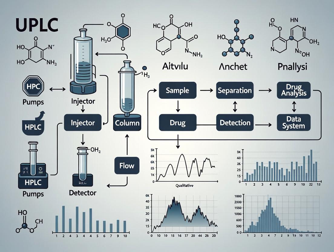
Accelerating Precision Medicine: How UPLC Is Revolutionizing High-Throughput Drug Analysis in Clinical Laboratories
This article provides a comprehensive guide to Ultra-Performance Liquid Chromatography (UPLC) for high-throughput drug analysis in clinical and research settings.
Accelerating Precision Medicine: How UPLC Is Revolutionizing High-Throughput Drug Analysis in Clinical Laboratories
Abstract
This article provides a comprehensive guide to Ultra-Performance Liquid Chromatography (UPLC) for high-throughput drug analysis in clinical and research settings. We explore the foundational principles that give UPLC its superior speed and resolution over HPLC. The piece details advanced methodological workflows for quantifying drugs and metabolites, including TDM, PK/PD studies, and toxicology screening. A dedicated troubleshooting section addresses common challenges like pressure spikes and method transfer, offering optimization strategies for robustness. Finally, we compare UPLC to traditional HPLC and newer techniques, examining validation protocols per ICH/FDA guidelines. Aimed at researchers and drug development professionals, this resource synthesizes current best practices to enhance laboratory efficiency and data reliability in modern pharmacoanalysis.
UPLC 101: The Core Principles Powering Fast, Sensitive Drug Analysis
The shift from High-Performance Liquid Chromatography (HPLC) to Ultra-Performance Liquid Chromatography (UPLC) represents a pivotal technological leap in separation science. Driven by the clinical demand for higher throughput, superior resolution, and reduced solvent consumption in drug analysis and therapeutic drug monitoring (TDM), UPLC has become integral to modern clinical research laboratories. This evolution directly supports the broader thesis that UPLC is indispensable for achieving the speed, sensitivity, and efficiency required for high-throughput drug analysis in clinical settings.
Comparative Performance Data
The quantitative advantages of UPLC over HPLC are summarized in the table below.
Table 1: Direct Comparison of HPLC vs. UPLC System Performance Parameters
| Parameter | Traditional HPLC (5 µm particles) | UPLC (<2 µm particles) | Improvement Factor |
|---|---|---|---|
| Particle Size | 3.5 - 5.0 µm | 1.7 - 1.8 µm | ~3x smaller |
| Optimal Flow Rate | 1.0 - 2.0 mL/min | 0.4 - 0.6 mL/min | ~70% reduction |
| Maximum Pressure | 400 - 600 bar | 1000 - 1500 bar | 2.5x higher |
| Analysis Time | 10 - 30 minutes | 2 - 5 minutes | 5x faster |
| Peak Capacity | 100 - 200 | 200 - 500 | 2.5x higher |
| Solvent Consumption per Run | 10 - 20 mL | 2 - 5 mL | 4x less |
Application Note: High-Throughput TDM for Antiepileptic Drugs
Objective: To simultaneously quantify levels of levetiracetam, lamotrigine, and valproic acid in human plasma with a cycle time under 3 minutes.
Protocol:
- Sample Preparation (Protein Precipitation):
- Pipette 100 µL of patient plasma into a microcentrifuge tube.
- Add 300 µL of acetonitrile containing internal standard (e.g., carbamazepine-d10).
- Vortex vigorously for 60 seconds.
- Centrifuge at 14,000 x g for 10 minutes at 4°C.
- Transfer 150 µL of the clear supernatant to a vial with insert for analysis.
Chromatographic Conditions:
- System: UPLC with PDA or tandem MS detection.
- Column: Acquity UPLC BEH C18 (2.1 x 50 mm, 1.7 µm).
- Mobile Phase A: 0.1% Formic acid in water.
- Mobile Phase B: 0.1% Formic acid in acetonitrile.
- Gradient: 5% B to 95% B over 1.5 minutes, hold for 0.5 min.
- Flow Rate: 0.5 mL/min.
- Column Temperature: 45°C.
- Injection Volume: 2 µL.
Data Analysis:
- Use a quadratic regression model for calibration curves (1-50 µg/mL).
- Apply internal standard normalization for peak area calculations.
- Report concentrations with appropriate clinical reference ranges.
Visualization of Method Evolution
Diagram Title: Clinical Drive for HPLC to UPLC Evolution
The Scientist's Toolkit: Key Reagents and Materials
Table 2: Essential Research Reagent Solutions for UPLC-Based TDM
| Item | Function & Rationale |
|---|---|
| Sub-2µm UPLC Columns (e.g., C18, phenyl) | Core separation media providing high efficiency and resolution under high pressure. |
| LC-MS Grade Solvents (Acetonitrile, Methanol, Water) | Minimize background noise and ion suppression in sensitive mass spectrometric detection. |
| Volatile Buffers (Ammonium formate, Formic acid) | Provide pH control and ion-pairing for separation while being compatible with MS detection. |
| Stable Isotope-Labeled Internal Standards (e.g., drug-analogues with 13C, 15N) | Correct for variability in sample preparation and ionization efficiency, ensuring quantitative accuracy. |
| Certified Drug-Free Human Plasma | Matrix for preparing calibration standards and quality controls to match patient samples. |
| Supported Liquid Extraction (SLE) or μElution SPE Plates | Enable rapid, clean, and high-throughput sample preparation in 96-well format. |
Detailed Protocol: Method Transfer from HPLC to UPLC
Objective: To successfully migrate an existing HPLC method for vitamin D metabolites to a UPLC platform while maintaining or improving data quality.
Experimental Workflow:
Diagram Title: UPLC Method Transfer and Optimization Protocol
Protocol Steps:
- Audit Original HPLC Method: Record column dimensions (e.g., 150 x 4.6 mm, 5 µm), flow rate (e.g., 1.2 mL/min), injection volume (e.g., 20 µL), and exact gradient profile.
- Apply Scaling Calculations:
- Flow Rate: F₂ = F₁ * (dc² / dc¹)², where dc is column diameter. For a shift from 4.6 mm to 2.1 mm ID: F₂ = 1.2 * (2.1/4.6)² ≈ 0.25 mL/min.
- Gradient Time: Adjust to maintain the same number of column volumes. tG2 = tG1 * (F₁/F₂) * (VC2/VC1), where VC is column volume.
- Injection Volume: Scale by column volume ratio, typically reducing to 1-5 µL.
- Column Selection: Choose a UPLC column with similar bonded phase (e.g., C18) but with 1.7-1.8 µm particles. Dimensions typically switch to 50-100 mm length.
- Initial Test Run: Execute the scaled method. Expect similar selectivity with higher backpressure and sharper peaks.
- Fine-Tuning Optimization:
- Slightly adjust gradient steepness (±5-10% organic) to optimize critical peak pairs.
- Increase column temperature (e.g., 45-60°C) to reduce backpressure and improve kinetics.
- Adjust flow rate (±0.1 mL/min) to fine-tune separation or pressure.
- Validation: Perform a system suitability test (resolution, tailing factor, repeatability) and a partial validation per ICH guidelines to confirm accuracy, precision, and linearity on the new UPLC platform.
Within the thesis on Ultra-Performance Liquid Chromatography (UPLC) for high-throughput drug analysis in clinical research laboratories, the adoption of sub-2µm particle technology represents a pivotal advancement. This foundational technology enables the dramatic increases in speed, resolution, and sensitivity required for modern pharmacodynamic, pharmacokinetic, and therapeutic drug monitoring studies. These Application Notes detail the core principles, experimental protocols, and practical implementation of sub-2µm particle columns in UPLC systems to optimize drug analysis workflows.
Core Principles and Data
Sub-2µm particle technology exploits the van Deemter equation, where reduced particle size (dp) minimizes the eddy diffusion (A) and mass transfer (C) terms, leading to a flatter curve and higher optimal linear velocity. This allows for faster separations without sacrificing—and often enhancing—chromatographic resolution (N). The trade-off is increased system pressure, necessitating instrumentation capable of operating at >15,000 psi.
Table 1: Comparative Performance of Particle Technologies in Pharmaceutical Analysis
| Parameter | Traditional HPLC (3-5µm) | UPLC (Sub-2µm) | Performance Gain |
|---|---|---|---|
| Typical Particle Size | 3.5 - 5 µm | 1.7 - 1.8 µm | ~2-3x reduction |
| Typical Plate Height (H) | ~10 µm | ~3-5 µm | 2-3x lower |
| Optimal Linear Velocity | ~1 mm/s | ~2-3 mm/s | 2-3x higher |
| Operational Pressure | 2000 - 4000 psi | 10,000 - 18,000 psi | 3-5x higher |
| Analysis Time (Typical Assay) | 10-20 min | 3-5 min | 60-80% reduction |
| Peak Capacity | 100 - 150 | 200 - 400 | ~2x increase |
| Sensitivity Gain (S/N) | Baseline | 3-5x | Significant |
Table 2: Impact on Key Drug Analysis Metrics in Clinical Research
| Analytical Metric | Effect of Sub-2µm Particles | Implication for High-Throughput Drug Analysis |
|---|---|---|
| Resolution (Rs) | Increased by up to 70% | Better separation of drugs/metabolites; cleaner MS spectra. |
| Run Time | Decreased by 50-80% | Higher sample throughput in TDM and PK studies. |
| Solvent Consumption | Reduced by 60-90% | Lower cost per analysis; reduced waste. |
| Detection Limit | Improved by 3-5x | Enables quantification of low-abundance drugs. |
| Data Density | Higher peaks per unit time | More confident identification in complex matrices. |
Detailed Experimental Protocols
Protocol 1: Method Transfer from HPLC to UPLC with Sub-2µm Particles
Objective: To successfully translate a legacy HPLC method for antiretroviral drug analysis (e.g., Lamivudine, Zidovudine, Nevirapine) from a 5µm, 150 x 4.6 mm column to a UPLC platform using a 1.7µm, 75 x 2.1 mm column while maintaining or improving resolution.
Materials: UPLC system (pressure capable to 18,000 psi), Acquity UPLC BEH C18 column (1.7µm, 75 x 2.1 mm), vial inserts, 0.22 µm PVDF syringe filters, mobile phases (10 mM ammonium formate in water, pH 3.5, and acetonitrile).
Procedure:
- System Preparation: Equilibrate UPLC system with starting mobile phase conditions (95% aqueous, 5% organic) at 0.4 mL/min. Ensure system pressure is within limits.
- Initial Scaling: Calculate the scaled gradient method. Key formula: t_UPLC = t_HPLC * (F_UPLC / F_HPLC) * (L_UPLC / L_HPLC) * (dc_UPLC² / dc_HPLC²), where F is flow rate, L is column length, dc is column inner diameter.
- For the specified columns: tUPLC ≈ tHPLC * (0.4/1.0) * (75/150) * (2.1²/4.6²) ≈ t_HPLC * 0.083.
- A 30-minute HPLC gradient scales to approximately a 2.5-minute UPLC gradient.
- Flow Rate Adjustment: Set initial flow rate to 0.4 mL/min (linear velocity equivalent to ~1 mL/min on 4.6 mm column).
- Injection Volume Scaling: Scale injection volume by column volume ratio. V_inj_UPLC = V_inj_HPLC * (dc_UPLC² * L_UPLC) / (dc_HPLC² * L_HPLC). A 10 µL HPLC injection scales to ~1.0 µL.
- Method Execution: Inject filtered patient serum sample extract (protein precipitated). Run the scaled gradient. Monitor pressure profile.
- Optimization: Adjust gradient slope or temperature if critical pair resolution is lost. Fine-tune flow rate (±0.05 mL/min) for optimal speed/resolution balance.
- Validation: Perform within-run precision (n=6), calibration linearity (5-5000 ng/mL), and carryover assessment per ICH M10 guidelines.
Protocol 2: Assessing System Band Broadening for Sub-2µm Columns
Objective: Quantify extra-column band broadening to ensure it does not degrade the efficiency of a 1.7µm particle column.
Materials: UPLC system, sub-2µm column (e.g., 1.7µm, 50 x 2.1 mm), a zero-dead-volume union, UV detector, 10 nL injection of 0.1% v/v acetone in mobile phase.
Procedure:
- System Dispersion Measurement (Without Column): Replace the column with a zero-dead-volume union. Set flow rate to 0.4 mL/min, isocratic conditions (80% ACN/20% water), detection at 254 nm. Make a 10 nL injection of acetone. Record the resulting peak width at 4.4% height (W₀.₀₄₄).
- Calculate Extra-column Variance (σ²ec): σ²ec = (W₀.₀₄₄)² / (2π), where W₀.₀₄₄ is in time units (seconds).
- Column Performance Measurement: Install the 1.7µm column under the same conditions. Inject the same acetone sample. Record the observed peak variance (σ²_obs).
- Calculate True Column Variance: σ²col = σ²obs - σ²_ec.
- Acceptance Criterion: The extra-column variance should contribute to less than 10% of the total observed variance for a well-retained peak (k' > 2). If contribution is higher, consider using narrower ID tubing, a smaller detector flow cell, or a column with larger i.d.
Mandatory Visualizations
Title: UPLC Method Transfer Workflow
Title: van Deemter Curve and Sub-2µm Impact
The Scientist's Toolkit: Research Reagent Solutions
Table 3: Essential Materials for UPLC with Sub-2µm Particle Columns
| Item & Example Product | Function in Sub-2µm UPLC Analysis |
|---|---|
| UPLC Column (e.g., Acquity UPLC BEH C18, 1.7µm) | The core separation device. Sub-2µm porous particles provide high efficiency. Hybrid silica (BEH) offers high pressure and pH stability (1-12). |
| MS-Compatible Buffers (e.g., Ammonium Formate, Ammonium Acetate) | Provide pH control and ion-pairing for reproducible retention of ionizable drugs without fouling the MS source. |
| LC-MS Grade Solvents (Water, Acetonitrile, Methanol) | Ultra-pure solvents minimize baseline noise, prevent system contamination, and ensure consistent ionization. |
| Solid-Phase Extraction (SPE) Plates (e.g., µElution Plate) | For rapid, parallelized sample cleanup of clinical samples (serum/plasma), concentrating analytes and removing phospholipids that cause matrix effects. |
| 0.22 µm PVDF Syringe Filters | Critical for filtering all mobile phases and sample extracts to prevent clogging of the sub-2µm column frits. |
| Low-Volume, Max Recovery Vials & Inserts | Minimize sample evaporation and adsorption, crucial for the low injection volumes (1-5 µL) typical of 2.1 mm i.d. columns. |
| Needle Wash Solvent (e.g., 50:50 Water:ACN with 0.1% Formic Acid) | Integrated in autosampler protocols to minimize carryover between injections of high-concentration clinical samples. |
| Column Heater/Chiller | Provides precise, stable temperature control (±0.5°C), critical for reproducible retention times and optimal efficiency. |
Ultra-Performance Liquid Chromatography (UPLC) has become the cornerstone of high-throughput drug analysis in modern clinical and pharmaceutical research laboratories. Its superior resolution, speed, and sensitivity compared to traditional HPLC directly address the need for rapid assay development, pharmacokinetic studies, and therapeutic drug monitoring. This application note, framed within a broader thesis on UPLC for clinical high-throughput analysis, details the critical system components, their operational principles, and provides actionable protocols for optimizing drug assays.
Core UPLC System Components and Quantitative Specifications
The performance of a UPLC system in drug analysis is defined by its key components, each contributing to the overall pressure, efficiency, and data fidelity. The following table summarizes the critical specifications for a state-of-the-art system.
Table 1: Key Specifications of Modern UPLC Components for High-Throughput Drug Assays
| System Component | Key Parameter | Typical Specification | Impact on Drug Assay |
|---|---|---|---|
| Solvent Manager (Pump) | Maximum Pressure | 15,000 - 18,000 psi | Enables use of sub-2µm particles for high resolution. |
| Flow Rate Range | 0.001 - 2.0 mL/min | Precise method scaling and micro-flow applications. | |
| Flow Precision | <0.075% RSD | Critical for retention time reproducibility in PK studies. | |
| Sample Manager (Injector) | Injection Volume Range | 0.1 - 50 µL | Allows low-volume injections for precious clinical samples. |
| Carryover | <0.005% | Essential for accuracy in sequential high/low concentration samples. | |
| Temperature Control | 4 - 40°C | Maintains sample integrity for labile compounds. | |
| Column Heater | Temperature Range | 10 - 90°C | Controls selectivity and backpressure; improves reproducibility. |
| Temperature Stability | ± 0.5°C | Vital for robust method transfer between labs. | |
| Detector (PDA) | Sampling Rate | Up to 80 Hz | Captures narrow peaks (<2 sec) without distortion. |
| Wavelength Range | 190 - 800 nm | Enables method development and peak purity assessment. | |
| Noise Level | <±0.5 x 10⁻⁵ AU | Improves limit of quantitation for low-abundance drugs. | |
| Detector (MS-ready) | Acquisition Speed | >10 spectra/sec | Sufficient data points across fast-eluting peaks for reliable ID/quant. |
| Dynamic Range | >4 orders of magnitude | Covers broad drug/metabolite concentrations in biological matrices. |
Detailed Experimental Protocols
Protocol 1: Method Development for a High-Throughput Antiretroviral Drug Panel Assay
Objective: To develop a fast, robust UPLC-PDA method for the simultaneous quantification of Lamivudine, Tenofovir, and Efavirenz in plasma for therapeutic drug monitoring.
Materials & Equipment:
- UPLC system with binary pump, autosampler (maintained at 10°C), column heater, and PDA detector.
- Column: C18, 1.7 µm, 2.1 x 50 mm.
- Mobile Phase A: 0.1% Formic acid in water.
- Mobile Phase B: 0.1% Formic acid in acetonitrile.
- Standard solutions of drugs in methanol:water (50:50, v/v).
Procedure:
- Sample Preparation: Perform protein precipitation of 100 µL plasma with 300 µL of acetonitrile containing internal standard. Vortex for 1 min, centrifuge at 14,000 x g for 10 min at 4°C. Transfer supernatant for analysis.
- Chromatographic Conditions:
- Flow Rate: 0.6 mL/min.
- Column Temperature: 45°C.
- Injection Volume: 2 µL.
- Gradient Program:
- 0-1.0 min: 5% B to 25% B.
- 1.0-2.5 min: 25% B to 80% B.
- 2.5-3.0 min: Hold at 80% B.
- 3.0-3.1 min: 80% B to 5% B.
- 3.1-4.0 min: Re-equilibrate at 5% B.
- PDA Detection: 260 nm for Lamivudine/Tenofovir; 245 nm for Efavirenz.
- Data Analysis: Generate a 5-point calibration curve (0.1 - 20 µg/mL) for each analyte using peak area ratio (analyte/IS). Apply linear regression with 1/x² weighting.
Protocol 2: System Suitability Test for Method Transfer to a Clinical Lab
Objective: To verify UPLC system performance meets pre-defined criteria before implementing a validated drug assay in a high-throughput clinical environment.
Procedure:
- Prepare a system suitability standard containing a model drug (e.g., caffeine) and related compound (e.g., theophylline) at known concentrations.
- Perform six consecutive injections using the established method.
- Acceptance Criteria (must be met):
- Retention Time RSD: ≤ 0.5%.
- Peak Area RSD: ≤ 1.0%.
- Tailing Factor: ≤ 1.5 for main peak.
- Theoretical Plates: > 10,000 per column.
- Resolution between two critical peaks: ≥ 2.0.
System Workflow and Signal Pathway
Diagram 1: UPLC System Analytical Workflow
Diagram 2: UPLC Detector Signal Pathways
The Scientist's Toolkit: Research Reagent Solutions
Table 2: Essential Materials for UPLC Drug Assay Development
| Item | Function & Importance | Example/Note |
|---|---|---|
| Sub-2µm UPLC Columns | Provides high-efficiency separation. The small particle size is key to achieving high resolution at high linear velocities. | C18, 1.7µm, 2.1 x 50-100 mm; HSS T3 for polar compounds. |
| LC-MS Grade Solvents | Minimizes baseline noise and system background, crucial for sensitive detection (especially MS). Reduces contaminant build-up. | Acetonitrile, Methanol, Water with <0.00001% non-volatile residue. |
| High-Purity Mobile Phase Additives | Modifies selectivity and improves ionization efficiency. Impurities cause ion suppression and column degradation. | Formic Acid, Ammonium Formate, Trifluoroacetic Acid (TFA). |
| Stable Isotope Labeled Internal Standards (SIL-IS) | Corrects for variability in sample prep and ionization. Essential for accurate bioanalysis (e.g., d3- or ¹³C-labeled drug). | Deuterated analog of the target analyte. |
| Protein Precipitation Plates | Enables rapid, parallel sample preparation for high-throughput clinical batches. Compatible with autosamplers. | 96-well plates with 0.2 µm filtration or deep-well plates. |
| System Suitability Test Mix | Validates system performance (pressure, injection, detection) before running critical samples. Ensures data integrity. | Contains compounds testing efficiency, tailing, and resolution. |
The escalating complexity of therapeutic drug monitoring (TDM) and pharmacokinetic (PK) studies, driven by precision medicine, biologic therapies, and complex dosing regimens, necessitates a paradigm shift towards high-throughput analytical platforms. Ultra-Performance Liquid Chromatography (UPLC) coupled with tandem mass spectrometry (MS/MS) has emerged as the cornerstone technology, enabling the rapid, precise, and simultaneous quantification of drugs and metabolites critical for clinical decision-making. This application note details protocols and data demonstrating the non-negotiable role of high-throughput UPLC-MS/MS in modern clinical pharmacology.
The High-Throughput Imperative: Quantitative Justification
The demand for faster turnaround times (TAT) in clinical labs is quantifiable. The following table summarizes key performance metrics comparing traditional HPLC with modern UPLC-MS/MS in TDM/PK applications.
Table 1: Performance Comparison: HPLC vs. UPLC-MS/MS for Clinical TDM/PK
| Parameter | Traditional HPLC-UV | Modern UPLC-MS/MS | Impact on Clinical Workflow |
|---|---|---|---|
| Average Run Time | 15-30 minutes/sample | 2-5 minutes/sample | Enables stat analysis and batch processing of large cohorts. |
| Sample Throughput (24h) | 48-96 samples | 288-720 samples | Supports large-scale studies and routine TDM for high-volume drugs. |
| Required Sample Volume | 100-500 µL | 10-50 µL | Essential for pediatric, geriatric, and critically ill patients. |
| Multiplexing Capacity | Typically 1-2 analytes | 10-50+ analytes per run | Allows for combinatorial TDM (e.g., antiretrovirals, antipsychotics) and PK profiling. |
| Method Development Time | Weeks | Days | Rapid response to new drug approvals and clinical needs. |
| Data Point Generation (per study) | Limited by throughput | 10-100x higher | Enhances PK model robustness and detection of rare subpopulations. |
Detailed Application Protocols
Protocol 1: High-Throughput Multiplexed TDM for Antipsychotics
Objective: Simultaneous quantification of 12 commonly prescribed atypical antipsychotics (e.g., aripiprazole, clozapine, olanzapine, risperidone + 9-OH-risperidone) in human plasma.
Workflow Diagram:
Diagram Title: High-Throughput TDM for Antipsychotics Workflow
Key Reagent Solutions:
| Research Reagent / Material | Function & Specification |
|---|---|
| Mass Spec Grade Acetonitrile & Methanol | Low UV absorbance and minimal ion suppression for sensitive MS detection. |
| Ammonium Formate (10mM) | Volatile buffer for mobile phase, compatible with MS ionization. |
| Stable Isotope-Labeled Internal Standards (IS) | e.g., Aripiprazole-d8, Clozapine-d4. Corrects for matrix effects and recovery variability. |
| Charcoal-Stripped Human Plasma | Used for preparation of calibration standards and quality controls. |
| 96-Well Protein Precipitation Plates | Enables parallel processing of 96 samples in <30 minutes. |
| UPLC C18 Column (1.7 µm particles) | Provides high resolution and peak capacity for rapid separations. |
Protocol 2: Rapid PK Profiling for Monoclonal Antibodies (mAbs) via Peptide Mapping
Objective: Quantify a therapeutic mAb in serum using a surrogate signature peptide after rapid enzymatic digestion.
Workflow Diagram:
Diagram Title: Rapid PK Workflow for mAb Analysis
Key Reagent Solutions:
| Research Reagent / Material | Function & Specification |
|---|---|
| Sequencing Grade Modified Trypsin | High-activity enzyme for rapid, reproducible digestion. |
| Tris(2-carboxyethyl)phosphine (TCEP) | Efficient reducing agent for disulfide bonds in fast protocols. |
| Iodoacetamide | Alkylating agent to cap cysteine residues. |
| Signature Peptide Standard (Synthetic) | Unlabeled peptide for calibration curve. |
| Stable Isotope-Labeled (SIL) Signature Peptide | Internal standard for absolute quantification. |
| Solid-Phase Extraction (SPE) Plates (C8) | For high-throughput sample cleanup to remove salts and phospholipids. |
Data Presentation: Throughput-Driven Clinical Outcomes
Table 2: Impact of High-Throughput UPLC-MS/MS on Clinical PK Study Timelines
| Study Phase | Conventional Analysis Timeline (weeks) | High-Throughput UPLC-MS/MS Timeline (weeks) | Key Enabler |
|---|---|---|---|
| Phase I SAD/MAD | 6-8 | 1-2 | Batch analysis of all subjects/time points in a single sequence. |
| Bioequivalence (BE) | 10-12 | 3-4 | Rapid analysis of thousands of samples from large cohorts. |
| Therapeutic Drug Monitoring (Routine) | 24-48 hr TAT | 2-6 hr TAT | Short run times and automated sample prep enable near-real-time reporting. |
| Population PK (PopPK) Modeling | Limited sampling points | Dense sampling feasible | High throughput allows for rich, dense PK profiles from each subject, improving model accuracy. |
The integration of high-throughput UPLC-MS/MS is no longer a luxury but a clinical imperative. It directly addresses the critical needs for speed, multiplexing, and minimal sample volume in modern TDM and PK studies. The protocols outlined provide a framework for implementing this technology, ultimately accelerating dose optimization, supporting personalized therapy, and improving patient outcomes.
Assessing UPLC's Impact on Sample Turnaround Time and Laboratory Workflow Efficiency
Application Notes
Ultra-Performance Liquid Chromatography (UPLC) represents a paradigm shift in chromatographic separations, leveraging sub-2µm particle columns and high-pressure fluidics to achieve superior resolution, speed, and sensitivity compared to traditional High-Performance Liquid Chromatography (HPLC). Within the context of high-throughput drug analysis for clinical research and development, UPLC directly addresses critical bottlenecks. Its implementation significantly compresses analytical run times, reduces solvent consumption, and increases data quality, thereby accelerating pharmacokinetic studies, therapeutic drug monitoring, and metabolomics profiling.
Recent live-search data from published studies and vendor application notes consistently demonstrate UPLC's operational advantages. The summarized quantitative impacts are presented below.
Table 1: Quantitative Comparison of HPLC vs. UPLC Performance Metrics in Drug Analysis
| Performance Metric | Traditional HPLC | UPLC System | % Improvement / Change |
|---|---|---|---|
| Typical Run Time | 10-30 minutes | 2-5 minutes | 70-85% reduction |
| Sample Throughput (per 24h) | 48-96 samples | 288-500+ samples | 300-500% increase |
| Solvent Consumption per Run | ~10 mL | ~2 mL | ~80% reduction |
| Peak Capacity / Resolution | 1x (Baseline) | 1.7x - 2x Increase | 70-100% improvement |
| Detection Sensitivity (Signal-to-Noise) | 1x (Baseline) | 3x - 5x Increase | 200-400% improvement |
| Backpressure Range | 100-400 bar | 600-1500 bar | - |
| Data Acquisition Rate | 1-5 Hz | 10-100 Hz | 10-20x increase |
Detailed Experimental Protocols
Protocol 1: High-Throughput Therapeutic Drug Monitoring (TDM) for Immunosuppressants
Objective: To quantify tacrolimus, cyclosporine A, sirolimus, and everolimus simultaneously from patient plasma with a sub-3-minute cycle time.
Materials & Reagents:
- UPLC System: Acquity or comparable, with PDA/UV and/or tandem MS detector.
- Column: C18 UPLC column (1.7µm, 2.1 x 50 mm).
- Mobile Phase A: 0.1% Formic acid in water.
- Mobile Phase B: 0.1% Formic acid in acetonitrile.
- Internal Standard: Ascomycin.
- Sample Prep: Protein precipitation with zinc sulfate in methanol.
Method:
- Sample Preparation: Aliquot 100 µL of calibrator, control, or patient plasma. Add 25 µL of internal standard working solution. Vortex. Add 300 µL of precipitant. Vortex vigorously for 1 minute and centrifuge at 13,000 x g for 5 minutes.
- Chromatography: Transfer supernatant to a UPLC vial. Inject 5 µL.
- Gradient Program:
- Flow Rate: 0.6 mL/min.
- Initial: 30% B.
- 0.5 min: Ramp to 70% B.
- 1.8 min: Ramp to 95% B. Hold until 2.2 min.
- 2.21 min: Return to 30% B. Re-equilibrate until 3.0 min.
- Detection: MS/MS detection in positive ESI MRM mode. Data acquisition at 20 Hz.
Protocol 2: Rapid Pharmacokinetic (PK) Profiling of Small Molecule Drug Candidates
Objective: To support high-throughput PK studies by analyzing drug and its major metabolite from serial mouse/rat plasma samples.
Materials & Reagents:
- UPLC System: As above.
- Column: HSS T3 UPLC column (1.8µm, 2.1 x 30 mm).
- Mobile Phase A: 10 mM Ammonium acetate in water.
- Mobile Phase B: 10 mM Ammonium acetate in 90:10 Acetonitrile:Methanol.
- Sample Prep: 96-well plate format solid-phase extraction (SPE).
Method:
- Sample Preparation: Using a 96-well SPE plate (cation exchange), condition with methanol, then water. Load 50 µL of plasma sample (diluted 1:1 with acidified water). Wash with 5% methanol. Elute with 80:20 methanol:ammonium hydroxide. Evaporate and reconstitute in initial mobile phase.
- Chromatography: Inject 2 µL.
- Gradient Program (Fast):
- Flow Rate: 0.5 mL/min.
- Initial: 5% B.
- 0.2 min: Ramp to 40% B.
- 1.0 min: Ramp to 95% B. Hold until 1.3 min.
- 1.31 min: Return to 5% B. Re-equilibrate until 1.8 min (total run time).
- Detection: UV at 254 nm and MS/MS for confirmation. High-speed data acquisition ensures adequate peak definition.
Visualizations
Title: UPLC-Based High-Throughput Drug Analysis Workflow
Title: UPLC Impact Pathway on Lab Efficiency
The Scientist's Toolkit: Key Research Reagent Solutions
Table 2: Essential Materials for UPLC-Based Drug Analysis
| Item | Function & Critical Role |
|---|---|
| 1.7µm ACQUITY UPLC BEH C18 Column | Provides the core separation power; sub-2µm particles enable high efficiency at high linear velocities. |
| Mass Spectrometry-Compatible Buffers (e.g., Ammonium Formate/Acetate, 0.1% FA) | Ensure volatile mobile phases for optimal ionization and detector sensitivity in LC-MS/MS. |
| High-Purity Gradient-Grade Solvents (ACN, MeOH) | Minimize baseline noise and system backpressure, crucial for stable UPLC performance. |
| 96-Well SPE Plates & Automated Liquid Handlers | Enable parallel processing of tens to hundreds of samples, aligning prep speed with UPLC analysis speed. |
| Stable Isotope-Labeled Internal Standards (SIL-IS) | Compensates for matrix effects and variability in extraction/ionization, ensuring quantitative accuracy. |
| Dedicated UPLC Vials & Caps (Low Volume, Low Adsorption) | Prevent sample loss and carryover, critical for small injection volumes (1-5 µL). |
| System Suitability Test (SST) Mixture | A cocktail of analytes to verify column performance, system pressure, and detector response daily. |
Building Robust UPLC Methods for TDM, Toxicology, and Biologics Analysis
Within the broader thesis on implementing Ultra-Performance Liquid Chromatography (UPLC) for high-throughput drug analysis in clinical laboratories, method development is the critical path to success. This protocol details the systematic development of robust, rapid, and reproducible UPLC-MS/MS methods for comprehensive drug panels, addressing the urgent need for fast and accurate toxicology and therapeutic drug monitoring in clinical research.
Core Column Selection Strategy
The choice of stationary phase is foundational. For broad-spectrum drug panels encompassing acidic, basic, and neutral compounds (e.g., opioids, benzodiazepines, stimulants, antidepressants), reversed-phase chemistry is standard. The current trend in high-throughput clinical labs favors superficially porous particle (SPP, e.g., Fused-Core) or sub-2µm fully porous particle columns for UPLC.
Table 1: Column Selection Guide for Drug Panel Analysis
| Column Type | Particle Size | Dimensions (mm) | Recommended For | Typical Plate Count | Max Pressure (psi) |
|---|---|---|---|---|---|
| C18 (AQ) | 1.7 - 1.8 µm | 50-100 x 2.1 | Broad-polar panels, includes hydrophilic drugs | ~200,000/m | 15,000-18,000 |
| Phenyl-Hexyl | 2.6 - 2.7 µm (SPP) | 50-100 x 2.1 | Isomeric separation, benzodiazepines | ~150,000/m | 6,000-9,000 |
| PFP (Pentafluorophenyl) | 1.8 - 1.9 µm | 50-75 x 2.1 | Challenging separations (e.g., catecholamines, structural analogs) | ~190,000/m | 15,000 |
| HILIC | 1.7 - 3.0 µm | 50-100 x 2.1 | Polar, hydrophilic bases (e.g., polar metabolites) | ~150,000/m | 15,000 |
Protocol 2.1: Initial Column Screening
- Setup: Acquire 3-4 columns (e.g., C18-AQ, PFP, Phenyl, HILIC) with identical dimensions (e.g., 50 x 2.1 mm).
- Conditioning: Flush each new column with 10-20 column volumes of starting mobile phase at 50% flow rate.
- Test Mix: Inject a standard mix of 5-10 drugs representing your panel's chemical diversity (e.g., morphine (polar base), diazepam (neutral), barbital (acid), amphetamine (base)).
- Isocratic Scout: Use a simple, moderate elution strength mobile phase (e.g., 40% acetonitrile in 10 mM ammonium formate, pH 3.5). Flow: 0.4 mL/min. Column temp: 40°C.
- Evaluation: Assess peak shape (asymmetry factor, 0.8-1.2 ideal), retention (k' between 1-10), and resolution. Select the column providing the best overall compromise.
Mobile Phase Optimization
Mobile phase composition dictates selectivity, peak shape, and MS sensitivity.
Table 2: Common Mobile Phase Additives for Drug Panels in UPLC-MS/MS
| Additive | Concentration | pH (approx.) | Primary Function | Best For | MS Compatibility |
|---|---|---|---|---|---|
| Ammonium Formate | 2-10 mM | 3.0-4.0 (acidic) | Volatile buffer; improves peak shape for bases | General drug panels, positive ESI | Excellent |
| Ammonium Acetate | 2-10 mM | 4.5-5.5 (mid) | Volatile buffer; useful for some amphoteric drugs | Broad-spectrum panels | Excellent |
| Formic Acid | 0.05 - 0.2% | ~2.7 | Protonation agent; enhances [M+H]+ signal | Basic/neutral drugs in +ESI | Excellent |
| Acetic Acid | 0.05 - 0.2% | ~3.8 | Milder acid than formic; alternative selectivity | Basic drugs prone to in-source fragmentation | Excellent |
| Ammonium Bicarbonate | 2-10 mM | ~8.0 (basic) | Volatile basic buffer for negative ESI or acidic drugs | NSAIDs, barbiturates, cannabinoids in -ESI | Good (requires careful source tuning) |
Protocol 3.1: pH and Buffer Scouting
- Prepare Buffers: Prepare Mobile Phase A (MPA) as water with (a) 0.1% formic acid, (b) 10 mM ammonium formate pH 3.0, (c) 10 mM ammonium acetate pH 5.0, (d) 0.1% acetic acid. Keep MPB as organic modifier (acetonitrile or methanol) with identical additive.
- Rapid Gradient: Run a fast, generic gradient from 5% to 95% MPB over 5 min on selected column. Flow: 0.4 mL/min.
- Analyze: Observe shifts in retention order and changes in peak shape (especially for early eluting polar bases). Acidic pH (formate/formic) typically yields best peak shape for most basic drugs.
Gradient Profile Development
The gradient is engineered for speed, resolution, and re-equilibration.
Protocol 4.1: Developing a High-Throughput Gradient
- Initial Steep Gradient: Using optimized MP A/B, start with 5% B, ramp to 95% B over 1.5-2.0 minutes for a 50-100mm column. Hold for 0.3 min.
- Identify Co-elutions: Use a drug panel mix of 50+ compounds. MS/MS detection in MRM mode helps identify co-eluting isobars.
- Insert Shallower Segments: For regions with critical co-elutions (e.g., isomers like oxycodone/hydrocodone), reduce gradient slope (e.g., 0.5-1.0% B/min) over that specific window.
- Re-equilibration: Program a rapid return to initial conditions (5% B) and hold for 0.5-1.0 column volumes (e.g., 0.5 min for 50 mm column). Confirm retention time stability across consecutive injections.
- Final Gradient Example for 50-Drug Panel:
- 0.0-0.2 min: 5% B (isocratic hold for loading)
- 0.2-3.0 min: 5% → 40% B (linear)
- 3.0-4.0 min: 40% → 55% B (shallower segment for critical pairs)
- 4.0-4.5 min: 55% → 95% B (linear)
- 4.5-5.0 min: 95% B (wash)
- 5.0-5.5 min: 95% → 5% B (rapid return)
- 5.5-6.5 min: 5% B (re-equilibration)
- Total Run Time: 6.5 min.
Systematic Method Validation Protocol
Protocol 5.1: Key Validation Experiments for Clinical Research
- Selectivity: Analyze 6 independent sources of blank matrix (e.g., human plasma). No significant interference at analytes' retention times (<20% of LLOQ).
- Calibration & Linearity: Prepare 8-point calibration curve in matrix. Use 1/x or 1/x² weighting. Acceptable range: R² > 0.99, accuracy 85-115%.
- Precision & Accuracy: QC samples at LLOQ, Low, Mid, High (n=6 per level, across 3 days). Criteria: Intra-/inter-day precision (CV) ≤15% (≤20% at LLOQ), accuracy 85-115%.
- Carryover: Inject blank after upper limit of quantification (ULOQ). Must be <20% of LLOQ.
- Matrix Effects & Recovery: Post-extraction addition vs. neat solution (for ionization efficiency) and comparison of extracted samples vs. post-extraction spiked (for recovery). Acceptable IS-normalized matrix factor CV <15%.
Title: UPLC Method Development Workflow for Drug Panels
Title: Gradient Optimization Decision Logic
The Scientist's Toolkit: Key Research Reagent Solutions
Table 3: Essential Materials for UPLC-MS/MS Drug Panel Method Development
| Item | Supplier Examples | Function in Method Development |
|---|---|---|
| Mixed Drug Standard Panels | Cerilliant, Lipomed, UTAK | Pre-mixed certified reference materials for 50+ drugs; saves preparation time and ensures accuracy. |
| SPE Cartridges (Mixed-Mode) | Waters Oasis MCX, Agilent Bond Elut Plexa | For robust sample clean-up; cationic exchange for basic/neutral drugs from biological matrix. |
| UPLC Columns (C18, PFP, HILIC) | Waters ACQUITY, Thermo Accucore, Phenomenex Kinetex | Core separation tools; sub-2µm or SPP for high efficiency and speed. |
| MS-Compatible Buffers | Fluka, Honeywell (LC-MS grade ammonium salts) | High-purity volatile buffers to maintain MS sensitivity and prevent source contamination. |
| LC-MS Grade Solvents | Fisher Optima, Honeywell Burdick & Jackson | Minimal background ions; essential for low LLOQs and clean baselines. |
| Surrogate/Internal Standards (Isotope-Labeled) | Cambridge Isotope Labs, Cerilliant | Correct for matrix effects and variability in extraction/ionization; crucial for quantification. |
| Artificial/Blank Matrices | BioreclamationIVT, UTAK | For preparing calibration curves without sourcing biological donor matrices. |
| Quality Control Materials | BIO-RAD, UTAK | Independent validation of method accuracy and precision across analytical runs. |
Within the broader thesis on Ultra-Performance Liquid Chromatography (UPLC) for high-throughput drug analysis in clinical laboratories, this document details specific Application Notes and Protocols for three critical TDM classes. The central thesis posits that UPLC-MS/MS, with its superior resolution, speed, and sensitivity compared to traditional HPLC, is the enabling technology for the efficient, multiplexed analysis required by modern personalized medicine. This section translates that thesis into practical methodologies for immunosuppressants, antiepileptics, and antibiotics, addressing the urgent need for rapid turnaround to guide dosing decisions.
Table 1: Key Analytical Parameters for High-Throughput UPLC-MS/MS TDM
| Drug Class | Exemplar Drugs | Therapeutic Range (Typical) | Sample Volume (µL) | UPLC Runtime (min) | Internal Standard (Example) | Critical Matrix Effect Consideration |
|---|---|---|---|---|---|---|
| Immunosuppressants | Tacrolimus, Cyclosporin A, Sirolimus, Everolimus | Tacro: 5-20 ng/mL; CsA: 100-400 ng/mL (C0) | 50-100 | 2.5 - 4.0 | Tacrolimus-13C,D2, Cyclosporin D | Severe ion suppression from phospholipids; requires robust protein precipitation/SPE. |
| Antiepileptics | Levetiracetam, Lamotrigine, Valproic Acid, Carbamazepine | Lamotrigine: 3-14 µg/mL; Levetiracetam: 12-46 µg/mL | 10-50 | 1.5 - 3.0 | Carbamazepine-D10, Levetiracetam-D6 | Wide polarity range necessitates versatile gradient; valproic acid requires negative ionization. |
| Antibiotics | Vancomycin, Gentamicin, Voriconazole, Piperacillin | Vancomycin: Trough 10-20 µg/mL; Gentamicin: Peak 5-10 µg/mL | 50-100 | 2.0 - 3.5 | Vancomycin-D6, Gentamicin C1a-D5 | Highly polar molecules (e.g., aminoglycosides) require HILIC or ion-pairing; β-lactams are thermally labile. |
Table 2: Representative High-Throughput UPLC-MS/MS Method Performance
| Parameter | Immunosuppressants (Multiplex) | Antiepileptics (Multiplex) | Antibiotics (Vanco/Gent/Voriconazole) |
|---|---|---|---|
| Linearity (R²) | >0.998 for all analytes | >0.995 for all analytes | >0.990 for all analytes |
| Precision (%CV) | Intra-run: <6%; Inter-run: <9% | Intra-run: <5%; Inter-run: <8% | Intra-run: <7%; Inter-run: <10% |
| Accuracy (% Bias) | ±12% across range | ±10% across range | ±15% at LLOQ |
| LLOQ | 0.5-1.0 ng/mL | 0.1-0.5 µg/mL | 0.2-1.0 µg/mL |
| Carryover | <0.5% of LLOQ | <0.2% of LLOQ | <0.5% of LLOQ |
| Sample Prep Method | Protein Precipitation + Phospholipid Removal Plate | Direct Protein Precipitation | Protein Precipitation (Acidified for β-lactams) |
Detailed Experimental Protocols
Protocol A: Multiplex Analysis of Immunosuppressants (Tacrolimus, Cyclosporin A, Sirolimus, Everolimus)
- Principle: Quantification via UPLC-MS/MS using stable isotope-labeled internal standards (SIL-IS) to correct for matrix effects and recovery losses.
- Materials: Whole blood (EDTA), calibrators/controls, SIL-IS working solution, zinc sulfate precipitant, methanol (LC-MS grade), acetonitrile (LC-MS grade), water (LC-MS grade), formic acid, 96-well phospholipid removal plates.
- Equipment: UPLC system (e.g., Waters ACQUITY, Thermo Vanquish), tandem quadrupole MS (e.g., Sciex 6500+, Waters Xevo TQ-S), positive electrospray ionization (ESI+), C18 column (e.g., 2.1 x 50 mm, 1.7 µm).
- Workflow:
- Aliquoting: Pipette 50 µL of whole blood calibrator, control, or patient sample into a 96-well plate.
- Internal Standard Addition: Add 100 µL of precipitant solution (0.1M ZnSO₄ in methanol/water containing SIL-IS).
- Protein Precipitation: Seal, vortex mix for 5 minutes, then centrifuge at 4000 x g for 10 min at 10°C.
- Phospholipid Removal: Transfer supernatant to a 96-well phospholipid removal plate. Apply positive pressure or centrifuge to collect filtrate.
- Injection: Inject 5-10 µL of filtrate onto the UPLC-MS/MS system.
- Chromatography:
- Column Temperature: 55°C
- Mobile Phase A: 2 mM ammonium acetate + 0.1% formic acid in water.
- Mobile Phase B: 2 mM ammonium acetate + 0.1% formic acid in methanol.
- Gradient: 40% B to 95% B over 2.0 min, hold 0.5 min, re-equilibrate. Total cycle time: 3.5 min.
- Mass Spectrometry (ESI+):
- Monitor 2-3 MRM transitions per analyte for quantification and confirmation.
- Example: Tacrolimus: 821.5 → 768.5 (quantifier); 821.5 → 786.5 (qualifier).
Protocol B: High-Throughput Serum Antiepileptic Drug Panel
- Principle: Fast, isocratic/gradient separation of chemically diverse AEDs with polarity switching ESI.
- Materials: Human serum, calibrators/controls, deuterated IS mix, acetonitrile (precipitation solvent).
- Equipment: UPLC-MS/MS, C18 or phenyl column, ESI capable of rapid polarity switching.
- Workflow:
- Precipitation: Mix 10 µL serum with 10 µL IS working solution and 200 µL cold acetonitrile.
- Centrifugation: Vortex, centrifuge at 15,000 x g for 5 min.
- Dilution: Dilute supernatant 1:1 with water.
- Injection: Inject 2 µL.
- Chromatography: Fast gradient from 5% to 95% organic phase (methanol/acetonitrile with 0.1% formic acid) over 1.2 min on a 2.1 x 30 mm column. Total cycle: 2.0 min.
- Mass Spectrometry: ESI+ for most AEDs (e.g., lamotrigine, carbamazepine); ESI- for valproic acid. MRM acquisition.
Protocol C: Simultaneous Quantification of Vancomycin, Gentamicin, and Voriconazole
- Principle: Combines analysis of polar (gentamicin) and less polar (vancomycin, voriconazole) antibiotics using a synergistic chromatography approach (e.g., mixed-mode or shallow HILIC gradient).
- Materials: Plasma/serum, calibrators/controls, appropriate deuterated IS, trichloroacetic acid or acidified acetonitrile for precipitation.
- Workflow:
- Precipitation: Add 50 µL sample to 150 µL precipitant (e.g., 3% trichloroacetic acid in water containing IS).
- Centrifugation: Vortex, centrifuge at high speed for 5 min.
- Injection: Transfer supernatant to vial and inject 5-10 µL.
- Chromatography: Use a charged surface hybrid (CSH) or HILIC column. Shallow gradient with mobile phase A: 10 mM ammonium formate in water (pH 3.0), B: 10 mM ammonium formate in acetonitrile. Runtime: ~3.0 min.
- Mass Spectrometry: ESI+ for all. Gentamicin is monitored as a sum of its major components (C1, C1a, C2). Careful optimization of cone voltage is needed for fragile β-lactams if included.
Visualization: Workflows and Pathways
UPLC-MS/MS Workflow for Immunosuppressant TDM
Thesis Framework Linking UPLC Tech to TDM Applications
The Scientist's Toolkit: Essential Research Reagents & Materials
Table 3: Key Research Reagent Solutions for High-Throughput TDM by UPLC-MS/MS
| Item / Reagent | Function & Rationale | Critical Specification |
|---|---|---|
| Stable Isotope-Labeled Internal Standards (SIL-IS) | Corrects for variability in extraction efficiency, ionization suppression/enhancement, and instrument drift. Essential for accurate quantification. | Isotopic purity (>99%), chemical stability, identical retention time to unlabeled analyte. |
| Matrix-Matched Calibrators & Controls | Provides the calibration curve and QC for accurate measurement. Must mimic patient sample matrix. | Prepared in pooled, analyte-free human plasma/whole blood. Value-assigned per CLSI guidelines. |
| Phospholipid Removal Plate (e.g., HybridSPE, Ostro) | Selectively removes phospholipids from protein precipitation supernatants, drastically reducing matrix effects in ESI-MS. | 96-well format for high throughput. Compatibility with organic supernatants. |
| LC-MS Grade Solvents & Additives | Minimizes background noise, ion suppression, and system contamination, ensuring method sensitivity and robustness. | Low UV absorbance, volatile acid/base purity (e.g., formic acid, ammonium hydroxide). |
| Solid-Phase Extraction (SPE) Plates (e.g., µElution) | For demanding applications (e.g., very low LLOQ, complex matrices). Provides clean-up and analyte concentration. | Chemistries: C18, mixed-mode cation/anion. 2-10 mg sorbent mass in 96-well format. |
| UPLC Columns (Sub-2µm Particle) | Provides the fast separations and high peak capacity required for high-throughput multiplex analysis. | Column chemistry: C18, phenyl, HILIC, CSH. Dimensions: 2.1 x 50-100 mm, 1.7-1.8 µm. |
| Mass Spectrometry Tuning & Calibration Solutions | Ensures optimal instrument sensitivity, mass accuracy, and resolution. Performed regularly. | Vendor-specific mixtures (e.g., sodium cesium iodide for quadrupoles). |
The quantitative analysis of drugs and their metabolites in biological matrices is a cornerstone of clinical pharmacology, therapeutic drug monitoring, and toxicology. The integration of Ultra-Performance Liquid Chromatography (UPLC) with tandem mass spectrometry (MS/MS) has revolutionized this field, enabling high-throughput, sensitive, and specific assays essential for modern clinical laboratory research. This application note details validated protocols for analyzing analytes in plasma, urine, and dried blood spots (DBS), framed within a thesis on optimizing UPLC for scalable clinical applications.
Key Research Reagent Solutions
The following table details essential materials and their functions for developing robust UPLC-MS/MS assays.
| Reagent/Material | Function & Rationale |
|---|---|
| Stable Isotope-Labeled Internal Standards (SIL-IS) | Corrects for matrix effects, ionization variability, and sample preparation losses. Essential for accurate quantification. |
| Hybrid SPE-PPT 96-Well Plates | Combines protein precipitation and solid-phase extraction for efficient phospholipid removal and cleaner extracts from plasma. |
| Weak Cation Exchange (WCX) SPE Cartridges | Selective extraction of basic drugs and metabolites from urine, reducing ionic interferences. |
| DBS Punches (3 mm) | Allows for volumetric sampling from dried blood spot cards, enabling miniaturized and remote sample collection. |
| Ammonium Formate Buffer (pH 3.0 & 10.0) | Provides volatile buffering for UPLC mobile phases, enhancing chromatographic peak shape and MS compatibility. |
| Methanol with 0.1% Formic Acid | Common organic modifier and reconstitution solvent; acid enhances positive-mode electrospray ionization (ESI+). |
Comparative Matrix Data & Method Performance
The table below summarizes quantitative performance data for a model panel of five drugs (e.g., Antiepileptic, Antidepressant, Immunosuppressant) across the three matrices.
Table 1: Method Performance Summary Across Matrices
| Matrix | Analytes | Linear Range (ng/mL) | LLOQ (ng/mL) | Extraction Recovery (%) | Matrix Effect (%CV) | Intra-day Precision (%RSD) |
|---|---|---|---|---|---|---|
| Plasma (EDTA) | Drug A-E | 1-1000 | 1.0 | 85-92 | 3-8 | ≤6 |
| Urine | Drug A-E | 10-5000 | 10.0 | 88-95 | 5-12 | ≤8 |
| Dried Blood Spot | Drug A-E | 5-2500 | 5.0 | 78-85 | 8-15 | ≤10 |
Detailed Experimental Protocols
Protocol 1: Plasma Sample Preparation (Hybrid SPE-PPT)
- Aliquot & Spike: Transfer 50 µL of calibrator, QC, or patient plasma to a 96-well hybrid SPE-PPT plate.
- Add Internal Standard: Add 100 µL of SIL-IS working solution in acetonitrile.
- Precipitate & Filter: Vortex mix for 5 minutes. Apply vacuum (≈5 inHg) to simultaneously precipitate proteins and filter the extract.
- Collect & Evaporate: Collect eluate into a clean 96-well collection plate. Evaporate to dryness under nitrogen at 40°C.
- Reconstitute & Analyze: Reconstitute in 100 µL of mobile phase A/B (50:50, v/v). Vortex and centrifuge. Inject 2-5 µL onto UPLC-MS/MS.
Protocol 2: Urine Sample Preparation (WCX SPE)
- Dilution: Dilute 100 µL of urine sample with 400 µL of 2% formic acid in water.
- Condition & Load: Condition a 30 mg WCX cartridge with 1 mL methanol, then 1 mL water. Load the diluted sample.
- Wash & Dry: Wash with 1 mL of 2% formic acid, followed by 1 mL methanol. Dry under full vacuum for 5 minutes.
- Elute: Elute analytes with 1 mL of 5% ammonium hydroxide in methanol.
- Evaporate & Reconstitute: Evaporate eluate to dryness. Reconstitute in 150 µL of reconstitution solvent. Centrifuge and inject.
Protocol 3: Dried Blood Spot (DBS) Sample Preparation
- Punch: Punch a single 3 mm disc from the center of the DBS sample into a 96-well plate.
- Extraction: Add 125 µL of extraction solvent (methanol:water:formic acid, 80:19:1, v/v/v) containing SIL-IS.
- Shake & Elute: Seal the plate and shake on a orbital mixer for 30 minutes at room temperature.
- Transfer: Transfer 100 µL of the supernatant to a clean 96-well analysis plate.
- Analyze: Inject directly onto the UPLC-MS/MS system.
Protocol 4: UPLC-MS/MS Analytical Conditions
- System: UPLC with binary pump, FTN autosampler (held at 10°C), column oven (40°C).
- Column: C18, 2.1 x 50 mm, 1.7 µm particle size.
- Mobile Phase: A) 0.1% Formic acid in water, B) 0.1% Formic acid in acetonitrile.
- Gradient: 5% B to 95% B over 2.5 minutes, hold for 0.5 min, re-equilibrate. Total run time: 4.0 min.
- Flow Rate: 0.6 mL/min.
- MS: Triple quadrupole with ESI+. MRM mode. Desolvation temperature: 500°C. Capillary voltage: 1.0 kV.
Visualized Workflows & Relationships
High-Throughput UPLC-MS/MS Bioanalysis Workflow
Matrix Challenges and UPLC Solutions
Within the broader thesis on UPLC for high-throughput drug analysis in clinical laboratories, UPLC-MS/MS emerges as the cornerstone technology. It enables the rapid, sensitive, and selective quantification of drugs and metabolites in complex biological matrices, directly fueling critical clinical research endpoints in PK/PD and bioequivalence (BE) studies. This application note details protocols and workflows central to this paradigm.
Key Applications & Quantitative Data
Pharmacokinetics: Quantification of Drug and Metabolites
UPLC-MS/MS enables the construction of concentration-time profiles for drugs and their major metabolites, which are essential for calculating PK parameters.
Table 1: Representative PK Parameters Derived from UPLC-MS/MS Data
| Parameter | Symbol | Typical Units | Description & Clinical Relevance |
|---|---|---|---|
| Maximum Concentration | C~max~ | ng/mL | Peak plasma concentration, indicating absorption rate and extent. |
| Time to C~max~ | T~max~ | hours | Time to reach peak concentration, reflecting absorption rate. |
| Area Under the Curve | AUC~0-t~, AUC~0-∞~ | ng·h/mL | Total drug exposure over time; primary measure of bioavailability. |
| Elimination Half-life | t~1/2~ | hours | Time for plasma concentration to reduce by 50%; dictates dosing interval. |
| Clearance | CL | L/h | Volume of plasma cleared of drug per unit time. |
| Volume of Distribution | V~d~ | L | Apparent volume into which the drug disperses. |
Bioequivalence Assessment
BE studies rely on comparing the rate and extent of absorption of a test (T) formulation against a reference (R) formulation. UPLC-MS/MS provides the precision and accuracy required for regulatory acceptance.
Table 2: Key Bioequivalence Criteria and Acceptance Ranges
| Metric | Parameter | Regulatory Acceptance Range (90% CI) | UPLC-MS/MS Role |
|---|---|---|---|
| Extent of Absorption | AUC~0-t~ | 80.00% - 125.00% | Primary endpoint; requires high accuracy and reproducibility. |
| Rate of Absorption | C~max~ | 80.00% - 125.00%* | Critical endpoint; demands high sensitivity for accurate T~max~ and C~max~. |
| Statistical Power | N/A | Typically >80% | Enabled by low inter- and intra-assay CVs (<15%), reducing required sample size. |
*Some agencies allow a wider range for highly variable drugs.
Pharmacodynamics: Exposure-Response & Biomarkers
Integrating PK data with PD endpoints (efficacy/safety biomarkers) enables exposure-response analysis, crucial for dose optimization.
Table 3: Common PD Biomarkers Quantified by UPLC-MS/MS in Clinical Trials
| Biomarker Class | Example Analytes | Matrix | Role in PK/PD Modeling |
|---|---|---|---|
| Target Engagement | Phosphorylated proteins, enzyme substrates/products | Plasma, PBMCs | Links drug concentration to proximal molecular effect. |
| Efficacy | Circulating lipids (PCSK9 inhibitors), glucose metabolites | Serum, Plasma | Correlates drug exposure to therapeutic effect. |
| Safety/Toxicity | Bile acids, creatinine, specific acyl-carnitines | Serum, Urine | Identifies exposure thresholds for adverse events. |
Experimental Protocols
Protocol: High-Throughput Quantitative Bioanalysis for a Small Molecule PK Study
Objective: To quantify Drug X and its major metabolite M1 in human plasma for a Phase I PK study.
I. Sample Preparation (Protein Precipitation)
- Thaw & Aliquot: Thaw frozen human plasma samples (K~2~EDTA) at room temperature. Vortex briefly.
- Pipette: Transfer 50 µL of calibrator, quality control (QC), or study sample into a 96-well plate.
- Internal Standard (IS) Addition: Add 100 µL of IS working solution (Drug X-d~6~ and M1-d~4~ at 50 ng/mL in acetonitrile) to all wells.
- Precipitate: Seal plate, vortex mix for 5 minutes, then centrifuge at 4,000 x g for 10 minutes at 4°C.
- Transfer: Transfer 100 µL of the supernatant to a fresh 96-well plate containing 100 µL of water. Seal and vortex for 2 minutes prior to UPLC-MS/MS injection.
II. UPLC-MS/MS Conditions
- System: UPLC coupled to a triple quadrupole mass spectrometer.
- Column: Acquity UPLC BEH C18 (1.7 µm, 2.1 x 50 mm).
- Mobile Phase: A: 0.1% Formic acid in water. B: 0.1% Formic acid in acetonitrile.
- Gradient:
Time (min) Flow (mL/min) %A %B 0.0 0.5 95 5 1.0 0.5 95 5 2.5 0.5 5 95 3.5 0.5 5 95 3.6 0.5 95 5 4.5 0.5 95 5 - Column Temp: 40°C. Injection Volume: 5 µL.
- MS Source: ESI positive mode. Capillary Voltage: 3.0 kV. Source Temp: 150°C.
- MRM Transitions:
- Drug X: 405.2 → 243.1 (Collision Energy: 20 eV)
- M1: 421.2 → 259.1 (Collision Energy: 18 eV)
- IS (Drug X-d~6~): 411.2 → 249.1 (CE: 20 eV)
- IS (M1-d~4~): 425.2 → 263.1 (CE: 18 eV)
III. Data Analysis
- Plot peak area ratio (analyte/IS) vs. nominal concentration for calibrators.
- Fit using a weighted (1/x²) linear regression model.
- Back-calculate concentrations of QCs and study samples using the calibration curve.
- Apply acceptance criteria: Calibrators within ±15% of nominal (±20% at LLOQ); ≥67% of QCs within ±15% of nominal.
Visualization
Diagram: UPLC-MS/MS Workflow in Clinical PK/PD Studies
Title: Workflow for UPLC-MS/MS in PK/PD and Bioequivalence Studies
Diagram: Integration of PK and PD Data for Modeling
Title: PK/PD Data Integration and Modeling Pathway
The Scientist's Toolkit: Key Research Reagent Solutions
Table 4: Essential Materials for UPLC-MS/MS Clinical Bioanalysis
| Item | Function & Importance |
|---|---|
| Stable Isotope-Labeled Internal Standards (SIL-IS) (e.g., Drug-d~6~) | Corrects for matrix effects and variability in extraction/ionization; essential for quantitative accuracy. |
| Blank Biological Matrix (e.g., charcoal-stripped human plasma) | Used for preparation of calibration standards and quality controls, ensuring matrix-matched quantification. |
| LC-MS Grade Solvents & Additives (Acetonitrile, Methanol, Formic Acid) | Minimizes background noise, prevents system contamination, and ensures optimal ionization efficiency. |
| High-Throughput Sample Prep Plates & Seals (96-well, protein precipitation plates) | Enables rapid, parallel processing of hundreds of clinical samples, critical for study throughput. |
| Quality Control (QC) Materials at Low, Mid, High concentrations | Monitors assay precision and accuracy throughout the batch run; key for GLP/GCP compliance. |
| Appropriate UPLC Columns (e.g., BEH C18, HSS T3 for polar analytes) | Provides the required resolution, speed, and peak shape for separating analytes from matrix interferences. |
Within the high-throughput drug analysis paradigm of modern clinical research laboratories, Ultra-Performance Liquid Chromatography (UPLC) has become indispensable. This note details protocols and data for the characterization of peptides, monoclonal antibodies (mAbs), and Antibody-Drug Conjugates (ADCs), emphasizing speed, resolution, and sensitivity critical for accelerating drug development timelines.
Table 1: UPLC Method Performance for Biotherapeutic Analysis
| Analyte Class | Column | Gradient Time | Key Performance Metric | Value |
|---|---|---|---|---|
| Therapeutic Peptide (GLP-1 analog) | C18, 1.7µm, 2.1x100mm | 5 min | Resolution (Critical Pair) | ≥ 2.1 |
| mAb (IgG1) Tryptic Map | C18, 1.7µm, 2.1x150mm | 15 min | Peptides Identified | > 200 |
| ADC Drug-Antibody Ratio (DAR) | Hydrophobic Interaction (HIC), 2.5µm, 2.1x50mm | 10 min | Average DAR (by UV) | 3.8 ± 0.1 |
| ADC Payload (Free small molecule) | C8, 1.6µm, 2.1x50mm | 3 min | LLOQ (UV Detection) | 10 ng/mL |
Table 2: High-Throughput Clinical Research Sample Analysis
| Sample Type | Analysis | Sample Prep Time | UPLC Run Time | Samples/Day (Est.) |
|---|---|---|---|---|
| Serum Peptide Biomarker | Quantification (SPE) | 20 min | 4 min | 180 |
| mAb Pharmacokinetics | Intact Mass (HRMS) | 10 min (precipitation) | 7 min | 120 |
| ADC Stability Monitoring | DAR Distribution (HIC) | 15 min (buffer exchange) | 10 min | 96 |
Detailed Experimental Protocols
Protocol 3.1: High-Resolution Peptide Mapping for mAb Primary Structure Verification Objective: Confirm amino acid sequence and detect post-translational modifications (PTMs). Materials:
- UPLC system with UV/PDA and HRMS detection.
- Column: C18, 1.7µm, 2.1x150mm, 45°C.
- Mobile Phase A: 0.1% Formic Acid in water.
- Mobile Phase B: 0.1% Formic Acid in acetonitrile.
- Trypsin/Lys-C mix, 50mM Tris-HCl buffer (pH 8.0), DTT, Iodoacetamide. Procedure:
- Denature 50 µg of mAb in 6M GuHCl for 30 min at 37°C.
- Reduce with 10mM DTT (45 min, 37°C) and alkylate with 25mM iodoacetamide (30 min, dark).
- Desalt via spin column and digest with enzyme (1:20 w/w) for 4 hours at 37°C.
- Quench with 1% FA. Inject 2 µL.
- UPLC Gradient: 2-35% B over 90 min at 0.25 mL/min.
- Acquire UV at 214 nm and HRMS data (data-dependent acquisition, m/z 300-1800).
Protocol 3.2: Routine DAR Determination for ADC Lot Release in Clinical Research Objective: Quantify average Drug-Antibody Ratio and distribution. Materials:
- UPLC system with UV/PDA.
- Column: HIC Butyl-NPR, 2.5µm, 2.1x50mm, 25°C.
- Mobile Phase A: 1.5M Ammonium Sulfate, 25mM Phosphate pH 7.0.
- Mobile Phase B: 25mM Phosphate pH 7.0, 25% Isopropanol. Procedure:
- Buffer exchange ADC sample into 1.5M Ammonium Sulfate, 25mM Phosphate pH 7.0 using spin filters.
- Dilute to 1 mg/mL final concentration.
- Inject 5 µL.
- UPLC Gradient: 0% B to 40% B over 10 min at 0.8 mL/min.
- Detect at 280 nm (antibody) and 254/365 nm (payload-specific).
- Calculate DAR by deconvolution of peak areas weighted by drug load.
Visualization of Workflows and Pathways
Title: Therapeutic mAb Peptide Mapping Workflow
Title: ADC DAR Analysis by HIC-UPLC
Title: Simplified ADC Mechanism of Action Pathway
The Scientist's Toolkit: Essential Research Reagent Solutions
| Item | Function in Analysis |
|---|---|
| C18 UPLC Columns (1.7µm) | High-resolution separation of peptides and small molecules. |
| HIC (Butyl) UPLC Columns | Intact separation of ADC species based on hydrophobicity from conjugated payloads. |
| Trypsin/Lys-C Enzymes | Specific proteolytic digestion for peptide mapping and primary structure analysis. |
| Stable Isotope-Labeled Peptides | Internal standards for absolute quantification of peptide biomarkers in complex matrices. |
| Reducing/Alkylating Agents (DTT, IAA) | Unfold and cap disulfide bonds for consistent, complete enzymatic digestion. |
| MS-Compatible Buffers (FA, TFA, AA) | Provide ionization for LC-MS while maintaining chromatographic peak shape. |
| HIC Mobile Phase Salts (Ammonium Sulfate) | Promote hydrophobic interactions for native protein/ADC separations. |
Solving Pressure, Peak, and Transfer Issues: A Practical UPLC Troubleshooting Guide
Within the framework of advancing Ultra-Performance Liquid Chromatography (UPLC) for high-throughput drug analysis in clinical research laboratories, system backpressure is a critical performance metric. Elevated or unstable backpressure directly compromises throughput, data reproducibility, and column integrity, ultimately hindering the rapid pharmacokinetic and metabolomic analyses essential for modern drug development. These application notes provide a structured diagnostic protocol and targeted solutions for researchers and scientists to maintain optimal UPLC system performance.
Common Culprits: Diagnostic Table
A systematic approach begins with identifying the source. The following table summarizes common causes of high backpressure, their typical symptoms, and initial diagnostic checks.
Table 1: Common Culprits of High Backpressure in UPLC Systems
| Culprit Category | Specific Cause | Typical Pressure Symptom | Key Diagnostic Indicators |
|---|---|---|---|
| Mobile Phase | Particulate contamination | Gradual, steady increase | Visible particles in solvent; clogged inlet frits. |
| Microbial growth (aqueous buffers) | Gradual increase over days | Cloudy buffer solution; presence in solvent reservoir. | |
| Incompatible solvent mixing | Sudden spike after change | Precipitate observed in lines or mixer. | |
| Sample | Particulate matter | Sudden spike during injection | Filtered vs. unfiltered sample comparison. |
| Matrix components (proteins, lipids) | Gradual rise over runs | Accumulation on guard column or pre-column frit. | |
| Flow Path | Blocked inlet frit | High initial pressure | Isolate column; pressure remains high in system. |
| Clogged guard column | Steady increase | Replace guard column; pressure returns to normal. | |
| Tubing obstruction (esp. at fittings) | Sudden, persistent high pressure | Disconnect sections sequentially to locate block. | |
| Column | Column frit blockage | High pressure, peak tailing | Pressure drops when column is bypassed. |
| Stationary phase collapse (C18 in low ionic strength) | Gradual, irreversible increase | History of using high aqueous/low ionic mobile phase. | |
| Particulate accumulation from samples | Gradual increase over use | Restoration after flushing with strong solvent. | |
| Instrument | Faulty pressure transducer | Erratic, inaccurate reading | Compare with a known-good transducer. |
| Clogged in-line filter | High pressure from start | Remove and clean or replace the filter. | |
| Faulty check valve | Fluctuating, irregular pressure | Stuttering flow; replace suspected valve. |
Experimental Diagnostic Protocol
This step-by-step protocol enables precise localization of the backpressure source.
Protocol 1: Systematic Isolation and Diagnosis of High Backpressure
Objective: To methodically identify the component causing elevated system pressure in a UPLC system. Materials: UPLC system, appropriate wrenches, sealable caps, waste container, spare frits/tubing as needed.
- Record Baseline Pressure: With the system plumbed for normal operation (but without column), pump pure weak solvent (e.g., water) at 0.5 mL/min. Record the system pressure. This is the instrument baseline.
- Isolate the Column:
- Disconnect the column. Cap the open tubing ends from the injector and detector.
- Run the pump at the same flow rate (0.5 mL/min). If pressure remains significantly higher than the recorded baseline, the problem is within the instrument flow path (proceed to Step 3).
- If pressure returns to near baseline, the problem is likely in the column or its connections (proceed to Step 4).
- Diagnose Instrument Flow Path:
- Locate by Section: Disconnect tubing sections sequentially starting from the pump outlet, moving toward the detector inlet. After each disconnection, cap the open end of the upstream section and run the pump.
- Check Components: Isolate and test individual components: in-line filter, purge valve, injection needle seat, and detector cell. A pressure drop after bypassing a component identifies it as the culprit.
- Diagnose Column and Sample:
- Test Column Alone: Connect the column directly to the pump (bypassing the autosampler) with appropriate tubing. Pump weak solvent at 0.2 mL/min. Record pressure.
- Reverse Flush: If pressure is high, carefully reverse-flush the column according to the manufacturer's instructions at a low flow rate (0.2 mL/min) with a strong solvent (e.g., 95% acetonitrile or methanol).
- Test Guard Column: If using a guard column, replace it with a new one. A pressure normalization indicates a clogged guard cartridge.
- Verify Sample & Mobile Phase:
- Centrifuge or filter (0.2 µm) a suspect sample and re-inject.
- Prepare fresh mobile phase from clean, high-quality solvents and degas. Filter all aqueous buffers through a 0.2 µm membrane.
Resolution Strategies & Preventative Protocols
Based on the diagnosis, execute the following targeted protocols.
Protocol 2: Column Cleaning and Restoration for Particulate or Matrix Fouling
Objective: To remove accumulated contaminants from a UPLC column and restore performance. Materials: UPLC column, appropriate cleaning solvents (e.g., isopropanol, 90% acetonitrile), UPLC system, column oven.
- Backflush Setup: Reverse the column direction in the flow path. Note: Ensure the column chemistry and hardware are compatible with backflushing.
- Gradient Clean: At 0.2 mL/min and elevated temperature (e.g., 40°C, check column limits), run a reversed-phase cleaning gradient:
- 20 column volumes (CV) of Water.
- 20 CV linear gradient to 50% Isopropanol/50% Water.
- 30 CV of 50% Isopropanol/50% Water.
- 20 CV linear gradient to 100% Acetonitrile.
- 30 CV of 100% Acetonitrile.
- 20 CV linear gradient back to initial mobile phase conditions.
- Re-equilibrate: Return column to normal flow direction and equilibrate with >30 CV of starting mobile phase.
- Pressure Check: Measure pressure under standard starting conditions. Compare to column's historical pressure.
Protocol 3: In-Line Filter and Frit Cleaning/Replacement
Objective: To clear blockages from system frits and filters. Materials: Sonicator, 10% nitric acid solution, HPLC-grade water, methanol, replacement frits/filters.
- Remove Component: Carefully remove the clogged inlet frit, guard column frit, or in-line filter holder.
- Sonicate: Sonicate the frit/filter for 15 minutes in 10% nitric acid. Caution: Use appropriate PPE.
- Rinse Thoroughly: Sonicate sequentially in HPLC-grade water (15 min) and then methanol (15 min).
- Dry and Re-install: Dry the component in a stream of inert gas or air and re-install.
- Alternative: For persistent blockage or routine maintenance, directly replace with a new frit/filter. Document the change in the instrument log.
Visual Diagnostic Workflows
Diagram Title: High Backpressure Diagnostic Decision Tree
Diagram Title: Primary Causes and Preventative Solutions Matrix
The Scientist's Toolkit: Essential Research Reagent Solutions
Table 2: Key Consumables for UPLC Maintenance and Troubleshooting
| Item | Function & Rationale |
|---|---|
| 0.2 µm Nylon Membrane Filters | For filtering aqueous and organic mobile phases to remove particulates and microbial contaminants, preventing frit blockages. |
| 0.2 µm PVDF Syringe Filters | For filtering complex biological samples (plasma, urine) prior to injection to remove proteins and particulates that foul the column. |
| UPLC In-Line Filter (0.5 µm) | Installed between injector and column to trap any particulates from the sample or system, protecting the column frit. |
| Guard Cartridge/Column | Contains the same stationary phase as the analytical column; sacrificially retains irreversibly adsorbed sample matrix components. |
| High-Purity LC-MS Grade Solvents | Minimize non-volatile impurities and background noise, reducing baseline drift and system contamination over time. |
| Seal Wash Solution | Appropriate solvent (often 10% isopropanol) for piston seal lubrication and washing, preventing salt crystallization and seal failure. |
| Column Cleaning Solvents | Dedicated, high-purity bottles of water, acetonitrile, methanol, and isopropanol for performing column regeneration protocols. |
| Certified Vacuum Degasser Inlet Filters | Protects the degasser from particulate contamination, ensuring efficient mobile phase degassing and stable baseline/pressure. |
Application Notes
In UPLC for high-throughput drug analysis within clinical research laboratories, peak shape integrity is paramount for accurate quantification, correct identification, and reliable method validation. Peak distortions—tailing, fronting, and splitting—directly compromise data quality, leading to inaccurate pharmacokinetic results and potential misinterpretation of patient samples. This document details the common causes, diagnostic parameters, and systematic troubleshooting protocols for these critical issues.
Diagnostic Parameters and Acceptance Criteria
| Peak Shape Parameter | Calculation Formula | Ideal Value (Clinical Assay) | Problem Indicator |
|---|---|---|---|
| Asymmetry Factor (As) | As = B/A (at 10% peak height) | 0.9 - 1.2 | Tailing (As > 1.2), Fronting (As < 0.9) |
| Tailing Factor (Tf) | Tf = (a+b)/2a (at 5% peak height) | ≤ 1.5 | Tailing (Tf > 1.5) |
| Plate Count (N) | N = 16 (tR/w)^2 | Method-dependent; consistent | Significant drop indicates broadening or splitting |
| Peak Splitting | Visual inspection of apex | Single, sharp apex | Shoulder or distinct multiple maxima |
Common Root Causes and Remedial Actions
| Problem | Primary Root Causes | Immediate Remedial Actions |
|---|---|---|
| Tailing | 1. Secondary interactions with active silanols (basic drugs).2. Column overload (mass/volume).3. Excessive void volume post-column. | 1. Use low-pH mobile phase or specialized BEH/C18 columns with enhanced endcapping.2. Reduce injection volume/mass.3. Check and tighten all fittings. |
| Fronting | 1. Column frit/head void.2. Sample solvent stronger than mobile phase.3. Overloaded column (less common). | 1. Replace column, use guard column.2. Ensure sample is in starting mobile phase or weaker solvent.3. Reduce injection amount. |
| Splitting | 1. Particle/void in column head.2. Two incompatible flow paths (frit issue).3. Injection solvent mismatch. | 1. Reverse-flush column per manufacturer's protocol.2. Replace column or frit.3. Match sample solvent to mobile phase initial conditions. |
Experimental Protocols
Protocol 1: Systematic Diagnosis of Peak Shape Issues
Objective: To identify the root cause of observed peak distortion in a UPLC-UV/MS method for drug analysis. Materials: See "Scientist's Toolkit" below. Procedure:
- Initial Assessment: Inject system suitability standard. Calculate As, Tf, and N. Compare to historical data.
- Diagnostic Injection: a. Column Performance Test: Inject a low-UV absorbing, neutral compound (e.g., uracil) to mark void volume and assess system dispersion. b. Sample Solvent Test: Re-inject analyte prepared in a solvent identical to the initial mobile phase composition. Note changes in shape. c. Load Test: Perform a series of injections at 50%, 100%, and 200% of the standard injection volume. Plot peak area and shape vs. load.
- Hardware Check: a. Examine system pressure trace for abnormal noise or drops. b. Check for loose fittings using the "wet wipes" method at connections. c. If available, substitute the column with a known-good, freshly opened column of identical type.
- Data Analysis: Use the decision tree (see Diagram 1) to correlate observations with the most probable cause.
Protocol 2: Mitigation of Silanol-Induced Tailing for Basic Drugs
Objective: To develop a robust mobile phase for a basic analyte (e.g., amitriptyline) exhibiting severe tailing (Tf > 2.0). Procedure:
- Mobile Phase Adjustment: a. Prepare mobile phase A: 0.1% Formic Acid in Water (v/v), pH ~2.7. b. Prepare mobile phase B: 0.1% Formic Acid in Acetonitrile. c. This low pH protonates basic analytes and suppresses silanol ionization.
- Column Selection: a. Compare performance on three columns: (1) Standard C18, (2) Charged Surface Hybrid (CSH) C18, (3) Polar Embedded C18. b. Use identical gradient: 5-95% B over 3 minutes, 0.5 mL/min, 40°C.
- Additive Screening (if needed): a. Prepare ammonium formate buffer (e.g., 10 mM, pH 3.5) as an alternative to formic acid. b. Test addition of 0.1% Triethylamine (TEA) as a competing base (Note: MS compatibility check required).
- Evaluation: Inject the analyte standard on each system. Calculate Tf at 5% height. Select the condition yielding Tf ≤ 1.5 with acceptable retention and MS response.
Visualization
Diagram 1: Peak Shape Problem Diagnosis Decision Tree
The Scientist's Toolkit: Key Research Reagent Solutions
| Item | Function in Troubleshooting |
|---|---|
| UPLC Grade Acetonitrile/Methanol | Low-UV absorbance, minimal impurities to prevent baseline noise and ghost peaks. |
| MS-Grade Additives (FA, AA, NH4Fa) | High-purity formic acid, acetic acid, and ammonium formate for reproducible ionization and pH control. |
| Charged Surface Hybrid (CSH) C18 Column | Specifically designed to mitigate tailing of basic compounds at low pH via electrostatic shielding. |
| Phenyl-Hexyl or Polar Embedded Phase Column | Alternative selectivity and different silanol activity for method robustness testing. |
| Certified Low-Volume, Max Pressure UPLC Vials/Inserts | Prevent volume overload and sample adsorption, critical for low-volume injections. |
| In-Line 0.1 µm Filter & Guard Column | Protects analytical column from particulates and matrix components in clinical samples. |
| System Suitability Standard Mix | Contains neutral and basic probes to diagnose column and system performance daily. |
| Precision Syringe & Vial Kits | For accurate, reproducible preparation of injection volume test series (load test). |
1. Introduction Within the broader thesis on implementing UPLC for high-throughput drug analysis in clinical research laboratories, the transfer of established HPLC methods is a critical, yet error-prone, step. Direct scaling often leads to inconsistencies in resolution, selectivity, and quantification, jeopardizing the integrity of clinical pharmacokinetic and therapeutic drug monitoring data. This application note details the primary pitfalls and provides validated protocols to ensure a robust, consistent method transfer.
2. Key Pitfalls and Quantitative Scaling Data The core challenge lies in the fundamental differences between HPLC and UPLC systems, primarily column particle size and system dwell volume. The following table summarizes the scaling calculations and common outcomes.
Table 1: Key System Parameters and Scaling Calculations for HPLC to UPLC Transfer
| Parameter | Typical HPLC System | Typical UPLC System | Scaling Law / Adjustment | Common Pitfall if Ignored |
|---|---|---|---|---|
| Column Particle Size | 3.5 - 5 µm | 1.7 - 2.5 µm | Direct translation of L/dp ratio. | Excessive backpressure, method failure. |
| Column Dimensions | e.g., 150 x 4.6 mm | Scaled to maintain L/dp and flow rate/volume ratios. | New Length = (Lold * dpnew) / dp_old. New ID maintains linear velocity. | Loss of efficiency or resolution. |
| Flow Rate | e.g., 1.0 mL/min | Scaled by column cross-sectional area. | Fnew = Fold * ( (IDnew²) / (IDold²) ). | Altered retention times & pressure profiles. |
| Injection Volume | e.g., 10 µL | Scaled by column volume. | Vinjnew = Vinjold * ( (IDnew² * Lnew) / (IDold² * Lold) ). | Band broadening or detector overload. |
| Gradient Time | e.g., 20 min | Scaled to maintain column volumes. | tgnew = tgold * ( (Fold / Fnew) * (Vnew / Vold) ). Must also adjust for system dwell volume. | Drastic shift in selectivity & elution order. |
| System Dwell Volume | ~1000 µL | ~100 - 150 µL | Critical: tgadj = tgcalc - ( (Dold - Dnew) / F_new ). | Severe retention time shifts, especially for early eluting peaks. |
Table 2: Example of a Scaled Method for Analgesics Analysis (HPLC to UPLC)
| Condition | Original HPLC Method | Direct Scaling (Ignoring Dwell) | Corrected UPLC Method (Dwell Adjusted) | Observed Impact |
|---|---|---|---|---|
| Column | 150 mm x 4.6 mm, 5 µm | 100 mm x 2.1 mm, 1.7 µm | 100 mm x 2.1 mm, 1.7 µm | Maintains L/dp ~30,000. |
| Flow Rate | 1.0 mL/min | 0.21 mL/min | 0.21 mL/min | Maintains linear velocity. |
| Inj. Volume | 10 µL | 2.1 µL | 2.1 µL | Maintains loading factor. |
| Gradient | 20-80% B in 20 min | 20-80% B in 4.2 min | 20-80% B in 3.5 min | Critical: Prevents elution shift of 0.7 min. |
| Dwell Vol. Comp. | Not Applied | No | Yes (∆Dwell = 850 µL) | Ensures identical gradient profile at column head. |
3. Experimental Protocol: Systematic Method Transfer and Verification
Protocol 1: Initial System Matching and Dwell Volume Determination
- Objective: Quantify the UPLC system's dwell volume to enable accurate gradient scaling.
- Materials: UPLC system with PDA detector, 0.1% acetone in water (v/v), water (mobile phase A), 0.1% acetone in acetonitrile (mobile phase B).
- Procedure:
- Install a zero-dead-volume union in place of the column.
- Set isocratic conditions at 100% A, 0.5 mL/min, 265 nm detection.
- Program a step gradient from 0% B to 100% B at 0.5 minutes, hold for 5 minutes.
- Inject a blank (water).
- In the chromatogram, measure the time from the gradient step command to the point of 50% maximum absorbance step (t50).
- Calculate Dwell Volume (Dnew): Dnew (µL) = t50 (min) * Flow Rate (µL/min).
Protocol 2: Scaled Method Development and Selectivity Check
- Objective: Generate an initial scaled UPLC method and verify critical peak pair resolution.
- Materials: UPLC system, scaled column (Table 2), test mixture containing drug and key metabolites/degradants, mobile phases as per original method.
- Procedure:
- Calculate scaled column dimensions, flow rate, and injection volume using Table 1.
- Calculate initial gradient time using the column volume scaling factor: tgcalc = tgold * (Fold/Fnew) * ( (IDnew² * Lnew) / (IDold² * Lold) ).
- Apply Dwell Volume Correction: tgfinal = tgcalc - ( (Dold - Dnew) / Fnew ). Use Dold from HPLC instrument specification or measurement.
- Install the scaled UPLC column. Set temperature as per original method.
- Run the scaled and corrected gradient with the test mixture.
- Key Verification: Compare the resolution (Rs) of the critical pair (two least-resolved peaks of interest) between the original HPLC and the new UPLC run. Rs must be ≥ 2.0 and not degrade by more than 25%.
Protocol 3: Validation of Performance Consistency
- Objective: Ensure the transferred method meets validation criteria for clinical analysis.
- Procedure: Perform a partial re-validation on the UPLC system, focusing on:
- Linearity: 5-point calibration curve, R² ≥ 0.995.
- Precision: Six replicate injections of a mid-range QC sample. %RSD of peak area ≤ 2.0%.
- Accuracy: Spike and recovery at three levels (Low, Mid, High QC). Mean recovery 98-102%.
- System Suitability: Document retention time stability (%RSD ≤ 1%), peak asymmetry (0.8-1.5), and plate count (typically 2-3x HPLC value).
4. Visualization of the Method Transfer Workflow
Title: UPLC Method Transfer and Optimization Workflow
5. The Scientist's Toolkit: Essential Research Reagent Solutions
Table 3: Key Materials for HPLC/UPLC Method Transfer in Clinical Drug Analysis
| Item / Reagent Solution | Function & Importance in Transfer |
|---|---|
| Pharmaceutical Secondary Standards | Certified reference materials for drug, metabolites, and internal standards. Critical for accurate retention time and peak identity confirmation during transfer. |
| Simulated/Spiked Matrix (e.g., Human Plasma) | Quality control materials to assess method performance in the clinical sample matrix. Verifies recovery and absence of matrix effects post-transfer. |
| UPLC-optimized columns (e.g., 1.7-2.5µm) | Columns packed with small, robust particles providing the efficiency and pressure tolerance required for UPLC. Brand/chemistry matching to original HPLC phase is essential. |
| LC-MS Grade Solvents & Buffers | High-purity mobile phase components to prevent system wear, baseline noise, and ion suppression in MS detection, which is more sensitive in UPLC. |
| System Suitability Test Mixture | A standardized blend of compounds to verify column efficiency (N), peak asymmetry (As), and resolution (Rs) before running clinical samples. |
| In-Line Filter & Guard Column | Protects the expensive UPLC column from particulates and matrix components in clinical samples, extending column life and maintaining performance. |
| Precision Injection Vials & Inserts | Minimizes volume variance and adsorption losses for the typically smaller injection volumes used in UPLC, ensuring reproducibility. |
Ultra-Performance Liquid Chromatography (UPLC) is the cornerstone of modern high-throughput drug analysis in clinical research laboratories. The central challenge lies in navigating the intrinsic trade-off between analytical speed (throughput) and chromatographic resolution (data quality). This application note, framed within a broader thesis on UPLC for clinical drug analysis, provides detailed protocols and data to guide researchers in achieving an optimal balance for efficient and reliable bioanalysis.
Quantitative Data: The Speed-Resolution Trade-off
The following tables summarize key experimental parameters and their impact on throughput and resolution.
Table 1: Impact of Column and Flow Rate on Analysis Time and Performance
| Parameter | Condition A (Speed-Optimized) | Condition B (Balanced) | Condition C (Resolution-Optimized) |
|---|---|---|---|
| Column Dimensions | 50 x 2.1 mm, 1.7 µm | 100 x 2.1 mm, 1.7 µm | 150 x 2.1 mm, 1.7 µm |
| Flow Rate (mL/min) | 0.6 | 0.4 | 0.25 |
| Gradient Time (min) | 3 | 5 | 10 |
| Backpressure (psi) | ~15,000 | ~12,000 | ~9,000 |
| Theoretical Plates (N) | ~12,000 | ~22,000 | ~32,000 |
| Cycle Time (min) | 4.5 | 7.0 | 12.5 |
| Peak Capacity | 45 | 75 | 125 |
Table 2: Comparative Method Performance for a Model Drug Panel (2023-2024 Data)
| Analytic Panel | Method Type | Avg. Resolution (Rs) | Total Run Time (min) | Throughput (Samples/Day)* | Key Quality Indicator (Matrix Effect %) |
|---|---|---|---|---|---|
| 5 Antivirals | Fast Gradient | 2.1 | 5.0 | 288 | 95-105 |
| 10 SSRIs/SNRIs | Standard Gradient | 4.5 | 12.0 | 120 | 97-103 |
| 15 Opioids & Metabolites | High-Resolution | >6.0 | 20.0 | 72 | 98-102 |
*Assumes 24-hour operation with 80% instrument utilization.
Experimental Protocols
Protocol 3.1: Rapid Screening for TDM (Therapeutic Drug Monitoring)
Objective: To quantify a panel of 5 first-line antivirals (e.g., Remdesivir, Molnupiravir) in human plasma with a cycle time < 6 minutes. Workflow Diagram:
Title: High-Throughput TDM Sample Preparation & Analysis Workflow
Procedure:
- Sample Preparation: To 100 µL of calibrator, QC, or patient plasma in a 1.5 mL microcentrifuge tube, add 300 µL of ice-cold acetonitrile (ACN) containing internal standards.
- Precipitation: Vortex vigorously for 60 seconds. Centrifuge at 16,000 x g for 5 minutes at 10°C.
- Dilution: Transfer 150 µL of the supernatant to a new vial containing 150 µL of Type I water. Cap and vortex for 30 seconds.
- Chromatography: Inject 2 µL onto the UPLC system.
- Column: C18, 50 x 2.1 mm, 1.7 µm.
- Mobile Phase A: 0.1% Formic acid in water.
- Mobile Phase B: 0.1% Formic acid in ACN.
- Gradient: 5% B to 95% B over 2.2 minutes, hold for 0.3 min, re-equilibrate for 1.5 min.
- Flow Rate: 0.6 mL/min. Column Temp.: 45°C.
- Detection: Use a tandem mass spectrometer in positive MRM mode.
Protocol 3.2: High-Resolution Metabolite Profiling
Objective: To separate and identify isobaric metabolites of a novel kinase inhibitor with baseline resolution (Rs > 1.5). Workflow Diagram:
Title: High-Resolution Metabolite ID Workflow
Procedure:
- Incubation Quench: Following in vitro microsomal incubation, add 200 µL of ice-cold ACN to 100 µL of incubation mixture. Vortex and centrifuge (16,000 x g, 10 min).
- SPE Clean-up: Load supernatant onto a pre-conditioned (1 mL methanol, 1 mL water) Oasis HLB 30 mg cartridge. Wash with 1 mL 5% ACN in water. Elute metabolites with 1 mL of 90% ACN.
- Concentration & Reconstitution: Evaporate eluent to dryness under nitrogen at 40°C. Reconstitute in 50 µL of 5% ACN in water.
- Chromatography: Inject 5 µL.
- Column: HSS T3, 150 x 2.1 mm, 1.7 µm.
- Mobile Phase A: 10 mM Ammonium formate, pH 4.5.
- Mobile Phase B: ACN.
- Gradient: 2% B to 40% B over 12 min, to 98% B by 13 min, hold for 2 min, re-equilibrate for 6 min.
- Flow Rate: 0.25 mL/min. Column Temp.: 35°C.
- Detection: Use a quadrupole time-of-flight (Q-TOF) mass spectrometer in data-dependent acquisition (DDA) mode.
The Scientist's Toolkit: Key Research Reagent Solutions
Table 3: Essential Materials for UPLC Method Development in Clinical Drug Analysis
| Item | Function & Rationale |
|---|---|
| 1.7 µm Ethylene-Bridged Hybrid (BEH) C18 Columns (e.g., 50-150 mm lengths) | Provides high efficiency and peak capacity. The 50 mm format is ideal for fast screening, while 150 mm is optimal for complex separations. |
| Mass Spectrometry-Grade Solvents & Additives (e.g., ACN, MeOH, FA, Ammonium Formate) | Minimizes background noise and ion suppression, ensuring MS sensitivity and reproducibility. |
| Stable Isotope-Labeled Internal Standards (SIL-IS) | Corrects for matrix effects and variability in sample preparation/MS ionization, crucial for accurate quantification in biological matrices. |
| Supported Liquid Extraction (SLE) or µElution SPE Plates | Enables efficient, reproducible clean-up of phospholipids and other interferents from plasma/serum in a 96-well format for high-throughput. |
| Ready-to-Use Matrix for Calibrators/QCs (Charcoal-Stripped Plasma) | Provides a consistent, analyte-free background for preparing calibration curves, essential for method validation. |
| LC-MS Compatible Vials & Caps with Pre-slit Septa | Prevents coring and extractable contamination, ensuring system robustness and data integrity in autosamplers. |
Preventive Maintenance Schedules to Ensure UPLC System Reliability and Uptime
Application Notes In the high-throughput environment of clinical drug analysis research, maximizing UPLC system uptime is critical for meeting stringent project timelines. A proactive, scheduled preventive maintenance (PM) strategy is superior to reactive, corrective maintenance, significantly reducing unexpected failures and data integrity risks. This protocol integrates scheduled tasks with performance verification to ensure system reliability, reproducibility, and compliance with regulatory standards.
Key Maintenance Protocols and Schedules
1. Daily/Per-Run Maintenance and Monitoring
- Mobile Phase Preparation: Use only HPLC/UPLC-grade solvents and high-purity (>18 MΩ-cm) water. Filter all aqueous buffers through 0.22 µm membranes. Prepare fresh daily for sensitive applications.
- System Suitability & Pressure Baseline: Record initial system pressure with a standard solvent mixture (e.g., 50:50 water:acetonitrile) at a fixed flow rate. Compare to the established baseline range.
- Autosampler Cleanliness: Visually inspect for spills. Wipe the exterior with a lint-free cloth dampened with water or isopropanol.
- Data Backup: Ensure all run data and methods are backed up at the end of the day.
2. Weekly Maintenance Protocols
- Solvent Line & Vacuum Degasser Inlet Filter Inspection: Check for discoloration or particulates. Soak in 35% nitric acid or 10% acetic acid for 30 minutes if clogged, then rinse thoroughly with water and methanol.
- Seal and Needle Wash: Run a strong needle wash (e.g., 50:50 water:isopropanol) for 10-15 cycles. Refill wash vials.
- Tray Cleanliness: Remove the autosampler tray and clean with a mild detergent solution, followed by rinses with water and methanol.
3. Monthly/Quarterly Maintenance Protocols
- Pump Seal Replacement: Primary seals should be replaced every 3-6 months, or 2,000-3,000 hours of operation, depending on buffer use.
- Autosampler Needle & Seal Inspection: Check for bent needles or worn needle seat seals. Replace if evidence of sample carryover exists or per manufacturer's recommendation (typically 6-12 months).
- In-line Filter and Frit Replacement: Replace the pre-column inline filter (0.2 µm) and column inlet frit guard (if applicable).
- Detector Lamp Intensity Check: For UV/PDA detectors, monitor lamp energy or reference wavelength intensity. Plan for replacement when intensity falls below 10% of initial or per manufacturer's alert.
4. Semi-Annual/Annual Maintenance Protocols
- Detector Flow Cell Cleaning: Remove and sonicate the flow cell sequentially in 6M nitric acid (10 min), water, methanol, and then dry.
- Valve Actuation Service: Clean and lubricate switching valves as per the vendor's manual.
- Comprehensive Performance Qualification (PQ): Execute a full PQ test per USP <1058> or equivalent, using a certified test mix to verify retention time repeatability, peak area precision, detector linearity, and pressure stability.
Quantitative Impact of Preventive Maintenance Table 1: Comparative Analysis of Maintenance Strategies
| Metric | Reactive Maintenance | Scheduled Preventive Maintenance | Data Source |
|---|---|---|---|
| Avg. System Downtime | 15-20% of operational time | 3-5% of operational time | Industry Benchmark Studies |
| Mean Time Between Failures (MTBF) | 400 - 600 hours | 1,800 - 2,200 hours | UPLC Manufacturer Field Data |
| Cost per Analysis | High (Spares + Downtime) | Reduced by ~35% | Lab Operational Audits |
| Data Repeatability (RSD) | Can degrade to >5% | Maintained at <1% | Internal QA/QC Tracking |
Experimental Protocol: Monthly System Performance Verification Objective: To quantitatively assess UPLC system performance and detect early signs of degradation. Materials: Certified test mix (e.g., caffeine, phenol, nitrobenzene, toluene in mobile phase), C18 analytical column (1.8 µm, 2.1 x 50 mm), mobile phase A (Water + 0.1% Formic Acid), mobile phase B (Acetonitrile + 0.1% Formic Acid). Procedure:
- Equilibrate system with 95% A / 5% B at 0.5 mL/min for 15 minutes.
- Set column oven to 30°C, detector wavelength to 254 nm.
- Inject 2 µL of the test mix in triplicate.
- Run a 10-minute gradient from 5% B to 95% B.
- Data Analysis: Calculate the %RSD for retention times and peak areas for each compound across triplicate runs. Measure asymmetry factor for a mid-retained peak. Record system pressure at 5% B.
- Acceptance Criteria: Retention time RSD < 0.5%; Peak area RSD < 2.0%; Asymmetry factor 0.9 - 1.5; Pressure fluctuation < 5% from baseline.
The Scientist's Toolkit: Key Reagent Solutions for UPLC Maintenance Table 2: Essential Maintenance Materials
| Item | Function & Purpose |
|---|---|
| HPLC/UPLC Grade Solvents | Minimize baseline noise, prevent detector contamination, and reduce salt precipitation in pumps. |
| Certified System Suitability Test Mix | Provides known benchmarks for column efficiency, detector response, and gradient performance. |
| Seal Wash Solution (10% Isopropanol) | Lubricates pump seals and prevents buffer crystallization, extending seal life. |
| Strong Needle Wash Solvent | Reduces autosampler carryover by dissolving residual analytes from the needle and seat. |
| 0.22 µm Nylon/PTFE Membrane Filters | Removes particulates from mobile phases and samples to protect column frits and fluidics. |
| Pump Seal & Valve Kit | Vendor-specific replacement parts for scheduled parts replacement to avoid unplanned downtime. |
| Column Storage Solution | Appropriate solvent (often 80% organic) to preserve stationary phase integrity during storage. |
Visualization: UPLC Preventive Maintenance Decision Workflow
Title: UPLC Maintenance Decision Workflow
Benchmarking UPLC: Validation Strategies and Comparative Analysis vs. HPLC and CE
Within a high-throughput clinical drug analysis framework, the validation of Ultra-Performance Liquid Chromatography (UPLC) methods is a critical regulatory and scientific requirement. This document provides detailed application notes and protocols for validating UPLC assays per ICH Q2(R1) and current FDA Bioanalytical Method Validation guidance, ensuring data integrity for pharmacokinetic and therapeutic drug monitoring studies.
Regulatory Framework and Validation Parameters
The ICH Q2(R1) guideline defines validation characteristics required for analytical procedures. For quantitative assays of small molecule drugs in biological matrices (e.g., plasma, serum), FDA expectations align and extend these principles.
Table 1: Core Validation Parameters and Acceptance Criteria
| Validation Parameter | ICH Q2(R1) Definition | Typical FDA/Clinical Acceptance Criteria | Example UPLC-UV Data for Drug 'X' in Plasma |
|---|---|---|---|
| Accuracy | Closeness to true value | Mean % bias within ±15% (±20% at LLOQ) | 98.5% Recovery (Range: 97.1-101.2%) |
| Precision | |||
| - Repeatability | Intra-assay variability | %RSD ≤15% (≤20% at LLOQ) | Intra-day %RSD: 4.2% |
| - Intermediate Precision | Inter-assay variability | %RSD ≤15% | Inter-day, Inter-analyst %RSD: 5.8% |
| Specificity | Ability to assess analyte unequivocally | No interference from matrix ≥20% of LLOQ analyte response | No interference at retention time from 6 different donor matrices. |
| Linearity & Range | Proportionality of response | Correlation coefficient (r) ≥0.99 | Range: 1-500 ng/mL, r² = 0.9991 |
| Limit of Detection (LOD) | Lowest detectable amount | Signal/Noise ≥ 3:1 | LOD = 0.3 ng/mL (S/N=3.5) |
| Limit of Quantification (LOQ) | Lowest quantifiable amount with accuracy and precision | Signal/Noise ≥ 10:1, meets accuracy/precision at LLOQ | LLOQ = 1.0 ng/mL (S/N=12, %Bias= -3.2%, %RSD=6.1%) |
| Robustness | Resilience to deliberate parameter variations | System suitability criteria met | Retention time shift <2% with ±0.1 pH, ±2°C variation. |
Detailed Experimental Protocols
Protocol 1: Method Development and System Suitability
Objective: Establish initial chromatographic conditions and system suitability tests (SST). Materials: See "Scientist's Toolkit" below. Procedure:
- Column Screening: Test three different C18 columns (e.g., 1.7µm, 2.1x50mm) at temperatures of 35°C, 40°C, and 45°C.
- Mobile Phase Optimization: Vary aqueous phase (0.1% Formic Acid vs. 10mM Ammonium Acetate) and organic modifier (Acetonitrile vs. Methanol) gradients.
- Flow Rate: Evaluate 0.3, 0.4, and 0.5 mL/min.
- Detection: Optimize UV wavelength or MS/MS parameters for target analyte and internal standard.
- SST Criteria Definition: Inject six replicates of a mid-range quality control (QC) sample. Calculate %RSD of retention time (<2%), peak area (<5%), and theoretical plates (>5000). Establish resolution from nearest interfering peak (>2.0).
Protocol 2: Specificity and Selectivity Assessment
Objective: Demonstrate the method's ability to differentiate the analyte from matrix components and co-administered drugs. Procedure:
- Prepare and analyze:
- Blank matrix from at least 6 individual sources.
- Blank matrix spiked with Internal Standard only.
- Blank matrix spiked with analyte at LLOQ.
- Blank matrix spiked with potential interfering substances (e.g., metabolites, common co-medications, hemolyzed/lipemic matrix) at high concentrations.
- Overlay chromatograms. Ensure analyte response at LLOQ is ≥5x the response of any interfering peak in the blank at the same retention time.
Protocol 3: Calibration Curve and LOQ Determination
Objective: Establish linear range and confirm LLOQ. Procedure:
- Prepare a minimum of 6 non-zero calibration standards covering the entire range (e.g., 1, 5, 25, 100, 250, 500 ng/mL).
- Analyze in triplicate. Use a weighted (1/x or 1/x²) least-squares regression of peak area ratio (analyte/IS) vs. concentration.
- The LLOQ is the lowest standard meeting: a) Signal/Noise ≥10, b) Accuracy within ±20%, c) Precision ≤20% RSD.
Protocol 4: Accuracy and Precision (Intra- and Inter-day)
Objective: Quantify the method's reliability. Procedure:
- Prepare QC samples at four levels: LLOQ, Low (3x LLOQ), Mid (50% of range), High (80% of range).
- Intra-day: Analyze six replicates of each QC level in a single analytical run.
- Inter-day: Analyze six replicates of each QC level across three separate runs on different days, by different analysts if possible.
- Calculate mean concentration, % accuracy (bias), and %RSD (precision) for each level.
Protocol 5: Stability Experiments
Objective: Evaluate analyte stability under conditions encountered during sample handling and analysis. Procedure: Prepare QC samples (Low and High) and subject them to:
- Bench-top stability: Room temperature for 24h.
- Post-preparative stability: In autosampler (e.g., 10°C) for 24-48h.
- Freeze-thaw stability: Through ≥3 cycles (-70°C/-20°C to room temp).
- Long-term stability: Storage at intended temperature for intended storage period.
- Compare mean response of stability samples against freshly prepared QCs. Acceptance: Mean within ±15% of nominal.
Visualizations
Title: UPLC Method Validation Workflow
Title: Specificity & Selectivity Experimental Flow
The Scientist's Toolkit
Table 2: Key Research Reagent Solutions for UPLC Assay Validation
| Item | Function/Description | Critical Notes for Clinical Methods |
|---|---|---|
| UPLC System | High-pressure chromatograph with low-dispersion fluidics and fast detector. | Enables high-resolution, high-speed separations essential for throughput. |
| BEH C18 Column (1.7µm) | Stationary phase for reversed-phase separation. | Provides high efficiency; batch-to-batch reproducibility is critical for validation transfer. |
| Mass Spectrometer (QqQ) | Detector for MS/MS quantification. | Offers superior specificity and sensitivity for low-concentration analytes in complex matrices. |
| Certified Reference Standard | High-purity analyte for preparation of stock solutions. | Purity and traceability documentation are mandatory for regulatory compliance. |
| Stable Isotope-Labeled Internal Standard (SIL-IS) | Isotopically labeled version of the analyte (e.g., ¹³C, ²H). | Corrects for matrix effects and variability in extraction/ionization; gold standard for bioanalysis. |
| Control Blank Matrix | Drug-free biological fluid (e.g., human plasma, serum). | Should match study samples; use anticoagulant consistent with clinical protocol. |
| Sample Preparation Kit | (e.g., Protein Precipitation, SPE, or SLE plates) | For reproducible and efficient analyte extraction. Automation-friendly 96-well formats are preferred for throughput. |
| LC-MS Grade Solvents | Acetonitrile, Methanol, Water with <0.1% additives (Formic Acid, Ammonium Acetate). | Minimizes baseline noise and ion suppression in MS detection. |
Application Notes
The transition from traditional High-Performance Liquid Chromatography (HPLC) to Ultra-Performance Liquid Chromatography (UPLC) is a cornerstone of modernizing clinical lab research for high-throughput drug analysis. UPLC leverages sub-2-micron particles and higher operating pressures (typically up to 15,000 psi) compared to HPLC's 3-5 micron particles and pressures up to 6,000 psi. This fundamental difference drives significant gains in analytical throughput, sensitivity, and reductions in solvent consumption, which are critical for processing large volumes of patient samples, accelerating pharmacokinetic studies, and improving sustainability in the lab.
The following tables summarize key performance metrics based on current literature and application data.
Table 1: System & Performance Parameters
| Parameter | Traditional HPLC | UPLC |
|---|---|---|
| Typical Particle Size | 3-5 µm | <2 µm (often 1.7 µm) |
| Operational Pressure | 2,000 - 6,000 psi | 7,000 - 15,000+ psi |
| Column Length | 50 - 150 mm | 50 - 100 mm |
| Column Internal Diameter | 2.1 - 4.6 mm | 1.0 - 2.1 mm |
| Optimal Flow Rate | 0.5 - 2.0 mL/min (for 4.6mm) | 0.2 - 0.6 mL/min (for 2.1mm) |
| Typical Injection Volume | 5 - 50 µL | 1 - 10 µL |
| System Dispersion Volume | ~ 50-100 µL | <10 µL |
Table 2: Analytical Performance Benchmarks
| Metric | Traditional HPLC | UPLC | Observed Improvement |
|---|---|---|---|
| Analysis Time (Typical) | 10 - 30 minutes | 3 - 10 minutes | 3-5x faster |
| Peak Capacity | ~ 100-200 | ~ 200-400 | ~ 2x higher |
| Sensitivity (Signal-to-Noise) | Baseline (1x) | 2 - 5x increase | 2-5x higher |
| Solvent Consumption per Run | ~ 10 - 30 mL | ~ 2 - 6 mL | 70-80% reduction |
| Resolution (Theoretical Plates) | ~ 10,000-15,000/column | ~ 20,000-40,000/column | ~ 2-3x higher |
Table 3: Economic & Operational Impact (Per 1000 Runs)
| Factor | Traditional HPLC | UPLC |
|---|---|---|
| Total Solvent Used | ~ 250 L | ~ 50 L |
| Solvent Purchase Cost* | ~ $12,500 | ~ $2,500 |
| Solvent Waste Disposal Cost* | ~ $5,000 | ~ $1,000 |
| Total Analysis Time | ~ 333 hours | ~ 100 hours |
*Estimates based on typical acetonitrile costs and disposal fees.
Experimental Protocols
Protocol 1: Method Transfer and Comparison for Antihypertensive Drug Panel
Objective: To directly compare throughput, sensitivity, and solvent use of HPLC and UPLC for the simultaneous quantification of five common antihypertensive drugs (e.g., amlodipine, lisinopril, valsartan, hydrochlorothiazide, metoprolol) in human plasma.
I. Materials & Reagents
- Analytes: Reference standards for target drugs.
- Internal Standard: Deuterated analogues (e.g., D4-amlodipine).
- Biological Matrix: Drug-free human plasma.
- Solvents: LC-MS grade water, acetonitrile, methanol, and formic acid.
- HPLC Column: C18, 150 x 4.6 mm, 5 µm.
- UPLC Column: C18, 50 x 2.1 mm, 1.7 µm.
- Sample Prep: Solid-phase extraction (SPE) plates or protein precipitation plates.
II. Sample Preparation
- Thaw plasma samples on ice.
- Aliquot 100 µL of plasma into a 96-well plate.
- Add 10 µL of internal standard working solution.
- Add 300 µL of ice-cold acetonitrile for protein precipitation.
- Seal, vortex mix for 5 minutes, then centrifuge at 4000 x g for 15 minutes at 4°C.
- Transfer 150 µL of supernatant to a new 96-well plate and dilute with 150 µL of water.
- Seal for LC analysis.
III. HPLC Method
- System: Traditional HPLC with UV or PDA detector.
- Mobile Phase A: 0.1% Formic acid in water.
- Mobile Phase B: 0.1% Formic acid in acetonitrile.
- Gradient: 5% B to 95% B over 20 minutes.
- Flow Rate: 1.2 mL/min.
- Column Temperature: 40°C.
- Detection: UV 230 nm.
- Injection Volume: 20 µL.
- Run Time: 25 minutes (including re-equilibration).
IV. UPLC Method
- System: UPLC with tandem quadrupole mass spectrometer (UPLC-MS/MS).
- Mobile Phase A: 0.1% Formic acid in water.
- Mobile Phase B: 0.1% Formic acid in acetonitrile.
- Gradient: 5% B to 95% B over 5 minutes.
- Flow Rate: 0.4 mL/min.
- Column Temperature: 50°C.
- Detection: MRM (Multiple Reaction Monitoring) in MS/MS.
- Injection Volume: 5 µL.
- Run Time: 7 minutes (including re-equilibration).
V. Data Analysis
- Plot chromatograms and measure peak widths at half height.
- Calculate theoretical plates (N), signal-to-noise (S/N) ratio for the lowest calibrator.
- Record total solvent volume used per run (Flow Rate x Run Time).
- Compare total run times for a batch of 96 samples.
Protocol 2: High-Throughput Pharmacokinetic Screening
Objective: To apply UPLC-MS/MS for the rapid quantification of a new chemical entity (NCE) in murine plasma across 500 time-point samples from a pharmacokinetic study.
I. Materials & Reagents
- UPLC-MS/MS System: Configured for high-throughput.
- Column: Acquity UPLC BEH C18, 2.1 x 50 mm, 1.7 µm.
- Plates: 96-well or 384-well injection plates.
- Autosampler Chiller: Maintained at 10°C.
- Other reagents as in Protocol 1.
II. Rapid Gradient Method
- Gradient: 5% B to 95% B in 1.5 minutes, hold for 0.3 min.
- Flow Rate: 0.6 mL/min.
- Column Temp: 55°C.
- Autosampler Wash: Strong/weak solvent wash between injections.
- Injection Cycle Time: <2.0 minutes total.
- Batch Duration: ~16.5 hours for 500 injections.
III. Data Processing Utilize automated data processing and review software to quantify concentrations and generate PK curves (AUC, Cmax, Tmax, t1/2) immediately upon batch completion.
Diagrams
Title: High-Throughput Drug Analysis Workflow Comparison
Title: UPLC vs HPLC: Cause and Effect Logic
The Scientist's Toolkit: Key Research Reagent Solutions
Table 4: Essential Materials for UPLC-based High-Throughput Drug Analysis
| Item | Function & Rationale |
|---|---|
| Sub-2µm UPLC Columns (e.g., C18, HSS, BEH) | Core of UPLC separation. Provides high efficiency and resolution at high pressures, enabling fast gradients and narrow peaks. |
| LC-MS Grade Solvents (Water, Acetonitrile, Methanol) | Minimizes baseline noise and ion suppression in sensitive MS detection, crucial for low-level analyte quantification in biological matrices. |
| Ammonium Formate/Acetate & Formic Acid (MS-grade) | Common volatile buffer additives for mobile phase pH control and ion pairing in positive/negative electrospray ionization modes. |
| Deuterated Internal Standards (IS) | Corrects for variability in sample preparation, injection, and ionization efficiency, ensuring assay accuracy and precision. |
| 96/384-well Protein Precipitation or SPE Plates | Enables parallel processing of dozens to hundreds of samples, matching the throughput capability of the UPLC system. |
| Polymeric SPE Sorbents (e.g., HLB) | Provide robust, reproducible extraction of a wide logP range of analytes from biological fluids with high recovery. |
| Low-Binding/Recovery Vials & Plates | Prevents adsorptive losses of hydrophobic or protein-bound drugs, especially critical at low concentrations. |
| Matrix-Matched Calibrators & QCs | Prepared in the same biological matrix as samples (e.g., human plasma) to accurately account for matrix effects in quantification. |
This application note details a cost-benefit analysis for implementing Ultra-Performance Liquid Chromatography (UPLC) within a clinical laboratory specializing in high-throughput therapeutic drug monitoring (TDM) and toxicology screening. The analysis is framed within the broader thesis that UPLC technology is a critical enabler for advancing clinical research and drug development, offering superior resolution, speed, and sensitivity compared to traditional High-Performance Liquid Chromatography (HPLC). The transition to UPLC represents a significant capital investment, and this document provides a framework for evaluating its return on investment (ROI) through quantifiable metrics in analytical performance and operational efficiency.
Table 1: Capital & Operational Cost Comparison (5-Year Period)
| Cost Category | Traditional HPLC System | UPLC System | Notes |
|---|---|---|---|
| Initial Capital Investment | $80,000 - $120,000 | $150,000 - $220,000 | Includes instrument, detector, and autosampler. |
| Annual Maintenance Contract | $12,000 | $18,000 | Typically 10-15% of capital cost. |
| Annual Solvent Consumption | $9,500 | $3,200 | UPLC uses ~70% less solvent due to smaller column and flow rates. |
| Annual Column & Consumables | $4,000 | $5,500 | UPLC columns are more expensive but last longer under optimized conditions. |
| Total 5-Year Cost of Ownership | ~$205,000 | ~$306,000 | Calculated: Capital + (5 x (Maintenance + Solvent + Consumables)) |
Table 2: Analytical Performance & Throughput Benefits
| Performance Metric | HPLC (5µm column) | UPLC (1.7µm column) | Improvement & Impact |
|---|---|---|---|
| Typical Run Time | 15-20 minutes | 3-5 minutes | 75% reduction in analysis time. |
| Samples per Day (Theoretical) | 48 | 240 | 5x increase in throughput capacity. |
| Peak Capacity / Resolution | Moderate | High | Improved separation of complex mixtures, reduces re-runs. |
| Signal-to-Noise Ratio | Baseline | 2-3x increase | Enables lower limit of quantitation (LLOQ), beneficial for micro-sampling. |
| Data Quality Impact | Standard | High | Fewer co-eluting interferences, more confident reporting. |
Table 3: ROI Calculation Key Metrics
| Metric | Formula | Example Value (UPLC) | Explanation |
|---|---|---|---|
| Annual Operational Savings | HPLC Solvent Cost - UPLC Solvent Cost | $6,300 | Direct cost savings from reduced solvent use. |
| Increased Revenue Capacity | (Extra samples/day * charge/sample * working days) | $187,500/yr | Assumes 192 extra samples/day, $25 charge, 250 days. |
| Payback Period (Years) | (UPLC CapEx - HPLC CapEx) / (Op Savings + New Revenue) | ~1.2 years | Time to recoup the incremental investment. |
| 5-Year Net ROI | (5-yr Benefit - 5-yr Cost Diff) / 5-yr Cost Diff | ~220% | ( [5*($6,300+$187,500)] - [$306k-$205k] ) / [$306k-$205k] |
Experimental Protocols for Validating UPLC Benefits
Protocol 1: Method Transfer and Efficiency Validation
Objective: To transfer a standard HPLC method for immunosuppressant drug panel (Tacrolimus, Sirolimus, Cyclosporin) to UPLC and compare efficiency metrics. Materials: See "Research Reagent Solutions" below. Workflow:
- Column Adaptation: Scale original HPLC method (e.g., 150mm x 4.6mm, 5µm) to UPLC (e.g., 50mm x 2.1mm, 1.7µm) using column length and particle size scaling equations to adjust flow rate (typically ~0.6 mL/min to 0.4 mL/min) and gradient time.
- System Equilibration: Equilibrate UPLC system with starting mobile phase for 10 column volumes.
- Sample Preparation: Process patient whole blood samples via identical protein precipitation extraction for both platforms.
- Parallel Analysis: Inject identical extracted calibrators and QCs on both HPLC and UPLC systems.
- Data Analysis: Compare run time, peak width, resolution of critical pairs (e.g., Tacrolimus vs. isomer), and signal-to-noise ratio at the LLOQ.
Protocol 2: High-Throughput Batch Processing Stress Test
Objective: To quantify maximum daily sample throughput and robustness. Workflow:
- Batch Preparation: Prepare a batch of 300 patient samples for a stat toxicology screen (opioids, benzodiazepines, barbiturates).
- UPLC-MS/MS Method: Utilize a fast 3-minute gradient on a C18 column.
- Injection Cycle: Set autosampler to inject sequentially with minimum needle wash. Use a 96-well plate format.
- System Monitoring: Record total batch completion time, instances of pressure anomalies (>15,000 psi), or carryover (>0.01%).
- Calculations: Throughput = (Total samples) / (Total batch time including system readiness). Compare to HPLC benchmark (typically 8-minute method).
Visualization of Workflow and Decision Logic
Title: UPLC Procurement ROI Decision Workflow
The Scientist's Toolkit: Research Reagent Solutions
| Item / Reagent | Function in UPLC Method Development & Analysis |
|---|---|
| Acquity UPLC BEH C18 Column (1.7µm, 2.1x50mm) | Core separation column providing high efficiency and pressure stability for small molecule drugs. |
| Mass Spectrometry Grade Acetonitrile & Methanol | Low-particulate, UV-absorbance compliant solvents for mobile phase, critical for sensitivity and low background. |
| Ammonium Formate / Formic Acid (LC-MS Grade) | Volatile buffers for mobile phase pH control and ion-pairing in positive ESI-MS mode. |
| Stable Isotope-Labeled Internal Standards (e.g., Tacrolimus-d3, Cyclosporin-d4) | Essential for accurate quantification via mass spectrometry, correcting for extraction and ionization variability. |
| Protein Precipitation Plates (e.g., 96-well, 2µm frit) | Enable high-throughput sample preparation directly in a plate format compatible with UPLC autosamplers. |
| Certified Reference Standards | Pure drug analytes for preparing calibration curves and quality control (QC) samples. |
| Control Matrices (Drug-free human plasma/whole blood) | For preparing calibrators and QCs to match patient sample matrix. |
Within a broader thesis on Ultra-Performance Liquid Chromatography (UPLC) for high-throughput drug analysis in clinical laboratory research, a comparative assessment of contemporary separation techniques is essential. This article provides detailed application notes and protocols, comparing UPLC with Capillary Electrophoresis (CE) and Supercritical Fluid Chromatography (SFC). The focus is on performance metrics critical for clinical drug analysis: speed, resolution, sensitivity, and method robustness.
Quantitative Comparison of Separation Techniques
Table 1: Core Performance Metrics for High-Throughput Drug Analysis
| Metric | UPLC | Capillary Electrophoresis (CE) | Supercritical Fluid Chromatography (SFC) |
|---|---|---|---|
| Typical Analysis Time | 1-10 minutes | 3-15 minutes | 2-10 minutes |
| Theoretical Plates | 150,000 - 300,000 | 100,000 - 500,000+ | 50,000 - 150,000 |
| Peak Capacity | High (100-500) | Very High (100-1000) | Moderate to High (50-300) |
| Sample Consumption | 1-10 µL | 1-50 nL (extremely low) | 1-10 µL |
| Mobile Phase | Aqueous/organic solvents | Aqueous buffers (often) | Supercritical CO₂ + co-solvents |
| Key Strength | Robustness, method transfer from HPLC | Exceptional efficiency for charged/ polar analytes | Fast, green chemistry for non-polar analytes |
| Key Limitation | High backpressure | Lower reproducibility (quantitative) | Polarity range limitations |
| Ideal for Clinical Drug Analysis of: | Small molecules, metabolites, peptides | Chiral drugs, ions, biomolecules (e.g., mAbs), illicit drugs | Chiral separations, lipid-soluble drugs, natural products |
Table 2: Application-Specific Suitability in Clinical Research
| Application | Preferred Technique | Rationale |
|---|---|---|
| High-Throughput Therapeutic Drug Monitoring (TDM) | UPLC | Superior robustness, reproducibility, and compatibility with complex biological matrices (serum, plasma). |
| Chiral Separation of Drug Enantiomers | SFC or CE | SFC offers rapid analysis with low solvent use; CE offers high resolution for charged species. |
| Analysis of Polar Metabolites & Ions | CE or UPLC (HILIC) | CE excels for ionic species; UPLC with HILIC columns is a robust alternative. |
| Rapid Lipid-Soluble Drug Screening | SFC | Faster than UPLC with different selectivity using CO₂-based mobile phases. |
| Biologics & Large Molecule Characterization | CE (especially cIEF, CE-SDS) | Unmatched for charge variant and size heterogeneity analysis of proteins/mAbs. |
Detailed Experimental Protocols
Protocol 1: UPLC-UV/MS for High-Throughput Serum Antiepileptic Drug Analysis
Objective: Simultaneous quantification of lamotrigine, levetiracetam, and carbamazepine in human serum.
Materials & Reagents:
- ACQUITY UPLC HSS T3 Column (1.8 µm, 2.1 x 100 mm): Provides high efficiency for small molecules.
- Mobile Phase A: 0.1% Formic acid in water.
- Mobile Phase B: 0.1% Formic acid in acetonitrile.
- Internal Standard Solution: Stable isotope-labeled analogs of each drug.
- Precipitation Reagent: Acetonitrile with 1% zinc sulfate.
- Calibrators & Controls: Prepared in pooled human serum.
Method:
- Sample Preparation: Mix 50 µL of serum with 10 µL of internal standard and 150 µL of cold precipitation reagent. Vortex for 1 min, centrifuge at 14,000 x g for 10 min (4°C). Transfer supernatant to a UPLC vial.
- UPLC Conditions:
- System: UPLC with PDA and/or TQD mass detector.
- Flow Rate: 0.4 mL/min.
- Gradient: 5% B (0-0.5 min), 5-95% B (0.5-4.5 min), hold 95% B (4.5-5.5 min), re-equilibrate to 5% B (5.5-7 min).
- Column Temp: 40°C.
- Injection Volume: 2 µL.
- Detection: UV at 210 nm and/or MRM on MS (ESI+). Use calibration curves (1-50 µg/mL) for quantification.
Protocol 2: CE-UV for Chiral Separation of Amphetamine Enantiomers in Urine
Objective: Determine D- and L-enantiomer ratios for forensic clinical research.
Materials & Reagents:
- Fused Silica Capillary: 50 µm ID, 60 cm total length (50 cm to detector).
- Background Electrolyte (BGE): 50 mM Tris-phosphate buffer, pH 2.5, containing 2% (w/v) sulfated β-cyclodextrin.
- Internal Standard: 3,4-Methylenedioxypropylamphetamine.
- Solid-Phase Extraction (SPE) Cartridges: C18 type.
Method:
- Sample Prep: Load 1 mL of urine onto conditioned SPE cartridge. Wash, then elute with 2 mL of methanol:ethyl acetate (1:1). Evaporate under nitrogen, reconstitute in 100 µL of deionized water.
- CE Conditions:
- System: Automated CE with UV detection.
- Capillary Conditioning: Flush with 1M NaOH (10 min), water (5 min), BGE (10 min) before first run. Between runs: flush with BGE (3 min).
- Injection: Hydrodynamic, 0.5 psi for 10 s.
- Separation Voltage: +25 kV.
- Temperature: 20°C.
- Detection: UV at 200 nm.
- Analysis: Identify peaks by migration time relative to standards. Quantify using internal standard calibration.
Protocol 3: SFC-UV for Rapid Analysis of Lipid-Soluble Vitamins (A, D, E)
Objective: Fast, solvent-efficient separation of fat-soluble vitamins.
Materials & Reagents:
- Chiralpak AD-3 Column (3 µm, 4.6 x 150 mm): Effective for non-polar separations.
- Mobile Phase: Supercritical CO₂ (≥99.99% purity) with ethanol as co-solvent (modifier).
- Back Pressure Regulator (BPR): Maintains supercritical state.
Method:
- Sample Prep: Dissolve standard or extracted sample in ethanol.
- SFC Conditions:
- System: Analytical SFC with UV detector and automated BPR.
- Column Temp: 40°C.
- BPR Pressure: 150 bar.
- Flow Rate: 2.5 mL/min.
- Gradient: 5% ethanol (0-2 min), 5-25% ethanol (2-8 min), hold (8-10 min).
- Injection Volume: 5 µL.
- Detection: UV at 265 nm (Vit D), 325 nm (Vit A), 294 nm (Vit E).
The Scientist's Toolkit: Research Reagent Solutions
Table 3: Essential Materials for Clinical Separations Research
| Item | Function in Research |
|---|---|
| Sub-2 µm UPLC Particles | Provides high efficiency and resolution; backbone of UPLC method development. |
| Stable Isotope-Labeled Internal Standards | Critical for accurate MS quantification in complex matrices, correcting for extraction and ionization variability. |
| Chiral Stationary Phases (CSPs) | For enantiomeric separation in all three techniques (UPLC, CE, SFC). |
| Sulfated Cyclodextrins (for CE) | Common chiral selectors added to BGE for high-resolution enantiomer separations. |
| Supercritical CO₂ Grade 4.5 | Primary mobile phase for SFC; high purity minimizes baseline noise. |
| Solid-Phase Extraction (SPE) Plates | Enables high-throughput sample cleanup for clinical samples prior to UPLC/CE/SFC analysis. |
| Low-Binding Vials & Tips | Prevents adsorption of analytes, especially critical for low-abundance drugs and proteins. |
| Mass Spectrometer-Compatible Buffers | e.g., Ammonium formate/acetate for UPLC-MS; volatile for easy nebulization and ion production. |
Visualized Workflows and Relationships
Decision Logic for Technique Selection
UPLC-MS/MS High-Throughput TDM Protocol
Ultra-Performance Liquid Chromatography (UPLC), with its superior resolution, speed, and sensitivity, has become indispensable in modern clinical research laboratories. Within the thesis framework of high-throughput drug analysis, UPLC serves as the critical nexus enabling the integration of multi-omics data (proteomics, metabolomics, lipidomics) and the transition of complex assays toward rapid, point-of-care (POC) applications. This document provides specific application notes and detailed protocols showcasing this dual role.
Application Note: UPLC-MS/MS for High-Throughput Pharmacometabolomics in Drug Response Profiling
Objective: To simultaneously quantify a panel of 50 endogenous metabolites and drug compounds from human plasma to stratify patient response to a novel cardiometabolic therapy within a 5-minute analytical run.
Key Results Summary: Table 1: UPLC-MS/MS Performance Metrics for a 50-Analyte Panel
| Metric | Value | Note |
|---|---|---|
| Chromatographic Runtime | 5.2 min | Enables >250 samples/day |
| Average Peak Width (FWHM) | 2.1 sec | At 15,000 PSI |
| Average Resolution (Rs) | 3.5 | Between critical isomer pairs |
| Linear Dynamic Range | 3-4 orders of magnitude | R² > 0.99 for all analytes |
| Intra-day Precision (%RSD) | < 8% | For 95% of analytes |
| LLOQ (typical) | 0.1-1.0 ng/mL | In 10 µL plasma |
Table 2: Differential Metabolite Findings in Responders vs. Non-Responders (n=100 patients)
| Metabolite Pathway | Fold-Change (Responder/Non-Responder) | p-value | Implication for Drug Mechanism |
|---|---|---|---|
| Bile Acid Synthesis | +2.5 | <0.001 | Upregulated FXR activation |
| Acylcarnitines (Long-Chain) | -0.4 | <0.01 | Improved mitochondrial β-oxidation |
| Kynurenine/Tryptophan Ratio | -0.6 | <0.05 | Attenuated inflammatory response |
Experimental Protocol: High-Throughput Plasma Metabolomics and Drug Analysis
I. Sample Preparation (96-well plate format)
- Materials: 10 µL of human plasma (K2EDTA), 40 µL of ice-cold methanol:acetonitrile (1:1, v/v) containing stable isotope-labeled internal standards (IS) for each analyte class.
- Procedure: Add precipitant/IS solution to plasma in a 96-well plate. Vortex-mix for 5 min at 4°C.
- Centrifugation: Centrifuge at 16,000 × g for 15 min at 4°C.
- Supernatant Transfer: Transfer 30 µL of clear supernatant to a fresh 96-well injection plate. Seal and place in autosampler at 10°C.
II. UPLC Conditions
- System: Acquity UPLC H-Class with 2.1 mm x 100 mm, 1.7 µm C18 column.
- Mobile Phase: A) 0.1% Formic acid in water; B) 0.1% Formic acid in acetonitrile.
- Gradient: 2% B to 98% B over 4.0 min, hold 0.5 min, re-equilibrate to 2% B for 0.7 min.
- Flow Rate: 0.6 mL/min.
- Column Temp: 55°C.
- Injection Volume: 2 µL (partial loop with needle overfill).
III. MS/MS Detection
- System: Triple quadrupole mass spectrometer with ESI source.
- Acquisition: Multiple Reaction Monitoring (MRM) mode, positive/negative polarity switching.
- Source Settings: Capillary Voltage: 3.0 kV (pos), 2.5 kV (neg); Desolvation Temp: 550°C; Cone Gas Flow: 150 L/hr.
IV. Data Analysis
- Process using vendor software (e.g., MassLynx, Skyline) for peak integration, IS normalization, and concentration calculation.
- Export data for multivariate statistical analysis (PCA, OPLS-DA) in platforms like MetaboAnalyst.
Application Note: Prototype UPLC-POC Device for Therapeutic Drug Monitoring (TDM)
Objective: To validate a compact, cartridge-based UPLC-UV prototype for the quantitation of four immunosuppressant drugs (Tacrolimus, Sirolimus, Everolimus, Cyclosporin A) from whole blood at the point-of-care, with a target turnaround time of <10 minutes.
Key Results Summary: Table 3: Performance of UPLC-POC Prototype vs. Central Lab LC-MS/MS
| Parameter | UPLC-POC Prototype | Central Lab LC-MS/MS | Acceptance Criteria Met? |
|---|---|---|---|
| Total Analysis Time | 8.5 min | 15 min | Yes |
| Correlation (R²) | 0.985 (average) | Reference | Yes |
| Bias at Clinical Cut-off | < 5% | N/A | Yes |
| Cartridge Precision (%CV) | < 7% (n=20) | < 5% | Yes (for POC) |
| On-cartridge Extraction Yield | 85-92% | >95% | Acceptable |
Experimental Protocol: On-Cartridge Sample Prep and UPLC-POC Analysis
I. Disposable Cartridge Preparation & Loading
- Cartridge Components: Integrated solid-phase extraction (SPE) sorbent (C8), pre-column filter, and a 5-cm monolithic C18 analytical column.
- Procedure: Pipette 50 µL of whole blood directly onto the cartridge inlet port. Apply 100 µL of pre-load reagent (containing zinc sulfate and IS) to lyse cells and precipitate proteins. Apply gentle vacuum (2-3 psi) to pull sample through SPE bed.
II. On-Cartridge Elution and Chromatography
- Wash: Pull 200 µL of 5% methanol in water through the cartridge to waste.
- Elution/Analysis: Automatically switch flow path. The integrated micro-pump delivers a fast organic gradient (20% to 95% acetonitrile in 5 min) directly through the SPE bed (eluting analytes) and onto the monolithic column for separation.
- Detection: Integrated micro-UV detector with a LED source at 210 nm.
III. Data Handling and Reporting
- The embedded microprocessor calculates peak areas, ratios to IS, and converts to concentration via stored calibration curve.
- Result is displayed on a touchscreen and transmitted via Wi-Fi to the electronic health record.
The Scientist's Toolkit: Key Research Reagent Solutions
Table 4: Essential Materials for Integrated Omics & POC UPLC Applications
| Item Name | Function / Role in Protocol | Critical Specification |
|---|---|---|
| Hybrid Solid Core C18 Columns (e.g., 1.7 µm) | High-resolution, high-speed separation for omics and drug panels. | Low dispersion, pH stable (1-12). |
| Stable Isotope-Labeled Internal Standards (13C, 15N, 2H) | Normalization for MS/MS quantitation; corrects for matrix effects. | Chemically identical to target analyte. |
| 96-well Protein Precipitation Plates | High-throughput sample prep for plasma/serum metabolomics. | Low analyte adsorption, compatible with automation. |
| Monolithic Silica Columns (Cartridge format) | Fast, low-backpressure separation for integrated POC devices. | High permeability for direct on-SPE elution. |
| Integrated SPE-Analytical Cartridges | All-in-one sample prep and separation for POC prototypes. | Consistent bed-to-bed reproducibility. |
| MS-Grade Solvents & Additives | Mobile phase preparation for high-sensitivity MS detection. | Low volatile organic acid/ base content. |
Visualized Workflows & Pathways
UPLC-Driven Integrated Multi-Omics Workflow
Integrated UPLC-POC Device Workflow
UPLC's Dual Role in the Thesis Framework
Conclusion
UPLC has unequivocally established itself as the cornerstone technology for high-throughput drug analysis in modern clinical and research laboratories. By mastering its foundational principles, laboratories can develop robust, application-specific methods that dramatically increase sample throughput without sacrificing data quality. As demonstrated, effective troubleshooting and rigorous validation are critical for translating this potential into reliable, routine operation. The comparative advantages over HPLC—in speed, resolution, and solvent use—offer a compelling return on investment, directly supporting the demands of precision medicine and accelerated drug development. Looking ahead, the integration of UPLC with advanced detection systems like high-resolution MS and its adaptation for novel biomolecule classes will further solidify its role. The future of clinical pharmacoanalysis lies in leveraging these optimized UPLC workflows to deliver faster, more precise data, ultimately guiding personalized therapeutic decisions and advancing biomedical discovery.