Advancing Drug Safety: 3D Cultured Hepatocytes for Predictive CYP Inhibition Studies
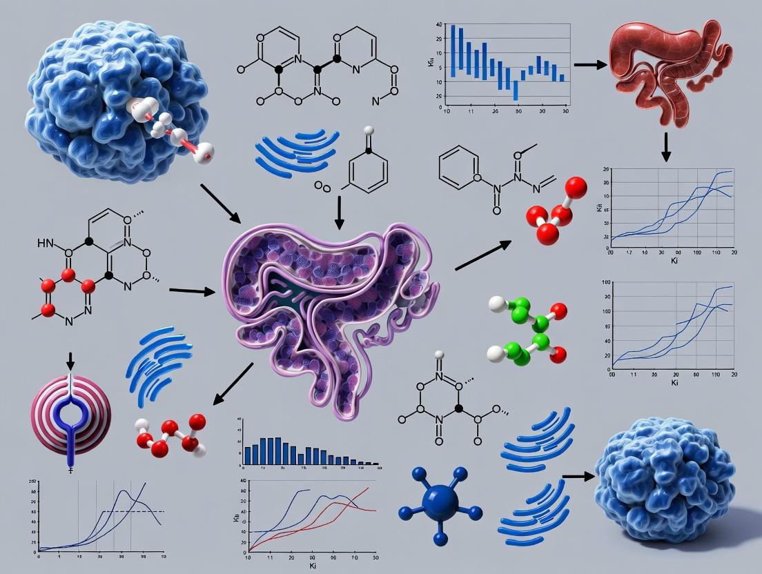
This article provides a comprehensive guide for researchers and drug development professionals on the application of 3D cultured hepatocytes in Cytochrome P450 (CYP) inhibition studies.
Advancing Drug Safety: 3D Cultured Hepatocytes for Predictive CYP Inhibition Studies
Abstract
This article provides a comprehensive guide for researchers and drug development professionals on the application of 3D cultured hepatocytes in Cytochrome P450 (CYP) inhibition studies. It explores the scientific rationale for moving beyond traditional 2D models, detailing modern methodologies for establishing and maintaining spheroids, organoids, and scaffold-based systems. The content addresses common experimental challenges and optimization strategies for functional longevity and reproducibility. Furthermore, it critically validates 3D models against clinical data and conventional in vitro systems, highlighting their superior predictive power for drug-drug interactions (DDIs) and hepatotoxicity. This synthesis aims to equip scientists with the knowledge to implement more physiologically relevant and reliable assays in preclinical drug safety assessment.
Beyond the Monolayer: Why 3D Hepatocyte Models Are Revolutionizing CYP Research
The Critical Role of CYP Enzymes in Drug Metabolism and Toxicity
Within the broader thesis exploring 3D cultured hepatocytes as advanced physiological models, understanding Cytochrome P450 (CYP) enzyme dynamics is paramount. CYP enzymes, primarily expressed in the liver, are responsible for the metabolism of approximately 70-80% of all clinically used drugs. This application note details the critical role of CYPs in drug metabolism and toxicity, providing protocols and data specifically framed for their study using 3D hepatocyte cultures, which offer superior metabolic functionality and longevity compared to 2D systems.
Key CYP Enzymes: Abundance and Metabolic Contribution
Table 1: Major Human Hepatic CYP Enzymes: Abundance and Drug Metabolism Share
| CYP Isoform | Approximate Hepatic Abundance (%) | Estimated Contribution to Drug Metabolism (%) | Notable Substrates (Examples) |
|---|---|---|---|
| CYP3A4/5 | ~30% | ~30% | Midazolam, Simvastatin, Tacrolimus |
| CYP2C9 | ~20% | ~15% | S-Warfarin, Phenytoin, Ibuprofen |
| CYP2C19 | ~5% | ~8% | Omeprazole, Clopidogrel |
| CYP2D6 | ~2-4% | ~20% | Codeine, Metoprolol, Tamoxifen |
| CYP1A2 | ~13% | ~10% | Caffeine, Theophylline, Clozapine |
| CYP2B6 | ~2-6% | ~4% | Efavirenz, Bupropion |
| CYP2E1 | ~7% | ~3% | Acetaminophen, Ethanol |
Research Reagent Solutions Toolkit
Table 2: Essential Materials for CYP Studies in 3D Hepatocytes
| Item | Function/Application |
|---|---|
| 3D Human Hepatocyte Spheroid Cultures (e.g., primary, iPSC-derived) | Physiologically relevant in vitro model with stable CYP expression for chronic studies. |
| CYP Isoform-Specific Probe Substrates (e.g., Phenacetin/CYP1A2, Bupropion/CYP2B6) | Selective compounds metabolized primarily by a single CYP to assess isoform-specific activity. |
| LC-MS/MS System | Gold standard for sensitive, specific quantification of probe metabolites and generated reactive intermediates. |
| CYP Inhibitor Cocktails (e.g., α-Naphthoflavone/CYP1A2, Ketoconazole/CYP3A4) | Chemical tools to delineate contribution of specific CYPs to overall metabolite formation. |
| NADPH Regenerating System | Provides essential co-factor (NADPH) for CYP oxidoreductase activity in cell lysates or microsomes. |
| CYP-Glo Assay Kits | Luminescence-based assays for high-throughput screening of CYP inhibition using recombinant enzymes. |
| Reactive Oxygen Species (ROS) Detection Dyes (e.g., DCFDA, CellROX) | Detect oxidative stress induced by CYP-mediated bioactivation leading to toxicity. |
Protocol: Assessing CYP Inhibition in 3D Hepatocyte Spheroids
Objective: To determine the inhibitory potential (IC₅₀) of a new chemical entity (NCE) on a major CYP isoform (e.g., CYP3A4) using 3D human hepatocyte spheroids.
Materials:
- 3D human hepatocyte spheroids (96-well ultra-low attachment plate, 7 days post-seeding).
- Test compound (NCE) in DMSO (final DMSO ≤0.1%).
- CYP3A4 probe substrate (Midazolam, 5 µM final).
- Positive control inhibitor (Ketoconazole).
- Warm Williams' E Medium.
- Stop solution: Acetonitrile with internal standard.
- LC-MS/MS for 1'-Hydroxymidazolam quantification.
Procedure:
- Pre-treatment: Prepare serial dilutions of NCE and Ketoconazole in culture medium. Aspirate old medium from spheroid plates and add 150 µL of inhibitor-containing medium. Incubate for 30 minutes at 37°C, 5% CO₂.
- Reaction Initiation: Add 50 µL of pre-warmed Midazolam solution (to achieve 5 µM final) directly to each well. Incubate for a predetermined linear time (e.g., 60 minutes).
- Reaction Termination: Transfer 100 µL of supernatant from each well to a deep-well plate containing 200 µL of ice-cold stop solution. Vortex and centrifuge (4000 x g, 15 min, 4°C).
- Analysis: Inject supernatant into LC-MS/MS to quantify 1'-Hydroxymidazolam formation.
- Data Analysis: Calculate % activity relative to vehicle control (DMSO only). Plot inhibitor concentration vs. % activity. Fit data to a sigmoidal dose-response model to calculate IC₅₀.
Protocol: Evaluating CYP-Mediated Toxicity via Bioactivation
Objective: To assess if cytotoxicity of a compound is dependent on CYP-mediated bioactivation using 3D hepatocytes with and without broad CYP inhibition.
Materials:
- 3D hepatocyte spheroids.
- Test compound.
- Pan-CYP inhibitor (1-Aminobenzotriazole, ABT, 1 mM).
- Cell viability assay (e.g., ATP-based luminescence).
- ROS detection dye.
Procedure:
- Experimental Groups: Set up four treatment conditions in quadrupicate: Vehicle, ABT alone, Test compound, Test compound + ABT.
- Pre-inhibition: Treat "ABT alone" and "Test compound + ABT" wells with ABT for 1 hour.
- Dosing: Add test compound at multiple concentrations to relevant wells. Incubate for 24-48 hours.
- Endpoint Analysis:
- Viability: Measure ATP content per manufacturer's protocol.
- ROS: Load parallel plates with ROS dye for the final 30 minutes of incubation, then measure fluorescence.
- Interpretation: A significant attenuation of cytotoxicity and ROS generation in the "Test compound + ABT" group versus "Test compound alone" indicates CYP-dependent bioactivation and toxicity.
Visualizing Key Concepts and Workflows
CYP-Mediated Metabolic Fate and Toxicity Pathway
Workflow for CYP Inhibition Assay in 3D Spheroids
Quantitative Data on CYP Variability and Inhibition
Table 3: Impact of Genetic Polymorphisms on Key CYP Activities
| CYP Isoform | Common Allelic Variant | Functional Consequence | Population Frequency (Varies) | Impact on Drug Exposure |
|---|---|---|---|---|
| CYP2D6 | *4 (rs3892097) | No Function | ~12-21% (European) | ↑ For substrates (e.g., Tamoxifen) |
| CYP2C9 | *2 (rs1799853) | Reduced Function | ~8-14% (European) | ↑ For S-Warfarin (bleeding risk) |
| CYP2C19 | *2 (rs4244285) | No Function | ~15% (European), ~29% (Asian) | ↓ Clopidogrel activation |
| CYP3A5 | *3 (rs776746) | No Function | ~80-95% (European) | ↓ Tacrolimus metabolism |
Table 4: Common Clinical CYP Inhibitors and Risk Classification
| Inhibitor | Primary CYP Target | Mechanism | Clinical Risk Rating (FDA) | Example Interaction |
|---|---|---|---|---|
| Ketoconazole | CYP3A4 | Reversible | Strong | ↑ Simvastatin AUC >10-fold (myopathy) |
| Ritonavir | CYP3A4 | Mechanism-based | Strong | Used pharmacokinetically to boost other drugs |
| Fluconazole | CYP2C9, CYP3A4 | Reversible | Moderate | ↑ S-Warfarin AUC ~2x (bleeding) |
| Paroxetine | CYP2D6 | Reversible | Strong | ↓ Tamoxifen activation to endoxifen |
| Amiodarone | CYP2C9 | Reversible | Moderate | ↑ S-Warfarin AUC ~1.5-2x |
Traditional two-dimensional (2D) monolayer cultures of primary hepatocytes remain a standard in vitro tool for early drug screening. However, within the context of advancing cytochrome P450 (CYP) inhibition studies, a critical limitation of these 2D systems is the rapid and precipitous loss of the native hepatic phenotype and function. This undermines their reliability for predicting drug metabolism and toxicity in humans. This application note details the quantitative decay of key functions in 2D cultures and provides protocols for their assessment, framing the necessity for more physiologically relevant 3D models.
Quantitative Loss of Function in 2D Cultures
The decline in hepatocyte-specific functions in 2D culture is well-documented. The following table summarizes key metrics of this phenotypic decay over time.
Table 1: Temporal Decay of Key Hepatic Functions in Traditional 2D Monolayer Culture
| Hepatic Function / Marker | Baseline (Freshly Isolated) | Day 3 in 2D Culture | Day 7 in 2D Culture | Measurement Method |
|---|---|---|---|---|
| Albumin Secretion | 100% (Reference) | 40-60% | 10-20% | ELISA |
| Urea Synthesis | 100% (Reference) | 30-50% | 5-15% | Urea Assay Kit |
| CYP3A4 Activity | 100% (Reference) | 20-40% | <5% | Luciferin-IPA / Testosterone 6β-hydroxylation |
| CYP1A2 Activity | 100% (Reference) | 25-45% | <10% | Phenacetin O-deethylation |
| CYP2C9 Activity | 100% (Reference) | 30-50% | <10% | Diclofenac 4'-hydroxylation |
| Gene Expression (CYP3A4 mRNA) | 100% (Reference) | 1-10% | <1% | qRT-PCR |
| Bile Canaliculi Formation | Polarized networks | Disrupted, fragmented | Absent | CLF secretion / Immunofluorescence |
| Transporter Activity (e.g., MRP2) | Fully functional | Significantly reduced | Negligible | CMFDA / CDFDA assay |
Detailed Experimental Protocols for Assessing 2D Limitations
Protocol 3.1: Assessing CYP450 Activity Decay Over Time in 2D Monolayers
Objective: To quantify the loss of major CYP450 enzyme activities in primary human hepatocytes maintained in standard 2D culture. Materials: See "Research Reagent Solutions" table. Procedure:
- Hepatocyte Plating: Seed cryopreserved primary human hepatocytes in collagen-I-coated 96-well plates at 50,000 viable cells/cm² in appropriate seeding medium.
- Culture Maintenance: 4-6 hours post-seeding, replace seeding medium with standard hepatocyte maintenance medium. Change medium daily.
- CYP Activity Assay (Day 1, 3, 5, 7):
- Prepare substrate working solutions in pre-warmed serum-free, phenol red-free maintenance medium.
- CYP3A4: 50 µM Luciferin-IPA.
- CYP1A2: 30 µM Phenacetin.
- CYP2C9: 10 µM Diclofenac.
- Aspirate culture medium and wash cells once with pre-warmed PBS.
- Add 100 µL of substrate solution per well. Incubate plate at 37°C, 5% CO₂ for 45-60 minutes.
- For Luciferin-IPA (CYP3A4): Transfer 50 µL of supernatant to a white assay plate. Add 50 µL of Luciferin Detection Reagent, incubate 20 min in the dark, and measure luminescence.
- For Phenacetin/Diclofenac (CYP1A2/2C9): Stop reaction by transferring supernatant to a tube containing 100 µL of ice-cold acetonitrile with internal standard. Vortex, centrifuge (10,000 x g, 10 min), and analyze supernatant via LC-MS/MS for metabolite formation (acetaminophen or 4'-hydroxydiclofenac).
- Prepare substrate working solutions in pre-warmed serum-free, phenol red-free maintenance medium.
- Data Analysis: Normalize metabolite formation or luminescence values to total cellular protein content (BCA assay) and express as percentage of Day 1 activity.
Protocol 3.2: Monitoring Polarization and Canalicular Function Loss
Objective: To visualize the disruption of hepatocyte polarization and bile canaliculi networks. Materials: See "Research Reagent Solutions" table. Procedure:
- Cell Seeding and Culture: Seed hepatocytes on collagen-I-coated glass coverslips in 24-well plates as in Protocol 3.1.
- Bile Canaliculi Staining (Day 2 and Day 5):
- Load cells with 5(6)-Carboxy-2',7'-Dichlorofluorescein Diacetate (CDFDA) by incubating with 5 µM CDFDA in maintenance medium for 30 min at 37°C.
- Wash cells 3x with warm PBS.
- Fix cells with 4% paraformaldehyde for 15 min at room temperature.
- Permeabilize with 0.1% Triton X-100 for 10 min.
- Stain actin cytoskeleton with Phalloidin (e.g., Alexa Fluor 568, 1:200) for 30 min. Perform DAPI nuclear counterstain.
- Mount coverslips and image using a confocal microscope.
- Analysis: On Day 2, observe interconnected, tubular CDF (cleaved product) networks indicative of functional canaliculi. By Day 5, note fragmentation and loss of these networks, correlating with loss of polarization.
Visualizing Key Pathways and Workflows
Diagram 1: 2D Culture-Induced Loss of Hepatic Phenotype
Diagram 2: Experimental Workflow: 2D vs 3D Comparison
The Scientist's Toolkit: Research Reagent Solutions
Table 2: Essential Materials for Assessing 2D Hepatocyte Limitations
| Item Name | Supplier Examples | Function in Protocol |
|---|---|---|
| Primary Human Hepatocytes (Cryopreserved) | Lonza, Thermo Fisher, BioIVT | The fundamental cellular model for studying human-relevant hepatic metabolism and toxicity. |
| Collagen I, Rat Tail | Corning, Thermo Fisher | Standard coating matrix for 2D hepatocyte adhesion, providing a baseline attachment surface. |
| Hepatocyte Maintenance Medium | Lonza (HCM), Thermo Fisher (Williams' E) | Serum-free medium formulation designed to support short-term hepatocyte function in 2D. |
| P450-Glo CYP3A4 Assay (Luciferin-IPA) | Promega | Luminescent, cell-based assay for convenient, high-throughput measurement of CYP3A4 activity. |
| CYP450 Substrate Cocktails | Corning (Gentest), BD Biosciences | Sets of isoform-specific probe substrates (e.g., Phenacetin, Diclofenac) for comprehensive CYP activity profiling via LC-MS/MS. |
| CDFDA (5(6)-Carboxy-2',7'-Dichlorofluorescein Diacetate) | Sigma-Aldrich, Cayman Chemical | Fluorescent probe for functional assessment of bile canalicular formation and MRP2 transporter activity. |
| Hepatocyte Nuclear Factor 4 Alpha (HNF4α) Antibody | Cell Signaling Technology, Abcam | Marker for hepatocyte differentiation and phenotype; used in immunostaining/WB to monitor dedifferentiation. |
| Human Albumin ELISA Kit | Bethyl Laboratories, Abcam | Quantifies albumin secretion, a key indicator of hepatocyte synthetic function and phenotypic stability. |
| Spheroid Microplate (U-bottom, Ultra-Low Attachment) | Corning, Greiner Bio-One | Cultureware used as the 3D comparator in validation workflows to form and maintain hepatocyte spheroids. |
Application Notes
Within the context of advancing in vitro models for CYP inhibition studies, three-dimensional (3D) cultured hepatocytes represent a paradigm shift from conventional two-dimensional (2D) monolayers. The 3D architecture recapitulates critical aspects of the native liver microenvironment, directly addressing the limitations of 2D systems in predicting drug metabolism and toxicity. This note details the key functional advantages conferred by 3D architecture.
1. Re-establishment of Apical-Basal Polarization In the liver, hepatocytes are polarized epithelial cells with distinct apical (canalicular) and basolateral (sinusoidal) membrane domains, a feature essential for directional bile secretion and uptake. 2D culture results in the rapid loss of this polarization. 3D spheroid or organoid models facilitate the re-establishment of this critical cytoarchitecture. The reformation of functional bile canaliculi networks within 3D structures enables more accurate assessment of drug transport and cholestatic potential, factors directly influencing CYP enzyme access and activity.
2. Restoration of Physiological Cell-Cell and Cell-ECM Contacts The liver is a highly structured tissue dependent on intricate cell-cell adhesion (e.g., via E-cadherin, tight junctions, gap junctions) and cell-extracellular matrix (ECM) interactions. 3D cultures restore these contacts, activating key signaling pathways (e.g., Hippo, Wnt/β-catenin) that regulate liver-specific function, proliferation, and survival. Enhanced gap junctional communication (Connexin 32) improves coordinated cellular responses. These interactions are minimal in 2D, leading to dedifferentiation.
3. Markedly Enhanced Long-Term Viability and Functional Stability The supportive 3D microenvironment mitigates anoikis (detachment-induced apoptosis) and reduces oxidative stress. This results in sustained viability and phenotypic stability for weeks, compared to the rapid decline in function observed in 2D cultures over days. This longevity is indispensable for chronic CYP inhibition studies, time-dependent inhibition (TDI) assessments, and evaluating metabolite-mediated toxicity.
Table 1: Comparative Functional Metrics of 2D vs. 3D Hepatocyte Cultures
| Metric | 2D Monolayer (Day 5-7) | 3D Spheroid (Day 21+) | Measurement Method |
|---|---|---|---|
| Albumin Secretion | 1 - 5 µg/day/million cells | 10 - 25 µg/day/million cells | ELISA |
| Urea Synthesis | 50 - 100 µg/day/million cells | 200 - 500 µg/day/million cells | Colorimetric assay (Berthelot) |
| CYP3A4 Activity | ~20-40% of in vivo | ~70-100% of in vivo | Luciferin-IPA / Testosterone 6β-hydroxylation |
| Viability (ATP content) | Sharp decline after Day 7 | Stable > 28 days | CellTiter-Glo 3D |
| Bile Canaliculi Formation | Disorganized, limited | Functional, networked | CLF accumulation / MRP2 staining |
| Gene Expression (CYP isoforms) | Rapid downregulation | Sustained near-physiological levels | qRT-PCR |
Table 2: Key Signaling Pathways Modulated by 3D Architecture
| Pathway | Effect in 3D vs. 2D | Functional Outcome |
|---|---|---|
| Hippo (YAP/TAZ) | Cytoplasmic retention / Inactivation | Suppressed proliferation, promoted differentiation |
| Wnt/β-catenin | Moderately active | Maintenance of hepatocyte identity |
| EGFR / Integrin | Balanced, matrix-dependent | Enhanced survival, reduced anoikis |
| NRF2 | Upregulated | Enhanced antioxidant response, cytoprotection |
Experimental Protocols
Protocol 1: Generation of Hepatic Spheroids for CYP Inhibition Studies
Objective: To produce uniform, functional 3D hepatocyte spheroids from primary human hepatocytes (PHHs) or hepatocyte-like cells (HLCs) for long-term enzyme activity and inhibition assays.
Materials:
- Primary Human Hepatocytes (PHHs, fresh or cryopreserved)
- Spheroid Formation Medium: Hepatocyte maintenance medium supplemented with 0.24% Methylcellulose or 1% Geltrex.
- Ultra-Low Attachment (ULA) 96-well round-bottom plates
- CYP Probe Substrates: e.g., Phenacetin (CYP1A2), Bupropion (CYP2B6), Testosterone (CYP3A4)
- LC-MS/MS system for metabolite quantification
Procedure:
- Cell Preparation: Thaw cryopreserved PHHs per vendor protocol. Count and resuspend in spheroid formation medium at 1.5–2.0 x 10³ cells/well in a volume of 100 µL.
- Spheroid Formation: Seed cell suspension into ULA 96-well plates. Centrifuge plates at 300 x g for 5 minutes to aggregate cells at the well bottom.
- Culture Maintenance: Incubate at 37°C, 5% CO₂. Spheroids will form within 24-48 hours. On day 3, gently replace 50% of the medium with fresh maintenance medium (without methylcellulose) every 48-72 hours.
- Functional Assessment (Day 7+): For CYP activity, incubate spheroids with CYP-specific probe substrates. Sample supernatant at timed intervals for metabolite analysis by LC-MS/MS.
- Inhibition Studies: Pre-incubate spheroids with test inhibitor for 30-60 minutes before adding probe substrate. Include positive controls (e.g., Ketoconazole for CYP3A4). Calculate IC₅₀ values.
Protocol 2: Immunofluorescence Analysis of Polarization and Cell Contacts
Objective: To visualize bile canaliculi and junctional complexes in 3D spheroids.
Materials:
- 4% Paraformaldehyde (PFA)
- Permeabilization Buffer (0.5% Triton X-100)
- Blocking Buffer (5% BSA, 0.1% Tween-20)
- Primary Antibodies: Anti-MRP2 (apical canaliculi), Anti-ZO-1 (tight junctions), Anti-E-cadherin (adherens junctions).
- Secondary Antibodies (conjugated to Alexa Fluor dyes)
- Confocal microscope
Procedure:
- Fixation: Transfer spheroids to a microtube, let settle. Fix with 4% PFA for 45 minutes at RT.
- Permeabilization & Blocking: Wash with PBS, permeabilize for 1 hour. Block for 2 hours at RT.
- Staining: Incubate with primary antibody cocktail overnight at 4°C. Wash extensively, then incubate with secondary antibodies for 2 hours at RT. Include DAPI for nuclei.
- Imaging: Mount spheroids on a glass-bottom dish. Acquire z-stacks using a confocal microscope. 3D reconstruction software can be used to visualize the canalicular network.
Visualizations
Diagram 1: 2D vs 3D Architecture Impact on Hepatocyte Function
Diagram 2: 3D Hepatocyte CYP Inhibition Assay Workflow
The Scientist's Toolkit
Table 3: Essential Research Reagents for 3D Hepatocyte CYP Studies
| Reagent / Material | Function & Rationale | Example Product |
|---|---|---|
| Ultra-Low Attachment (ULA) Plates | Prevents cell adhesion, forcing self-aggregation into spheroids. Critical for consistent size and shape. | Corning Spheroid Microplates |
| Hepatocyte Maintenance Medium | Formulated to support hepatocyte function, typically containing dexamethasone, insulin, and growth factors. | Williams' E Medium + ITS, HCM |
| Basement Membrane Matrix | Provides physiological ECM components (laminin, collagen IV) to enhance cell contacts and signaling. | Geltrex, Matrigel (diluted) |
| CYP Isoform-Specific Probe Substrates | Selective compounds metabolized by specific CYP enzymes to quantify isoform activity. | Luciferin-IPA (CYP3A4), Phenacetin (CYP1A2) |
| 3D Viability Assay Kit | Optimized lytic reagents for penetration and ATP quantification in dense 3D structures. | CellTiter-Glo 3D |
| Primary Human Hepatocytes (PHHs) | Gold standard cell source with full complement of human drug-metabolizing enzymes. | Multiple commercial vendors (e.g., BioIVT, Lonza) |
| CYP Inhibitor Positive Controls | Pharmacological standards for validating inhibition assays (e.g., Ketoconazole for CYP3A4). | Commercial chemical inhibitors |
Three-dimensional (3D) culture models have become indispensable for advancing in vitro hepatotoxicity and drug metabolism studies, offering a more physiologically relevant environment than traditional 2D monolayers. For Cytochrome P450 (CYP) inhibition studies—a critical component of drug-drug interaction (DDI) assessment—these models provide superior phenotypic stability, prolonged culture longevity, and recapitulation of cell-cell and cell-matrix interactions that govern metabolic function. This application note details three primary 3D model types—spheroids, organoids, and scaffold-based systems—within the context of culturing hepatocytes for reliable, high-content CYP enzyme activity and inhibition profiling.
Table 1: Quantitative Comparison of 3D Hepatocyte Model Characteristics
| Feature | Hepatocyte Spheroids | Hepatocyte Organoids | Scaffold-Based Hepatocyte Systems |
|---|---|---|---|
| Typical Size Range | 50 - 500 µm | 100 - 1000+ µm | Variable; often >1 mm constructs |
| Key Cellular Components | Primary hepatocytes +/- NPCs* | Hepatoblasts/progenitors or iPSC-derived cells; may self-organize | Primary hepatocytes or cell lines; often with supporting stromal cells |
| Self-Assembly | Yes (cell-aggregation) | Yes (directed differentiation & self-organization) | No (cells seeded into pre-formed matrix) |
| Extracellular Matrix | Minimal, endogenous secretion | Often embedded in Matrigel/BME for growth | High, provided by natural (collagen) or synthetic scaffold |
| Culture Longevity | 3-5 weeks (functional) | Several weeks to months (expanding) | 2-4 weeks (varies with scaffold) |
| Throughput for Screening | High (U/L-plate formats) | Medium (requires embedding) | Low to Medium |
| CYP Expression & Activity | High, stable for 2+ weeks | Variable; can achieve mature phenotypes | Good, dependent on scaffold porosity & signaling |
| Cost & Technical Demand | Low to Moderate | High (specialized media, growth factors) | Moderate to High |
| Primary Use in CYP Studies | High-throughput DDI screening, chronic inhibition | Disease modeling, developmental toxicity, personalized DDI | Mechanistic studies, zonation modeling, implantable devices |
*NPCs: Non-Parenchymal Cells (e.g., Kupffer, stellate cells).
Detailed Application Notes & Protocols
Hepatocyte Spheroids for CYP Inhibition Screening
Application Note: Hepatic spheroids, particularly those formed from primary human hepatocytes (PHHs), are the current gold standard for high-fidelity, high-throughput CYP inhibition studies. They maintain Phase I/II metabolic activity closer to in vivo levels for several weeks, enabling the study of time-dependent inhibition and metabolite-mediated toxicity.
Protocol: Generation of PHH Spheroids in Ultra-Low Attachment (ULA) Plates for CYP3A4 Inhibition Assay
Objective: To form uniform, functional spheroids for assessing inhibitor potency (IC50) against a major CYP enzyme, CYP3A4.
Materials (Research Reagent Solutions):
- Cryopreserved Primary Human Hepatocytes (PHHs): Metabolically competent cells from a reputable supplier (e.g., BioIVT, Lonza). Thaw using recommended systems.
- Hepatocyte Maintenance Medium: Williams' E Medium supplemented with 5% FBS, 1% Insulin-Transferrin-Selenium (ITS), 100 nM dexamethasone, 100 U/mL penicillin, and 100 µg/mL streptomycin.
- ULA Round-Bottom 96-Well Plates: Plates treated to prevent cell adhesion, forcing self-aggregation.
- CYP3A4 Substrate: Midazolam or a luminogenic probe like Luciferin-IPA (Promega).
- Test Inhibitors: e.g., Ketoconazole (strong), Verapamil (moderate). Dissolved in DMSO (<0.1% final concentration).
- LC-MS/MS System or Luminescence Plate Reader: For quantifying metabolite formation (1'-OH-midazolam) or luminescent output.
Methodology:
- Thawing & Viability Check: Rapidly thaw cryopreserved PHHs per supplier protocol. Determine viability via trypan blue exclusion (>80% required).
- Seeding: Resuspend PHHs at 1.0 x 10^6 cells/mL in maintenance medium. Seed 100 µL/well (1.0 x 10^5 cells/well) into the ULA 96-well plate using a multichannel pipette.
- Spheroid Formation: Centrifuge plate at 100 x g for 3 min to aggregate cells at the well bottom. Incubate at 37°C, 5% CO2.
- Culture Maintenance: After 72 hours, carefully replace 50% of the medium with fresh maintenance medium every 2-3 days. Mature, compact spheroids form by day 5-7.
- Inhibition Assay (Day 7):
- Prepare serial dilutions of test inhibitors in serum-free incubation medium.
- Aspirate medium from spheroids and add 100 µL of inhibitor solution per well. Include vehicle (DMSO) and positive control (e.g., 10 µM Ketoconazole) wells.
- Pre-incubate for 30 min at 37°C.
- Add CYP3A4 substrate directly to each well (final midazolam concentration: 5 µM; Luciferin-IPA per manufacturer).
- Incubate for 2 hours.
- Terminate reaction: for LC-MS/MS, transfer supernatant to analysis plate; for luminescence, add detection reagent and read immediately.
- Data Analysis: Calculate % activity remaining vs. vehicle control. Plot inhibitor concentration vs. response to determine IC50 values.
Diagram Title: Workflow for CYP Inhibition in Hepatocyte Spheroids
Hepatic Organoids for Advanced Modeling
Application Note: Hepatic organoids derived from adult stem cells (ASCs) or induced pluripotent stem cells (iPSCs) offer a renewable, patient-specific model. They are valuable for studying genetic determinants of CYP expression variability and idiosyncratic DDI.
Protocol: Differentiating iPSC-Derived Hepatic Organoids for CYP Induction/Inhibition Studies
Objective: To generate metabolically mature hepatic organoids capable of responding to CYP inducers and inhibitors.
Key Reagent Solutions:
- iPSC Line: Maintained in feeder-free conditions.
- Defined Differentiation Media: Sequential media for definitive endoderm, hepatoblast, and hepatic specification.
- Basement Membrane Extract (BME): Liquid at 4°C, gels at 37°C to provide a 3D scaffold.
- Maturation Factors: Including glucocorticoids (Dexamethasone), FGF19, and NOTCH inhibitors to promote metabolic maturation.
- CYP Inducer: Rifampicin (for CYP3A4/CYP2B6 induction).
Methodology (Abbreviated):
- iPSC to Hepatoblast Differentiation: Follow established 2D monolayer protocols to generate hepatic progenitor cells (HPCs) over ~10 days.
- 3D Organoid Formation: Dissociate HPCs to single cells. Mix 5.0 x 10^4 cells with 20 µL BME and plate as dome droplets in pre-warmed plates. Polymerize at 37°C for 30 min.
- Hepatic Maturation: Overlay droplets with organoid maturation medium (containing maturation factors). Culture for 21-28 days, with medium changes every 3-4 days.
- CYP Induction/Inhibition: Treat mature organoids with inducers (e.g., 10 µM Rifampicin for 48h) prior to inhibition assays (as in Protocol 3.1) to assess the impact of pre-induction on inhibitor potency.
Scaffold-Based Systems for Zonation and Co-Culture
Application Note: Porous scaffolds (e.g., collagen, polyester) allow for the creation of larger tissue constructs that can model hepatic zonation—a gradient of oxygen, nutrients, and CYP expression (e.g., periportal vs. perivenous). This is critical for studying zonal-specific toxicity.
Protocol: Seeding Hepatocytes in Collagen Scaffolds for Zonal CYP Analysis
Objective: To create a 3D hepatic construct with controlled cell distribution for compartmentalized analysis.
Key Reagent Solutions:
- Porous Collagen Scaffold Discs: ~5 mm diameter x 2 mm thickness.
- Hepatocyte Seeding Medium: High-serum medium to promote attachment.
- Perfusion Bioreactor (Optional): For creating nutrient/oxygen gradients.
- Laser Capture Microdissection (LCM) or Sectioning: For spatially resolved analysis.
Methodology:
- Scaffold Preparation: Hydrate collagen scaffolds in culture medium for 1 hour.
- Cell Seeding: Use a dynamic seeding method. Place scaffold in a low-volume chamber, add 2.0 x 10^6 PHHs in 50 µL medium, and centrifuge at 50 x g for 10 min to drive cells into pores. Repeat if needed.
- Culture: Transfer seeded scaffolds to a 24-well plate with maintenance medium. Use orbital shaking or a perfusion system to enhance nutrient exchange.
- Analysis: After 7-14 days, fix and section constructs. Use immunohistochemistry for zonal markers (e.g., CYP3A4 - perivenous, CYP2E1 - periportal) or microdissect zones for RNA/protein extraction to assess localized inhibitor effects.
Diagram Title: PXR-Mediated CYP3A4 Induction Pathway
The Scientist's Toolkit: Essential Reagents for 3D Hepatocyte CYP Studies
Table 2: Key Research Reagent Solutions
| Item | Function in 3D CYP Studies | Example Product/Brand |
|---|---|---|
| Primary Human Hepatocytes (PHHs) | Gold-standard cell source with full complement of human CYPs and transporters. Essential for clinically relevant DDI data. | BioIVT Hepatocytes, Lonza Hepatocytes |
| iPSC-Derived Hepatocyte Cells | Renewable, patient-specific source for genetic studies and personalized pharmacology. | Cellular Dynamics (CDI) iCell Hepatocytes, Stemcell Technologies kits |
| Ultra-Low Attachment (ULA) Plates | Enable forced floating aggregation for consistent, high-throughput spheroid formation. | Corning Spheroid Microplates, Nunclon Sphera plates |
| Basement Membrane Extract (BME) | Complex, natural matrix supporting organoid growth and polarization. | Corning Matrigel, Cultrex BME |
| Defined Hepatocyte Maintenance Medium | Supports long-term phenotypic stability and CYP activity in 3D cultures. | William's E based supplements, HepatoZYME-SFM |
| CYP-Specific Luminescent Substrates | Enable high-throughput, real-time kinetic analysis of CYP activity in intact 3D models. | Promega P450-Glo Assays |
| PXR/CAR Receptor Agonists | Positive controls for studying CYP induction, a key regulatory mechanism in DDIs. | Rifampicin (PXR), CITCO (CAR) |
| Porous 3D Scaffolds | Provide structural support for larger constructs and allow modeling of zonation. | Collagen I scaffolds (e.g., Avitene), Synthetic PET scaffolds |
| CYP Isoform-Selective Inhibitors | Controls for validating assay specificity in complex 3D systems. | Ketoconazole (CYP3A4), Sulfaphenazole (CYP2C9) |
| 3D Cell Viability/Cytotoxicity Assays | Optimized for penetration and accuracy in dense 3D structures. | CellTiter-Glo 3D, MultiTox-Fluor Multiplex Assay |
Core CYP Enzymes (e.g., 3A4, 2D6, 2C9) and Their Expression in 3D vs. 2D Cultures
Application Notes
Cytochrome P450 (CYP) enzymes, predominantly CYP3A4, CYP2D6, and CYP2C9, are critical for the oxidative metabolism of approximately 70-80% of clinically used drugs. Accurate assessment of CYP-mediated metabolism and inhibition is paramount in drug development to predict drug-drug interactions (DDIs). Traditional in vitro models, primarily two-dimensional (2D) monolayer cultures of hepatocytes, suffer from rapid dedifferentiation and loss of native hepatic phenotype, including a precipitous decline in CYP expression and activity within hours to days. This limits their utility for chronic inhibition studies and mechanistic investigations.
Three-dimensional (3D) hepatocyte cultures—including spheroids, organoids, and scaffold-based systems—emerge as a physiologically relevant alternative. By restoring cell-cell and cell-extracellular matrix interactions, 3D cultures promote the maintenance of hepatocyte polarity, bile canaliculi formation, and sustained expression of drug-metabolizing enzymes and nuclear receptors (e.g., PXR, CAR). This application note details the superior expression profiles of core CYP enzymes in 3D cultures compared to 2D and provides protocols for their use in CYP inhibition studies, framed within a thesis on advancing in vitro DDI prediction models.
Quantitative Comparison of CYP Expression & Activity
Table 1: Expression and Activity of Core CYP Enzymes in 2D vs. 3D Hepatocyte Cultures Over Time
| CYP Enzyme | Culture Format | Measurement Type | Day 1 Value (vs. Fresh PHH) | Day 7 Value (vs. Fresh PHH) | Key Supporting Technology |
|---|---|---|---|---|---|
| CYP3A4 | 2D Monolayer | mRNA | 60-80% | <20% | qPCR, RNA-Seq |
| Protein (pmol/mg) | ~50-100 | ~5-20 | LC-MS/MS, WB | ||
| Activity (Testosterone 6β-hydroxylation) | ~40-60% | <10% | LC-MS/MS | ||
| 3D Spheroid | mRNA | 70-90% | 50-80% | qPCR, RNA-Seq | |
| Protein (pmol/mg) | ~80-120 | ~60-150 | LC-MS/MS, WB | ||
| Activity | 50-70% | 40-70% | LC-MS/MS | ||
| CYP2D6 | 2D Monolayer | mRNA | 50-70% | <15% | qPCR |
| Activity (Bufuralol 1'-hydroxylation) | ~30-50% | <5% | LC-MS/MS | ||
| 3D Spheroid | mRNA | 60-85% | 40-70% | qPCR | |
| Activity | 40-60% | 30-60% | LC-MS/MS | ||
| CYP2C9 | 2D Monolayer | mRNA | 55-75% | <20% | qPCR |
| Activity (Diclofenac 4'-hydroxylation) | ~35-55% | <10% | LC-MS/MS | ||
| 3D Spheroid | mRNA | 65-90% | 45-75% | qPCR | |
| Activity | 45-65% | 35-65% | LC-MS/MS |
Note: PHH = Primary Human Hepatocytes. Values are approximate ranges synthesized from recent literature. 3D cultures demonstrate significantly superior maintenance of phenotype.
Table 2: IC50 Shift Analysis for Mechanism-Based Inhibitors in 2D vs. 3D Systems
| Inhibitor (CYP Target) | Culture Format | Pre-incubation Time | Apparent IC50 (µM) | Shift from 2D (No Pre-incub) | Implication for DDI Risk |
|---|---|---|---|---|---|
| Ketoconazole (CYP3A4) | 2D | 0 min | 0.02 | Reference (Reversible) | Standard reversible inhibition. |
| 3D | 0 min | 0.015-0.03 | ~1x | Similar reversible inhibition detected. | |
| Erythromycin (CYP3A4) | 2D | 0 min | >100 | Reference | Missed mechanism-based inhibition (MBI). |
| 2D | 30 min | ~40 | N/A | Some MBI detected. | |
| 3D | 30 min | ~5-10 | >10x lower than 2D (0 min) | Enhanced MBI detection due to sustained CYP3A4 and NADPH. | |
| Paroxetine (CYP2D6) | 2D | 0 min | 1.5 | Reference | Partial MBI potential. |
| 3D | 30 min | 0.2 | ~7.5x lower | More accurate prediction of clinical MBI. |
Experimental Protocols
Protocol 1: Generation of 3D Hepatocyte Spheroids for CYP Studies
Objective: To establish long-term, functional 3D hepatocyte spheroid cultures from cryopreserved primary human hepatocytes (PHHs) for CYP expression and inhibition profiling.
Materials: See "The Scientist's Toolkit" below.
Procedure:
- Thawing & Viability Assessment:
- Rapidly thaw cryopreserved PHHs in a 37°C water bath.
- Transfer cells to pre-warmed hepatocyte thawing medium. Centrifuge at 100 x g for 10 minutes.
- Resuspend pellet in hepatocyte plating medium. Determine viability via trypan blue exclusion (>80% required).
Spheroid Formation (Ultra-Low Attachment Plates):
- Adjust cell density to 1.0–1.5 x 10⁶ viable cells/mL in plating medium.
- Seed 100 µL of cell suspension per well (1.0–1.5 x 10⁵ cells/well) into a 96-well ultra-low attachment (ULA), round-bottom plate.
- Centrifuge the plate at 100 x g for 5 minutes to aggregate cells at the well bottom.
- Incubate at 37°C, 5% CO₂ for 3-7 days. Spheroids will form within 24-48 hours.
Long-Term Maintenance:
- After 48-72 hours, replace 50% of the medium with pre-warmed hepatocyte maintenance medium.
- Perform 50% medium changes every 48 hours thereafter. Spheroids remain viable and functional for 4+ weeks.
Protocol 2: Assessing CYP Activity in 2D vs. 3D Cultures
Objective: To quantify the functional activity of CYP3A4, 2D6, and 2C9 in both culture formats using isoform-specific probe substrates.
Procedure:
- Culture Preparation:
- 2D: Seed PHHs in collagen-coated 24-well plates at 0.7 x 10⁶ cells/well. Allow to attach for 4-6 hours, then replace with maintenance medium. Treat on Day 2 or 4.
- 3D: Use mature spheroids (Day 7-10 post-seeding) in 96-well ULA plates.
CYP Activity Assay:
- Prepare incubation cocktail containing isoform-specific probes in serum-free, phenol-red free maintenance medium:
- CYP3A4: 100 µM Testosterone
- CYP2D6: 5 µM Bufuralol
- CYP2C9: 10 µM Diclofenac
- Aspirate medium from wells and wash cultures once with pre-warmed PBS.
- Add 200 µL (for 24-well) or 100 µL (for 96-well) of probe cocktail.
- Incubate for 30-60 minutes at 37°C. Ensure the incubation time is within the linear range for metabolite formation.
- Terminate the reaction by transferring the supernatant to a tube containing an equal volume of ice-cold acetonitrile with internal standard.
- Prepare incubation cocktail containing isoform-specific probes in serum-free, phenol-red free maintenance medium:
Sample Analysis:
- Vortex, centrifuge (10,000 x g, 10 min, 4°C), and analyze supernatant via LC-MS/MS.
- Quantify specific metabolites: 6β-hydroxytestosterone (CYP3A4), 1'-hydroxybufuralol (CYP2D6), 4'-hydroxydiclofenac (CYP2C9).
- Normalize metabolite formation to total cellular protein (via BCA assay) per well.
Protocol 3: Time-Dependent CYP Inhibition (TDI) Study in 3D Spheroids
Objective: To evaluate mechanism-based inhibition (MBI) by comparing IC50 values with and without a pre-incubation phase, leveraging the metabolic competence of 3D spheroids.
Procedure:
- Pre-Incubation Phase:
- Prepare serial dilutions of the test inhibitor (e.g., Erythromycin) and a control reversible inhibitor (e.g., Ketoconazole) in maintenance medium.
- Aspirate medium from Day 7 spheroids and add inhibitor-containing medium.
- Include a NADPH-generating system (1 mM NADP⁺, 10 mM Glucose-6-Phosphate, 1 U/mL G6PDH) to support CYP activity during pre-incubation.
- Incubate for 30 minutes at 37°C.
Probe Incubation Phase:
- Without removing the pre-incubation medium, add a concentrated probe substrate solution directly to each well (final concentration as in Protocol 2).
- Incubate for an additional 30 minutes.
- Terminate and process samples as in Protocol 2, Step 3.
Data Analysis:
- Plot % residual CYP activity vs. inhibitor concentration on a log scale.
- Fit data to a sigmoidal dose-response model to calculate IC50 values.
- An IC50 shift (pre-incubation IC50 / no pre-incubation IC50) of ≥1.5-fold is indicative of TDI/MBI.
Diagrams
Title: 2D vs 3D Hepatic Culture Outcomes for CYP Studies
Title: Workflow for CYP Studies in 3D Hepatocyte Spheroids
The Scientist's Toolkit
Table 3: Essential Research Reagent Solutions for 3D Hepatic CYP Studies
| Item | Function & Rationale | Example Vendor/Product |
|---|---|---|
| Cryopreserved Primary Human Hepatocytes (PHHs) | Gold-standard cell source with full complement of human CYP enzymes and nuclear receptors. Lot-to-lot variability requires pooling or careful characterization. | BioIVT, Lonza, Thermo Fisher |
| Ultra-Low Attachment (ULA) Spheroid Microplates | Surface modification prevents cell attachment, forcing aggregation and enabling consistent, reproducible spheroid formation in each well. | Corning Spheroid Microplates, Greiner CELLSTAR |
| Hepatocyte Maintenance Medium | Specialized serum-free medium supplemented with growth factors, hormones, and metabolites to support long-term hepatocyte function and CYP expression. | Williams' E Medium + ITS, dexamethasone, gentamicin. Commercial: Hepatocyte Maintenance Medium (Lonza). |
| NADPH-Generating System | Provides essential cofactor (NADPH) for CYP enzyme activity during inhibition pre-incubation phases. Critical for accurate MBI assessment. | Prepared fresh from NADP+, Glucose-6-Phosphate, and G6PDH, or commercial solutions. |
| Isoform-Specific Probe Substrates | Selective drug molecules metabolized primarily by a single CYP isoform, allowing specific activity measurement. | Testosterone (CYP3A4), Bufuralol (CYP2D6), Diclofenac (CYP2C9). Available from Sigma, TRC. |
| LC-MS/MS System with UPLC | Gold-standard analytical platform for separating and quantifying specific CYP metabolites (and parent drugs) with high sensitivity and specificity in complex biological matrices. | Waters, Agilent, Sciex systems. |
| Mechanism-Based Inhibitor Controls | Positive control compounds known to cause time-dependent inhibition (TDI) for assay validation (e.g., Erythromycin for CYP3A4). | Available from pharmaceutical suppliers or Sigma. |
Building Better Liver Models: Protocols for 3D Hepatocyte Culture in CYP Assays
Within the thesis research on 3D cultured hepatocytes for CYP inhibition studies, the selection of the cellular source is a foundational decision. This application note provides a comparative analysis and detailed protocols for working with Primary Human Hepatocytes (PHHs) and hepatic cell lines (HepaRG, HepG2) in 3D culture formats, specifically for cytochrome P450 (CYP) enzyme inhibition assays—a critical component of drug-drug interaction (DDI) prediction in preclinical development.
Comparative Analysis: Key Parameters for CYP Inhibition Studies
Table 1: Source Characteristics & Relevance to 3D CYP Inhibition Studies
| Parameter | Primary Human Hepatocytes (PHHs) | HepaRG Cells | HepG2 Cells |
|---|---|---|---|
| Physiological Relevance | Gold standard; full complement of human DMEs, transporters, and NRs. | High; inducible expression of major CYPs and transporters upon differentiation. | Low; basal expression of some CYPs (e.g., 3A4) is very low; lack many liver-specific functions. |
| Inter-Donor Variability | High (genetic, environmental). Represents population diversity for translation. | Low (clonal origin). Ensures experimental reproducibility. | Very Low (clonal origin). High reproducibility. |
| CYP Expression & Activity | Physiological levels & ratios. All major CYP isoforms (1A2, 2B6, 2C9, 2C19, 2D6, 3A4) present. | Differentiated cells show high, inducible activity (CYP3A4, 2C9, 2C19, 1A2). Requires 2-week differentiation. | Constitutively low/absent for most CYPs (e.g., negligible CYP3A4). Not suitable for direct inhibition studies. |
| Cost & Availability | High cost; limited availability; short lifespan. | Moderate cost; unlimited supply; long culture possible. | Low cost; unlimited supply; easy to culture. |
| Suitability for 3D Culture | Excellent; form functional spheroids/organs-on-chips with enhanced stability (4+ weeks). | Very Good; form polarized, bile canaliculi-containing structures in 3D. | Good; readily form spheroids, but with limited metabolic functionality. |
| Best Use in Thesis Context | Final, translationally relevant CYP inhibition & DDI studies in a sophisticated 3D model. | Mid-stage, reproducible screening of CYP inhibition in a competent, tractable 3D system. | Preliminary 3D culture protocol optimization, cytotoxicity assessments. |
Table 2: Typical CYP Enzyme Activity in 2D vs. 3D Culture Systems (Representative Values)
| Cell Source | Culture Format | CYP3A4 Activity (pmol/min/mg protein) | CYP2C9 Activity (pmol/min/mg protein) | Stable Function (Duration) |
|---|---|---|---|---|
| PHHs | 2D Monolayer | 100-500 | 50-200 | 5-7 days |
| PHHs | 3D Spheroid | 300-1000+ | 150-400 | 21-28+ days |
| Differentiated HepaRG | 2D Monolayer | 50-200 | 20-100 | Long-term |
| Differentiated HepaRG | 3D Spheroid | 150-400 | 50-150 | Long-term |
| HepG2 | 2D/3D | <5 | <5 | N/A |
Detailed Experimental Protocols
Protocol 1: Generation of 3D PHH Spheroids for CYP Inhibition Time-Dependent Inhibition (TDI) Assay
Objective: To create long-term stable 3D PHH spheroids for assessing time-dependent CYP inhibition.
Materials: See "Scientist's Toolkit" Section 5. Procedure:
- Thawing & Plating: Rapidly thaw cryopreserved PHHs (donor-specific or pooled) using a 37°C water bath. Use recommended supplier thawing medium. Centrifuge at 100g for 5 mins.
- Viability Check: Determine viability via Trypan Blue exclusion. Proceed if viability >80%.
- 3D Seeding: Resuspend PHHs at 0.5–1.0 x 10^6 cells/mL in spheroid formation medium (e.g., Williams' E + 10% FBS + Primary Hepatocyte Maintenance Supplements + 0.1% DMSO).
- Plate: Seed 1000-1500 cells/well (50 μL droplet) into an ultra-low attachment (ULA) round-bottom 96-well plate.
- Spheroid Formation: Centrifuge plate at 100g for 5 mins. Incubate at 37°C, 5% CO₂ for 3-5 days. Spheroids should form within 24-48 hours.
- Maintenance: On day 3, replace 50% of medium with Hepatocyte Maintenance Medium (without DMSO). Subsequently, perform 50% medium changes every 2 days.
- Maturation: Allow spheroids to mature and stabilize CYP expression for 7-10 days before inhibition studies.
- CYP Inhibition Assay: Pre-incubate spheroids with inhibitor (or vehicle) for a designated time (e.g., 0-60 min for reversible inhibition; 30 min pre-inc + 24h washout for TDI). Then, incubate with a CYP-specific probe substrate (e.g., Midazolam for CYP3A4, Diclofenac for CYP2C9).
- Sample Analysis: Collect supernatant. Quantify metabolite formation (e.g., 1'-OH-midazolam, 4'-OH-diclofenac) using LC-MS/MS.
- Data Analysis: Calculate IC₅₀ or Kᵢ values. Compare metabolite formation rates to vehicle controls.
Protocol 2: Differentiation & 3D Culture of HepaRG Spheroids for CYP Induction/Inhibition
Objective: To differentiate HepaRG cells and form 3D spheroids for CYP inhibition screening.
Procedure:
- 2D Expansion & Differentiation:
- Culture undifferentiated HepaRG cells in growth medium (Williams' E + 10% FBS, 5 μg/mL Insulin, 50 μM Hydrocortisone Hemisuccinate, 100 U/mL Pen/Strep) for 14 days.
- At confluence (~day 14), switch to differentiation medium (add 1.7% DMSO to growth medium) for 2 additional weeks. Change medium bi-weekly.
- 3D Spheroid Formation:
- Differentiate cells as above. Detach cells using trypsin.
- Seed 5000-10000 cells/well into ULA round-bottom plates in differentiation medium with DMSO.
- Centrifuge at 100g for 5 mins. Incubate.
- Mature 3D spheroids for 7 days with medium changes every 2-3 days.
- CYP Inhibition Assay: Follow steps 8-10 from Protocol 1. Confirm CYP activity with prototypical inducers (e.g., Rifampicin for CYP3A4) prior to inhibition studies.
Visualizations
Diagram Title: Workflow for 3D Hepatocyte CYP Inhibition Study
Diagram Title: Key CYP Inhibition Pathways in Hepatocytes
The Scientist's Toolkit: Essential Research Reagents & Materials
Table 3: Key Reagents for 3D Hepatocyte CYP Inhibition Studies
| Item | Function & Importance | Example Product/Catalog |
|---|---|---|
| Cryopreserved PHHs | Biologically relevant cell source. Multiple donors recommended for variability assessment. | ThermoFisher Scientific (Hu4190), BioIVT, Lonza. |
| HepaRG Cells | Differentiable cell line with high metabolic competence. | ThermoFisher Scientific (HPRGC10). |
| Ultra-Low Attachment (ULA) Plates | Promotes 3D spheroid formation via forced cell aggregation. | Corning Spheroid Microplates (4515), Elplasia plates. |
| Hepatocyte Maintenance Medium | Serum-free, hormonally defined medium for long-term PHH function. | Williams' E Medium + GlutaMAX + HCM SingleQuots (Lonza). |
| DMSO (Cell Culture Grade) | Induces and maintains differentiation in HepaRG cells; used at low % in PHH culture. | Sigma-Aldrich (D2650). |
| CYP-Specific Probe Substrates | Selective metabolized compounds to measure isoform-specific activity. | Midazolam (CYP3A4), Diclofenac (CYP2C9), Bupropion (CYP2B6). |
| LC-MS/MS System | Gold-standard for sensitive, specific quantification of metabolites from inhibition assays. | Agilent 6495C, Sciex QTRAP 6500+. |
| CYP Inhibitor Controls | Prototypical inhibitors to validate assay system (e.g., Ketoconazole for CYP3A4). | Commercially available from Sigma, Tocris. |
Step-by-Step Guide to Forming and Maintaining 3D Hepatocyte Spheroids
Within the scope of advancing in vitro models for drug metabolism and toxicity (DMT) studies, 3D cultured hepatocyte spheroids represent a paradigm shift. This guide is framed within a broader thesis on utilizing these spheroids for Cytochrome P450 (CYP) inhibition studies. Compared to 2D monolayers, 3D spheroids better preserve native hepatic morphology, cell polarity, and metabolic function for extended periods, leading to more physiologically relevant and predictive data for drug development.
Key Reagent & Equipment Toolkit
Table 1: The Scientist's Toolkit for 3D Hepatocyte Spheroid Culture
| Category | Item/Reagent | Function & Rationale |
|---|---|---|
| Cell Source | Cryopreserved Primary Human Hepatocytes (PHHs) or HepaRG cells | Gold-standard metabolically competent cells; HepaRG offer a proliferative progenitor alternative. |
| Culture Medium | Hepatocyte Maintenance Medium (e.g., Williams' E) | Specifically formulated to support hepatocyte viability and function. |
| Essential Supplements | L-Glutamine, HEPES Buffer, Penicillin/Streptomycin | Support cell metabolism, maintain pH in CO₂ fluctuation, prevent bacterial contamination. |
| Critical Additives | ITS (Insulin-Transferrin-Selenium), Dexamethasone, Matrigel (or other ECM) | ITS: Supports survival and function. Dexamethasone: Induces CYP expression. ECM: Promotes aggregation and mimics native microenvironment. |
| Formation Platform | Ultra-Low Attachment (ULA) 96-well Plates, Hanging Drop Plates, or Agitation-Based Bioreactors | Prevents cell adhesion, forcing cell-cell contact and spontaneous spheroid formation. |
| Assessment Kits | CellTiter-Glo 3D, Albumin ELISA Kit, CYP450-Glo Assays | Measure viability (ATP), hepatic function (albumin synthesis), and CYP enzyme activity. |
Protocol: Spheroid Formation via ULA Plates
Aim: To generate uniform, size-controlled hepatocyte spheroids for long-term culture.
Materials:
- Thawed and viable primary human hepatocytes (≥85% viability post-thaw)
- Pre-warmed Hepatocyte Maintenance Medium, supplemented with 100 nM dexamethasone
- 96-well ULA round-bottom plates
- Multichannel pipettes and reagent reservoirs
Method:
- Cell Preparation: Thaw hepatocytes according to supplier protocol. Centrifuge and resuspend in complete maintenance medium. Perform a viable cell count (e.g., Trypan Blue exclusion).
- Cell Seeding: Calculate volume for a density of 1,500 - 3,000 cells per spheroid. For a 96-well plate, prepare a single-cell suspension at 5.0 x 10⁴ cells/mL. Seed 100 µL per well (resulting in 5,000 cells/well, forming one dense spheroid). For smaller spheroids, use 1,500-2,000 cells/well.
- Centrifugal Aggregation: Centrifuge the seeded plate at 100 x g for 3 minutes at room temperature to gently pellet cells into the well center.
- Initial Incubation: Place the plate in a humidified incubator (37°C, 5% CO₂). Do not disturb for 72 hours.
- Media Exchange: After 72h, carefully aspirate 50% of the medium (50 µL) from each well without disturbing the formed spheroid. Gently replace with 50 µL of fresh, pre-warmed complete medium. Perform half-medium changes every 48-72 hours thereafter.
Protocol: CYP Inhibition Study in Mature Spheroids
Aim: To assess the inhibitory potential of a test compound on a specific CYP isoform (e.g., CYP3A4) in 3D hepatocyte spheroids.
Materials:
- 7-10 day old hepatocyte spheroids
- Test compound (inhibitor) and known CYP isoform-specific probe substrates (see Table 2)
- CYP450-Glo Assay kits or LC-MS/MS for metabolite quantification
- Positive control inhibitors (e.g., Ketoconazole for CYP3A4)
Method:
- Pre-treatment: Prepare serial dilutions of the test compound in maintenance medium. Aspirate medium from spheroid wells and add 100 µL of each inhibitor concentration. Include vehicle (DMSO ≤0.1%) and positive control wells. Incubate for 60 minutes.
- Substrate Addition: Without removing the inhibitor medium, add the isoform-specific luminogenic or fluorescent probe substrate at its known Km concentration (e.g., Luciferin-IPA for CYP3A4). Incubate for the recommended time (typically 30-90 minutes).
- Reaction Termination & Measurement:
- For Luminescence Assays: Transfer an aliquot of supernatant to a white plate. Add reconstituted Luciferin Detection Reagent, incubate, and measure luminescence.
- For LC-MS/MS: Collect and freeze supernatant for later analysis of metabolite formation.
- Data Analysis: Normalize metabolite formation/luminescence to vehicle control (100% activity). Plot % activity vs. log(inhibitor concentration) to determine IC₅₀ values.
Table 2: Example CYP Isoform-Specific Probe Substrates for Inhibition Studies
| CYP Isoform | Typical Probe Substrate | Common Positive Control Inhibitor | Typical IC₅₀ Range in 3D Models (µM)* |
|---|---|---|---|
| CYP1A2 | Phenacetin → Acetaminophen | α-Naphthoflavone | 0.01 - 0.1 |
| CYP2C9 | Diclofenac → 4'-Hydroxydiclofenac | Sulfaphenazole | 0.3 - 1.0 |
| CYP2D6 | Bufuralol → 1'-Hydroxybufuralol | Quinidine | 0.01 - 0.05 |
| CYP3A4 | Testosterone → 6β-Hydroxytestosterone | Ketoconazole | 0.01 - 0.03 |
Note: IC₅₀ ranges are illustrative and can vary based on cell source and culture duration.
Title: 3D Spheroid Formation and CYP Assay Workflow
Key Signaling Pathways in Functional Spheroids
The enhanced functionality in spheroids is driven by reactivated cell-cell contact signaling and improved polarity.
Title: Key Pathways Driving Hepatic Function in Spheroids
Incorporating Non-Parenchymal Cells (NPCs) for a More Complex Liver Microenvironment
Application Notes
Within the broader thesis on utilizing 3D cultured hepatocytes for cytochrome P450 (CYP) inhibition studies, the incorporation of Non-Parenchymal Cells (NPCs) is a critical advancement. Primary hepatocytes alone, even in 3D spheroids, lack the complex cell-cell interactions of the native liver, leading to the rapid decline of metabolic functions, including CYP450 expression and activity. Integrating NPCs—such as hepatic stellate cells (HSCs), liver sinusoidal endothelial cells (LSECs), and Kupffer cells (KCs)—creates a more physiologically relevant microenvironment. This co-culture approach enhances hepatocyte longevity, stabilizes CYP450 isoenzyme expression and induction responses, and improves the prediction of drug-induced liver injury (DILI) by modeling inflammatory and fibrotic responses. The following data and protocols outline the establishment and validation of a 3D heterotypic liver spheroid model for enhanced CYP inhibition studies.
Table 1: Impact of NPC Co-culture on Hepatic Function in 3D Spheroids
| Functional Metric | Hepatocytes Alone (Day 7) | Hepatocytes + NPCs (Day 7) | Improvement Factor | Key NPC Contributor |
|---|---|---|---|---|
| Albumin Secretion (μg/day/million cells) | 12.5 ± 2.1 | 28.7 ± 3.5 | 2.3x | HSC, LSEC |
| Urea Production (μg/day/million cells) | 45.3 ± 5.6 | 92.8 ± 8.9 | 2.0x | HSC, LSEC |
| CYP3A4 Activity (RLU/mg protein) | 1.0 x 10⁵ | 3.5 x 10⁵ | 3.5x | All NPCs |
| CYP1A2 Induction (Fold over Control) | 5.2 ± 0.8 | 18.7 ± 2.4 | 3.6x | LSEC |
| ATP Content (nmol/mg protein) | 25.1 ± 3.3 | 52.4 ± 4.7 | 2.1x | All NPCs |
| Viability (LDH Release, % of Total) | 15.2% | 8.5% | 1.8x (reduction) | KC, LSEC |
Table 2: Common NPC Types and Ratios in 3D Hepatic Co-culture Models
| NPC Cell Type | Primary Function in Liver | Typical Seeding Ratio (Hepatocyte : NPC) | Contribution to Microenvironment |
|---|---|---|---|
| Hepatic Stellate Cell (HSC) | ECM deposition, vitamin A storage, fibrosis. | 10:1 to 5:1 | Provides essential ECM components; stabilizes spheroid structure. |
| Liver Sinusoidal Endothelial Cell (LSEC) | Fenestrated endothelium, filtration, signaling. | 4:1 to 2:1 | Secretes paracrine factors (e.g., VEGF, HGF) crucial for hepatocyte function. |
| Kupffer Cell (KC) | Resident macrophage, immune response. | 20:1 to 10:1 | Models inflammatory DILI; can be pre-activated for toxicity studies. |
Experimental Protocols
Protocol 1: Generation of 3D Heterotypic Liver Spheroids via Hanging Drop Method Objective: To form consistent, multicellular spheroids comprising primary human hepatocytes (PHHs) and NPCs for long-term culture.
- Cell Preparation: Thaw cryopreserved PHHs and NPCs (e.g., HSCs, LSECs). Culture separately for 24 hours in hepatocyte maintenance medium.
- Cell Counting and Mixing: Detach and count cells. Prepare a co-culture suspension at a total density of 1,000 cells/µL. Use a PHH:NPC ratio of 4:1 (e.g., 800 PHHs : 200 NPCs per spheroid).
- Hanging Drop Setup: Pipette 25 µL drops of the cell suspension onto the lid of a sterile tissue culture dish. Carefully invert the lid and place it over a dish filled with PBS to maintain humidity.
- Spheroid Formation: Incubate the hanging drop plate for 72 hours at 37°C, 5% CO₂. Spheroids will form via self-aggregation at the bottom of each drop.
- Spheroid Transfer: After 72 hours, gently wash spheroids from the lid using culture medium and transfer to an ultra-low attachment (ULA) 96-well round-bottom plate, one spheroid per well, in 150 µL of 3D liver culture medium.
- Maintenance: Culture spheroids for up to 28 days, with 50% medium changes every 48 hours. Conduct functional assays on designated days.
Protocol 2: Assessment of CYP450 Inhibition in 3D Heterotypic Spheroids Objective: To evaluate the inhibitory effect of a test compound on CYP3A4 activity in the enhanced liver model.
- Spheroid Maturation: Culture heterotypic spheroids (from Protocol 1) for 7 days to allow full functional maturation.
- Compound Exposure: Prepare serial dilutions of the test inhibitor (e.g., ketoconazole) and a known inducer (e.g., rifampicin for induction studies) in fresh culture medium. Aspirate old medium from spheroids and add 150 µL of compound-containing medium. Incubate for 48 hours (for induction) or 1 hour (for direct inhibition).
- CYP Activity Assay (Luminescent):
- Prepare a luminogenic CYP substrate cocktail (e.g., Luciferin-IPA for CYP3A4).
- Aspirate compound medium and wash spheroids once with PBS.
- Add 100 µL of substrate solution to each well.
- Incubate plate for 60 minutes at 37°C.
- Transfer 50 µL of reaction supernatant to a white-walled plate.
- Add 50 µL of luciferin detection reagent, incubate for 20 minutes, and measure luminescence.
- Data Analysis: Normalize luminescence readings to total protein content (via BCA assay). Calculate IC₅₀ values by plotting inhibitor concentration vs. normalized activity (% of control).
The Scientist's Toolkit: Key Research Reagent Solutions
| Reagent/Material | Function in Protocol |
|---|---|
| Primary Human Hepatocytes (PHHs) | The parenchymal cell foundation for all metabolic studies, including CYP450 activity. |
| Cryopreserved NPCs (HSCs, LSECs, KCs) | Provides the non-parenchymal compartment to reconstitute cell-cell signaling. |
| 3D Liver Culture Medium | Specialized serum-free medium designed to support both hepatocyte and NPC viability and function. |
| Ultra-Low Attachment (ULA) Plates | Prevents cell adhesion, forcing cells to aggregate and form 3D spheroids. |
| Luminogenic CYP450 Substrates (e.g., P450-Glo) | Cell-permeable probes that produce luminescence upon CYP-specific metabolism, enabling high-throughput activity measurement. |
| Toxicity Assay Kits (LDH, ATP) | Quantify cell viability and cytotoxic responses in 3D cultures. |
| Recombinant Human HGF & VEGF | Key paracrine factors used to pre-condition media or supplement cultures to enhance hepatocyte function. |
Visualizations
NPC Crosstalk Enhances Hepatocyte Function
Workflow for 3D Heterotypic CYP Inhibition Assay
Within the broader thesis on advancing CYP inhibition studies using 3D cultured hepatocytes, this application note details the design of robust in vitro experiments. The physiological relevance of 3D hepatocyte models, such as spheroids or organoids, provides a superior platform for predicting drug-drug interactions (DDIs) by maintaining native-like CYP enzyme activity and expression profiles over prolonged culture. This protocol focuses on critical experimental design elements: substrate probe cocktails, incubation parameters, and sampling techniques optimized for 3D culture systems.
Key Considerations for 3D Hepatocyte Models
3D cultured hepatocytes exhibit enhanced metabolic competence and longevity compared to 2D monolayers. This necessitates specific adaptations in study design:
- Penetration Dynamics: Test articles and substrates must diffuse into the 3D structure.
- Long-term Stability: Cultures can be maintained for weeks, enabling chronic inhibition studies.
- Non-Parenchymal Cell Co-culture: Some models include Kupffer or stellate cells, requiring confirmation of CYP activity localization.
Substrate Cocktail Design
The use of CYP-selective probe substrates in a cocktail approach increases throughput. The following table summarizes a recommended 5-probe cocktail for major CYPs, with validated LC-MS/MS detection.
Table 1: Recommended CYP Probe Substrate Cocktail for 3D Hepatocyte Studies
| CYP Enzyme | Probe Substrate | Typical [Final] in Incubation | Primary Metabolite | Km (µM) Range (Literature) |
|---|---|---|---|---|
| 1A2 | Phenacetin | 50 µM | Acetaminophen | 20 - 100 |
| 2B6 | Bupropion | 100 µM | Hydroxybupropion | 50 - 150 |
| 2C9 | Diclofenac | 10 µM | 4'-Hydroxydiclofenac | 5 - 20 |
| 2D6 | Dextromethorphan | 5 µM | Dextrorphan | 0.5 - 10 |
| 3A4 | Midazolam | 5 µM | 1'-Hydroxymidazolam | 1 - 5 |
Protocol 1.1: Cocktail Stock Solution Preparation
- Prepare individual 1000X stock solutions of each probe substrate in DMSO or methanol.
- Combine appropriate volumes of each stock to create a 100X master cocktail mix in an organic solvent (total organic solvent in final incubation ≤0.1% v/v).
- Verify cocktail compatibility by comparing metabolite formation rates from individual substrates vs. the cocktail in control 3D hepatocyte incubations (deviation should be <20%).
Incubation Parameters & Experimental Workflow
Incubation conditions must preserve the viability and functionality of 3D hepatocyte aggregates.
Protocol 2.1: Direct Incubation of 3D Hepatocyte Spheroids
- Culture Preparation: Plate 3D hepatocyte spheroids (e.g., 200-300 µm diameter) in a 96-well ultra-low attachment plate. Maintain in appropriate culture medium for at least 7 days to stabilize CYP expression.
- Pre-Incubation: Aspirate culture medium. Wash spheroids gently with 200 µL/well of pre-warmed, serum-free incubation buffer (e.g., Krebs-Henseleit or Williams' E buffer).
- Dosing:
- Inhibitor Pre-Incubation: For time-dependent inhibition (TDI) assessment, pre-incubate spheroids with inhibitor (or vehicle) in buffer for 30 min. Remove and add fresh buffer containing inhibitor and substrate cocktail.
- Direct Reversible Inhibition: Add incubation buffer containing both the substrate cocktail and a range of inhibitor concentrations (typically 8 concentrations, e.g., 0.1 µM to 100 µM).
- Incubation: Place plate on an orbital shaker (≥300 rpm) in a 37°C, 5% CO₂ incubator to ensure adequate oxygenation and mixing. Duration is critical; perform a linearity-of-formation test (e.g., 15, 30, 60, 90, 120 min) to determine the optimal time point within the linear range for each metabolite (typically 60-120 min for 3D models).
- Termination: At designated time points, transfer an aliquot of the incubation buffer (e.g., 50 µL) to a pre-chilled microcentrifuge tube containing 100 µL of stop solution (acetonitrile with internal standards). Vortex immediately.
- Sample Processing: Centrifuge at >10,000 x g for 10 min at 4°C. Transfer supernatant to an HPLC vial for LC-MS/MS analysis.
Diagram 1: Experimental Workflow for CYP Inhibition in 3D Hepatocytes
Sampling and Analytical Considerations
Accurate sampling from 3D cultures is crucial. The protocol above uses buffer sampling, which is non-destructive, allowing potential longitudinal assessment from the same well. Alternatively, whole spheroids can be lysed for intracellular metabolite measurement if transporter effects are under investigation.
Table 2: Key Incubation Parameters for 3D Hepatocyte CYP Studies
| Parameter | Recommended Condition | Rationale & Notes |
|---|---|---|
| Spheroid Size | 150 - 300 µm diameter | Optimizes nutrient/oxygen diffusion while maintaining 3D architecture. |
| Cell Density | 500 - 2000 cells/spheroid | Model-dependent. Ensure consistency across experiments. |
| Incubation Volume | 100 - 200 µL per well (96-well) | Minimizes volume for sufficient analyte concentration while preventing drying. |
| Agitation | Orbital shaking, ≥300 rpm | Enhances compound diffusion and gas exchange; critical for reproducibility. |
| Incubation Duration | 60 - 120 minutes (validate) | Must be within linear range for ALL metabolites. Longer possible with 3D models. |
| Inhibitor Concentrations | 8 points, spanning 0.1xIC₅₀ to 100xIC₅₀ | Include a positive control inhibitor (e.g., Ketoconazole for CYP3A4). |
| Sampling Method | Buffer aliquot transfer | Non-destructive. Use multi-channel pipettes for consistency across time points. |
Protocol 2.2: Determining IC₅₀ in 3D Cultures
- Perform Protocol 2.1 with at least 8 different inhibitor concentrations in triplicate.
- Quantify metabolite peak area ratios (analyte/internal standard) via LC-MS/MS.
- Calculate % Activity remaining relative to vehicle control (0% inhibition).
- Fit data using non-linear regression (e.g., four-parameter logistic model) to determine IC₅₀ value.
The Scientist's Toolkit
Table 3: Essential Research Reagent Solutions for CYP Inhibition in 3D Hepatocytes
| Item / Reagent | Function & Application | Key Consideration for 3D Models |
|---|---|---|
| 3D Hepatocyte Co-culture Kit | Provides primary or stem-cell derived hepatocytes & non-parenchymal cells for forming physiologically relevant spheroids. | Select kits validated for stable CYP expression >7 days. |
| Ultra-Low Attachment (ULA) Microplates | Promotes and maintains 3D aggregate formation via inhibition of cell-surface adhesion. | Round-bottom wells (96- or 384-well) enhance spheroid uniformity. |
| CYP Probe Substrate Cocktail | Simultaneously assesses the activity of multiple major CYP isoforms in a single incubation. | Verify non-interference and linearity in the specific 3D model used. |
| LC-MS/MS Stable Isotope Internal Standards | (¹³C or ²H-labeled metabolites) Normalize for extraction efficiency and matrix effects in MS analysis. | Essential for accurate quantitation in complex biological matrices. |
| Positive Control Inhibitors | (e.g., α-Naphthoflavone (CYP1A2), Quinidine (CYP2D6), Ketoconazole (CYP3A4)) Validate system sensitivity and experimental correctness. | Use at single, selective concentrations to confirm expected inhibition. |
| Cryopreserved Human Hepatocytes (Suspension) | Traditional 2D/suspension controls for benchmarking 3D model performance. | Batch-match with the donor used for 3D model if possible. |
| ATP or LDH Viability Assay Kit | Assesses compound cytotoxicity concurrently with inhibition. | Use assays compatible with 3D formats (e.g., luminescent ATP). |
| Orbital Plate Shaker (for incubator) | Ensures consistent agitation during incubation to prevent settling and promote diffusion. | Speed must be optimized to not disrupt spheroid integrity. |
Diagram 2: Core Concept of Competitive CYP Enzyme Inhibition
Designing CYP inhibition studies for 3D cultured hepatocyte models requires careful optimization of cocktail compositions, incubation parameters that support spheroid health, and appropriate sampling techniques. The protocols outlined here provide a framework for generating high-quality, physiologically relevant data on drug-drug interaction potential, contributing significantly to the thesis that 3D models offer a more predictive in vitro tool for hepatic metabolism studies.
Within the broader thesis on employing 3D cultured hepatocytes for cytochrome P450 (CYP) inhibition studies, robust analytical endpoints are critical. Three-dimensional (3D) hepatocyte models, such as spheroids or organoids, offer a more physiologically relevant platform for predicting drug-drug interactions (DDIs) compared to traditional 2D cultures. This application note details the integration of Liquid Chromatography-Tandem Mass Spectrometry (LC-MS/MS) for quantifying specific CYP probe metabolite formation, enabling the accurate determination of half-maximal inhibitory concentration (IC50) and inhibition constant (Ki) values. These quantitative endpoints are essential for assessing the inhibitory potential of new chemical entities on major CYP enzymes (e.g., CYP3A4, 2D6, 2C9) in a more in vivo-like system.
Research Reagent Solutions (The Scientist's Toolkit)
| Item | Function in 3D CYP Inhibition Assay |
|---|---|
| 3D Human Hepatocyte Spheroids | Physiologically relevant in vitro model maintaining CYP enzyme expression and activity better than 2D cultures over longer durations. |
| CYP Isoform-Specific Probe Substrates | Compounds metabolized selectively by a single CYP enzyme (e.g., Midazolam for CYP3A4, Bupropion for CYP2B6). |
| Stable Isotope-Labeled Internal Standards (SIL-IS) | Deuterated or 13C-labeled analogs of target metabolites. Corrects for matrix effects and variability in MS ionization efficiency. |
| LC-MS/MS Mobile Phase Additives | Ammonium formate/Formic acid or Ammonium acetate/Acetic acid. Essential for efficient chromatographic separation and ionization in MS. |
| CYP Inhibitor Positive Controls | Known potent inhibitors (e.g., Ketoconazole for CYP3A4, Quinidine for CYP2D6) for assay validation and comparison. |
| Cryopreserved Hepatocyte Recovery Media | Optimized media for thawing and recovering hepatocyte function prior to 3D spheroid formation. |
| Ultra-Low Attachment Microplates | Plates with specially coated wells to promote 3D spheroid formation via forced floating or hanging-drop methods. |
| Mass Spectrometry Calibration Standards | Pure, quantified analyte solutions for constructing the calibration curve to ensure accurate metabolite quantification. |
Detailed Experimental Protocols
Protocol 1: 3D Hepatocyte Spheroid Formation & CYP Inhibition Assay
Objective: To incubate 3D hepatocyte spheroids with a test compound and CYP probe substrate for IC50/Ki determination.
Materials: 3D human hepatocytes, ultra-low attachment 96-well plate, warm assay medium (Williams' E), test compound (8 concentrations, typically 0.1-100 µM), CYP probe substrate, positive control inhibitor, phosphate-buffered saline (PBS), stop solution (80% acetonitrile with SIL-IS).
Procedure:
- Spheroid Formation: Seed hepatocytes in ultra-low attachment plates at 1,000-5,000 cells/well in seeding medium. Centrifuge plates gently (100-200 x g, 2-5 min) to aggregate cells. Incubate at 37°C, 5% CO₂ for 3-7 days to form mature spheroids, with medium changes every 48 hours.
- Pre-Incubation (Mechanism-Based Inhibition Check): For reversible inhibition studies, proceed to step 3. For time-dependent inhibition (TDI) assessment, pre-incubate spheroids with test compound (without probe) for 30 min. Otherwise, pre-incubate with medium only.
- Dosing: Aspirate medium. Add fresh assay medium containing the CYP probe substrate at a concentration near its Km (see Table 1) and serial dilutions of the test compound. Include vehicle control (0% inhibition) and a positive control inhibitor (100% inhibition) wells.
- Incubation: Incubate plates at 37°C, 5% CO₂ for a predetermined time (e.g., 2 hours for CYP3A4 activity).
- Reaction Termination: Transfer an aliquot of the incubation supernatant (or the entire well content for spheroid lysis) to a deep-well plate containing ice-cold stop solution (with SIL-IS). Vortex vigorously.
- Sample Processing: Centrifuge at 4,000 x g for 15 min at 4°C to precipitate proteins. Transfer clarified supernatant to a clean plate for LC-MS/MS analysis.
Protocol 2: LC-MS/MS Analysis of CYP Metabolites
Objective: To quantify the formation rate of specific CYP probe metabolites from inhibition assay samples.
Materials: Clarified sample supernatants, calibration standards (metabolite in matrix), UHPLC system, tandem quadrupole mass spectrometer, analytical column (e.g., C18, 2.1 x 50 mm, 1.7-1.8 µm).
Procedure:
- Chromatography: Inject 5-10 µL of sample. Use a binary gradient: Mobile Phase A (0.1% Formic acid in water), Mobile Phase B (0.1% Formic acid in acetonitrile). Employ a fast gradient (e.g., 5% B to 95% B over 2-3 minutes) at 0.4-0.6 mL/min flow rate.
- Mass Spectrometry: Operate MS in positive/negative electrospray ionization (ESI) mode with Multiple Reaction Monitoring (MRM). Optimize MS parameters for each metabolite and its corresponding SIL-IS.
- Quantification: Integrate peak areas for analyte and IS. Plot calibration curve (analyte/IS area ratio vs. nominal concentration) using linear regression with 1/x² weighting. Calculate metabolite concentration in unknown samples from the curve.
Protocol 3: IC50 & Ki Calculation
Objective: To determine the potency of CYP inhibition from the metabolite formation data.
Procedure:
- Data Normalization: Express metabolite formation rates in test compound wells as a percentage of the vehicle control (mean) rate.
- IC50 Curve Fitting: Plot % activity vs. log10[test compound]. Fit data to a four-parameter logistic (4PL) model:
Activity = Bottom + (Top - Bottom) / (1 + 10^((LogIC50 - X)*HillSlope))where X is log10[inhibitor]. - Ki Determination (for Reversible Inhibition): Conduct incubations with at least two different probe substrate concentrations (near 0.5xKm and 2xKm). Plot data as a Dixon plot (1/v vs. [I]) or fit globally to the appropriate inhibition model (competitive, non-competitive, uncompetitive) using nonlinear regression software to derive Ki.
Quantitative Data Presentation
Table 1: Example CYP Probe Substrates, Metabolites, and LC-MS/MS MRM Transitions
| CYP Enzyme | Probe Substrate | Metabolite (Quantified) | Typical Km (µM) | Example MRM Transition (Quantifier) | Internal Standard (IS) MRM |
|---|---|---|---|---|---|
| CYP3A4 | Midazolam | 1'-Hydroxymidazolam | 2 - 5 | 342.1 > 203.1 | d4-1'-OH-Midazolam: 346.1 > 207.1 |
| CYP2D6 | Dextromethorphan | Dextrorphan | 5 - 10 | 258.2 > 157.1 | d3-Dextrorphan: 261.2 > 160.1 |
| CYP2C9 | Diclofenac | 4'-Hydroxydiclofenac | 5 - 15 | 312.0 > 231.0 | 13C6-4'-OH-Diclofenac: 318.0 > 237.0 |
| CYP1A2 | Phenacetin | Acetaminophen | 50 - 100 | 152.1 > 110.1 | d4-Acetaminophen: 156.1 > 114.1 |
| CYP2C19 | S-Mephenytoin | 4'-Hydroxymephenytoin | 40 - 80 | 235.1 > 150.1 | d3-4'-OH-Mephenytoin: 238.1 > 153.1 |
Table 2: Example IC50 & Ki Results from a 3D Hepatocyte CYP3A4 Inhibition Assay
| Test Compound | IC50 (µM) | 95% CI (µM) | Inhibition Model (from Ki study) | Ki (µM) | Positive Control (Ketoconazole) IC50 (nM) |
|---|---|---|---|---|---|
| Compound A | 1.8 | (1.3 - 2.5) | Competitive | 0.9 | 15 |
| Compound B | 25.1 | (19.4 - 32.5) | Mixed-type | 12.5 | 18 |
| Compound C | >100 | N/A | No Inhibition | N/A | 20 |
Visualizations
Diagram Title: Workflow for 3D Hepatocyte CYP Inhibition & LC-MS/MS Analysis
Diagram Title: Logical Flow from Thesis Objective to Analytical Endpoints
Solving the Puzzle: Troubleshooting Common Issues in 3D CYP Inhibition Assays
The use of three-dimensional (3D) cultured hepatocyte models, such as spheroids and organoids, has revolutionized in vitro drug metabolism and toxicity testing. These systems better recapitulate the native liver architecture, cell-cell interactions, and polarized functionality compared to traditional two-dimensional (2D) monolayers. However, a persistent challenge in maintaining 3D hepatocyte cultures beyond 7-14 days is the gradual loss of mature hepatic phenotype—specifically the decline in cytochrome P450 (CYP) enzyme expression and activity—through a process of dedifferentiation. This application note provides detailed protocols and strategies, framed within CYP inhibition research, to ensure long-term functional stability of primary human hepatocyte (PHH) spheroids for reliable, extended-duration studies.
Key Strategies for Functional Maintenance
Optimization of the 3D Microenvironment
The extracellular matrix (ECM) and biochemical milieu are critical determinants of hepatocyte stability.
Protocol 2.1.A: Preparation of Matrigel-Supplemented Medium for Spheroid Maintenance
- Objective: To create a supportive 3D matrix that mimics the native liver microenvironment.
- Materials:
- Phenol-red free, growth factor reduced Matrigel (Corning, #356231)
- Chilled pipette tips and tubes
- Basal hepatocyte culture medium (e.g., Williams' E)
- Pre-warmed culture plates
- Procedure:
- Thaw Matrigel overnight at 4°C on ice.
- Prepare a 5% (v/v) Matrigel-supplemented medium by diluting the required volume of Matrigel into cold basal medium. Maintain everything on ice to prevent polymerization.
- Gently mix by pipetting slowly. Do not vortex.
- Add the Matrigel-supplemented medium to pre-formed spheroids (e.g., in ultra-low attachment plates). A final concentration of 2-5% in the culture medium is typical.
- Transfer the plate to a 37°C incubator. The Matrigel will form a soft hydrogel around spheroids.
- Note: Refresh 75% of the Matrigel-supplemented medium every 48-72 hours.
Targeted Modulation of Key Signaling Pathways
Preventing dedifferentiation requires active inhibition of pro-dedifferentiation signals and promotion of pro-maturation pathways.
Protocol 2.2.B: Small Molecule Cocktail Treatment to Stabilize Phenotype
- Objective: To inhibit TGF-β and Notch signaling while activating the glucocorticoid receptor to maintain hepatic function.
- Reagents:
- A-83-01 (TGF-β receptor inhibitor): Stock 5 mM in DMSO.
- DAPT (γ-secretase/Notch inhibitor): Stock 10 mM in DMSO.
- Dexamethasone (Glucocorticoid receptor agonist): Stock 100 µM in ethanol.
- CHIR99021 (GSK-3β inhibitor/Wnt activator): Stock 10 mM in DMSO (optional, for potential proliferation boost).
- Procedure:
- Prepare a 1000X concentrated stock mixture in DMSO: A-83-01 (5 µM final), DAPT (10 µM final), Dexamethasone (100 nM final).
- Add 1 µL of the 1000X stock per 1 mL of pre-warmed, complete hepatocyte maintenance medium. Final DMSO concentration = 0.1%.
- Apply this medium to 3D hepatocyte spheroids from day 3 post-seeding onwards.
- Perform medium changes with the supplemented medium every 48 hours.
Table 1: Effect of Small Molecule Cocktail on CYP Activity in Long-Term 3D PHH Spheroids
| Culture Condition | CYP3A4 Activity (pmol/min/mg protein) | CYP1A2 Activity (pmol/min/mg protein) | Albumin Secretion (µg/day/mg protein) |
|---|---|---|---|
| 2D Monolayer (Day 7) | 45.2 ± 12.1 | 18.5 ± 4.3 | 5.1 ± 1.2 |
| 3D Control (Day 21) | 82.3 ± 15.6 | 35.7 ± 6.8 | 25.4 ± 3.5 |
| 3D + Cocktail (Day 21) | 215.4 ± 28.9 | 78.9 ± 9.2 | 42.8 ± 5.1 |
| Fold Change (Ctrl vs. +Cocktail) | 2.6x | 2.2x | 1.7x |
Data are representative of published and internally validated studies. Activity measured via luciferin-IPA (CYP3A4) and luciferin-CEE (CYP1A2) assays.
Core Experimental Protocol: Long-Term CYP Inhibition Study in Stabilized 3D Hepatocytes
Protocol 3.A: Extended-Duration Reversible CYP Inhibition Assessment
- Objective: To evaluate time-dependent inhibition (TDI) of CYP3A4 in stable 3D PHH spheroids over a 14-day exposure period.
- Workflow Overview: See Diagram 1.
- Detailed Steps: Day -4: Seed PHHs in ultra-low attachment 96-well plates to form spheroids (e.g., 1500 cells/spheroid). Day 0: Begin maintenance with Matrigel-supplemented medium and small molecule cocktail (Protocol 2.1.A & 2.2.B). Day 7: Initiate inhibitor exposure. Prepare serial dilutions of test compound (e.g., known TDI like erythromycin) and time-dependent control (reversible inhibitor like ketoconazole) in maintenance medium. Day 7-21: Continuously expose spheroids to inhibitors, refreshing medium + compounds every 48h. Day 21: Wash spheroids 3x with warm PBS to remove all compounds. Day 22: Perform CYP3A4 Activity Recovery Assay: 1. Treat spheroids with a non-inhibitory concentration of testosterone (250 µM) for 60 min. 2. Collect supernatant and quantify 6β-hydroxytestosterone formation via LC-MS/MS. 3. Normalize data to total cellular protein content (Bradford assay). Analysis: Compare CYP3A4 activity in treated spheroids to vehicle controls. A persistent >50% reduction in activity after washout in the TDI group, but not in the reversible inhibitor group, confirms mechanism-based inactivation maintained only in a stable long-term model.
Diagram 1: 14-Day CYP Inhibition Study Workflow in Stabilized 3D Hepatocytes.
The Scientist's Toolkit: Key Research Reagent Solutions
Table 2: Essential Materials for Long-Term 3D Hepatocyte Culture Stabilization
| Reagent / Material | Supplier (Example) | Catalog # (Example) | Primary Function in Protocol |
|---|---|---|---|
| Primary Human Hepatocytes (Cryopreserved) | BioIVT, Lonza | Various Donors | Primary cellular model for metabolically relevant CYP studies. |
| Ultra-Low Attachment (ULA) Spheroid Microplate | Corning, Bio-Techne | #4515, #3830 | Promotes 3D self-assembly of cells into spheroids. |
| Matrigel (GFR, Phenol Red-free) | Corning | #356231 | Provides laminin-rich ECM for polarization and stability. |
| Hepatocyte Maintenance Supplement Cocktail | Thermo Fisher | #CM4000 | Provides growth factors, hormones, and trace elements. |
| A-83-01 (TGF-β RI Inhibitor) | Tocris | #2939 | Suppresses epithelial-to-mesenchymal transition (EMT). |
| DAPT (γ-Secretase Inhibitor) | Cayman Chemical | #13197 | Inhibits Notch signaling, a dedifferentiation driver. |
| Dexamethasone | Sigma-Aldrich | #D4902 | Potent glucocorticoid agonist; induces CYP expression and gluconeogenic enzymes. |
| P450-Glo CYP3A4 Assay | Promega | #V9001 | Luminescent, high-throughput assay for CYP3A4 activity screening. |
| Cryo-recoverable Spheroid Media | Stemcell Technologies | #100-0195 | Enables spheroid formation without centrifugation. |
Monitoring Functional Stability: Key Assays
Protocol 5.A: Multiplexed CYP Activity Profiling using LC-MS/MS
- Objective: Simultaneously quantify the metabolic capacity of multiple CYP isoforms.
- Substrate Cocktail (Final Concentration in medium):
- Phenacetin (CYP1A2): 50 µM
- Bupropion (CYP2B6): 100 µM
- Amodiaquine (CYP2C8): 5 µM
- Diclofenac (CYP2C9): 10 µM
- S-Mephenytoin (CYP2C19): 50 µM
- Dextromethorphan (CYP2D6): 5 µM
- Testosterone (CYP3A4): 250 µM
- Procedure:
- Incubate spheroids (in a 96-well format) with substrate cocktail for 2 hours at 37°C.
- Collect supernatant and quench with an equal volume of cold acetonitrile containing internal standards.
- Centrifuge, dilute, and analyze via a validated LC-MS/MS method for specific metabolite quantification (e.g., acetaminophen, hydroxybupropion, etc.).
- Normalize metabolite formation rates to spheroid protein content.
Table 3: Longitudinal CYP Isoform Activity Profile in Stabilized 3D Spheroids
| CYP Isoform | Day 7 Activity | Day 14 Activity | Day 21 Activity | % Retention (Day 21 vs. Day 7) |
|---|---|---|---|---|
| CYP1A2 | 100% (Ref) | 118% ± 15% | 105% ± 12% | 105% |
| CYP2B6 | 100% | 95% ± 18% | 88% ± 16% | 88% |
| CYP2C9 | 100% | 110% ± 10% | 102% ± 11% | 102% |
| CYP2D6 | 100% | 92% ± 20% | 85% ± 14% | 85% |
| CYP3A4 | 100% | 240% ± 35% | 260% ± 40% | 260% |
Activity normalized to Day 7 levels (set as 100%). Data illustrates selective induction/maintenance, particularly of CYP3A4, under stabilization protocols.
Diagram 2: Signaling Pathways Targeted for Phenotype Stability.
Optimizing Nutrient and Oxygen Diffusion in Dense 3D Structures
Within the broader thesis on developing physiologically relevant 3D cultured hepatocyte models for Cytochrome P450 (CYP) inhibition studies, optimizing mass transport is the critical path to success. Dense 3D cellular architectures, such as spheroids, organoids, and tissue-engineered constructs, inherently suffer from diffusion-limited nutrient and oxygen supply, leading to necrotic cores and aberrant metabolic function. This application note details protocols and strategies to overcome these barriers, ensuring that hepatocytes in the core of 3D structures maintain high viability and CYP expression for reliable, predictive toxicology and drug interaction assays.
Key Challenges and Quantitative Analysis
The primary limitation in 3D culture is the diffusion limit of oxygen, which has a lower diffusion coefficient and solubility in culture media compared to nutrients. The table below summarizes critical parameters affecting diffusion in hepatocyte spheroids.
Table 1: Key Physicochemical Parameters for Diffusion in 3D Hepatocyte Cultures
| Parameter | Typical Value / Range | Impact on 3D Culture | Reference / Measurement Method |
|---|---|---|---|
| Oxygen Diffusion Coefficient (in water, 37°C) | ~2.4-3.0 x 10⁻⁵ cm²/s | Defines the rate of O₂ penetration into tissue. | Polarographic electrode; computational modeling. |
| Critical Oxygen Tension for Hepatocytes | ~2-5% (15-38 µM) | Minimum permissive level for aerobic metabolism and CYP function. | Fluorescent probes (e.g., Image-iT Hypoxia Reagent). |
| Practical Diffusion Limit for Viability | 150-200 µm | Approximate maximum radius for a spheroid without a hypoxic/necrotic core. | Histology (H&E, pimonidazole staining). |
| Hepatocyte Oxygen Consumption Rate (OCR) | 1-5 nmol/10⁶ cells/min | Dictates the steepness of the oxygen gradient. | Seahorse XF Analyzer in 3D culture mode. |
| Glucose Diffusion Coefficient | ~6.0 x 10⁻⁶ cm²/s | Less limiting than oxygen but crucial for glycolysis. | Assayed media depletion kits. |
| Lactate Accumulation in Core | Can be 2-3x higher than periphery | Acidifies microenvironment, inhibiting enzymatic activity. | Micro-sensor probes; fluorescence lifetime imaging. |
Core Optimization Strategies and Protocols
Strategy 1: Enhancing Diffusion via Scaffold and Matrix Engineering
Protocol 1.1: Incorporating Porogens into Extracellular Matrix (ECM) Hydrogels
- Objective: Create micro-channels in ECM (e.g., Matrigel, collagen) to enhance convective flow and diffusion.
- Materials: Rat tail Collagen I (5 mg/mL), sterile PBS, 1M NaOH, 37°C incubator, microcarrier beads (e.g., Cytodex 3, 100-200 µm) or sucrose crystals as sacrificial porogens.
- Method:
- Prepare a neutralized collagen solution on ice (e.g., 8 parts collagen, 1 part 10x PBS, 1 part NaOH to adjust pH to 7.4).
- Mix in sterile porogens (e.g., 20% v/v sucrose crystals) uniformly.
- Plate the mixture in a multi-well plate and incubate at 37°C for 1 hour to polymerize.
- Gently add warm culture medium to dissolve the sucrose, leaving behind a porous network.
- Seed primary human hepatocytes or HepaRG cells suspended in a thin layer of collagen on top of the porous scaffold.
- Validation: Measure diffusion using fluorescent dextran tracers of varying molecular weights via confocal microscopy and calculate effective diffusion coefficients.
Strategy 2: Inducing Angiogenesis and Vascular Mimicry
Protocol 1.2: Co-culture with Endothelial Cells to Form Primitive Vasculature
- Objective: Promote self-organization of endothelial cells into tube-like structures that facilitate perfusion.
- Materials: Primary human hepatocytes, Human Umbilical Vein Endothelial Cells (HUVECs), fibroblasts (optional), advanced DMEM/F12 medium, endothelial growth supplement, biocompatible scaffold (e.g., Puramatrix).
- Method:
- Create a co-culture suspension at a defined ratio (e.g., Hepatocyte:HUVEC:Fibroblast = 10:7:3).
- Mix cells with a self-assembling peptide hydrogel (e.g., 0.5% Puramatrix) and plate.
- Allow construct to solidify, then add medium containing both hepatocyte and endothelial growth factors.
- Culture for 7-14 days, refreshing medium every 48 hours.
- Image network formation using CD31 immunofluorescence. Functional perfusion can be tested using microbead injection in a microfluidic chip setup.
Strategy 3: Active Perfusion using Microfluidic Bioreactors
Protocol 1.3: Establishing a Perfused 3D Hepatocyte Chamber for CYP Studies
- Objective: Maintain constant nutrient supply and waste removal via continuous medium flow.
- Materials: Commercially available microfluidic 3D culture chip (e.g., AIM Biotech DAX-1, CN Bio PhysioMimix), peristaltic or pneumatic pump, tubing set, hepatocyte spheroids, William's E medium with supplements.
- Method:
- Load pre-formed hepatocyte spheroids (diameter ~150 µm) into the central gel chamber of the chip.
- Mix and inject a dilute collagen I gel around the spheroids to immobilize them.
- Connect the chip's medium channels to the pump system and reservoirs.
- Initiate continuous perfusion at a low shear stress rate (e.g., 0.5-2 dyne/cm²). Optimize flow rate by monitoring glucose consumption and albumin secretion.
- For CYP inhibition studies: Perfuse with test compound for 7 days, followed by a marker substrate (e.g., Phenacetin for CYP1A2). Collect effluent for LC-MS/MS analysis of metabolite formation.
Table 2: Comparison of Optimization Strategies for 3D Hepatocyte Culture
| Strategy | Key Mechanism | Max Construct Size | Suitability for HTS | Primary Readout for CYP Function |
|---|---|---|---|---|
| Scaffold Porogen | Passive diffusion enhancement | ~500 µm thickness | Moderate (plate-based) | Metabolite production from probe substrates (e.g., 7-ethoxycoumarin). |
| Endothelial Co-culture | Vasculogenic priming | 1-2 mm (with potential perfusion) | Low | Immunostaining for CYP3A4; activity via Luciferin-IPA assay. |
| Microfluidic Perfusion | Active convective transport | >5 mm (theoretically) | Medium (chip-based platforms) | Real-time metabolite kinetics in effluent; transcriptomics (CYP induction). |
The Scientist's Toolkit: Research Reagent Solutions
Table 3: Essential Materials for Optimizing Diffusion in 3D Hepatocyte Cultures
| Item | Function & Rationale | Example Product (for reference) |
|---|---|---|
| Oxygen-Sensitive Probes | Visualize and quantify hypoxia gradients in live spheroids. | Image-iT Red Hypoxia Reagent; Luminescent Oxygen Sensor Spots. |
| Tunable Hydrogels | Provide structural support with controllable stiffness and porosity. | Gibco Geltrex (Basement Membrane Matrix); Corning Matrigel; PEG-based hydrogels. |
| Microfluidic 3D Culture Chips | Enable precise perfusion and mimic in vivo shear forces. | Emulate (Organ-on-a-chip); Mimetas OrganoPlate; 3D Biomatrix Spherofilm. |
| Multiplexed Metabolic Assay Kits | Simultaneously measure key metabolites (glucose, lactate, glutamine) from limited 3D culture supernatant. | Biocrates MxP Quant 500 kit; Agilent Seahorse XFp 3D Spheroid Kits. |
| LC-MS/MS CYP Probe Substrate Cocktail | Quantitatively assess the activity of multiple major CYP isoforms (e.g., 1A2, 2C9, 2D6, 3A4) from a single sample. | "Phenotyping cocktail" assays validated for 3D culture lysates/effluent. |
| Decellularized Liver ECM | Provides a biologically relevant, tissue-specific scaffold with native biochemical cues. | BioInks derived from decellularized porcine or human liver. |
Visualizations
Diagram 1: Oxygen Diffusion Gradient in a Hepatocyte Spheroid
Diagram 2: Experimental Workflow for Perfused CYP Inhibition Study
Diagram 3: Signaling Pathways Modulated by Hypoxia in Hepatocytes
Challenges in Compound Penetration and Achieving Uniform Exposure
Within the thesis investigating 3D cultured hepatocyte spheroids as a superior model for cytochrome P450 (CYP) inhibition studies, a central technical hurdle is the limited and non-uniform penetration of test compounds into the spheroid core. This challenge directly compromises the accuracy of IC50 determinations and in vitro-in vivo extrapolations (IVIVE). These Application Notes detail protocols to quantify and mitigate penetration barriers.
Quantitative Data on Penetration Gradients
Table 1: Measured Penetration Metrics of Model Compounds in Hepatocyte Spheroids
| Compound / Probe | Log P | Molecular Weight (Da) | Spheroid Diameter (µm) | Core Concentration (% of Media) | Penetration Half-time (t½, hours) | Assay Method |
|---|---|---|---|---|---|---|
| 7-Hydroxycoumarin (7-HC) | 1.9 | 162.1 | 200 | 95 ± 5 | 0.5 | Fluorescence Microscopy |
| Caffeine | -0.1 | 194.2 | 200 | 88 ± 7 | 1.2 | LC-MS/MS |
| Dextran (3kDa FITC) | N/A | ~3000 | 200 | 25 ± 10 | >6.0 | Fluorescence Microscopy |
| Lapatinib | 5.5 | 581.1 | 200 | 15 ± 5 | >8.0 | LC-MS/MS |
| Test Compound A | 3.2 | 450.0 | 150 | 65 ± 8 | 2.5 | LC-MS/MS |
| Test Compound A | 3.2 | 450.0 | 300 | 30 ± 6 | 6.8 | LC-MS/MS |
Table 2: Impact of Penetration on CYP3A4 Inhibition (Midazolam 1'-Hydroxylation)
| Condition | Nominal IC50 (µM) | Corrected Core IC50 (µM) | Underestimation Factor | Assay Format |
|---|---|---|---|---|
| 2D Monolayer Hepatocytes | 2.1 ± 0.3 | 2.1 ± 0.3 | 1.0 | 96-well plate |
| 3D Spheroids (150µm) | 5.8 ± 1.1 | 3.9 ± 0.8 | 1.5 | ULA 96-well plate |
| 3D Spheroids (300µm) | 15.2 ± 2.7 | 4.6 ± 1.0 | 3.3 | ULA 96-well plate |
Experimental Protocols
Protocol 1: Quantifying Compound Penetration via LC-MS/MS
Objective: To measure the spatio-temporal concentration of a test compound within 3D hepatocyte spheroids.
- Spheroid Formation: Seed primary human hepatocytes (PHH) or HepaRG cells at 1,000 cells/well in ultra-low attachment (ULA) round-bottom 96-well plates. Culture for 5-7 days to form compact spheroids (~150-200µm).
- Dosing: Prepare a 100X stock of test compound in DMSO (final DMSO ≤0.1%). Dilute in maintenance medium (e.g., Williams' E). Aspirate old medium and add 150µL of dosing medium per well.
- Time-Course Sampling:
- At designated times (e.g., 1, 4, 8, 24h), transfer 6 replicate spheroids per time point to a microfuge tube.
- Wash: Gently rinse spheroids 3x with cold PBS.
- Segmentation (Optional): For large spheroids (>250µm), use a commercial spheroid microdissector or serial trypsinization to separate core from periphery.
- Lysis: Lyse spheroids in 100µL of 70:30 MeOH:Water containing internal standard. Vortex vigorously for 5 minutes.
- Sample Analysis: Centrifuge lysate at 15,000g for 10 min. Analyze supernatant via LC-MS/MS using a validated method. Quantify against a matrix-matched calibration curve.
- Data Analysis: Model concentration vs. time and radial position using Fick's laws of diffusion to estimate apparent permeability (Papp) and penetration half-time.
Protocol 2: Functional Assessment of Uniform CYP Inhibition
Objective: To determine the IC50 of a CYP inhibitor in 3D spheroids with correction for penetration limits.
- Inhibitor Pre-incubation & Metabolism: Pre-incubate spheroids (day 5) with a 10-point serial dilution of the test inhibitor for 2 hours. Then, add a CYP-specific probe substrate (e.g., 5µM Midazolam for CYP3A4) directly to the same well. Incubate for a predetermined linear time (e.g., 60 min).
- Reaction Termination: Transfer 100µL of supernatant to a plate containing 50µL of stop solution (acetonitrile with internal standard). Mix and centrifuge.
- Metabolite Quantification: Analyze supernatant by LC-MS/MS to quantify the formation rate of the specific metabolite (e.g., 1'-Hydroxymidazolam).
- Corrected IC50 Calculation:
- Step 1: Plot nominal inhibitor concentration vs. % remaining activity. Fit to a 4-parameter logistic model to get the apparent IC50.
- Step 2: Using core concentrations measured in Protocol 1 at the pre-incubation endpoint, plot corrected core concentration vs. % activity. Fit to the same model to obtain the corrected core IC50.
- Step 3: Calculate the Underestimation Factor = Apparent IC50 / Corrected Core IC50.
Visualization of Workflows and Relationships
Title: 3D Spheroid CYP Inhibition Challenge & Solution Workflow
Title: Factors Influencing Compound Penetration into 3D Spheroids
The Scientist's Toolkit
Table 3: Essential Research Reagent Solutions for 3D Penetration Studies
| Item | Function / Rationale | Example Product/Catalog |
|---|---|---|
| Ultra-Low Attachment (ULA) Plates | Prevents cell adhesion, forcing self-assembly into 3D spheroids. Round-bottom wells promote a single spheroid per well. | Corning Spheroid Microplates (Cat# 4515) |
| Primary Human Hepatocytes (PHH) | Gold-standard cells with full metabolic competence and physiologically relevant expression of transporters and CYPs. | BioIVT Human Hepatocytes |
| HepaRG Differentiated Cells | Progenitor cell line that differentiates into hepatocyte-like and biliary-like cells, forming polarized structures with functional bile canaliculi. | HepaRG (Biopredic International) |
| LC-MS/MS System | Essential for sensitive, specific quantification of test compounds and metabolites in sparse spheroid lysates and media. | Sciex Triple Quad 6500+ |
| Fluorescent Penetration Probes | Visual, qualitative assessment of diffusion and penetration kinetics (e.g., FITC-dextrans, CellTracker dyes). | Thermo Fisher CellTracker Green CMFDA |
| Spheroid Lysis Buffer | Efficiently liberates intracellular and membrane-bound compounds for LC-MS analysis without causing degradation. | 70:30 Methanol:Water with 0.1% Formic Acid |
| CYP-Glo Assay Kits | Luminescent, substrate-depletion assays for high-throughput CYP activity screening in intact spheroids. | Promega CYP3A4 Assay (Luciferin-IPA) |
| Micro-Spheroid Dissection Tool | Allows for physical separation of spheroid core from periphery for zone-specific analysis. | The Micro- dissector (Precision Instruments) |
Within the broader thesis on advancing 3D cultured hepatocytes for Cytochrome P450 (CYP) inhibition studies, this document addresses the critical challenges of standardization and reproducibility. The transition from 2D monocultures to complex 3D models—such as spheroids, organoids, and scaffold-based systems—introduces significant sources of batch-to-batch variability. This variability can compromise the reliability of high-stakes drug development data, particularly for chronic toxicity and drug-drug interaction assessments. These Application Notes provide detailed protocols and quality control (QC) metrics to ensure robust, reproducible outcomes in 3D hepatocyte culture systems used for CYP enzyme activity and inhibition profiling.
The complexity of 3D culture systems multiplies potential variability sources compared to traditional 2D cultures. Key factors include:
- Primary Cell Source Variability: Donor-specific genetic backgrounds, health status, and isolation efficiency directly impact basal CYP expression and metabolic competence.
- Matrix and Scaffold Consistency: Variability in extracellular matrix (ECM) components (e.g., Matrigel lot-to-lot differences in growth factor concentration), polymer scaffold porosity, and mechanical properties.
- 3D Structure Formation: Inconsistencies in spheroid size, cell number per aggregate, and necrotic core development due to seeding density or agitation methods.
- Differentiation and Maturation Media: Stability and activity of differentiation cocktails, including concentrations of growth factors (HGF, OSM), glucocorticoids, and small molecules.
- Functional Assay Conditions: Substrate concentration, incubation time, and analytical detection sensitivity for CYP activity assays.
Essential Quality Control (QC) Metrics
A multi-parametric QC approach is required to qualify each batch of 3D cultured hepatocytes before their use in CYP inhibition studies. The following table summarizes the core QC metrics, recommended methods, and target acceptance criteria.
Table 1: Mandatory QC Metrics for 3D Cultured Hepatocytes in CYP Studies
| QC Category | Specific Metric | Recommended Assay/Method | Target Acceptance Criteria (Example Range) | Measurement Timepoint |
|---|---|---|---|---|
| Viability & Morphology | Aggregate Viability | Calcein-AM (live)/EthD-1 (dead) staining, ATP assay | >85% viability | Days 3, 7, 14 post-seeding |
| Spheroid/Organoid Size & Uniformity | Bright-field microscopy, image analysis (diameter) | Diameter: 150-250 µm; CV < 20% | Days 3, 7, 14 post-seeding | |
| Hepatocyte Identity | Albumin Secretion | ELISA of supernatant | >5 µg/mL/day/10^6 cells | Day 7-10 (steady state) |
| Urea Production | Colorimetric assay (UREA/BUN) | >50 µg/mL/day/10^6 cells | Day 7-10 (steady state) | |
| CYP Metabolic Competence | Basal CYP3A4 Activity | Luciferin-IPA substrate (P450-Glo) or Testosterone 6β-hydroxylation LC-MS/MS | RLU > 3x background or >20 pmol/min/mg protein | Day 7-10 post-differentiation |
| Basal CYP1A2 Activity | Luciferin-CEE substrate (P450-Glo) or Phenacetin O-deethylation LC-MS/MS | RLU > 3x background | Day 7-10 post-differentiation | |
| CYP2C9 Activity | Diclofenac 4'-hydroxylation LC-MS/MS | >5 pmol/min/mg protein | Day 7-10 post-differentiation | |
| Inducibility & Inhibition Response | CYP3A4 Induction (Positive Control) | Rifampicin (10 µM, 48h) treatment, fold-change in activity | Induction Ratio > 3-fold over vehicle | Day 5-7 treatment |
| CYP3A4 Inhibition (Positive Control) | Ketoconazole (1 µM) co-incubation with substrate | Inhibition > 90% of control activity | Day 10 assay | |
| Gene Expression | Key CYP & Nuclear Receptor mRNA | qRT-PCR (TaqMan assays) for CYP3A4, CYP1A2, CYP2C9, NR1I2 (PXR), NR1I3 (CAR) | Ct values within 1 cycle of historical positive batch | Day 7-10 post-differentiation |
Detailed Experimental Protocols
Protocol 4.1: Standardized Generation of Hepatocyte Spheroids in Ultra-Low Attachment Plates
Objective: To produce uniform, high-viability 3D hepatocyte spheroids for CYP inhibition studies.
Materials:
- Cryopreserved primary human hepatocytes (PHHs) or hepatocyte-like cells (HLCs) from iPSCs
- Complete hepatocyte maintenance medium (e.g., Williams' E + supplements)
- Pre-warmed recovery or seeding medium
- 96-well round-bottom ultra-low attachment (ULA) microplate
- Inverted phase-contrast microscope with camera
- Hemocytometer or automated cell counter
Procedure:
- Thawing & Viability Assessment: Rapidly thaw PHHs per supplier's protocol. Determine viability using Trypan Blue exclusion. Only proceed if viability >80%.
- Cell Seeding Calculation: Calculate cell suspension volume to deliver 5,000 – 10,000 cells per well in a final volume of 150 µL complete medium. Optimize density for specific cell source.
- Plate Seeding: Pipette 150 µL of cell suspension into each well of the ULA plate. Gently tap plate sides to ensure the cell droplet is centered at the bottom.
- Spheroid Formation: Centrifuge the plate at 100 x g for 3 minutes using a low-acceleration setting to aggregate cells at the well bottom.
- Incubation: Place plate in a humidified incubator (37°C, 5% CO2). Spheroids should form within 24-48 hours.
- QC Check (Day 3): Image 10 random spheroids per plate batch. Measure diameters using image analysis software (e.g., ImageJ). Calculate the average diameter and coefficient of variation (CV). Accept batch if average diameter is within 150-250 µm and CV < 20%.
Protocol 4.2: CYP3A4 Activity Assay using Luminescent Substrate (P450-Glo)
Objective: To quantitatively measure baseline and inhibited CYP3A4 activity in 3D hepatocyte spheroids.
Materials:
- 3D hepatocyte spheroids (Day 7-10 post-seeding) in 96-well ULA plate
- P450-Glo CYP3A4 Assay Kit (Luciferin-IPA substrate)
- Positive control inhibitor: Ketoconazole stock solution (e.g., 10 mM in DMSO)
- Assay Buffer (PBS, pH 7.4)
- Luciferin Detection Reagent
- Orbital shaker placed inside incubator
- Luminescence plate reader
Procedure:
- Preparation: Pre-warm complete medium and assay buffer to 37°C.
- Inhibitor Pre-treatment (Optional): For inhibition assays, prepare serial dilutions of test compound/control inhibitor in complete medium. Aspirate old medium from spheroid wells and add 150 µL of inhibitor-containing medium. Pre-incubate for 15-30 minutes.
- Substrate Addition: Prepare Luciferin-IPA working solution in assay buffer per kit instructions (typical final concentration in well: 50 µM). Add 50 µL of substrate working solution directly to each 150 µL well (final volume 200 µL). Swirl gently.
- Enzymatic Reaction: Incubate plate for 60 minutes at 37°C on an orbital shaker (low speed, ~50 rpm) to ensure substrate penetration.
- Termination & Detection: Transfer 50 µL of the incubation mixture from each well to a corresponding well of a white-walled, clear-bottom 96-well assay plate. Add 50 µL of Luciferin Detection Reagent to each well. Mix briefly and incubate at room temperature for 20 minutes.
- Measurement: Read luminescence on a plate reader (integration time: 0.5-1 second).
- Data Analysis: Subtract background luminescence (no-cell control). Normalize luminescence of inhibitor-treated wells to vehicle control wells (0% inhibition) to calculate % inhibition. Generate IC50 curves using non-linear regression (e.g., four-parameter logistic model).
Protocol 4.3: Assessment of CYP mRNA Expression via qRT-PCR
Objective: To monitor the expression levels of key CYP enzymes and nuclear receptors as a batch qualification metric.
Materials:
- Pooled 3D hepatocyte spheroids (minimum 10 spheroids per replicate)
- RNA isolation kit (designed for small samples/cells)
- DNase I treatment kit
- High-capacity cDNA reverse transcription kit
- TaqMan Gene Expression Assays (e.g., CYP3A4: Hs00604506m1, PXR/NR1I2: Hs01114267m1, GAPDH: Hs02786624_g1)
- TaqMan Fast Advanced Master Mix
- Real-time PCR system (384-well format compatible)
Procedure:
- Sample Collection: Collect spheroids into a microcentrifuge tube. Let spheroids settle or centrifuge briefly at 100 x g. Aspirate medium completely.
- RNA Isolation: Lyse spheroids directly in the tube using the RNA lysis buffer. Follow the manufacturer's protocol for RNA isolation, including the on-column DNase I digestion step. Elute RNA in 15-20 µL nuclease-free water.
- Quantification & Quality Control: Measure RNA concentration using a spectrophotometer (e.g., Nanodrop). Accept samples with A260/A280 ratio of 1.9-2.1.
- cDNA Synthesis: Reverse transcribe 500 ng of total RNA per sample in a 20 µL reaction using the high-capacity cDNA kit. Use a thermal cycler: 25°C for 10 min, 37°C for 120 min, 85°C for 5 min.
- qPCR Setup: Prepare 10 µL reactions per well: 5 µL TaqMan Master Mix, 0.5 µL 20x TaqMan Assay, 3.5 µL nuclease-free water, 1 µL cDNA (diluted 1:5). Run in triplicate.
- qPCR Run: Use fast-cycling conditions on the real-time PCR system: Hold: 50°C for 2 min, 95°C for 2 min; 40 Cycles: 95°C for 1 sec, 60°C for 20 sec.
- Data Analysis: Calculate ΔΔCt values using GAPDH or another stable housekeeper as the endogenous control. Compare Ct values to a reference sample (e.g., a validated previous batch) to confirm expression levels are within an acceptable range (e.g., ΔCt within ±1 cycle).
Visualizations
Title: 3D Hepatocyte Batch Qualification Workflow
Title: Key Pathways in 3D Hepatocyte CYP Regulation & Inhibition
The Scientist's Toolkit: Essential Research Reagents & Materials
Table 2: Key Reagent Solutions for 3D Hepatocyte CYP Inhibition Studies
| Item Name | Supplier Examples | Critical Function & Role in Standardization |
|---|---|---|
| Primary Human Hepatocytes (PHHs) | Lonza (CryoHepatocytes), BioIVT, CellzDirect | Gold-standard cell source. Use pooled donors to minimize genetic variability. Pre-qualified lots for CYP activity are essential. |
| iPSC-Derived Hepatocyte-Like Cells | Fujifilm CDI (iCell Hepatocytes), Stemcell Technologies | Renewable, genetically defined source. Requires rigorous batch QC for maturity and CYP expression. |
| Matrigel GFR / Cultrex BME | Corning, Bio-Techne | Basement membrane extract for embedding or overlay cultures. Major variability source. Requires pre-aliquoting and consistent lot testing. |
| 96-Well ULA Spheroid Microplates | Corning, Greiner Bio-One, PerkinElmer | Promote consistent, scaffold-free spheroid formation. Round-bottom geometry standardizes aggregation. |
| P450-Glo Luminescence Assay Kits | Promega | Homogeneous, high-throughput assays for CYP activity (3A4, 1A2, 2C9, etc.). Use same substrate lot for a study series. |
| LC-MS/MS CYP Probe Substrates | Sigma-Aldrich, Cayman Chemical | Chemical substrates (testosterone, phenacetin, diclofenac) for gold-standard analytical quantification of metabolite formation. |
| Hepatocyte Maintenance & Induction Media | Thermo Fisher (Williams' E), Lonza (HCM), BioIVT | Chemically defined media supplements (e.g., ITS, dexamethasone) reduce serum-induced variability. Use same base medium batch. |
| Reference CYP Inducers & Inhibitors | Sigma-Aldrich, Tocris | Pharmacological positive controls (Rifampicin, Omeprazole for induction; Ketoconazole, Furafylline for inhibition) for system validation. |
| TaqMan Gene Expression Assays | Thermo Fisher | Gold-standard qPCR assays for quantifying mRNA of CYPs, transporters, and nuclear receptors (PXR, CAR). |
Adapting High-Throughput and Automated Screening for 3D Formats
Application Notes
Within the thesis context of advancing 3D cultured hepatocytes for CYP inhibition studies, adapting High-Throughput Screening (HTS) and automation is critical. This shift addresses the limitations of 2D monolayers, which fail to recapitulate the native hepatic architecture, leading to rapid dedifferentiation, loss of cytochrome P450 (CYP) expression, and unreliable metabolic data. 3D formats, such as spheroids, organoids, and scaffold-based systems, restore cell-polarity, enhance cell-cell interactions, and maintain stable CYP enzyme activity over weeks, providing more physiologically relevant and predictive toxicology and drug-drug interaction data.
Automated liquid handlers, robotic imagers, and integrated analytical systems are now being configured to handle the unique challenges of 3D models: variable size, penetration barriers for reagents, and complex image analysis. The integration of these tools enables robust, reproducible screening of compound libraries for CYP inhibition potential in a high-fidelity in vitro model, directly supporting the thesis aim of establishing a next-generation platform for hepatic safety pharmacology.
Protocols
Protocol 1: Automated Generation and Maintenance of Hepatic Spheroids in 384-Well Plates
Aim: To reproducibly generate 3D hepatocyte spheroids for CYP inhibition screening. Materials: Primary human hepatocytes (PHHs) or HepaRG cells, HTS-compatible 384-well ultra-low attachment (ULA) round-bottom plates, automated liquid handler, collagen I, Williams' E Medium supplemented with Hepatocyte Maintenance Supplements. Procedure:
- Cell Suspension Preparation: Using an automated liquid handler, prepare a single-cell suspension of hepatocytes at 1.5 x 10³ cells/mL in supplemented Williams' E Medium containing 0.25 mg/mL collagen I.
- Dispensing: Dispense 50 µL of cell suspension per well into a 384-well ULA plate (resulting in ~75 cells/well).
- Centrifugal Aggregation: Seal the plate and centrifuge at 300 x g for 3 minutes to pellet cells at the well bottom.
- Culture: Incubate plate at 37°C, 5% CO₂ for 72 hours to allow spheroid formation. Media is not changed during this period.
- Long-term Maintenance: On day 3, using an automated media exchanger, perform a 50% medium exchange every 48 hours.
Protocol 2: Automated CYP Inhibition Assay in 3D Spheroids
Aim: To quantify CYP3A4 inhibition using a luminescent substrate in an HTS format. Materials: 7-day-old hepatic spheroids, test compounds, Luciferin-IPA (CYP3A4 substrate), automation-compatible cell viability reagent, BioTek Cytation or comparable automated imager/plate reader, multichannel pipette or liquid handler. Procedure:
- Compound Dosing: Using a liquid handler, treat spheroids with a 10-point, 1:3 serial dilution of test compounds (e.g., 0.001-10 µM Ketoconazole as control inhibitor) in maintenance medium. Incubate for 30 minutes.
- Substrate Addition: Add an equal volume of medium containing 25 µM Luciferin-IPA. Final substrate concentration is 12.5 µM.
- Kinetic Incubation: Incubate plate for 60 minutes at 37°C.
- Reaction Termination & Detection: Automatically add an equal volume of Luciferin Detection Reagent. Incubate for 20 minutes at room temperature protected from light.
- Readout: Measure luminescence on a plate reader. Normalize data to vehicle control (0% inhibition) and 10 µM Ketoconazole (100% inhibition).
Data Presentation
Table 1: Comparison of CYP3A4 Activity & Inhibition IC₅₀ in 2D vs 3D Hepatocyte Models
| Model Format | CYP3A4 Basal Activity (RLU/sec) | Stability (Days >70% Activity) | Ketoconazole IC₅₀ (nM) | Rifampicin Induction (Fold-Change) |
|---|---|---|---|---|
| 2D Monolayer (PHHs, Day 3) | 1.2 x 10⁵ | 5-7 | 18 ± 5 | 3.5 |
| 3D Spheroid (PHHs, Day 7) | 8.5 x 10⁵ | 21-28 | 32 ± 8 | 12.8 |
| 3D Spheroid (HepaRG, Day 14) | 6.3 x 10⁵ | 28+ | 25 ± 6 | 8.5 |
Table 2: Automated Screening Performance Metrics for 3D Spheroid CYP Assay
| Parameter | Value | Acceptability Criterion |
|---|---|---|
| Z'-Factor (CYP3A4 assay) | 0.72 | >0.5 (Excellent) |
| Coefficient of Variation (CV) | <10% | <15% |
| Throughput (Plates/Day) | 40 x 384-well | N/A |
| Minimum Significant Ratio (MSR) | 1.8 | <2.5 |
Diagrams
Title: Automated 3D Spheroid Assay Workflow
Title: CYP3A4 Inhibition Assay Mechanism
The Scientist's Toolkit: Essential Research Reagents & Materials
| Item | Function in 3D CYP Inhibition Studies |
|---|---|
| Ultra-Low Attachment (ULA) Plates | Promotes spontaneous 3D aggregation by preventing cell adhesion. Critical for spheroid formation in HTS format. |
| Primary Human Hepatocytes (PHHs) | Gold-standard cell type offering full CYP complement and human-relevant metabolism. Essential for translational relevance. |
| HepaRG Differentiated Cells | Progenitor cell line that differentiates into hepatocyte-like cells with stable CYP expression. Offers reproducibility for screening. |
| Luciferin-IPA (CYP3A4 probe) | Cell-permeable, luminogenic substrate. Metabolism by CYP3A4 produces luciferin, generating a luminescent signal proportional to enzyme activity. |
| Matrigel / Collagen I | Extracellular matrix components. Enhance spheroid formation, stability, and maintenance of polarized phenotype and function. |
| Hepatocyte Maintenance Supplements | Defined cocktail (e.g., ITS, dexamethasone) essential for maintaining hepatocyte viability and CYP expression in long-term culture. |
| Automated Liquid Handler | Enables precise, reproducible dispensing of cells, compounds, and reagents into high-density microplates, removing manual variability. |
| Multimode Microplate Imager | Combines brightfield/fluorescence imaging for spheroid morphology assessment with luminescence detection for kinetic CYP readouts. |
Proving Predictive Power: Validating 3D Hepatocyte Models Against Clinical Outcomes
Thesis Context: These protocols support the broader thesis that 3D cultured hepatocytes (e.g., spheroids, bioreactor-based systems) provide a superior in vitro model for predicting cytochrome P450 (CYP)-mediated drug-drug interactions (DDIs) by more accurately reflecting human hepatic physiology, enzyme activity, and transporter expression compared to traditional 2D models. The core hypothesis is that inhibitory potency (IC50) generated in 3D culture systems demonstrates a stronger correlation with the magnitude of clinical DDIs (AUC ratio).
1. Key Experimental Protocol: Determination of Time-Dependent IC50 (IC50,shift) in 3D Hepatocyte Cultures
Objective: To quantify the reversible and time-dependent inhibition (TDI) potential of a test compound against a key CYP enzyme (e.g., CYP3A4) in a 3D hepatocyte model.
Materials & Pre-Conditions:
- 3D Hepatocytes: Cryopreserved human hepatocytes (e.g., 5-donor pool) cultured in a spheroid or scaffold-based 3D format for 5-7 days to stabilize metabolic function.
- Culture Medium: Hepatocyte maintenance medium, serum-free, supplemented with 3D culture-specific additives (e.g., Matrigel).
- Test Compounds: Diluted in DMSO (final concentration ≤0.1% v/v).
- Probe Substrates: Midazolam (CYP3A4), Bupropion (CYP2B6), Phenacetin (CYP1A2). Prepared in buffer.
- Inhibitor: Positive control inhibitor (e.g., Ketoconazole for reversible CYP3A4 inhibition, Verapamil for TDI).
- Analytical System: LC-MS/MS for metabolite quantification.
Procedure:
- Pre-Incubation: Prepare 3D hepatocyte cultures in a 96-well spheroid microplate. Aspirate medium.
- Dosing for TDI Assessment:
- Add fresh medium containing seven serial concentrations of test compound (e.g., 0.1–100 µM) or vehicle (control) to the 3D cultures.
- Incubate for a pre-defined period (typically 30 minutes for reversible inhibition; 24 hours for TDI assessment) at 37°C, 5% CO₂.
- Wash: After pre-incubation, carefully wash each well 2-3 times with pre-warmed buffer to remove the test compound.
- Probe Reaction:
- Add medium containing a low, non-saturating concentration of the CYP-specific probe substrate.
- Incubate for a predetermined time (e.g., 60 minutes) to allow metabolite formation.
- Termination & Analysis:
- Transfer aliquots of the incubation medium to a stop plate containing acetonitrile with internal standard.
- Centrifuge, dilute supernatant, and analyze metabolite formation via LC-MS/MS.
- Data Analysis:
- Calculate % activity remaining relative to vehicle control for each test compound concentration.
- Plot % activity vs. log[inhibitor] and fit data to a four-parameter logistic model to calculate the IC50.
- IC50,shift: Calculate the ratio of IC50 after short pre-incubation (reversible) to IC50 after long pre-incubation (TDI). A shift ≥1.5-fold suggests time-dependent inhibition.
2. Key Experimental Protocol: Clinical DDI Magnitude (AUC Ratio) Prediction using 3D IC50 Data
Objective: To predict the clinical area-under-the-curve ratio (AUCi/AUC) using the in vitro parameters generated from 3D hepatocyte studies and compare predictions to observed clinical data.
Procedure:
- Obtain 3D System Parameters: From the IC50,shift assay, determine the reversible IC50 or the enzyme inactivation parameters (KI and kinact) if TDI is confirmed.
- Determine [I]: Estimate the relevant systemic inhibitor concentration ([I]). For reversible inhibition, use maximum unbound plasma concentration ([I]max,u). For mechanism-based inhibitors, use the average unbound steady-state concentration ([I]avg,u).
- Apply Mechanistic Static Model:
- For Reversible Inhibition: AUC Ratio = 1 + ([I]max,u / IC50,u)
- For Time-Dependent Inhibition: Use the comprehensive model incorporating enzyme degradation rate (kdeg) and inactivation parameters.
- Benchmarking: Compile a dataset of clinically observed AUC ratios (from published DDI studies) for inhibitors studied in the 3D system.
- Correlation Analysis: Perform linear regression analysis between the predicted AUC ratio (from 3D data) and the observed clinical AUC ratio.
Quantitative Data Summary
Table 1: Comparison of CYP3A4 Inhibition Predictions from 2D vs. 3D Hepatocyte Models Against Clinical DDI Data
| Inhibitor (CYP3A4) | 2D IC50 (µM) | 3D IC50 (µM) | Predicted AUC Ratio (3D) | Observed Clinical AUC Ratio | Prediction Accuracy (3D) |
|---|---|---|---|---|---|
| Ketoconazole | 0.012 | 0.031 | 5.2 | 5.0 – 7.5 | Within 2-fold |
| Verapamil | 0.85 | 2.10 | 1.8 | 1.5 – 2.2 | Within 2-fold |
| Ritonavir | 0.15* | 0.08* | 12.5 | 8.0 – 12.0 | Within 2-fold |
| Fluconazole | 5.50 | 12.30 | 1.3 | 1.5 – 2.4 | Within 2-fold |
*Represents KI value (µM) due to dominant TDI mechanism.
Table 2: Key "Research Reagent Solutions" for 3D CYP Inhibition Studies
| Item | Function & Rationale |
|---|---|
| Primary Human Hepatocytes (Pooled Donors) | Biologically relevant source of human CYP enzymes and transporters; donor pooling reduces inter-individual variability. |
| 3D Culture Matrices (e.g., Matrigel, BME) | Provides a biomimetic extracellular environment to maintain polarized morphology and sustained hepatic function. |
| Spheroid Formation Microplates (U/L-bottom) | Enables consistent, scaffold-free self-assembly of hepatocytes into 3D spheroids with defined size. |
| CYP-Specific Luminescent or LC-MS/MS Probe Kits | For high-throughput or definitive quantitative assessment of CYP enzyme activity via metabolite formation. |
| Mechanistic Static Model Calculators (e.g., in Phoenix WinNonlin) | Software tools to integrate in vitro parameters ([I], IC50, KI/kinact) and predict clinical DDI magnitude. |
| Cryopreserved Human Hepatocyte Media (3D Optimized) | Specialized formulation to support long-term viability and metabolic stability in 3D architecture. |
Visualizations
Title: Experimental & Prediction Workflow for 3D DDI Studies
Title: 2D vs. 3D Model Attributes for DDI
Application Notes
Within the thesis context of advancing 3D cultured hepatocytes for cytochrome P450 (CYP) inhibition studies, selecting the appropriate in vitro model is critical. This analysis compares four primary systems: 3D hepatocyte cultures (e.g., spheroids, organoids), 2D hepatocyte monolayers, liver microsomes, and recombinant CYP enzymes. Each system offers distinct advantages and limitations in predicting drug-drug interactions (DDIs).
Table 1: Characteristics of In Vitro Systems for CYP Inhibition Studies
| Parameter | 3D Hepatocyte Cultures | 2D Hepatocyte Monolayers | Liver Microsomes | Recombinant CYP Enzymes |
|---|---|---|---|---|
| Biological Complexity | High (Polarized cells, 3D matrix, sustained viability >4 weeks) | Moderate (Rapid dedifferentiation, loss of function in days) | Low (Vesicles of ER, containing CYPs & reductase) | Minimal (Single human CYP isoform + CPR) |
| CYP Isoform Coverage | Full native complement | Full native complement (initially) | Full native complement | Single isoform per preparation |
| Metabolic Competence | High (Phase I & II, physiological Km/Vmax) | Low to Moderate (Declining rapidly) | High (Phase I focused) | High for specific isoform |
| Predictive Value for DDI | High (Intact cellular context, transporter interplay) | Moderate to Low (Limited longevity) | High for direct inhibition | High for isoform-specific Ki determination |
| Throughput | Low to Moderate | High | Very High | Very High |
| Cost | High | Moderate | Low | Low |
| Key Application in Thesis | Mechanistic DDI in physiological context | Early screening (if fresh) | High-throughput IC50 screening | Definitive Ki determination & reaction phenotyping |
Table 2: Typical Experimental Parameters for IC50 Determination
| System | Protein Concentration (mg/ml) | Incubation Time | Typical Probe Substrate (CYP) | Common [S] / Km |
|---|---|---|---|---|
| 3D Hepatocyte Spheroid | N/A (cells per spheroid) | 1-24 hours (chronic exposure possible) | Testosterone (3A4), Bupropion (2B6) | Near physiological |
| 2D Hepatocyte Monolayer | N/A (cells per well) | 1-4 hours | Same as 3D | 1-5 x Km |
| Human Liver Microsomes | 0.1-0.5 mg/ml | 5-30 minutes | Phenacetin (1A2), Diclofenac (2C9), Midazolam (3A4) | At or below Km |
| rCYP Enzymes | 5-50 pmol CYP/ml | 5-30 minutes | Isoform-specific substrate | At or below Km |
Research Reagent Solutions Toolkit
Table 3: Essential Materials for Featured 3D Hepatocyte CYP Inhibition Assay
| Reagent/Material | Function & Explanation |
|---|---|
| Cryopreserved Human Hepatocytes | Primary cells that retain metabolic capacity; the biological core of 3D and 2D models. |
| Spheroid Formation Plate (ULA) | Ultra-Low Attachment plate enables self-aggregation of hepatocytes into 3D spheroids. |
| Hepatocyte Maintenance Medium | Chemically defined medium supporting long-term CYP expression and function in culture. |
| CYP-Isoform Specific Probe Substrates | Selective compounds metabolized primarily by a single CYP to monitor isoform-specific activity. |
| LC-MS/MS System | Gold-standard for quantitative analysis of metabolite formation with high sensitivity. |
| Positive Control Inhibitors | Potent, selective inhibitors (e.g., Ketoconazole for CYP3A4) for assay validation. |
| NADPH Regenerating System | Supplies essential cofactor (NADPH) for CYP enzymatic activity in cell-free systems. |
| Quenching Solution (ACN/MeOH) | Stops enzymatic reaction and precipitates protein for sample analysis. |
Experimental Protocols
Protocol 1: CYP Inhibition in 3D Hepatocyte Spheroids Objective: To determine the IC50 value of a test compound for a specific CYP isoform in a physiologically relevant 3D model.
- Spheroid Generation: Seed cryopreserved human hepatocytes into a 96-well ULA plate at 1,000-2,000 cells/well in seeding medium. Centrifuge (100 x g, 3 min) to promote aggregation. Culture for 5-7 days, with medium changes every 48h, to form stable, functional spheroids.
- Inhibitor Treatment: Prepare serial dilutions of the test compound and positive control inhibitor in maintenance medium. Aspirate old medium and add 150 µL of inhibitor-containing medium per well. Incubate for a predetermined time (e.g., 24h for mechanism-based inhibition assessment).
- Probe Substrate Incubation: After inhibitor pre-incubation, add a specific CYP probe substrate at a concentration near its Km. Incubate for 2-4 hours.
- Reaction Termination: Transfer 100 µL of supernatant to a deep-well plate containing 100 µL of ice-cold acetonitrile with internal standard. Vortex and centrifuge (4000 x g, 15 min, 4°C) to precipitate proteins.
- Metabolite Quantification: Analyze the supernatant by LC-MS/MS to quantify the formation rate of the specific metabolite.
- Data Analysis: Calculate remaining enzyme activity relative to vehicle control (DMSO). Fit data to a log(inhibitor) vs. response model to calculate IC50.
Protocol 2: High-Throughput IC50 Screen Using Human Liver Microsomes Objective: To rapidly screen compound libraries for direct CYP inhibition.
- Reaction Mixture: Prepare master mix containing human liver microsomes (0.25 mg/mL final) and probe substrate at ~Km concentration in phosphate buffer (pH 7.4). Keep on ice.
- Inhibitor Addition: Dispense 80 µL of master mix into 96-well plates pre-dispensed with 10 µL of test compound (various concentrations) or controls.
- Reaction Initiation: Pre-warm plate for 5 min at 37°C. Initiate reactions by adding 10 µL of NADPH Regenerating System (final 1 mM NADP+, isocitrate, isocitrate dehydrogenase).
- Incubation & Termination: Incubate at 37°C for 10 min. Stop reactions by adding 100 µL of ice-cold acetonitrile with internal standard.
- Sample Processing: Seal, vortex, centrifuge (4000 x g, 15 min, 4°C). Transfer supernatant for LC-MS/MS analysis.
- Data Analysis: Calculate % inhibition and IC50 as in Protocol 1.
Protocol 3: Ki Determination Using Recombinant CYP Enzymes Objective: To obtain definitive kinetic parameters (Ki) for competitive inhibition of a single CYP isoform.
- Kinetic Experiment Setup: Set up reactions with rCYP enzyme (e.g., rCYP2C9) at 5-10 pmol/mL. Use 6-8 concentrations of probe substrate (e.g., diclofenac) spanning 0.2-5 x Km, in the absence and presence of 3-4 fixed concentrations of the inhibitor.
- Reaction Execution: Follow steps similar to Protocol 2 (initiation with NADPH, incubation, quenching).
- Data Modeling: Plot metabolite formation rate (v) vs. substrate concentration [S] for each inhibitor concentration. Fit data globally to the competitive inhibition model: v = (Vmax * [S]) / (Km * (1 + [I]/Ki) + [S]). The fit yields Ki.
Visualizations
Application Note: Validating 3D Hepatocyte Models with Benchmark CYP Inhibitors
This application note details the use of advanced 3D cultured hepatocyte systems to accurately predict the Cytochrome P450 (CYP) inhibition potential of known, potent inhibitors. The successful recapitulation of clinical drug-drug interaction (DDI) outcomes for agents like ketoconazole and ritonavir validates these physiologically relevant models as superior tools for preclinical safety assessment.
Table 1: Experimental IC50 Values of Benchmark Inhibitors in 3D Hepatocyte Models vs. Human Clinical Outcomes
| Inhibitor (CYP Target) | Mean IC50 in 3D Hepatocytes (µM) | Clinical Potency Classification | Reported Clinical Interaction (AUC Increase) | Reference System Used |
|---|---|---|---|---|
| Ketoconazole (3A4) | 0.015 ± 0.003 | Strong >5-fold (Midazolam) | Spheroid (HepG2/HepaRG) | |
| Ritonavir (3A4) | 0.18 ± 0.04 | Strong >5-fold (Multiple substrates) | Microfluidic Liver-on-Chip (Primary) | |
| Paroxetine (2D6) | 0.4 ± 0.1 | Strong >5-fold (Desipramine) | 3D Bioprinted Co-culture | |
| Quinidine (2D6) | 0.21 ± 0.05 | Strong >5-fold (Debrisoquine) | Spheroid (Primary Human Hepatocytes) | |
| Montelukast (2C8) | 0.92 ± 0.2 | Moderate 2-5 fold (Repaglinide) | 3D Aggregated Co-culture | |
| Sulfaphenazole (2C9) | 0.6 ± 0.15 | Moderate 2-5 fold (Tolbutamide) | Spheroid (iPSC-derived) |
Table 2: Key Advantages of 3D Hepatocyte Models over 2D for CYP Inhibition Studies
| Parameter | 2D Monolayer Culture | 3D Culture (Spheroid/Chip) | Impact on Inhibition Assay Relevance |
|---|---|---|---|
| CYP Enzyme Expression | Declines rapidly (<72h) | Stable for 2+ weeks | Enables longer-term/repeat-dose studies |
| Basolateral & Canalicular Polarity | Limited | Re-established | Accurate parent/metabolite compartmentalization |
| Albumin/Urea Production | Low, transient | High, sustained | Indicator of robust metabolic function |
| Co-factor Regeneration | Impaired | Physiologically maintained | Sustains CYP activity for kinetic assays |
| [ATP] & Viability | Declines quickly | High, stable | Reduces false-positive toxicity artifacts |
Experimental Protocols
Protocol 1: CYP3A4 Time-Dependent Inhibition (TDI) Assay in 3D Hepatocyte Spheroids
Objective: To assess the metabolism-dependent inhibition by ketoconazole and ritonavir.
Materials:
- 3D human hepatocyte spheroids (e.g., HepaRG or primary)
- Test compounds: Ketoconazole, Ritonavir (positive controls), vehicle control
- Probe substrate: Midazolam or Testosterone
- LC-MS/MS system for quantification
- Pre-warmed Williams' E Medium
Procedure:
- Pre-incubation: Treat triplicate spheroids with inhibitor (0.01-30 µM) or vehicle in serum-free medium for 0, 15, 30, and 60 minutes.
- Wash: Gently wash spheroids 3x with warm PBS to remove inhibitor.
- Probe Incubation: Incubate spheroids with probe substrate (e.g., 50 µM Midazolam) for 60 minutes.
- Sample Collection: Collect supernatant. Lyse spheroids for intracellular metabolite analysis.
- LC-MS/MS Analysis: Quantify metabolite formation (1'-OH-midazolam or 6β-OH-testosterone).
- Data Analysis: Calculate remaining activity (%) relative to vehicle control. Plot IC50 shift; a >1.5-fold leftward shift indicates TDI.
Protocol 2: Quantitative Prediction of Clinical DDI Magnitude (Ritonavir Case Study)
Objective: To calculate the predicted increase in victim drug AUC ([I]/Ki method) using parameters derived from 3D systems.
Procedure:
- Determine [I]max,u: Measure unbound maximum hepatic inlet concentration of ritonavir in the 3D system using its perfusion rate and protein binding.
- Determine Ki,u: Perform reversible inhibition kinetics (Dixon plot) in 3D hepatocytes under physiological flow to derive unbound inhibition constant.
- Apply Mechanistic Static Model: Use the equation: AUCi/AUC = 1 / ( [Ig]/Kig + [Ih]/Kih ), where [Ig] and [Ih] are gut and hepatic concentrations, and Kig and Kih are inhibition constants for each compartment, derived from 3D intestinal and liver co-cultures.
- Validate Prediction: Compare predicted AUC ratio to the clinically observed AUC increase for a co-administered drug like simvastatin.
Mandatory Visualization
Diagram Title: Mechanism-Based CYP Inactivation Leading to DDI
Diagram Title: 3D Hepatocyte CYP Inhibition Assay Workflow
The Scientist's Toolkit: Research Reagent Solutions
Table 3: Essential Materials for 3D Hepatocyte CYP Inhibition Studies
| Item & Supplier Example | Function in the Experiment | Critical Specification |
|---|---|---|
| Primary Human Hepatocytes (e.g., BioIVT, Lonza) | Biologically relevant metabolic enzyme source | High viability (>80%), plateable, cryopreserved |
| HepaRG Cells (Biopredic/ATCC) | Differentiated hepatoma cell line with stable CYP expression | Require 2-week differentiation prior to assay |
| Ultra-Low Attachment (ULA) Plates (Corning) | Promote 3D spheroid self-assembly | Round-bottom wells, hydrogel-coated |
| Matrigel Basement Membrane Matrix (Corning) | Provide physiological extracellular matrix for 3D culture | Growth factor reduced, lot-to-lot consistency |
| Williams' E Medium (Thermo Fisher) | Serum-free culture medium for hepatocytes | Supplements: ITS, dexamethasone, penicillin/streptomycin |
| CYP-Specific Probe Substrates (e.g., BD Biosciences) | Selective markers for individual CYP enzyme activity | e.g., Bupropion (2B6), Diclofenac (2C9) |
| LC-MS/MS Grade Solvents (e.g., Merck) | Sample preparation and mobile phase for metabolite quantification | Low UV absorbance, high purity, minimal ion suppression |
| Recombinant CYP Enzymes & NADPH (e.g., Sigma) | Positive controls for inhibition assays in initial screening | Human, supersomes, co-factor supplied separately |
| Microfluidic Liver-on-Chip Platform (e.g., Emulate, CN Bio) | Physiologically relevant perfusion model for advanced studies | Perfusable, supports co-culture with NPCs |
Application Notes
Within the broader thesis on 3D cultured hepatocytes for cytochrome P450 (CYP) inhibition studies, a critical and often overlooked factor is the functional interplay between drug-metabolizing enzymes and efflux transporters, notably within the context of a polarized architecture. In standard 2D hepatocyte models, the expression and polarization of transporters like P-glycoprotein (P-gp, MDR1), BCRP, and MRP2 are suboptimal, leading to an uncoupled enzyme-transporter relationship. Advanced 3D models, such as spheroids or scaffold-based cultures, promote the spontaneous formation of bile canalicular (BC) networks—specialized apical membrane domains between hepatocytes. This structural maturation is essential for accurate in vitro to in vivo extrapolation (IVIVE) of hepatic clearance, drug-drug interactions (DDIs), and cholestasis risk.
The primary application of this 3D model is the simultaneous assessment of CYP activity (via probe substrate metabolism) and transporter function (via the biliary excretion of metabolites or model substrates). For instance, the metabolism of a compound by CYP3A4 followed by the active efflux of its metabolite into BC spaces can be quantified. This integrated approach reveals potential interplay, such as transporter-mediated reuptake for further metabolism or the shielding of metabolites from intracellular degradation. Data from recent studies validate the superiority of 3D cultures in this domain.
Table 1: Quantitative Comparison of Key Functional Markers in 2D vs. 3D Hepatocyte Models
| Functional Marker | 2D Sandwich Culture (Day 5-7) | 3D Spheroid Culture (Day 7-10) | Measurement Method |
|---|---|---|---|
| Albumin Secretion | 5-15 µg/day/mg protein | 20-40 µg/day/mg protein | ELISA |
| Urea Production | 50-150 µg/day/mg protein | 100-300 µg/day/mg protein | Colorimetric assay |
| CYP3A4 Activity (Testosterone 6β-hydroxylation) | 50-150 pmol/min/mg protein | 100-300 pmol/min/mg protein | LC-MS/MS |
| Bile Canaliculi Formation (% of cells with polarized structures) | 20-40% | 60-90% | CLSM imaging (5(6)-CFDA) |
| P-gp/MRP2 Functional Activity (CDFDA excretion into BC) | Low, diffuse fluorescence | High, punctuated canalicular fluorescence | Quantitative fluorescence imaging |
| Functional Lifespan (Maintained CYP activity) | 7-10 days | 21-28 days | Longitudinal activity assays |
Research Reagent Solutions Toolkit
| Reagent/Solution | Function in Experiment |
|---|---|
| Primary Human Hepatocytes (PHHs) or HepaRG cells | Biologically relevant cell source with constitutive expression of CYPs and transporters. |
| Matrigel or Cultrex BME | Basement membrane extract for embedding 3D spheroids or providing a 3D matrix to support polarization. |
| 5(6)-Carboxy-2',7'-Dichlorofluorescein Diacetate (CDFDA) | Non-fluorescent substrate hydrolyzed intracellularly to fluorescent CDF, which is actively excreted by MRP2 into BC; used to visualize and quantify BC networks. |
| Rhodamine 123 or Digoxin | Prototypical fluorescent or non-fluorescent substrates for P-glycoprotein (P-gp/MDR1) efflux activity assays. |
| LC-MS/MS Probe Substrate Cocktails | Sets of isoform-specific CYP substrates (e.g., midazolam for CYP3A4, bupropion for CYP2B6) for concurrent activity quantification. |
| CLSM-Compatible Live-Cell Dyes (e.g., CellMask, Hoechst) | For staining plasma membranes and nuclei to delineate 3D cellular structures during confocal laser scanning microscopy (CLSM). |
| Specific Chemical Inhibitors (e.g., Ketoconazole, Cyclosporin A) | To inhibit specific CYPs or transporters (P-gp, BSEP) for interaction/ inhibition studies. |
Experimental Protocols
Protocol 1: Generation of 3D Hepatocyte Spheroids for BC Formation Studies Objective: To establish consistent, high-functioning 3D hepatocyte spheroids with in vivo-like bile canaliculi networks.
- Cell Seeding: Suspend cryopreserved primary human hepatocytes (PHHs) or differentiated HepaRG cells at 1.5-2.0 x 10⁶ cells/mL in complete hepatocyte maintenance medium.
- Spheroid Formation: Dispense 100 µL of cell suspension per well into a 96-well ultra-low attachment (ULA), round-bottom plate. Centrifuge the plate at 200 x g for 5 minutes to aggregate cells at the well bottom.
- Culture Maintenance: Incubate at 37°C, 5% CO₂. After 24-48 hours, spheroids will form. Gently replace 50% of the medium every 48 hours.
- BC Network Visualization (Day 7-10): Add 5(6)-CFDA to a final concentration of 2 µM. Incubate for 30 minutes at 37°C.
- Live-Cell Imaging: Using a confocal microscope, acquire z-stacks of spheroids (ex/em ~492-495/517-527 nm). Functional BC appear as bright, tubular networks between cells.
- Quantification: Use image analysis software (e.g., ImageJ) to quantify the total area or intensity of CDF fluorescence within canalicular structures per spheroid volume.
Protocol 2: Integrated CYP-Transporter Activity Assay Objective: To simultaneously measure CYP3A4 metabolic activity and the subsequent biliary excretion of its fluorescent metabolite.
- Spheroid Pre-treatment: On culture day 7, transfer spheroids to a clear-bottom, poly-D-lysine coated 96-well plate for adherence.
- Dual Substrate Loading: Incubate spheroids with a cocktail containing:
- CYP3A4 Substrate: 10 µM Dibenzylfluorescein (DBF). It is metabolized by CYP3A4 to fluorescent benzylfluorescein.
- Transporter Tracer: 2 µM 5(6)-CFDA (as in Protocol 1).
- Incubate for 60-90 minutes at 37°C.
- Inhibition Controls: Include control wells with 10 µM ketoconazole (CYP3A4 inhibitor) and/or 10 µM cyclosporin A (P-gp/MRP2 inhibitor).
- Wash and Imaging: Wash twice with pre-warmed PBS. Immediately image using confocal microscopy with appropriate filter sets:
- Channel 1: CDF (BC network, ex/em ~492/517 nm).
- Channel 2: Benzylfluorescein (CYP3A4 product, ex/em ~500/535 nm).
- Analysis: Quantify: a. CYP Activity: Total intracellular (benzylfluorescein) fluorescence intensity per spheroid. b. Transporter Interplay: Co-localization analysis or ratio of benzylfluorescein signal within CDF-positive BC structures versus cytoplasm.
Visualizations
3D Spheroid Generation & BC Analysis Workflow
Enzyme-Transporter Interplay in a 3D Hepatocyte
Cost-Benefit Analysis and Regulatory Perspectives on 3D Model Adoption
Application Notes
The adoption of 3D cultured hepatocyte models, particularly for Cytochrome P450 (CYP) inhibition studies, represents a paradigm shift in preclinical drug development. These systems offer a more physiologically relevant microenvironment compared to traditional 2D cultures, leading to improved predictive accuracy for drug-drug interactions (DDIs). The primary cost-benefit analysis hinges on the trade-off between higher initial setup and operational costs against the long-term value derived from reduced late-stage attrition, more accurate pharmacokinetic predictions, and alignment with evolving regulatory expectations for human-relevant systems. Major regulatory agencies, including the U.S. FDA and EMA, are increasingly providing supportive guidances that recognize the potential of novel in vitro systems, though formal validation standards for 3D hepatic models in regulatory submissions are still under development.
Protocols
Protocol 1: Establishment of 3D Primary Human Hepatocyte Spheroid Cultures for CYP Inhibition
Objective: To generate metabolically stable 3D hepatocyte spheroids for repeat-dose CYP enzyme inhibition studies.
- Cell Seeding: Thaw cryopreserved primary human hepatocytes (PHHs) and suspend in spheroid formation medium (e.g., Williams' E medium supplemented with 10% FBS, 4.5 g/L glucose, 0.5% Penicillin-Streptomycin, 4 μg/mL insulin, 1 μM dexamethasone). Seed cells into ultra-low attachment (ULA) 96-well round-bottom plates at a density of 1,500-2,000 cells/well in 150 μL.
- Spheroid Formation: Centrifuge plates at 100 x g for 3 minutes to aggregate cells at the well bottom. Incubate at 37°C, 5% CO₂ for 3-5 days. Compact, spherical structures should form within 72 hours.
- Maintenance: On day 3-4, carefully replace 100 μL of spent medium with fresh hepatocyte maintenance medium (Williams' E medium with 5% FBS, 6.25 μg/mL insulin, 6.25 μg/mL transferrin, 6.25 ng/mL selenous acid, 1.25 mg/mL BSA, 5.35 μg/mL linoleic acid, 1 μM dexamethasone, 0.5% Penicillin-Streptomycin). Perform 50% medium exchanges every 48 hours thereafter. Spheroids are typically ready for experimentation by day 7-10, demonstrating stable albumin secretion and CYP3A4 activity.
Protocol 2: CYP450 Time-Dependent Inhibition (TDI) Assay in 3D Hepatocyte Spheroids
Objective: To assess time-dependent (mechanism-based) inhibition of CYP3A4 in a 3D model.
- Pre-incubation with Inhibitor: Transfer one mature spheroid (day 7-10) per well to a 96-well ULA plate. Treat spheroids with the test compound (at multiple concentrations, e.g., 0.1, 1, 10 μM) or vehicle control (DMSO ≤0.1%) in maintenance medium. Incubate for a pre-defined pre-incubation period (e.g., 0, 15, 30, 60 minutes) at 37°C, 5% CO₂.
- Probe Substrate Reaction: Following pre-incubation, without removing the inhibitor, add a CYP3A4-specific probe substrate (e.g., midazolam at 5 μM or testosterone at 50 μM). Incubate for a further 60-120 minutes.
- Termination & Analysis: Terminate the reaction by transferring the entire well content to a deep-well plate containing an equal volume of acetonitrile with internal standard. Vortex, centrifuge, and analyze the supernatant via LC-MS/MS to quantify the formation of the specific metabolite (1'-hydroxymidazolam or 6β-hydroxytestosterone).
- Data Calculation: Calculate remaining enzyme activity relative to vehicle control for each pre-incubation time. A significant decrease in activity with increasing pre-incubation time indicates time-dependent inhibition.
Data Tables
Table 1: Cost-Benefit Comparison: 2D vs. 3D Hepatocyte Models for CYP Studies
| Parameter | Traditional 2D Hepatocytes | 3D Hepatocyte Spheroids | Commentary |
|---|---|---|---|
| Initial Setup Cost | $5,000 - $15,000 | $25,000 - $50,000 | 3D requires specialized plates, imaging, and potentially perfusion bioreactors. |
| Cost per CYP Inhibition Assay | $800 - $1,500 | $2,000 - $3,500 | Higher cell and reagent use per data point in 3D. |
| Culture Longevity | 3-7 days | 21-35+ days | 3D models enable long-term & repeat-dose studies. |
| CYP Enzyme Stability | Rapid decline (>50% in 72h) | Maintained >80% for 14+ days | Major benefit for TDI and induction studies. |
| Predictive Accuracy (IVIVE) | Moderate (Often under-predicts CL) | High (Improved in vitro-in vivo extrapolation) | Reduces risk of costly late-stage DDI failure. |
| Regulatory Acceptance | Standard, well-characterized | Emerging, case-by-case basis | 3D data is compelling supportive evidence. |
Table 2: Regulatory Guideline References for Advanced In Vitro Hepatic Models
| Agency | Guideline/Concept Paper | Relevance to 3D Models | Status |
|---|---|---|---|
| U.S. FDA | "Microphysiological Systems (MPS)" (2022-2025) | Encourages use of MPS for human-relevant pharmacology/toxicology. | Active benchmarking programs. |
| EMA | "Guideline on the investigation of drug interactions" (2023) | Acknowledges that emerging models may provide better DDI predictions. | In effect; mentions novel systems. |
| ICH | ICH S12: Nonclinical Biodistribution (Draft, 2024) | Opens discussion on use of complex in vitro models for biodistribution. | Under development. |
Diagrams
3D Hepatic Spheroid TDI Assay Workflow
CYP3A4 Regulation & Inhibition Pathways
The Scientist's Toolkit: Key Research Reagent Solutions
| Item | Function in 3D CYP Studies |
|---|---|
| Primary Human Hepatocytes (PHHs) | Gold-standard cell source with full complement of human drug-metabolizing enzymes and transporters. Cryopreserved formats are essential. |
| Ultra-Low Attachment (ULA) Plates | Microplates with covalently bonded hydrogel coating to prevent cell adhesion, forcing cell aggregation into spheroids. |
| Hepatocyte Maintenance Medium | Chemically defined medium optimized for long-term survival and phenotypic maintenance of hepatocyte function. |
| CYP-Isozyme Specific Probe Substrates | Fluorogenic or LC-MS/MS compatible substrates (e.g., Midazolam for CYP3A4, Bupropion for CYP2B6) to quantify isoform-specific activity. |
| LC-MS/MS System | Essential analytical platform for sensitive and specific quantification of drug compounds and their metabolites from complex 3D culture matrices. |
| Mechanism-Based Inhibitor Controls | Positive controls (e.g., Furafylline for CYP1A2, Troleandomycin for CYP3A4) to validate TDI assay performance. |
Conclusion
The integration of 3D cultured hepatocytes into CYP inhibition studies represents a paradigm shift towards more physiologically relevant and predictive preclinical safety testing. By recapitulating critical aspects of the native liver microenvironment, these models address the fundamental shortcomings of 2D systems, offering enhanced metabolic function, stable CYP expression, and the capacity to study complex interactions. While challenges in standardization, throughput, and cost remain, the robust validation against clinical data underscores their superior ability to forecast drug-drug interactions and hepatotoxicity risk. Future directions will involve further model sophistication through immune component integration, patient-derived organoids for personalized medicine applications, and alignment with regulatory guidelines. The adoption of 3D hepatocyte models is poised to significantly de-risk drug development pipelines, leading to safer and more effective therapies.