Developing Robust Tag-lite Binding Assays: A Step-by-Step Protocol Guide for Drug Discovery and Protein Interaction Analysis
This comprehensive guide details the development of robust Tag-lite binding assay protocols for researchers, scientists, and drug development professionals.
Developing Robust Tag-lite Binding Assays: A Step-by-Step Protocol Guide for Drug Discovery and Protein Interaction Analysis
Abstract
This comprehensive guide details the development of robust Tag-lite binding assay protocols for researchers, scientists, and drug development professionals. It explores the foundational principles of Tag-lite technology, provides a detailed methodological workflow for assay setup and execution, addresses common troubleshooting and optimization challenges, and offers validation strategies with comparative analysis against traditional binding methods. This article serves as a complete resource for implementing this versatile, homogeneous time-resolved FRET (HTRF)-based platform to study protein-protein and small molecule-protein interactions in live cells or cell lysates.
Understanding Tag-lite Technology: Principles, Components, and Applications in Modern Biophysics
What is Tag-lite? An Introduction to the HTRF-Based Binding Assay Platform.
Tag-lite is a homogeneous, no-wash assay platform developed by Cisbio Bioassays (a Revvity company) that leverages Homogeneous Time-Resolved Fluorescence (HTRF) technology to measure molecular interactions in live cells or cell lysates. It is specifically designed for studying G protein-coupled receptor (GPCR) ligand binding, protein-protein interactions, and receptor dimerization. The core principle involves labeling targets with specific fluorescent tags (SNAP-tag, CLIP-tag, or HaloTag) and using cell-impermeable HTRF donor and acceptor fluorophores. Upon excitation, energy transfer from the donor to the acceptor occurs only when the labeled molecules are in close proximity (<10 nm), providing a ratiometric, time-resolved signal that minimizes background fluorescence. This platform is central to modern drug discovery, enabling high-throughput screening and detailed kinetic analysis of binding events within a physiologically relevant cellular context.
Application Note: Quantifying Competitive Binding at a Labeled GPCR
Objective: To determine the half-maximal inhibitory concentration (IC₅₀) of an unlabeled test compound competing with a fluorescent tracer for binding to a SNAP-tagged GPCR expressed on the surface of live cells.
Protocol: Live Cell Competitive Binding Assay
Cell Preparation:
- Seed adherent cells (e.g., HEK293) expressing the SNAP-tagged receptor of interest in a 96-well or 384-well microplate. Culture until 70-90% confluent.
- Wash cells once with assay buffer (e.g., HBSS, pH 7.4).
- Label cells by adding 100 µL of SNAP-Lumi4-Tb donor conjugate (1:1000 dilution in labeling medium) per well. Incubate for 1 hour at 37°C, protected from light.
- Wash cells four times with 200 µL of assay buffer to remove excess donor conjugate.
Competition Reaction:
- Prepare a serial dilution of the unlabeled test compound (typically 11 concentrations in duplicate, e.g., from 10 µM to 0.1 nM) in assay buffer.
- Prepare the fluorescent tracer solution (e.g., red acceptor-labeled ligand) at its predetermined Kd concentration in assay buffer.
- To each well, add 50 µL of the test compound dilution (or buffer for control wells) followed by 50 µL of the tracer solution. The final well volume is 100 µL.
- Incubate the plate for 1-2 hours at room temperature or 4°C (to minimize internalization) on a plate shaker.
HTRF Detection & Data Analysis:
- Read the plate on a compatible microplate reader (e.g., Revvity's PHERAstar, CLARIOstar) equipped with HTRF optics. Measure time-resolved fluorescence at 620 nm (donor emission) and 665 nm (acceptor emission).
- Calculate the HTRF ratio for each well: (Signal665nm / Signal620nm) * 10,000.
- Normalize data: 0% inhibition = mean ratio of wells with tracer only (maximal binding). 100% inhibition = mean ratio of wells with a saturating concentration of reference compound (non-specific binding).
- Fit the normalized data to a four-parameter logistic (4PL) model to determine the IC₅₀ value of the test compound.
Key Data from a Model Competitive Binding Experiment
Table 1: Representative IC₅₀ Data for Unlabeled Antagonists at a SNAP-Tagged GPCR (Model: β2-Adrenergic Receptor).
| Compound | Mean IC₅₀ (nM) | Std. Deviation | Hill Slope | n (replicates) |
|---|---|---|---|---|
| Reference Antagonist (Propranolol) | 1.2 | 0.3 | -1.1 | 6 |
| Test Compound A | 5.5 | 1.1 | -1.0 | 6 |
| Test Compound B | 25.3 | 4.7 | -0.9 | 6 |
| Tracer Kd (determined separately) | 0.8 nM |
Table 2: Key Assay Performance Metrics (Z'-factor and Signal Window).
| Parameter | Calculation | Value | Interpretation |
|---|---|---|---|
| Signal-to-Background (S/B) | Mean(Max Binding) / Mean(Min Binding) | 12.5 | Robust signal. |
| Signal-to-Noise (S/N) | (Mean(Max) - Mean(Min)) / SD(Min) | 45.2 | Excellent signal clarity. |
| Z'-factor | 1 - [3*(SDMax + SDMin) / |MeanMax - MeanMin|] | 0.78 | Excellent assay quality for HTS. |
The Scientist's Toolkit: Essential Tag-lite Reagents
Table 3: Core Research Reagent Solutions for Tag-lite Binding Assays.
| Item | Function | Example (Cisbio/Revvity) |
|---|---|---|
| SNAP-Lumi4-Tb | Cell-permeable donor reagent. Covalently labels SNAP-tagged proteins with the Terbium cryptate (donor) fluorophore. | SNAP-Lumi4-Tb (Cat# SSNAPABE) |
| Red Tracer | Cell-impermeable acceptor reagent. A fluorescently labeled ligand (agonist/antagonist) that binds the target receptor. | Tag-lite Red Ligand (custom or catalog) |
| Labeling Medium | Optimized, serum-free medium for efficient labeling of SNAP-tag proteins. Minimizes non-specific binding. | Tag-lite Labeling Medium (Cat# LABMED) |
| Assay Buffer | Physiological buffer for binding reactions. Often supplemented to reduce non-specific interactions. | Tag-lite Binding Buffer (Cat# LABBUF) |
| Reference Compound | A well-characterized, high-affinity unlabeled ligand for defining non-specific binding and validating assay performance. | Compound-specific (e.g., Propranolol) |
| Microplate, White | Low-volume, white plates optimized for HTRF signal collection. | 384-well, small volume, white (Cat# 784075) |
Diagram 1: Tag-lite Workflow & HTRF Competitive Binding Principle.
Diagram 2: HTRF Energy Transfer Mechanism.
Within the broader thesis on Tag-lite binding assay protocol development, a critical evaluation of detection technologies is paramount. Time-Resolved FRET (TR-FRET) and its commercial implementation as HTRF (Homogeneous Time-Resolved Fluorescence) are cornerstone methodologies for studying bimolecular interactions in high-throughput screening (HTS) and lead optimization. This note delineates their core principles, applications, and provides optimized protocols, framing them as essential tools for robust, homogeneous assay development in drug discovery.
Core Principles and Comparative Analysis
TR-FRET is a two-step fluorescence technique. First, a long-lifetime lanthanide cryptate (e.g., Europium, Terbium) donor is excited by a pulsed light source. After a delay (typically 50-150 µs), short-lived autofluorescence has decayed. Energy transfer to a suitable acceptor (e.g., XL665, d2) occurs only if the donor and acceptor are in close proximity (<10 nm) due to a biological interaction. The time-resolved measurement of acceptor emission provides a specific FRET signal.
HTRF is a specific, commercialized form of TR-FRET (Cisbio Bioassays) that combines patented lanthanide cryptates (Eu³⁺ or Tb³⁺) with matched acceptors. It is optimized for homogeneous (no-wash) assays in microplates, offering exceptional robustness against compound interference.
Key Quantitative Comparison:
Table 1: Core Technical Comparison of TR-FRET and HTRF
| Parameter | Generic TR-FRET | HTRF |
|---|---|---|
| Donor Probes | Eu³⁺, Tb³⁺ chelates/cryptates | Patented Eu³⁺ Cryptate (Lumi4-Tb), Tb Cryptate |
| Acceptor Probes | Allophycocyanin (APC), Cy5, Alexa Fluor 647, d2 | XL665 (modified allophycocyanin), d2 (fluorescent organic molecule) |
| Donor Lifetime | ~100 µs to >1 ms | ~800 µs (Eu), ~2.5 ms (Tb) |
| Measurement Delay | 50-150 µs | 50-100 µs |
| Key Advantage | Flexible probe pairing | Ultra-high stability, optimized for HTS, reduced quenching |
| Assay Format | Can require washes | Fully homogeneous (mix-and-read) |
| Primary Application | Custom assays, imaging | High-throughput screening, cytokine detection, GPCR, kinase assays |
Table 2: Performance Metrics in a Typical Binding Assay
| Metric | Typical HTRF/TR-FRET Performance |
|---|---|
| Dynamic Range (Z'-factor) | 0.5 - 0.9 (Excellent for HTS) |
| Assay Volume | 5 - 25 µL (384/1536-well plates) |
| Incubation Time | 1 hour to overnight |
| Signal Stability | > 6 hours post-incubation |
| Detection Limit | Low pM to nM for protein-protein interactions |
Detailed Application Notes
A. Tag-lite Binding Assay Development: The thesis focuses on using SNAP-tag, CLIP-tag, or HALO-tag technology to specifically label proteins of interest with TR-FRET probes. This enables precise, cell-surface, quantitative binding studies (e.g., GPCR-ligand, protein-protein).
B. Key Applications:
- GPCR Pharmacology: Ligand binding (competition, saturation), conformational changes, dimerization.
- Kinase Activity: Detection of phosphorylated substrates using anti-phospho antibodies labeled with TR-FRET probes.
- Cytokine & Biomarker Quantification: Immunoassays in cell supernatants or lysates.
- Protein-Protein Interactions (PPI): In-cell or biochemical PPI screening.
Experimental Protocols
Protocol 1: Generic TR-FRET/Tag-lite Saturation Binding Assay Objective: Determine Kd of a labeled ligand binding to a cell-surface tagged receptor. Workflow Diagram:
Title: Tag-lite Saturation Binding Assay Workflow
Materials:
- SNAP-tagged receptor-expressing cells.
- SNAP-Lumi4-Tb (Donor) substrate (Cisbio).
- Red fluorescent ligand (SNAP-tag compatible acceptor ligand).
- Assay buffer (HBSS, 0.1% BFA, 0.1% BSA).
- Low-volume, white microplates (384-well).
- Compatible TR-FRET plate reader (e.g., BMG PHERAstar, PerkinElmer EnVision).
Procedure:
- Cell Seeding: Seed cells expressing the SNAP-tagged receptor into a 384-well plate (e.g., 10,000 cells/well in 20 µL). Centrifuge.
- Receptor Labeling: Add 10 µL of SNAP-Lumi4-Tb substrate (diluted in buffer to final recommended concentration, e.g., 100 nM). Incubate 1 hour at 37°C or 2 hours at RT.
- Ligand Binding: Add 10 µL of the red fluorescent ligand prepared in a serial dilution across the plate. Include wells for total binding (ligand only) and nonspecific binding (ligand + excess unlabeled competitor).
- Incubation: Incubate plate for 3 hours at RT or 4°C overnight in the dark.
- Reading: Read on a TR-FRET-compatible microplate reader. Standard HTRF settings: Excitation 337nm, Emission 620nm (donor) and 665nm (acceptor) with a 50-80 µs delay.
- Analysis: Calculate the TR-FRET ratio: (Emission665nm / Emission620nm) * 10⁴. Plot ratio vs. ligand concentration to determine Kd.
Protocol 2: HTRF Kinase Activity Assay (Generic) Objective: Measure inhibition of kinase activity by a test compound. Workflow Diagram:
Title: HTRF Kinase Activity Assay Workflow
Materials:
- HTRF Kinase Kit (e.g., Cisbio) or separate components: Eu³⁺-labeled anti-phospho-antibody, XL665-labeled streptavidin, biotinylated substrate.
- Purified kinase, ATP, test compounds.
- Reaction buffer.
Procedure:
- Compound Addition: Dispense test compounds/controls in 2 µL into a low-volume 384-well plate.
- Kinase Reaction: Add 4 µL of kinase/substrate mixture and 4 µL of ATP solution to start the reaction. Incubate for desired time (e.g., 60 min at RT).
- Detection: Stop the reaction by adding 10 µL of detection mix containing the Eu³⁺ anti-phospho-antibody and XL665-streptavidin.
- Incubation: Incubate for 1 hour at RT in the dark.
- Reading: Read HTRF signal (Ratio 665/620 * 10⁴).
- Analysis: Calculate % inhibition relative to controls (100% activity, 0% inhibition) and determine IC₅₀ values.
The Scientist's Toolkit: Key Research Reagent Solutions
Table 3: Essential Reagents for TR-FRET/HTRF Assay Development
| Reagent/Material | Function & Description | Example Vendor |
|---|---|---|
| SNAP-Lumi4-Tb | Cell-surface donor labeling. Covalently labels SNAP-tag with Terbium cryptate. | Cisbio |
| Red Fluorescent Ligand (SNAP) | Acceptor probe for binding studies. Binds SNAP-tag with red fluorophore (acceptor). | Cisbio, custom synthesis |
| HTRF Kinase Kit | Complete optimized kit for phospho-substrate detection. Includes Eu-antibody & XL665. | Cisbio |
| Anti-tag Antibodies (Eu/XL665) | For detection of tagged proteins (e.g., GST, His, HA). Enable generic assays. | Cisbio, Thermo Fisher |
| Low-Volume White Plates | Optimal for 5-20 µL assay volumes, minimal signal crosstalk. | Corning, Greiner |
| TR-FRET Microplate Reader | Equipped with pulsed laser (337nm), time-resolved detection, dual PMTs. | BMG Labtech, PerkinElmer, Tecan |
Within the broader thesis on Tag-lite binding assay protocol development, engineered self-labeling protein tags are pivotal reagents. Tag-lite is a homogeneous time-resolved fluorescence (HTRF) based technology used for studying biomolecular interactions in living cells and cell lysates without wash steps. The SNAP-tag, CLIP-tag, and HaloTag systems enable the specific, covalent labeling of target proteins with fluorescent or other functional probes. This facilitates the investigation of protein-protein interactions (PPIs), receptor-ligand binding, and cellular localization with high specificity and signal-to-noise ratio, forming the cornerstone of sensitive, mix-and-read assay formats critical for modern drug discovery.
Core Characteristics and Mechanism
SNAP-tag: A 20 kDa engineered mutant of the human DNA repair protein O6-alkylguanine-DNA alkyltransferase (AGT). It covalently transfers a benzylguanine (BG)-linked substrate to its active site cysteine residue, releasing guanine.
CLIP-tag: A 20 kDa engineered derivative of SNAP-tag, evolved to react specifically with O2-benzylcytosine (BC) derivatives. It allows orthogonal labeling alongside SNAP-tag in the same cell.
HaloTag: A 33 kDa engineered haloalkane dehalogenase that forms a covalent bond between its active site aspartate residue and a chloroalkane (HA)-linked ligand.
Quantitative Comparison of Key Properties
Table 1: Comparative Properties of Self-Labeling Protein Tags
| Property | SNAP-tag | CLIP-tag | HaloTag |
|---|---|---|---|
| Size | 20 kDa | 20 kDa | 33 kDa |
| Parent Enzyme | Human O6-alkylguanine-DNA alkyltransferase | Engineered variant of SNAP-tag | Rhodococcus haloalkane dehalogenase |
| Covalent Substrate | Benzylguanine (BG) | Benzylcytosine (BC) | Chloroalkane (HA) |
| Reactive Residue | Cysteine | Cysteine | Aspartate |
| Labeling Kinetics (k₂) | ~10³ - 10⁴ M⁻¹s⁻¹ | ~10³ - 10⁴ M⁻¹s⁻¹ | ~10⁶ M⁻¹s⁻¹ |
| Orthogonality | Compatible with CLIP-tag | Compatible with SNAP-tag | Orthogonal to SNAP/CLIP |
| Common Applications | PPI, receptor trafficking, FRET | Dual-color imaging with SNAP, PPI | Protein immobilization, long-term tracking |
Detailed Experimental Protocols for Tag-lite Assay Development
Protocol: Cell Surface Receptor Labeling for a Tag-lite Binding Assay
This protocol details the labeling of a SNAP-tag fusion receptor expressed in HEK293 cells for subsequent ligand binding analysis using Tag-lite HTRF detection.
Materials:
- HEK293 cells expressing SNAP-tag fusion receptor of interest.
- SNAP-Cell 647 (or equivalent BG-linked fluorescent dye compatible with Tag-lite acceptor/donor).
- Tag-lite Labeling Buffer (Cisbio Bioassays).
- Assay Buffer (e.g., HBSS with 0.1% BSA, 20 mM HEPES, pH 7.4).
- White, low-volume, 384-well microplate.
- Tag-lite-compatible HTRF reader.
Procedure:
- Cell Preparation: Seed and culture HEK293 cells expressing the SNAP-tag receptor to 80-90% confluency.
- Labeling Reaction: Harvest cells gently. Resuspend 2x10⁶ cells in 1 mL of pre-warmed Tag-lite Labeling Buffer containing 1 µM SNAP-Cell 647 substrate.
- Incubation: Incubate the cell suspension for 30 minutes at 37°C under gentle agitation, protected from light.
- Washing: Pellet cells (300 x g, 5 min) and wash three times with 5 mL of Assay Buffer to remove excess, unreacted dye.
- Plate Seeding: Resuspend labeled cells in Assay Buffer to a density of 1x10⁶ cells/mL. Dispense 10 µL (10,000 cells) per well into a 384-well plate.
- Ligand Addition: Add 10 µL per well of serially diluted ligand (unlabeled or Tag-lite donor-labeled competitor).
- HTRF Measurement: Centrifuge plate briefly (500 rpm, 1 min). Incubate for 1-4 hours at RT or 4°C. Read HTRF signal on a compatible plate reader (e.g., excitation at 337 nm, measure emission at 620 nm and 665 nm). Calculate the 665 nm/620 nm ratio.
Protocol: Orthogonal Dual-Labeling with SNAP-tag and CLIP-tag for Complex Studies
This protocol enables simultaneous labeling of two different proteins in the same system for co-localization or interaction studies.
Materials:
- Cells co-expressing SNAP-tag and CLIP-tag fusion proteins.
- SNAP-Cell Oregon Green (BG-derivative).
- CLIP-Cell 647 (BC-derivative).
- Serum-free culture medium.
Procedure:
- Prepare Labeling Medium: Dilute SNAP-Cell and CLIP-Cell substrates in serum-free medium to a final concentration of 5 µM each.
- Label Live Cells: Replace the culture medium on live, adherent cells with the prepared labeling medium.
- Incubate: Incubate cells for 30 minutes at 37°C, 5% CO₂, protected from light.
- Wash: Remove labeling medium. Wash cells three times with complete growth medium or PBS.
- Imaging/Analysis: Proceed with live-cell imaging or harvest cells for Tag-lite assays. The orthogonal chemistry ensures minimal cross-reactivity.
Visualization of Key Concepts and Workflows
Title: Covalent Labeling Mechanisms of SNAP, CLIP, and HaloTag
Title: Tag-lite Binding Assay Workflow Using SNAP-tag
The Scientist's Toolkit: Essential Research Reagent Solutions
Table 2: Key Reagents and Materials for Tag-based Assay Development
| Reagent/Material | Supplier Examples | Function in Protocol |
|---|---|---|
| SNAP-tag Vector Series | NEB, Promega | Mammalian expression plasmids for generating N- or C-terminal SNAP-tag fusions. |
| HaloTag CMV Vector | Promega | Expression vector for creating HaloTag fusion constructs. |
| SNAP-Cell / CLIP-Cell Substrates | NEB, Tocris | Cell-permeable, fluorescent BG/BC derivatives for live-cell labeling. |
| HaloTag Ligands (Janelia Fluor) | Promega | High-performance, cell-permeable fluorescent chloroalkane ligands for HaloTag. |
| Tag-lite Labeling Buffer | Cisbio Bioassays | Optimized buffer for efficient, specific labeling of SNAP-tag proteins on cell surface. |
| Tag-lite Certified Plates | Cisbio Bioassays | White, low-volume, 384-well plates optimized for HTRF signal detection. |
| HTRF-Compatible Anti-tag Antibodies | Cisbio Bioassays | Donor (Terbium)-conjugated antibodies for detecting non-covalent tags (e.g., HA, Flag). |
| Time-Resolved Fluorescence Plate Reader | PerkinElmer, BMG Labtech | Instrument capable of exciting at ~337 nm and measuring time-gated emission at two wavelengths. |
Within the context of developing robust, homogeneous Tag-lite binding assays for drug discovery, understanding the signal generation mechanism of the Lumi4-Tb donor and fluorescent acceptor system is paramount. This Application Note details the principle of time-resolved Förster Resonance Energy Transfer (TR-FRET) using this specific donor-acceptor pair, providing the foundational knowledge and protocols necessary for researchers to implement and optimize binding assays for targets such as GPCRs, kinases, and protein-protein interactions.
Signal Generation Mechanism: TR-FRET
The Lumi4-Tb complex is a photostable, luminescent lanthanide cryptate donor. When excited by a pulsed light source (typically ~337 nm), it emits long-lived luminescence (lifetime ~1-3 ms) at several specific wavelengths. A key emission peak is at 620 nm. If a suitable fluorescent acceptor (e.g., d2, Alexa Fluor 488, GFP) is brought into close proximity (<10 nm) via a biomolecular binding event, the energy from the excited Lumi4-Tb donor is transferred non-radiatively to the acceptor. The acceptor then emits its characteristic fluorescence at a longer wavelength (e.g., 665 nm for d2). The critical readout is the time-resolved measurement of the acceptor emission after a delay (typically 50-100 µs), which eliminates short-lived background fluorescence, resulting in a highly specific and sensitive signal proportional to the binding event.
Diagram 1: TR-FRET Signal Generation Principle
Key Quantitative Parameters
Table 1: Key Spectral and Physical Properties of the Lumi4-Tb/d2 System
| Parameter | Lumi4-Tb Donor | d2 Acceptor | Notes |
|---|---|---|---|
| Excitation Max | ~337 nm | N/A (FRET only) | Direct acceptor excitation should be minimal. |
| Emission Max | 490, 545, 620 nm | ~665 nm | 620 nm peak is used for FRET to d2. |
| Lifetime | 1-3 milliseconds | Nanoseconds | Long donor lifetime enables time-gated detection. |
| FRET Distance (R₀) | ~7-9 nm (with d2) | Distance for 50% FRET efficiency. | |
| Assay Z' Factor | >0.5 | Typical for well-optimized Tag-lite binding assays. | |
| Detection Window | Delay: 50-100 µs, Integration: 200-1000 µs | Post-excitation timing to reject background. |
Detailed Protocol: Tag-lite Saturation Binding Assay
This protocol determines the affinity (Kd) of a fluorescent ligand for a target protein labeled with Lumi4-Tb.
A. Materials & Reagent Setup
- Buffer: Tag-lite assay buffer (Cisbio).
- Labeled Target: Recombinant protein (e.g., GPCR) SNAP-tagged or HaloTagged, labeled with Lumi4-Tb substrate (e.g., SNAP-Lumi4-Tb).
- Tracer: Target-specific fluorescent ligand (acceptor-conjugated).
- Microplate: Low-volume, white 384-well plate.
- Reader: Compatible TR-FRET plate reader (e.g., PHERAstar, CLARIOstar).
B. Procedure
- Labeling Verification: Confirm labeling efficiency of the target protein with Lumi4-Tb by reading donor signal (620 nm) after excitation.
- Plate Preparation: In the assay plate, add 10 µL of Tag-lite buffer to all wells.
- Tracer Dilution: Prepare a 2X serial dilution of the fluorescent tracer (acceptor) in buffer, typically spanning a range from ~0.1 nM to 50 nM.
- Addition: Add 10 µL of each tracer dilution to the assay plate in triplicate.
- Protein Addition: Add 10 µL of the Lumi4-Tb-labeled target protein (final concentration typically 1-5 nM) to all wells. For non-specific binding (NSB) control wells, add buffer instead of protein or include a large excess of unlabeled competitor ligand.
- Incubation: Seal the plate and incubate protected from light at room temperature for 1-4 hours (or as optimized).
- Reading: Measure the plate using a TR-FRET protocol on the microplate reader:
- Excitation: 337 nm (or appropriate laser/diode).
- Emission 1 (Donor): 620 nm, 50 µs delay, 200 µs integration.
- Emission 2 (Acceptor): 665 nm, 50 µs delay, 200 µs integration.
- Data Analysis: Calculate the FRET ratio (Acceptor Emission / Donor Emission) x 10⁴. Plot the ratio versus tracer concentration. Fit data to a one-site specific binding model to determine Kd.
Diagram 2: Saturation Binding Assay Workflow
The Scientist's Toolkit
Table 2: Essential Research Reagent Solutions for Tag-lite Assays
| Item | Function & Explanation |
|---|---|
| SNAP-Lumi4-Tb / HaloTag-Lumi4-Tb | Covalent labeling substrates. Fuse the SNAP or HaloTag protein to your target; this reagent specifically labels it with the Tb cryptate donor. |
| Tag-lite Assay Buffer | Optimized homogeneous buffer. Contains components to reduce non-specific interactions and autofluorescence, ensuring robust FRET signals. |
| d2-labeled Tracer Ligands | Acceptor probes for direct binding assays. Pre-conjugated with the d2 acceptor dye, these are the "fluorescent ligands" used to probe target binding sites. |
| Anti-SNAP / Anti-HaloTag Lumi4-Tb Antibodies | Alternative labeling strategy. Use these for tagging endogenous or overexpressed proteins with SNAP/Halo tags in live cells prior to lysis. |
| TR-FRET Compatible Microplate Reader | Detection instrument. Equipped with a pulsed excitation source (laser or flash lamp) and time-gated detectors capable of reading at 620 nm and 665 nm. |
| Low-Volume White Plates | Assay vessel. White plates enhance signal collection for low-volume, homogeneous assays (e.g., 384-well, 10-20 µL final volume). |
This application note, framed within a broader thesis on Tag-lite binding assay protocol development research, details the implementation of HTRF-based Tag-lite platforms for three critical applications in drug discovery and molecular biology. The standardized, no-wash, homogenous format of Tag-lite assays provides a robust framework for high-throughput screening and characterization, central to the thesis's goal of developing optimized, universally applicable protocols.
Application Note 1: Measuring GPCR Ligand Binding
Tag-lite enables the study of ligand-GPCR interactions in a native membrane environment using SNAP-tag or CLIP-tag technology. A GPCR is labeled with a fluorescent donor (e.g., terbium cryptate), and a fluorescently tagged ligand serves as the acceptor. Binding brings the donor and acceptor into proximity, generating a FRET signal.
Key Research Reagent Solutions
| Item | Function |
|---|---|
| SNAP-Lumi4-Tb / CLIP-Lumi4-Tb | Covalently labels SNAP/CLIP-tagged GPCRs with a time-resolved FRET donor. |
| Red-labeled Ligand (e.g., JNJ-5207852-red) | High-affinity, fluorescent acceptor probe for the target GPCR. |
| Tag-lite Buffer | Optimized buffer for binding, minimizing non-specific interactions. |
| White, low-volume microplates | Maximizes signal detection for HTRF/FRET assays. |
| Unlabeled test compounds | For competitive binding studies to determine Ki values. |
Experimental Protocol: Competitive Binding Assay
- Cell Preparation & Labeling: Harvest cells expressing the SNAP-tagged GPCR. Label 5 million cells with 100 nM SNAP-Lumi4-Tb in Tag-lite labeling buffer for 1 hour at 37°C under gentle agitation.
- Wash & Resuspend: Wash cells twice in Tag-lite buffer and resuspend at a density of 1,000 cells/µL.
- Plate Setup: In a white 384-well plate, add 5 µL of serially diluted unlabeled competitor compound. Add 5 µL of the red-labeled ligand at its predetermined Kd concentration.
- Reaction Initiation: Add 10 µL of labeled cell suspension (10,000 cells) to each well. Final assay volume: 20 µL.
- Incubation & Reading: Incubate plate for 1-2 hours at RT. Read time-resolved FRET signal on a compatible microplate reader (e.g., PHERAstar). Excitation: 337 nm, Donor Emission: 620 nm, Acceptor Emission: 665 nm.
- Data Analysis: Calculate specific binding and fit competitive displacement curves to determine IC50 and Ki.
| Parameter | Typical Value/Range | Notes |
|---|---|---|
| Cell Number per Well | 5,000 - 20,000 | Optimize for signal-to-background. |
| SNAP-Lumi4-Tb Labeling Concentration | 50 - 200 nM | Avoid receptor saturation. |
| Assay Incubation Time | 1 - 4 hours | Time to equilibrium. |
| Z'-factor for HTS | >0.5 | Indicates excellent assay robustness. |
| Signal-to-Noise Ratio | Often >10:1 | For a well-optimized assay. |
Diagram Title: Tag-lite GPCR Competitive Binding Workflow
Application Note 2: Measuring Protein-Protein Interactions (PPIs)
Tag-lite quantifies PPIs in living cells by labeling two putative interacting partners (e.g., via SNAP-tag and CLIP-tag) with donor and acceptor fluorophores. Interaction generates a FRET signal proportional to complex formation.
Key Research Reagent Solutions
| Item | Function |
|---|---|
| SNAP-Lumi4-Tb & CLIP-red | Pair for orthogonal labeling of two proteins of interest. |
| GFP-Lumi4-Tb / Anti-GFP-d2 | For detecting interactions with GFP-fusion proteins. |
| PPI Positive/Negative Control Plasmids | Validated interacting/non-interacting protein pairs. |
Experimental Protocol: Direct PPI Assay
- Cell Transfection: Co-transfect cells with plasmids encoding Protein A-SNAP-tag and Protein B-CLIP-tag. Include negative controls (non-interacting mutants).
- Labeling (Live Cells): 24-48h post-transfection, label cells with 100 nM SNAP-Lumi4-Tb and 50 nM CLIP-red in culture medium for 1 hour at 37°C.
- Wash & Read: Wash cells once with buffer, trypsinize, resuspend in Tag-lite buffer, and dispense into a microplate. Read TR-FRET immediately.
- Signal Calculation: The specific FRET signal is the ratio of acceptor emission (665 nm) over donor emission (620 nm), multiplied by 10^4 (ΔF%).
| Parameter | Typical Value/Range | Notes |
|---|---|---|
| Transfection Method | Transient (PEI, Lipofectamine) | Ensure high co-transfection efficiency. |
| Labeling Time | 60-90 min | For live-cell labeling. |
| Assay Format | Live-cell suspension or adhered cells | Adhered format possible with compatible readers. |
| Specific ΔF% | >10% over background | Indicates a positive interaction. |
| Coefficient of Variation (CV) | <10% | For a reproducible assay. |
Diagram Title: Direct PPI Detection via SNAP/CLIP Tag-lite
Application Note 3: Epitope Binning
Tag-lite enables high-throughput, sandwich-format epitope binning of monoclonal antibodies (mAbs) to group them based on their binding to identical or non-overlapping epitopes on a target antigen.
Key Research Reagent Solutions
| Item | Function |
|---|---|
| SNAP-tagged Antigen | Purified antigen labeled with SNAP-Lumi4-Tb. |
| Anti-Tag (d2) Antibody | Acceptor-labeled antibody for quantification. |
| Biotinylated & Streptavidin-red | Alternative labeling strategy for capturing antigen. |
| Unlabeled Candidate mAbs (Biotinylated & Native) | For competition and detection. |
Experimental Protocol: Sandwich Binning Assay
- Antigen Labeling: Label purified SNAP-antigen with SNAP-Lumi4-Tb (100 nM, 1h, RT). Purify via size-exclusion column.
- Capture Assay Setup: In a streptavidin-coated plate, immobilize a biotinylated reference mAb (2 µg/mL, 1h). Block with assay buffer.
- Antigen Binding: Add labeled antigen (5 nM, 1h) to capture via the reference mAb. Wash.
- Competitor Incubation: Add an excess of unlabeled candidate mAb (200 nM, 30 min) as competitor.
- Detector Incubation: Add the anti-tag-d2 antibody (or a red-labeled detector mAb) to detect remaining antigen. Incubate 1h.
- Read & Analyze: Read TR-FRET. If the candidate mAb competes with the reference/detector pair, FRET signal decreases, placing it in the same bin.
| Parameter | Typical Value/Range | Notes |
|---|---|---|
| Antigen Concentration | 1-10 nM | Near Kd for sensitive competition. |
| Competitor mAb Concentration | 10x - 100x Kd | Ensure saturation for effective competition. |
| Assay Format | 384-well streptavidin plate | For high-throughput screening. |
| Bin Classification Threshold | >50% signal inhibition | Suggests overlapping epitope. |
| Throughput | 100s-1000s of mAbs per screen | Enables rapid binning campaigns. |
Diagram Title: Epitope Binning Assay Logic Flow
These protocols demonstrate the versatility of the Tag-lite platform within the thesis framework, providing robust, homogeneous solutions for measuring ligand binding, PPIs, and antibody epitope binning. The standardized approach facilitates protocol transfer and high-throughput implementation across diverse drug discovery programs.
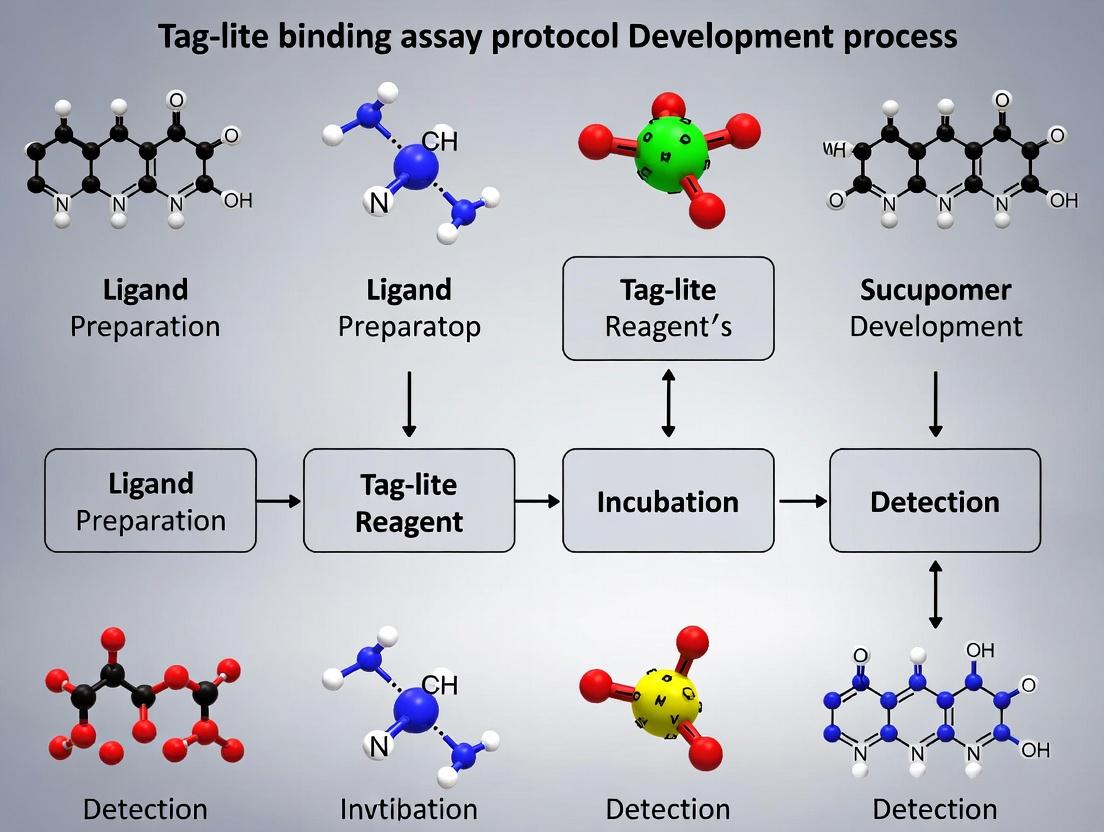
1.0 Introduction
This application note details the development and optimization of a Tag-lite binding assay protocol, a critical component of a broader thesis focused on advancing high-throughput screening (HTS) methodologies for G Protein-Coupled Receptors (GPCRs). The core innovation lies in exploiting the advantages of Tag-lite technology over traditional radiometric and other heterogeneous assay formats. The primary benefits are Homogeneity (no-wash, mix-and-read format), Speed (greatly reduced assay time), and Miniaturization Potential (compatibility with 384- and 1536-well plates), which collectively enhance throughput, reduce reagent consumption, and improve data quality.
2.0 Comparative Advantages: Quantitative Summary
Table 1: Comparative Analysis of Binding Assay Platforms
| Parameter | Traditional Radioligand Binding | Time-Resolved Fluorescence Resonance Energy Transfer (Tag-lite) |
|---|---|---|
| Assay Format | Heterogeneous (requires filtration/separation) | Homogeneous (no-wash) |
| Assay Time (hands-on) | ~2-4 hours | ~1-2 hours |
| Assay Time (incubation) | 60-120 minutes | 30-60 minutes |
| Miniaturization | Limited (typically 96-well) | Excellent (384-, 1536-well) |
| Throughput | Low to Moderate | High to Very High |
| Reagent Consumption | High | Low (µL volumes) |
| Signal Detection | Radioactive (scintillation) | Fluorescence (TR-FRET) |
| Safety Concerns | Yes (radioactive waste) | No (non-radioactive) |
| Z'-Factor (Typical) | 0.5 - 0.7 | 0.7 - 0.9 |
3.0 Core Experimental Protocol: Tag-lite SNAP-tag GPCR Ligand Binding Assay
Table 2: Research Reagent Solutions Toolkit
| Item | Function |
|---|---|
| SNAP-tag GPCR Cell Line | Recombinant cells expressing the GPCR of interest fused to the SNAP-tag. |
| Fluorescent Ligand (Lumi4-Tb conjugate) | Tracer ligand that binds to the GPCR's orthosteric or allosteric site, donor in TR-FRET. |
| Cell Membrane Preparation | Source of SNAP-tag GPCR receptors; enables stable, consistent receptor presentation. |
| SNAP-Lumi4-Tb Substrate | Cell-impermeant substrate that covalently labels the SNAP-tag with the Terbium cryptate donor. |
| Tag-lite Buffer | Optimized physiological buffer for labeling and binding reactions, minimizing background. |
| Reference Compound (e.g., antagonist) | High-affinity unlabeled ligand for determining non-specific binding (NSB). |
Protocol 3.1: Receptor Labeling and Binding Assay
3.1.1 Materials Preparation
- Thaw Tag-lite Buffer, SNAP-Lumi4-Tb substrate, and cell membrane preparation on ice.
- Prepare a 100 nM working solution of SNAP-Lumi4-Tb substrate in Tag-lite Buffer.
- Prepare serial dilutions of test compounds in assay buffer.
- Prepare a 10 µM stock of reference compound for NSB wells.
3.1.2 SNAP-tag Labeling (Pre-assay)
- In a low-volume microplate (e.g., 384-well), add 10 µL of cell membrane preparation per well.
- Add 10 µL of the 100 nM SNAP-Lumi4-Tb substrate solution. Final [Substrate] = 50 nM.
- Seal the plate, incubate for 1 hour at 37°C protected from light.
- Critical Step: After incubation, add 60 µL of Tag-lite Buffer to each well to stop the labeling reaction. Centrifuge at 4°C (2000 x g, 10 min). Carefully aspirate 70 µL of supernatant, leaving the labeled membrane pellet.
3.1.3 Ligand Binding Reaction
- Resuspend the labeled membrane pellet in 20 µL of Tag-lite Buffer.
- Add 10 µL of test compound (or buffer for total binding, or reference compound for NSB).
- Initiate the binding reaction by adding 10 µL of fluorescent ligand at the predetermined Kd concentration (typically 1-10 nM). Final assay volume = 40 µL.
- Seal the plate, incubate for 30-60 minutes at room temperature, protected from light.
3.1.4 TR-FRET Measurement & Data Analysis
- Read the plate using a compatible microplate reader (e.g., PHERAstar, CLARIOstar) equipped with TR-FRET optics.
- Measure donor emission at 620 nm and acceptor emission at 665 nm following excitation at 337 nm.
- Calculate the TR-FRET ratio: Acceptor (665 nm) emission / Donor (620 nm) emission.
- Calculate specific binding: Specific Signal = TR-FRET Ratio(Total) - TR-FRET Ratio(NSB).
- Fit data to a non-linear regression model (e.g., one-site competitive binding) to determine IC50/Ki values.
4.0 Signaling Pathway and Experimental Workflow Visualization
Diagram 1: Tag-lite TR-FRET Binding Principle
Diagram 2: Homogeneous Assay Workflow
Step-by-Step Protocol: From Construct Design to Data Acquisition in a 384-Well Format
This application note constitutes Phase 1 of a comprehensive thesis on Tag-lite binding assay protocol development. It focuses on the foundational steps of selecting an appropriate SNAP-tag or CLIP-tag system and establishing a robust, reproducible cell line via transfection. The choice of tag and transfection method critically influences signal-to-noise ratio, assay robustness, and suitability for high-throughput screening (HTS) in drug discovery.
Key Considerations for Tag Selection
The selection between SNAP-tag and CLIP-tag hinges on the experimental requirements. Both are engineered variants of the human DNA repair protein O⁶-alkylguanine-DNA alkyltransferase that irreversibly react with specific benzylguanine (BG) or benzylcytosine (BC) substrates, respectively.
Table 1: Comparative Analysis of SNAP-tag vs. CLIP-tag
| Feature | SNAP-tag | CLIP-tag | Implication for Assay Development |
|---|---|---|---|
| Size | 20 kDa | 20 kDa | Comparable; minimal steric hindrance. |
| Substrate | Benzylguanine (BG) derivatives | Benzylcytosine (BC) derivatives | Orthogonal chemistry enables dual-labeling. |
| Reaction Kinetics (k₂) | ~10⁴ M⁻¹s⁻¹ | ~10³ M⁻¹s⁻¹ | SNAP-tag reacts ~10x faster than CLIP-tag. |
| Commercial Substrate Variety | Extensive (Fluorescent, Biotin, Beads) | Good, but less than SNAP-tag | SNAP offers more flexibility for detection. |
| Background | Very low cellular activity | Very low cellular activity | High specificity in mammalian cells. |
| Ideal Use Case | Single target labeling, Fast kinetics needed | Simultaneous dual-target labeling with SNAP | CLIP enables complex, multiplexed studies. |
Table 2: Common Transfection Methods for Stable Cell Line Generation
| Method | Principle | Max. Efficiency (HEK-293) | Key Advantage | Key Limitation | Best For |
|---|---|---|---|---|---|
| Lipofection | Cationic lipid-DNA complex fusion | >90% (transient) | High efficiency, easy to use, low cytotoxicity (new gens) | Cost for large scale, serum can interfere | Fast transient and stable line development |
| Electroporation | Electrical pulse creates pores | 70-80% | Effective for "hard-to-transfect" cells (e.g., primary) | Higher cell death, requires optimization | Suspension cells, primary cells |
| Lentiviral Transduction | Viral vector integration | >95% (with selection) | Very high efficiency, stable integration, broad cell tropism | Biosafety Level 2+ required, more complex | Generating homogeneous, long-term stable pools |
| PEI-based | Polymeric DNA compaction | 80-90% (transient) | Very low cost, effective for large-scale prep | Can be cytotoxic at high concentrations | Large-scale transient transfection for protein prod. |
Detailed Experimental Protocols
Protocol 1: Mammalian Expression Vector Construction for SNAP/CLIP-Tag Fusion Proteins
Objective: To clone the gene of interest (GOI) in-frame with the SNAP-tag or CLIP-tag into a mammalian expression vector.
Materials:
- SNAP-tag or CLIP-tag mammalian vector (e.g., pSNAPf, pCLIPf from New England Biolabs).
- cDNA for your target receptor/Protein of Interest (POI).
- Restriction enzymes or infusion cloning reagents.
- Competent E. coli.
- LB-Ampicillin agar plates.
- Plasmid Miniprep and Maxiprep kits.
Method:
- Design: Decide on fusion orientation (N-terminal or C-terminal to POI). Ensure a flexible linker (e.g., (GGGGS)₂) is encoded between tag and POI to minimize folding interference.
- Amplification: PCR-amplify the POI cDNA with primers adding appropriate 15-20 bp homology arms for Infusion cloning or specific restriction sites.
- Digestion & Ligation: Linearize the tag vector. For restriction cloning, digest both vector and insert, purify, and ligate using T4 DNA ligase. For infusion cloning, mix linearized vector and insert with recombinase enzyme.
- Transformation: Transform the reaction into competent E. coli. Plate on LB-Ampicillin agar. Incubate overnight at 37°C.
- Screening: Pick 5-10 colonies, grow in mini-cultures, and isolate plasmid DNA. Verify constructs by restriction digest and Sanger sequencing using tag-specific and POI-specific primers.
- Preparation: Isolate high-purity, endotoxin-free plasmid DNA using a maxiprep kit for transfection.
Protocol 2: Generation of Stable Cell Lines Expressing SNAP-tag Fusion Proteins via Lipofection and Antibiotic Selection
Objective: To create a monoclonal or polyclonal mammalian cell line stably expressing the SNAP/CLIP-tag fusion protein.
Materials:
- Validated plasmid DNA (from Protocol 1).
- HEK-293T or CHO-K1 cells (recommended for high transfectability).
- Complete growth medium (DMEM/F12 + 10% FBS).
- Serum-free Opti-MEM medium.
- Lipofection reagent (e.g., Lipofectamine 3000).
- Appropriate selection antibiotic (e.g., Geneticin/G418, Hygromycin B).
- 6-well plates, 10 cm dishes.
- Cloning rings (for monoclonal line isolation).
Method:
- Day 1 – Seeding: Seed HEK-293T cells in a 6-well plate at 30-50% confluence in antibiotic-free complete medium. Incubate at 37°C, 5% CO₂ overnight.
- Day 2 – Transfection: a. For one well, dilute 2.5 µg plasmid DNA in 125 µL Opti-MEM. Add 5 µL P3000 enhancer reagent (if using Lipofectamine 3000). b. In a separate tube, dilute 3.75 µL Lipofectamine 3000 in 125 µL Opti-MEM. Incubate for 5 min. c. Combine diluted DNA and diluted lipofectamine. Mix gently and incubate for 15-20 min at RT. d. Add the 250 µL DNA-lipid complex dropwise to the well. Gently rock the plate.
- Day 3 – Passage: 24h post-transfection, trypsinize cells and transfer to a 10 cm dish in complete medium.
- Day 4 – Selection: Begin selection by adding the pre-determined optimal concentration of antibiotic (e.g., 500 µg/mL G418 for HEK-293). Change medium with antibiotic every 2-3 days.
- Days 10-14 – Colony Formation: After 7-10 days, non-transfected cells will die. Surviving colonies will become visible.
- Monoclonal Isolation: a. For polyclonal pools, simply trypsinize and expand all surviving cells. b. For monoclonal lines, rinse plate with PBS, place a sterile cloning ring dipped in grease around a single colony. Trypsinize cells within the ring and transfer to a 24-well plate. Expand and screen.
- Validation: Screen clones for expression level and functionality via Western blot (anti-SNAP/CLIP antibody) and live-cell labeling with fluorescent substrate (e.g., SNAP-Surface 549).
Visualizations
Title: Phase 1 Workflow: From Tag Choice to Cell Line
Title: SNAP-tag Covalent Labeling Chemistry
The Scientist's Toolkit
Table 3: Essential Research Reagent Solutions for Phase 1
| Reagent/Category | Example Product/Type | Primary Function in Phase 1 |
|---|---|---|
| Tag Vectors | pSNAPf, pCLIPf (NEB), pFC vectors (Promega) | Mammalian expression backbones with codon-optimized SNAP/CLIP tags for N- or C-terminal fusions. |
| Cloning Reagents | Infusion HD Cloning Kit (Takara), Gibson Assembly | Enable seamless, restriction-site-independent construction of fusion gene plasmids. |
| Transfection Reagents | Lipofectamine 3000 (Invitrogen), PEI MAX (Polysciences), Fugene HD (Promega) | Form complexes with plasmid DNA to facilitate its entry into mammalian cells with high efficiency and low toxicity. |
| Selection Antibiotics | Geneticin (G418), Hygromycin B, Puromycin | Kill non-transfected cells, allowing for the survival and expansion of stably integrated clones. |
| Validation Substrates | SNAP-Surface 549 (NEB), SNAP-Cell Oregon Green | Cell-permeable fluorescent BG substrates for confirming tag expression and localization via microscopy or flow cytometry. |
| Cell Culture Media | Opti-MEM (Gibco) | Serum-free medium used for diluting DNA and transfection reagents to maximize complex formation and uptake. |
| Detection Antibodies | Anti-SNAP-tag mAb (NEB), Anti-CLIP-tag pAb | For Western blot validation of fusion protein expression and size. |
This application note details Phase 2 of a comprehensive Tag-lite binding assay protocol development thesis. This phase focuses on the critical steps between cell plating and the final detection readout: seeding cells for optimal confluency, labeling cell-surface SNAP-tag or CLIP-tag fusion proteins with fluorescent dyes, and executing wash steps to minimize non-specific background signal. Optimizing these conditions is paramount for achieving robust, reproducible data in live-cell ligand-binding and internalization studies.
The following parameters were systematically investigated to define optimal assay conditions. Data is pooled from internal validation studies and current literature.
Table 1: Optimization of Cell Seeding Density for 96-well Plates
| Cell Line Type | Recommended Seeding Density (cells/well) | Seeding Volume (µL) | Target Confluence at Assay (% ) | Optimal Attachment Time (hours) |
|---|---|---|---|---|
| Adherent (HEK293, CHO) | 40,000 - 60,000 | 100 | 80-90 | 18-24 |
| Suspension (Jurkat, K562) | 150,000 - 200,000 | 100 | N/A | Immediate (coated plates) |
| Neuronal (SH-SY5Y) | 70,000 - 100,000 | 100 | 60-80 | 24-48 |
Table 2: Optimization of Labelling Parameters for SNAP/CLIP-tags
| Parameter | Tested Range | Optimal Value | Impact on Signal-to-Background Ratio (S/B) |
|---|---|---|---|
| Label (BG-778, BG-Lumi4-Tb) Concentration | 50 nM - 500 nM | 100 nM | S/B peaks at 100 nM; higher conc. increases background |
| Labelling Incubation Time | 30 min - 2 hours | 1 hour | >1 hour yields minimal S gain but increases background |
| Labelling Temperature | 4°C, 22°C, 37°C | 4°C (surface) / 37°C (total) | 4°C minimizes internalization during label; 37°C labels total pool |
| Quenching Agent (e.g., Bromophenol Blue) | 0 - 100 µM | 10 µM | Reduces non-covalent dye binding by >50% |
Table 3: Wash Buffer Composition Comparison
| Buffer Component | Purpose | Tested Formulations | Optimal Formulation (1x) |
|---|---|---|---|
| Physiological Saline | Maintain osmolarity, cell viability | PBS, HBSS | HBSS (with Ca2+/Mg2+) |
| Serum/Protein | Reduce non-specific binding | 0.1-1% BSA, 0.1% Pluronic F-68 | 0.1% BSA |
| pH Stabilizer | Maintain physiological pH | 10-25 mM HEPES | 20 mM HEPES |
| Recommended Final | HBSS + 20 mM HEPES + 0.1% BSA, pH 7.4 |
Detailed Experimental Protocols
Protocol 3.1: Optimized Cell Seeding for Adherent Cells
Objective: To achieve uniform, sub-confluent monolayers for consistent labeling and ligand access. Materials: Sterile cell culture hood, humidified CO2 incubator (37°C, 5% CO2), multichannel pipette, sterile reservoir, 96-well microplate (white, clear-bottom), complete growth medium, trypsin-EDTA, hemocytometer. Procedure:
- Cell Preparation: Harvest cells in mid-log phase via trypsinization. Neutralize with complete medium, centrifuge (300 x g, 5 min), and resuspend in fresh pre-warmed medium.
- Counting & Dilution: Determine cell density using a hemocytometer. Dilute cell suspension to a concentration of 4-6 x 10^5 cells/mL in complete medium.
- Seeding: Using a multichannel pipette, dispense 100 µL of cell suspension per well into the 96-well plate (final: 40,000-60,000 cells/well). Gently tap plate sides to disperse cells evenly.
- Incubation: Place plate in the humidified CO2 incubator for 18-24 hours. Visually inspect confluence prior to proceeding (target 80-90%). Note: For suspension cells, use plates pre-coated with Poly-D-Lysine and seed cells directly in assay buffer.
Protocol 3.2: Live-Cell SNAP-tag Labelling
Objective: To specifically label cell-surface SNAP-tag fusion proteins with minimal background and internalization. Materials: Labelling buffer (HBSS/HEPES), SNAP-tag substrate (e.g., BG-778, BG-Lumi4-Tb), 10 µM Bromophenol Blue stock, plate centrifuge, microplate shaker. Procedure:
- Prepare Labelling Solution: Dilute the SNAP-tag substrate to 100 nM in cold (4°C) labelling buffer supplemented with 10 µM Bromophenol Blue. Keep on ice.
- Cell Preparation: Remove growth medium from the seeded 96-well plate by gently inverting. Wash cells once with 150 µL of cold labelling buffer (without substrate).
- Labelling: Add 50 µL/well of the prepared labelling solution. Incubate the plate on a microplate shaker (gentle orbit) at 4°C for 60 minutes, protected from light.
- Quenching & Washing: After incubation, add 100 µL/well of cold labelling buffer containing 0.1% BSA. Centrifuge plate at 300 x g for 5 min at 4°C. Carefully aspirate supernatant. Repeat this wash step twice more (total of 3 washes).
- Proceed to Assay: Cells are now ready for ligand addition in Phase 3 (Binding Reaction).
Visualizations
Diagram 1: Live-Cell Labeling Workflow (50 chars)
Diagram 2: Labeling Specificity & Wash Mechanism (80 chars)
The Scientist's Toolkit
Table 4: Essential Research Reagent Solutions for Phase 2
| Item | Function in Phase 2 | Key Consideration |
|---|---|---|
| SNAP-tag Substrate (e.g., BG-Lumi4-Tb) | Covalently binds SNAP-tag for target detection. | Choose fluorophore (e.g., red) or lanthanide (e.g., Tb) based on assay type (FRET vs. direct). |
| CLIP-tag Substrate (e.g., BC-Lumi4-Tb) | Covalently binds CLIP-tag for orthogonal labeling. | Use for co-expression studies with SNAP-tag. |
| HEPES-Buffered HBSS | Provides physiological ion balance and pH stability outside a CO2 incubator. | Always supplement with Ca2+/Mg2+ for cell adhesion integrity. |
| Bovine Serum Albumin (BSA), Fraction V | Blocks non-specific binding sites on plastic and cell surfaces during washes. | Use at 0.1% in buffer; higher concentrations may interfere with some ligands. |
| Pluronic F-68 | Non-ionic surfactant that reduces hydrophobic interactions and cell clumping. | Alternative to BSA, especially for sensitive binding interactions. |
| Bromophenol Blue (BPB) | Competitive agent that quenches non-covalent binding of dye to serum proteins/cell surfaces. | Critical for reducing background in SNAP/CLIP assays; use at ~10 µM. |
| Poly-D-Lysine | Coats plate surface to enhance adhesion of sensitive or suspension cell lines. | Essential for neuronal cells and assays requiring firm attachment. |
| Cell Dissociation Reagent (Trypsin-EDTA) | Gently detaches adherent cells for seeding at uniform density. | Neutralize completely with serum-containing medium to avoid cell damage. |
Within the broader thesis on Tag-lite binding assay protocol development, Phase 3 represents the core experimental step where molecular interactions are quantitatively measured. This phase involves the precise addition of labeled ligands and unlabeled competitors to cell samples expressing the target receptor of interest, followed by a controlled incubation to reach binding equilibrium. The successful execution of this phase is critical for generating robust data for both saturation binding (to determine receptor affinity (Kd) and density (Bmax)) and competition binding (to determine competitor compound affinity (Ki)) experiments. This application note details the protocols and considerations for this decisive phase.
Key Concepts and Quantitative Parameters
The following table summarizes the core quantitative parameters determined in Phase 3 experiments and their significance.
Table 1: Key Quantitative Parameters from Binding Experiments
| Parameter | Experiment Type | Definition | Typical Range/Units |
|---|---|---|---|
| Kd | Saturation | Equilibrium dissociation constant of the labeled ligand. Measure of affinity. | pM to µM |
| Bmax | Saturation | Maximum number of binding sites. Measure of receptor density. | fmol/mg protein or sites/cell |
| Ki | Competition | Inhibition constant of the unlabeled competitor. Measure of its affinity for the target. | pM to µM |
| IC50 | Competition | Concentration of competitor that inhibits 50% of specific labeled ligand binding. | nM to µM |
| Non-specific Binding (NSB) | Both | Binding not displaced by a high concentration of competitor. | Ideally <10-30% of total binding |
| Z'-Factor | Both | Statistical parameter for assay quality and robustness. | >0.5 indicates excellent assay |
Detailed Experimental Protocols
Protocol 3.1: Saturation Binding Experiment Setup
Objective: To determine the affinity (Kd) and density (Bmax) of a receptor for a fluorescent ligand.
Materials & Reagents:
- Tag-lite labeled cells expressing the target GPCR (from Phase 2: Cell Preparation).
- Serial dilutions of the SNAP- or CLIP-tagged fluorescent ligand (e.g., red-emitting ligand for Lumi4-Tb donor cells).
- Saturation Buffer: Hanks’ Balanced Salt Solution (HBSS) or assay-specific buffer, 0.1% BSA, pH 7.4.
- Unlabeled antagonist at high concentration (for NSB determination).
- Low-volume, non-binding, white 384-well or 96-well assay plates.
- Multichannel pipettes and reagent reservoirs.
Methodology:
- Plate Preparation: Distribute Tag-lite cells (prepared in Phase 2) into two identical sets of wells in a white assay plate (e.g., 5,000 cells/well in 20 µL). One set is for Total Binding, the other for Non-Specific Binding (NSB).
- Ligand Dilution Series: Prepare a 12-point, 1:2 or 1:3 serial dilution of the fluorescent ligand in Saturation Buffer, typically covering a range from ~0.1 x Kd to 10 x Kd (pilot range may be 0.1 nM to 100 nM).
- Competitor for NSB: To the NSB well set, add a fixed, high concentration (e.g., 1-10 µM) of an unlabeled competitor to define non-specific binding. Add an equivalent volume of buffer to the Total Binding well set.
- Ligand Addition: Add the serial dilutions of the fluorescent ligand to both the Total and NSB well sets. Final assay volume is typically 40-50 µL.
- Incubation: Seal the plate and incubate in the dark at the predetermined temperature (often 4°C to minimize internalization, or room temperature) for 2-4 hours to reach equilibrium. Agitation is recommended.
- Signal Measurement: Proceed to Phase 4 (Signal Detection). Specific binding is calculated as (Total Binding signal) - (NSB signal at corresponding ligand concentration).
Protocol 3.2: Competition Binding Experiment Setup
Objective: To determine the affinity (Ki) of an unlabeled test compound for the receptor.
Materials & Reagents:
- Tag-lite labeled cells expressing the target GPCR.
- Fixed concentration of the fluorescent ligand (near its Kd value, e.g., 2-5 nM).
- Serial dilutions of unlabeled test/competitor compounds.
- Reference compound (known high-affinity antagonist/agonist).
- Assay Buffer: HBSS, 0.1% BSA, pH 7.4.
- Low-volume, non-binding, white 384-well or 96-well assay plates.
Methodology:
- Plate Preparation: Distribute Tag-lite cells into assay plates (e.g., 5,000 cells/well in 20 µL).
- Competitor Dilution Series: Prepare 10-point, 1:3 serial dilutions of each test compound and the reference compound in Assay Buffer, typically from 10 µM to 0.1 nM (or beyond expected Ki).
- Competitor Addition: Add the compound dilution series to the plate. Include control wells for Total Binding (buffer only, no competitor) and NSB (high concentration of reference compound).
- Ligand Addition: Add a fixed concentration of the fluorescent ligand to all wells. The final concentration should be near the Kd of the ligand (determined in Protocol 3.1).
- Incubation: Seal the plate and incubate in the dark under optimized conditions (temperature, time as in Protocol 3.1) to reach equilibrium.
- Signal Measurement: Proceed to Phase 4. Percent inhibition is calculated relative to Total and NSB controls.
Visualizing Experimental Workflows
Title: Phase 3 Experimental Setup Workflow
Title: Competitive Binding Equilibrium at Target Receptor
The Scientist's Toolkit: Research Reagent Solutions
Table 2: Essential Materials for Phase 3 Experiments
| Item | Function in Phase 3 | Key Considerations |
|---|---|---|
| SNAP-/CLIP-tagged Fluorescent Ligands (e.g., red-emitting) | High-affinity probe that binds specifically to the labeled target receptor, generating the TR-FRET signal. | Must be spectrally compatible with donor (Tb). Affinity (Kd) should suit assay range. Purity is critical. |
| Unlabeled Reference Compound (e.g., known antagonist) | Defines non-specific binding (NSB) at high concentration. Used as control in competition experiments. | Should have high affinity and selectivity for the target. |
| Test Compounds/Competitors | Unlabeled molecules whose affinity (Ki) for the target is to be determined. | Require serial dilution in DMSO/buffer. Stability and solubility must be assessed. |
| Low-Volume, White Assay Plates (384-well) | Platform for the binding reaction and subsequent TR-FRET reading. | White plates enhance signal. Non-binding surface minimizes adsorption. Low volume reduces reagent costs. |
| Multichannel Electronic Pipette | Enables rapid, precise, and reproducible transfer of ligand/competitor dilution series and cells. | Essential for minimizing well-to-well variability and plate preparation time. |
| Assay Buffer with BSA (e.g., HBSS + 0.1% BSA) | Provides physiological pH and ionic strength for binding. BSA reduces non-specific adsorption to plates and tubes. | Must be optimized for the specific receptor-ligand pair. Chelators (e.g., EDTA) may be added. |
| Plate Sealer & Microplate Shaker | Seal prevents evaporation during incubation. Shaker ensures homogeneous mixing and facilitates equilibrium. | Adhesive seals are preferred. Orbital shaking at 300-600 rpm is typical. |
| Temperature-Controlled Incubator | Maintains consistent temperature (4°C, RT, or 37°C) during the binding equilibrium period. | Choice affects kinetics, internalization, and final binding parameters. |
Within the broader thesis on Tag-lite binding assay protocol development, Phase 4 represents the critical data acquisition stage. This phase translates the biological interactions of previous steps (cell preparation, labeling, and compound addition) into quantifiable, high-quality data. Homogeneous Time-Resolved Fluorescence (HTRF) is a robust, proximity-based assay technology combining FRET with time-gated detection to eliminate short-lived background fluorescence. The selection and correct configuration of a compatible multi-mode microplate reader are paramount for achieving optimal signal-to-noise ratios (S/N) and assay robustness. This note details the protocols and considerations for this final measurement step.
Key Principles of HTRF Measurement
HTRF utilizes a donor fluorophore (typically Europium cryptate, Eu³⁺) with a long fluorescence lifetime and an acceptor (XL665 or d2) that emits at 665 nm upon FRET. Time-resolved detection (after a delay of 50-150 µs) allows the short-lived autofluorescence (ns range) to decay, leaving only the specific, long-lived signal. The primary calculated metric is the Ratio (665 nm / 620 nm), which normalizes the FRET signal (665 nm) against the donor emission (620 nm), correcting for well-to-well variations, compound interference, and pipetting errors.
Compatible Multi-mode Reader Specifications
Not all readers are equipped for HTRF. Essential features include:
- Time-Resolved Fluorometry (TRF/TR-FRET) capability.
- Dual-emission detection for 620 nm and 665 nm.
- Appropriate light sources (e.g., Xenon flash lamp or laser).
- High-quality filters or monochromators.
- Pre-optimized HTRF application settings.
Examples of widely compatible readers include the PerkinElmer EnVision, Revvity (formerly BioTek) Synergy Neo2, Tecan Spark Cyto, and BMG LABTECH PHERAstar.
Comparative Reader Specifications & Performance
Table 1: Key Specifications of Compatible Multi-mode Readers for HTRF
| Reader Model | Light Source | Detection Method | Time-Gate Delay (Typical) | Z-Height Adjustment | Pre-configured HTRF Protocols |
|---|---|---|---|---|---|
| PerkinElmer EnVision | Xenon flash lamp | PMT (with filters) | 50-100 µs | Yes | Extensive library |
| BMG LABTECH PHERAstar | Xenon flash lamp or Laser | PMT (with filters) | 60-80 µs | Yes | Yes, with optimization |
| Revvity Synergy Neo2 | Quad monochromators + filters | PMT / CCD | Adjustable (50-150 µs) | Yes | Available |
| Tecan Spark Cyto | Xenon flash lamp + monochromator | PMT | 50-100 µs | Yes | Yes |
Table 2: Typical Assay Performance Metrics (Using a Tag-lite SNAP-tag Binding Assay)
| Performance Metric | Target Value | Acceptable Range | Notes |
|---|---|---|---|
| Ratio (665/620 nm) | Varies by assay | ≥ 2 for positive control | System-specific baseline. |
| Signal-to-Noise (S/N) | > 10 | Minimum 5 | (Signalpositive - Signalnegative) / SDnegative |
| Signal-to-Background (S/B) | > 5 | Minimum 3 | Signalpositive / Signalnegative |
| Z'-Factor | > 0.5 | 0.5 - 1.0 | Indicator of assay robustness. |
| CV (% of Ratios) | < 10% | < 15% | For replicate positive controls. |
Detailed Experimental Protocol for Plate Reading
Protocol 4.1: Instrument Setup and Pre-Read Validation
Objective: To configure the multi-mode reader for optimal HTRF signal detection. Materials: Compatible multi-mode reader, calibration plate (if available), experimental microplate. Procedure:
- Power and Initialize: Turn on the reader and associated software. Allow lamps to warm up for the recommended time (typically 15-30 min).
- Protocol Selection: Load the pre-configured "HTRF" or "TR-FRET" protocol. If creating new:
- Measurement Type: Select "Time-Resolved Fluorescence" or "TR-FRET".
- Excitation: Set to 320-340 nm (for Eu cryptate).
- Emission 1: 620 nm (±10 nm, bandwidth ~15 nm) for Donor.
- Emission 2: 665 nm (±10 nm, bandwidth ~15 nm) for Acceptor.
- Delay Time: Set to 50-100 µs.
- Integration Time (Window): Set to 100-500 µs.
- Number of Flashes: 50-100 flashes per well.
- Plate Definition: Define plate type (e.g., 384-well, low-volume, white). Set the measurement height (Z-height) optimally for the assay volume (e.g., 7.5 µL for 384-well low-volume plates). Critical: Confirm the plate bottom type (e.g., ProxiPlate) is correctly selected.
- Calibration (Optional but Recommended): Run a system suitability test using an HTRF positive control calibration plate, if available, to verify optical alignment and performance.
- Save Protocol: Save the validated protocol with a unique name.
Protocol 4.2: Plate Loading, Reading, and Data Export
Objective: To acquire raw fluorescence data from the assay plate. Procedure:
- Plate Preparation: After the final assay incubation (Phase 3), centrifuge the plate briefly (e.g., 1000 rpm, 1 min) to settle contents and remove bubbles.
- Plate Loading: Wipe the plate bottom with a lint-free cloth and ethanol to remove fingerprints/dust. Load the plate into the reader carriage.
- Read Sequence: Initiate the reading protocol. The reader will typically perform a scan, recording time-gated fluorescence intensities at 620 nm (Donor) and 665 nm (Acceptor) for every well.
- Data Inspection: Visually inspect the raw fluorescence values post-read. Check for edge effects, obvious outliers, or reading errors.
- Data Export: Export the raw data (620 nm and 665 nm intensities for all wells) in a standard format (.csv, .xlsx) for downstream analysis. Include well identifiers and any plate maps.
Protocol 4.3: Post-Read Data Normalization and Analysis
Objective: To convert raw fluorescence into biologically meaningful metrics. Procedure:
- Calculate the Ratio: For each well, compute: [ \text{Ratio} = \frac{\text{Fluorescence}{665 nm} \times 10^4}{\text{Fluorescence}{620 nm}} ] (The 10⁴ multiplier is conventional to bring the ratio to a convenient scale).
- Calculate Assay Controls: Determine the average Ratio for:
- Positive Control (PC): Wells with known binding interaction.
- Negative Control (NC): Wells with no binding interaction (e.g., donor-only, acceptor-only, or unlabeled).
- Normalize Data (for dose-response): Express compound wells as a percentage of the control response. [ \% \text{Inhibition} = \frac{(\text{Avg. Ratio}{PC} - \text{Ratio}{Sample})}{(\text{Avg. Ratio}{PC} - \text{Avg. Ratio}{NC})} \times 100 ] [ \% \text{Signal} = 100 - \% \text{Inhibition} ]
- Calculate QC Parameters:
- S/B = Avg. Ratio{PC} / Avg. Ratio{NC}
- S/N = (Avg. Ratio{PC} - Avg. Ratio{NC}) / SD_{NC}
- Z' Factor = 1 - [ (3 * SD{PC} + 3 * SD{NC}) / |Avg. Ratio{PC} - Avg. Ratio{NC}| ]
- Curve Fitting: Fit normalized dose-response data to a 4-parameter logistic (4PL) model to determine IC₅₀/EC₅₀ values.
The Scientist's Toolkit: Key Research Reagent Solutions
Table 3: Essential Materials for Tag-lite HTRF Assay and Measurement
| Item | Function/Description | Example Product (Supplier) |
|---|---|---|
| Tag-lite Labeling Reagents | Cell-impermeable fluorophores that covalently label SNAP-tag or CLIP-tag proteins on live cells. | SNAP-Lumi4-Tb, RED-tris-NTA (Cisbio) |
| Multi-mode Reader | Instrument capable of time-resolved, dual-emission detection for HTRF. | EnVision, PHERAstar, Synergy Neo2 |
| Low-Volume Microplates | White, solid-bottom plates optimized for low assay volumes and HTRF signal. | 384-well ProxiPlate (PerkinElmer) |
| Assay Buffer | Provides physiological pH and ionic strength; often HEPES-based with low autofluorescence. | Tag-lite Labeling Buffer (Cisbio) |
| Positive/Negative Control Compounds | Validates assay performance; provides reference signals for normalization. | Target-specific reference ligand (e.g., antagonist), buffer/DMSO. |
| Data Analysis Software | For curve fitting, plate visualization, and statistical analysis of HTRF ratios. | GraphPad Prism, Microsoft Excel with XLFit, Reader-native software. |
Visualizations
HTRF Signal Measurement Workflow
HTRF Principle and Detection Logic
Within the framework of Tag-lite binding assay protocol development, robust data processing is critical for accurate interpretation of ligand-receptor interactions. This Application Note details the methodology for calculating emission ratios (665 nm / 620 nm) and normalizing signals to generate reliable, quantitative binding data, essential for drug discovery professionals.
The Scientist's Toolkit: Key Research Reagent Solutions
| Item | Function in Tag-lite Assay |
|---|---|
| SNAP-Tag or CLIP-Tag Recombinant Protein | Enables covalent, specific labeling of the target receptor with a fluorescent dye. |
| Terbium (Tb) Cryptate-Conjugated Substrate | Acts as the long-lifetime donor fluorophore (excitation ~337 nm, emission ~620 nm). |
| Fluorescent Acceptor (e.g., d2, GFP) | Acts as the acceptor, attached to the ligand or a secondary binder (emission ~665 nm). |
| Tag-lite Buffer | Optimized assay buffer to minimize autofluorescence and maintain protein stability. |
| Multiwell Microplate (White) | Used for homogenous, time-resolved FRET (TR-FRET) signal detection. |
| Plate Reader with TR-FRET Capability | Must be capable of pulsed excitation and time-gated detection at 620 nm and 665 nm. |
Core Protocol: Ratio Calculation and Data Normalization
Raw Data Acquisition
- Instrument: Use a time-resolved fluorescence plate reader.
- Settings: Following a delay after a pulsed excitation (~337 nm), integrate the emission signals at 620 nm (Tb Cryptate donor) and 665 nm (acceptor) using appropriate time-gating (e.g., 50-500 µs delay, 400-1000 µs integration).
- Output: For each experimental well, obtain two intensity values: I~620~ and I~665~.
Calculating the 665 nm / 620 nm Ratio (R)
For each well, calculate the emission ratio to correct for well-to-well variability in protein concentration and donor labeling efficiency.
Normalization of Ratio Data
Normalization translates raw ratios into interpretable biological parameters (e.g., % specific binding, % inhibition).
Common Normalization Methods:
| Method | Formula | Application in Tag-lite |
|---|---|---|
| Signal-to-Background | (R~sample~ - R~blank~) / R~blank~ | Assessing total binding signal strength. |
| % of Specific Binding | 100 * (R~sample~ - R~NSB~) / (R~Total~ - R~NSB~) | For saturation or competition binding assays. |
| % Inhibition | 100 * [1 - (R~sample~ - R~NSB~) / (R~Max~ - R~NSB~)] | For competition assays with a reference ligand. |
- R~sample~: Ratio for the test condition.
- R~blank~: Ratio from wells with donor-labeled receptor only (no acceptor).
- R~NSB~: Ratio for non-specific binding (e.g., + excess unlabeled ligand).
- R~Total~ or R~Max~: Ratio for total specific binding (e.g., + saturating labeled ligand or vehicle control in competition assays).
Data Presentation: Example from a Competition Binding Assay
The following table summarizes processed data from a hypothetical Tag-lite competition assay for a novel antagonist.
Table 1: Normalized Data for Compound X Dose-Response.
| [Compound X] (M) | Raw I₆₂₀ | Raw I₆₆₅ | Ratio (665/620) | Specific Binding (R - R_NSB) | % Inhibition |
|---|---|---|---|---|---|
| NSB Control | 105,000 | 12,800 | 0.122 | 0.000 | 100%* |
| 0 (Max Ctrl) | 98,500 | 45,200 | 0.459 | 0.337 | 0% |
| 1.00E-11 | 99,100 | 44,500 | 0.449 | 0.327 | 3.0% |
| 1.00E-10 | 97,800 | 40,100 | 0.410 | 0.288 | 14.5% |
| 1.00E-09 | 101,200 | 32,900 | 0.325 | 0.203 | 39.8% |
| 1.00E-08 | 102,500 | 21,500 | 0.210 | 0.088 | 73.9% |
| 1.00E-07 | 103,800 | 14,100 | 0.136 | 0.014 | 95.8% |
| 1.00E-06 | 104,200 | 12,900 | 0.124 | 0.002 | 99.4% |
*NSB is defined as 100% inhibition. RNSB = 0.122. RMax = 0.459.
Detailed Experimental Protocol: Tag-lite Saturation Binding with Data Processing
Objective: Determine the binding affinity (K~D~) of a fluorescent ligand.
Protocol Steps:
- Labeling: Seed cells expressing SNAP-tagged receptor in a white 96-well plate. Label with Tb cryptate-conjugated SNAP substrate according to manufacturer's protocol.
- Ligand Addition: Prepare a serial dilution of the fluorescent acceptor ligand. Add to labeled cells in triplicate, including wells for total binding (all ligand concentrations) and non-specific binding (NSB, high ligand concentration + 1000x unlabeled competitor).
- Incubation: Incubate plate for equilibrium binding (typically 1-2h at RT or 4°C).
- Reading: Measure time-resolved fluorescence at 620 nm and 665 nm.
- Data Processing:
- Calculate the 665/620 ratio (R) for each well.
- For each ligand concentration, calculate specific binding: R~specific~ = R~total~ - R~NSB~ (using the average R~NSB~ from high competitor wells).
- Normalize specific binding values as a percentage of the maximum specific binding (from the highest ligand concentration).
- Fit the normalized data vs. log[ligand] to a four-parameter logistic (4PL) or one-site specific binding model to derive K~D~.
1. Introduction and Thesis Context
Within the broader thesis on Tag-lite binding assay protocol development research, this application note details the implementation of a homogeneous, time-resolved fluorescence resonance energy transfer (TR-FRET) competitive binding assay for G protein-coupled receptor (GPCR) drug screening. The Tag-lite platform leverages SNAP-tag or HaloTag technology to specifically label receptors with a fluorescent donor, enabling precise, cell-based quantification of ligand binding without the need for radioactive tracers or washing steps. This protocol exemplifies the core thesis aim of developing robust, generic, and high-throughput-compatible binding assays.
2. Key Research Reagent Solutions
Table 1: Essential Materials for Tag-lite Competitive Binding Assays
| Reagent / Solution | Function in the Assay |
|---|---|
| SNAP-tag or HaloTag-labeled GPCR Cell Line | Engineered cell line expressing the GPCR of interest fused to the SNAP or HaloTag protein. Provides the target for ligand binding. |
| Terbium (Tb) Cryptate-conjugated Substrate (e.g., SNAP-Lumi4-Tb or HaloTag-Lumi4-Tb) | FRET donor. Covalently binds to the tag on the GPCR, allowing stable, specific receptor labeling. |
| Fluorescently Labeled Tracer Ligand (Red acceptor, e.g., d2 dye) | FRET acceptor. Binds competitively with test compounds to the receptor's orthosteric or allosteric site. Serves as the displaceable probe. |
| Reference Ligand (e.g., known high-affinity antagonist) | Used to determine non-specific binding (NSB) and validate assay performance. |
| Assay Buffer (e.g., HBSS with 0.1% BSA or proprietary Tag-lite buffer) | Maintains cell viability and provides optimal conditions for ligand-receptor interaction. |
| Low-Volume, White Multiwell Plates (e.g., 384-well) | Optimized for homogeneous assays and sensitive fluorescence detection. |
3. Experimental Protocol: Competitive Binding Assay
Day 1: Cell Seeding
- Harvest SNAP-tagged GPCR-expressing cells in log growth phase.
- Count cells and adjust density to 1-2 x 10⁶ cells/mL in growth medium.
- Seed 5,000-10,000 cells per well in a 384-well white microplate in 20 µL of growth medium.
- Incubate plates overnight (16-24 h) at 37°C, 5% CO₂ for cell adhesion.
Day 2: Receptor Labeling and Assay Execution
- Prepare Labeling Solution: Dilute the SNAP-Lumi4-Tb substrate in assay buffer to a final recommended concentration (typically 100 nM). Protect from light.
- Label Receptors: Remove growth medium from cells. Add 20 µL of labeling solution per well. Incubate for 1 hour at 37°C protected from light.
- Prepare Compound/Tracer Plates: In a separate plate, serially dilute test and reference compounds in assay buffer. Pre-mix the fluorescent tracer ligand at its predetermined Kd concentration (e.g., 5-10 nM) with assay buffer.
- Initiate Competitive Binding: Remove the labeling solution and gently wash cells twice with 40 µL of assay buffer. Add 10 µL of the compound dilution (or buffer for total binding controls) to appropriate wells. Immediately add 10 µL of the tracer ligand solution to all wells. The final assay volume is 20 µL.
- Incubation: Incubate the plate for 1-2 hours at room temperature or 4°C (to minimize internalization) protected from light.
- Detection: Read the plate on a TR-FRET compatible microplate reader (e.g., PHERAstar, EnVision). Excitation: 337 nm. Emission: measure donor signal at 620 nm and acceptor FRET signal at 665 nm.
Data Analysis:
- Calculate the ratio of acceptor emission (665 nm) to donor emission (620 nm) x 10⁴ for each well. This is the normalized TR-FRET signal.
- Determine specific binding: Specific Signal = Total Signal (buffer control) - NSB Signal (saturating reference ligand control).
- For each test compound, calculate % Inhibition: 100 * [1 - (Signal_compound - Signal_NSB) / (Signal_Total - Signal_NSB)].
- Fit dose-response data using a four-parameter logistic equation to determine IC₅₀ values.
4. Quantitative Data Summary
Table 2: Typical Assay Performance Metrics and Data Output
| Parameter | Typical Target Value / Output | Description |
|---|---|---|
| Z'-Factor | > 0.5 | Statistical parameter reflecting assay robustness and suitability for HTS. |
| Signal-to-Background (S/B) | > 5 | Ratio of total binding signal to non-specific binding signal. |
| Coefficient of Variation (CV) | < 10% | Measure of well-to-well reproducibility for control wells. |
| Tracer Kd | 1 - 20 nM | Experimentally determined dissociation constant of the fluorescent tracer for the target GPCR. |
| Reference Ligand IC₅₀ | Consistent with literature | Validates correct assay pharmacology. |
| Test Compound IC₅₀ / Ki | Primary screening output | Concentration for half-maximal inhibition. Ki (inhibition constant) is calculated using the Cheng-Prusoff equation. |
5. Visualized Pathways and Workflows
Diagram 1: Tag-lite Competitive Binding Assay Principle
Diagram 2: Competitive Binding Assay Workflow
Troubleshooting Tag-lite Assays: Solving Common Issues and Optimizing Signal-to-Noise Ratio
Abstract Within the broader thesis on Tag-lite binding assay protocol development, optimizing Signal-to-Noise (S/N) ratio is paramount for achieving robust, sensitive, and reliable data. This application note details the primary causes of low S/N in Tag-lite assays, which utilize HaloTag and SNAP-tag technology for studying biomolecular interactions in a homogenous, time-resolved fluorescence resonance energy transfer (TR-FRET) format. We provide actionable solutions and detailed protocols to systematically diagnose and rectify sensitivity issues, enabling researchers to develop high-performance binding assays for drug discovery.
1. Introduction: The S/N Challenge in Tag-lite Assays Tag-lite assays offer a versatile platform for studying protein-protein interactions (PPIs) and receptor-ligand binding. The assay relies on specific labeling of targets with HaloTag or SNAP-tag ligands, followed by measurement of TR-FRET between a terbium cryptate (donor) and a compatible fluorophore (acceptor). A low S/N ratio, characterized by a weak specific signal (low FRET) relative to high background noise, compromises the assay window (Z'-factor) and the reliability of IC50/Kd determinations. Identifying the root cause is essential for effective troubleshooting.
2. Top Causes of Low S/N Ratio: A Diagnostic Table The following table summarizes the major contributors to poor S/N, their manifestations, and underlying mechanisms.
Table 1: Primary Causes of Low Signal-to-Noise Ratio in Tag-lite Assays
| Category | Specific Cause | Effect on Signal | Effect on Noise | Diagnostic Check |
|---|---|---|---|---|
| Protein & Labeling | Insufficient protein expression or degradation | Drastically Reduced | Increased (non-specific binding) | Measure expression via fluorescence; gel electrophoresis. |
| Suboptimal labeling ratio (donor/acceptor) | Reduced or Absent | Increased (donor bleed-through) | Titrate labeling reagents; measure absorbance/fluorescence. | |
| Tag accessibility or steric hindrance | Reduced | Unchanged | Test different tag positions (N- vs C-terminal). | |
| Reagent & Protocol | Inadequate terbium cryptate donor concentration | Reduced | Unchanged | Titrate donor from 1-10 nM. |
| Quenching agents (e.g., azides, heavy metals) in buffer | Reduced | Unchanged | Use ultrapure water and assay-optimized buffers. | |
| Non-optimized plate or incubation time | Reduced | Increased | Test assay plates (white, low-binding); kinetic read. | |
| Instrument & Read | Incorrect instrument settings (delay/time-gate) | Reduced | Drastically Increased | Verify TR-FRET-specific settings on plate reader. |
| Plate reader optic or lamp issues | Reduced | Increased | Perform calibration with reference dyes. | |
| Assay Design | Acceptor fluorophore proximity/orientation issue | Reduced | Unchanged | Use validated Tag-lite labeling pairs (e.g., Lumi4-Tb/d2, Green). |
| High compound/detergent autofluorescence | Unchanged | Increased | Include control wells with compound only. |
3. Detailed Experimental Protocols for Diagnosis & Optimization
Protocol 1: Determining Optimal Protein Labeling Ratio Objective: To establish the correct stoichiometry of donor (HaloTag-Tb cryptate) and acceptor (SNAP-tag fluorophore) labels for maximal FRET efficiency. Materials: Tagged protein(s), HaloTag Ligand-Tb (Donor), SNAP-tag Ligand-Acceptor (e.g., d2, Green), Assay Buffer (Cisbio Tag-lite buffer or equivalent low-autofluorescence buffer), 384-well low-volume white plate. Procedure:
- Prepare Labeling Series: Prepare a constant concentration of the purified, tagged protein (e.g., 50 nM in 50 µL assay buffer) in a series of microcentrifuge tubes.
- Titrate Acceptor: To each tube, add a varying molar excess of the SNAP-tag Ligand-Acceptor (e.g., 0.5x, 1x, 2x, 5x relative to protein). Incubate in the dark for 1 hour at RT or per manufacturer's recommendation.
- Add Donor: Add a constant, saturating concentration of the HaloTag Ligand-Tb donor (e.g., 5x molar excess) to all tubes. Incubate for 1 hour in the dark.
- Dilution & Plate: Dilute each reaction mixture to a final protein concentration suitable for your assay (e.g., 5 nM) in assay buffer. Dispense 20 µL/well into the 384-well plate, in quadruplicate.
- Read Plate: Measure TR-FRET on a compatible plate reader (e.g., PHERAstar, CLARIOstar). Standard settings: Excitation: 337 nm; Emission 1 (Donor): 620 nm, 50 µs delay; Emission 2 (Acceptor): e.g., 665 nm for d2, 50 µs delay; integration time: 100-400 µs.
- Analysis: Calculate the FRET ratio (Acceptor Emission / Donor Emission * 10^4) for each condition. Plot FRET ratio vs. acceptor labeling excess. The plateau point indicates the optimal labeling ratio.
Protocol 2: Systematic Buffer & Additive Screening Objective: To identify buffer components that quench TR-FRET signal or increase background fluorescence. Materials: Optimally labeled protein complex from Protocol 1, 10x concentrated stocks of test buffers/additives (e.g., PBS, Tris-HCl, HEPES, DTT, EDTA, CHAPS, NP-40, Glycerol), Assay Buffer (control). Procedure:
- Prepare Buffer Conditions: Dilute 10x stock buffers/additives to 1x final concentration in ultrapure water. Adjust pH identically.
- Prepare Protein Mix: Dilute the pre-labeled protein complex into each test buffer to the final assay concentration.
- Plate & Read: Dispense 20 µL of each protein-buffer mix into a 384-well plate (n=6). Include a "buffer-only" background control for each condition. Read TR-FRET signal as in Protocol 1.
- Analysis: For each condition, calculate the specific signal (FRET ratio of protein wells minus FRET ratio of buffer-only wells) and the associated noise (standard deviation of buffer-only wells). Compute S/N (Specific Signal / Noise). Compare to the standard assay buffer control.
4. Visualization of Key Concepts
Diagram 1: Tag-lite TR-FRET Assay Workflow
Diagram 2: Major Pathways Leading to Low S/N Ratio
5. The Scientist's Toolkit: Key Research Reagent Solutions
Table 2: Essential Materials for Tag-lite Assay Development
| Reagent/Material | Function & Role in S/N Optimization |
|---|---|
| HaloTag Ligand-Tb (Terbium Cryptate) | TR-FRET donor. Provides long-lived fluorescence for time-gated detection, reducing short-lived background noise. Concentration must be optimized. |
| SNAP-tag Ligand-Acceptor (e.g., d2, Green, Red) | TR-FRET acceptor. Accepts energy from Tb donor. Must be paired correctly and its labeling ratio optimized for maximum FRET. |
| Tag-lite Certified Assay Buffer | Low-fluorescence, optimized buffer. Minimizes quenching and autofluorescence, a common source of noise. |
| Low-Volume, White 384-Well Plates | Maximize signal collection (white reflects light) and reduce reagent consumption. Must have low-binding surfaces to prevent non-specific loss. |
| Time-Gated Fluorescence Plate Reader | Essential hardware. Uses a delay after excitation to measure only the long-lived Tb signal, filtering out compound/protein autofluorescence. |
| Purified, Tagged Protein (HaloTag/SNAP-tag) | High-quality, properly folded protein with accessible tag is the foundation. Insufficient expression/purity is a primary cause of low signal. |
| Precision Liquid Handling System | Ensures reproducibility in dispensing small volumes of reagents, critical for minimizing well-to-well variability (noise). |
Within the broader thesis on Tag-lite binding assay protocol development, managing high background fluorescence (HBF) is a critical determinant of success. HBF directly compromises the signal-to-noise ratio (S/N), obscuring specific binding events and leading to inaccurate quantification of ligand-receptor interactions. This document outlines the systematic identification of HBF sources and provides optimized protocols for its minimization, ensuring robust, publication-quality data.
HBF in Tag-lite assays arises from multiple sources, broadly categorized as follows.
Table 1: Common Sources of High Background Fluorescence
| Source Category | Specific Cause | Characteristic Impact on Assay |
|---|---|---|
| Reagent-Derived | Impure fluorescent ligands (SNAP/CLIP-tag substrates). | High, uniform signal across all wells, including controls. |
| Fluorescent compounds in assay buffer (e.g., phenol red, certain preservatives). | Buffer-dependent increase in baseline counts. | |
| Non-specific binding of ligands to plate or non-target proteins. | Elevated signal in negative control wells (e.g., untagged cells). | |
| Sample-Derived | Cellular autofluorescence (e.g., from flavins, NADPH). | Wavelength-dependent, cell-type-specific background. |
| Cell debris or unhealthy cells. | High well-to-well variability and speckled signal patterns. | |
| Instrument/Protocol | Plate reader optics contamination. | Consistent positional artifacts across different plates. |
| Inadequate wash steps. | Decreasing signal with successive washes indicates removable background. | |
| Light exposure/photo-bleaching of reagents. | Unstable signals over time. |
Diagnostic Protocol: Systematic Identification of HBF Source Objective: To pinpoint the dominant source of HBF in your Tag-lite assay. Materials: White, opaque 384-well assay plate; Tag-lite compatible cell line (expressing target of interest) and isogenic untagged parental line; complete Tag-lite buffer; fluorescent ligand; plate sealant. Procedure:
- Plate Cells: Seed both tagged and untagged cell lines in separate wells. Include wells for "cells only" (no ligand) and "buffer only" (no cells).
- Prepare Ligand Dilutions: Prepare the fluorescent ligand in Tag-lite buffer at the working concentration.
- Assay Setup: Create the following conditions in quadruplicate: a. Tagged cells + ligand. b. Tagged cells + buffer. c. Untagged cells + ligand. d. Untagged cells + buffer. e. Buffer only + ligand. f. Buffer only + buffer.
- Incubation & Reading: Follow standard Tag-lite protocol. Read fluorescence (e.g., at 620 nm emission for Lumi4-Tb donor) using a time-resolved fluorescence (TR-FRET) compatible plate reader.
- Data Analysis: Compare signals across conditions.
- High signal in (e) & (f) → Buffer/Reagent contamination.
- High signal in (c) vs. (d) → Non-specific binding to cells/plate.
- High signal in (b) & (d) → Cellular autofluorescence.
- Signal in (a) only marginally > (c) → Specific signal is low relative to background.
Optimization Protocols for Minimizing Interference
Protocol 3.1: Reagent Purification and Validation
Objective: Ensure fluorescent ligands are of high purity and specificity. Materials: SNAP-Lumi4-Tb or HTRF-certified fluorescent ligand, desalting column (e.g., Zeba Spin), Tag-lite buffer. Procedure:
- Spin Purification: Reconstitute lyophilized ligand as per manufacturer's instructions. Use a 7kDa molecular weight cut-off desalting spin column pre-equilibrated with ice-cold Tag-lite buffer to remove free, unconjugated fluorophore.
- Quality Control: Measure the absorbance at 280 nm (protein) and 337 nm (Lumi4-Tb). Calculate the degree of labeling (DoL). A DoL of ~1 is optimal. High DoL can increase non-specific binding.
- Aliquot & Store: Aliquot purified ligand, flash-freeze, and store at -80°C. Avoid freeze-thaw cycles.
Protocol 3.2: Cell Preparation and Plating for Minimal Autofluorescence
Objective: Reduce background from cells. Materials: Healthy, low-passage cells, phenol red-free growth medium, Tag-lite assay buffer (commercial or: HBSS, 20 mM HEPES, 0.1% BSA, pH 7.4), opaque white microplates. Procedure:
- Culture Conditions: Maintain cells in phenol red-free medium for at least two passages prior to assay. This reduces medium-derived fluorescence.
- Harvesting: Use gentle dissociation reagents (e.g., enzyme-free solutions). Avoid trypsin for extended periods.
- Wash: Pellet cells and resuspend in pre-warmed, serum-free, phenol red-free assay buffer twice to remove serum proteins and media components.
- Plating Optimization: Titrate cell density (typically 10,000-50,000 cells/well in 384-well format). Use the lowest density that yields a robust specific signal. Allow cells to settle for 15 min before assay.
Protocol 3.3: Enhanced Washing Protocol
Objective: Remove unbound ligand efficiently. Materials: Multichannel pipette or plate washer, chilled Tag-lite wash buffer (e.g., PBS + 0.1% BSA or proprietary Tag-lite washing solution). Procedure:
- After ligand incubation, gently invert the plate to discard liquid.
- Add 50 µL/well (for 384-well) of chilled wash buffer using a multichannel pipette. Do not direct stream onto cell monolayer.
- Incubate on a rocking platform for 2 minutes at 4°C.
- Invert plate and blot firmly on lint-free paper towels. Repeat steps 2-4 for a total of 3 washes.
- After final wash, add a fixed volume of fresh Tag-lite buffer for reading.
The Scientist's Toolkit: Essential Research Reagent Solutions
Table 2: Key Reagents for Managing Background in Tag-lite Assays
| Reagent/Material | Function & Role in Reducing Background |
|---|---|
| SNAP-Lumi4-Tb or CLIP-Lumi4-Tb | Donor probe. Use fresh, purified aliquots to minimize free fluorophore contamination. |
| Tag-lite Assay Buffer (Commercial) | Optimized for low autofluorescence and stable FRET. Contains proprietary quenching agents. |
| HTRF/GTRF Certified Low-Fluorescence Microplates | White plates with high reflectivity and minimal well-to-well crosstalk. Critical for S/N. |
| Zeba Spin Desalting Columns | For rapid buffer exchange and purification of fluorescent ligands, removing aggregates. |
| Bovine Serum Albumin (BSA), Fatty-Acid Free | Used in wash buffers to block non-specific binding sites on plates and cells. |
| Probenecid | Anion transport inhibitor; included in buffer for live-cell assays to prevent probe internalization, reducing intracellular background. |
| Anti-fading Reagents / Plate Sealants | Non-fluorescent sealants prevent evaporation and signal drift during reading. |
Visualization of Concepts and Workflows
Diagram 1: Hierarchy of High Background Fluorescence Sources
Diagram 2: Diagnostic Workflow for Identifying HBF Source
Diagram 3: FRET Signaling and Background Interference Pathways
1. Introduction & Context Within the broader thesis on Tag-lite binding assay protocol development, optimizing Tag expression and labeling is paramount. This assay platform, based on Homogeneous Time-Resolved Fluorescence (HTRF), uses SNAP-tag or CLIP-tag technology to specifically label membrane proteins (e.g., GPCRs) with a fluorescent acceptor (e.g., Lumi4-Tb). The specific signal-to-noise ratio (S/N) and assay window (Z’-factor) are directly correlated with two interdependent factors: the surface density of the tagged receptor and the efficiency of its covalent labeling. These notes detail protocols to quantify, troubleshoot, and optimize these critical parameters for robust binding assays.
2. Quantitative Data Summary: Impact of Expression & Labeling
Table 1: Key Performance Indicators vs. Expression & Labeling
| Parameter | Low Expression/Poor Labeling | Optimal Expression/Labeling | Measurement Method |
|---|---|---|---|
| Specific HTRF Signal (ΔF) | < 5,000 counts | > 15,000 counts | HTRF reader (615 nm / 665 nm) |
| Non-Specific Signal | High (>50% of total) | Low (<20% of total) | Signal with excess cold ligand |
| Assay Window (Z’-factor) | < 0.5 | > 0.7 | Calculated from control wells |
| Labeling Saturation | < 60% | > 90% | Flow cytometry or HTRF signal plateau |
| Cell Surface Expression (molecules/cell) | < 50,000 | 100,000 - 500,000 | Flow cytometry with anti-tag Ab |
Table 2: Common Transfection Reagents & Observed Expression Efficiency
| Reagent | Typical Transfection Efficiency (HEK293) | Recommended DNA (µg/mL) | Impact on Labeling Efficiency |
|---|---|---|---|
| Linear PEI (Polyethylenimine) | 75-90% | 1.0 | High expression, consistent labeling. |
| Calcium Phosphate | 60-80% | 10-20 | Can be variable; requires optimization. |
| Lipofectamine 3000 | >90% | 1.0-2.0 | High efficiency, but costlier for scale-up. |
| Electroporation | 70-95% | 5-10 | Highest single-cell expression, critical for labeling uniformity. |
3. Core Protocols
Protocol 3.1: Quantifying Cell Surface Tag Expression via Flow Cytometry Objective: Determine the mean number of SNAP-tag fusion proteins on the cell surface. Materials: Transfected cells, non-transfected cells, anti-SNAP-tag primary antibody (e.g., monoclonal), fluorescent secondary antibody, PBS + 2% FBS (FACS buffer), flow cytometer. Procedure:
- 48h post-transfection, harvest cells using non-enzymatic dissociation buffer.
- Wash cells 2x with cold FACS buffer.
- Incubate 1x10⁶ cells with anti-SNAP-tag antibody (1:500 dilution) in 100 µL FACS buffer for 1h at 4°C.
- Wash 3x with FACS buffer.
- Incubate with fluorescent secondary antibody (1:1000) for 45 min at 4°C in the dark.
- Wash 3x, resuspend in 500 µL buffer, and analyze on flow cytometer.
- Use quantification beads to convert Median Fluorescence Intensity (MFI) to approximate molecules/cell.
Protocol 3.2: Optimizing and Assessing Labeling Efficiency Objective: Achieve >90% covalent labeling of surface SNAP-tags with the fluorescent substrate. Materials: Tag-expressing cells, SNAP-Lumi4-Tb substrate, labeling medium (assay buffer or serum-free medium), HTRF-compatible microplate, plate centrifuge. Procedure:
- Seed transfected cells into a white, low-volume 384-well plate at 20,000 cells/well in 20 µL culture medium. Centrifuge (300 x g, 1 min).
- Prepare a dilution series of SNAP-Lumi4-Tb substrate in labeling medium (e.g., 25 nM to 1200 nM).
- Replace culture medium with 20 µL of each substrate concentration. Include a "no substrate" control.
- Incubate plate for 1-2 hours at 37°C or 2-4 hours at room temperature, protected from light.
- Wash cells 3x with 50 µL assay buffer using a plate washer or manual pipetting.
- Add 20 µL assay buffer to each well.
- Read HTRF signal on a compatible reader (excitation: 337 nm, emission: 615 nm & 665 nm).
- Analysis: Plot 665 nm/615 nm ratio (or ΔF) vs. substrate concentration. Fit a saturation curve. The plateau indicates maximal labelable surface receptors. The EC₅₀ reflects labeling kinetics efficiency.
Protocol 3.3: Integrated Workflow for Assay Development Objective: A sequential protocol to establish a Tag-lite binding assay from transfection to data acquisition.
- Transfection Optimization: Titrate DNA (0.1-2.0 µg/mL) and transfection reagent ratio. Use Protocol 3.1 at 48h to select conditions yielding 100,000-500,000 tags/cell.
- Labeling Optimization: Using optimal cells, perform Protocol 3.2. Select the lowest substrate concentration yielding >90% saturation (e.g., 200-400 nM often sufficient).
- Binding Assay Execution: Plate cells labeled as in step 2. Add serial dilutions of unlabeled competitor ligand and a fixed concentration of fluorescent tracer ligand (e.g., redagonist). Incubate to equilibrium (often 1h at RT or 4°C).
- HTRF Reading & Analysis: Read HTRF signal. Calculate specific binding. Fit a sigmoidal curve to determine IC₅₀ of the competitor.
4. The Scientist's Toolkit: Essential Research Reagents & Materials
| Item | Function & Rationale |
|---|---|
| SNAP-tag or CLIP-tag Vector | Genetic fusion to target protein; enables specific, covalent labeling with benzylguanine or benzylcytosine substrates. |
| SNAP-Lumi4-Tb / CLIP-Lumi4-Tb | Donor fluorescent substrate. Covalently binds tag, providing the TR-FRET energy donor. Critical for homogeneous assay format. |
| Tag-lite Compatible Tracer Ligand | Acceptor fluorescent ligand (e.g., redagonist) that binds the receptor, enabling TR-FRET upon donor excitation. |
| Linear PEI Max (Transfection) | High-efficiency, low-cost transfection reagent for robust, scalable protein expression in HEK293 or CHO cells. |
| Non-Enzymatic Cell Dissociation Buffer | Preserves cell surface protein integrity during harvesting for flow cytometry or plating. |
| Anti-SNAP-tag Antibody (Alexa Fluor conjugate) | For direct quantification of surface expression via flow cytometry without secondary labeling steps. |
| HTRF-compatible 384-well Microplate | Low-volume, white plates optimized for signal collection and minimal meniscus effects in HTRF readings. |
| Plate Washer (e.g., BioTek ELx405) | Ensures consistent and efficient removal of unbound substrate or ligand, reducing background variability. |
5. Visualizations
Diagram 1: Tag-lite TR-FRET Binding Assay Principle
Diagram 2: Optimization Workflow for Tag-lite Assays
Diagram 3: Key Factors Influencing Assay Signal & Noise
Within the broader thesis on Tag-lite binding assay protocol development, optimizing the assay buffer is a critical step to maximize signal-to-noise ratio, ensure target stability, and minimize non-specific interactions. Tag-lite assays, utilizing SNAP-tag or HaloTag technology with time-resolved fluorescence resonance energy transfer (TR-FRET), are highly sensitive to the biochemical environment. This application note details the systematic optimization of key buffer components—Bovine Serum Albumin (BSA), salts, and reducing agents—to improve assay performance for drug discovery applications targeting GPCRs and other membrane proteins.
The Scientist's Toolkit: Essential Reagents for Tag-lite Buffer Optimization
| Reagent / Material | Primary Function in Tag-lite Assays |
|---|---|
| BSA (Fraction V, Fatty Acid-Free) | Blocks non-specific binding to plates and biomolecules; stabilizes proteins in solution. |
| Hepes or Tris Buffer | Maintains physiological pH (typically 7.0-7.5) for optimal protein function and ligand binding. |
| Sodium Chloride (NaCl) | Modulates ionic strength to influence electrostatic protein-protein and protein-ligand interactions. |
| Potassium Chloride (KCl) | Often used to mimic intracellular or physiological salt conditions. |
| Magnesium Chloride (MgCl₂) | Essential cofactor for many GPCR-ligand interactions and nucleotide-dependent processes. |
| Dithiothreitol (DTT) or TCEP | Reducing agents that maintain cysteine residues in a reduced state, preventing disulfide-mediated aggregation. |
| CHAPS or n-Dodecyl-β-D-Maltoside | Mild detergents to solubilize membrane proteins and prevent non-specific hydrophobic interactions. |
| EDTA or EGTA | Chelators that bind divalent cations to inhibit metalloproteases or study cation-dependent binding. |
| Tag-lite Labeling Substrates | Fluorescent (e.g., Terbium cryptate) or acceptor dyes conjugated to SNAP or HaloTag substrates. |
| White, Low-Volume, 384-Well Plates | Optically optimal plates for TR-FRET signal detection, minimizing crosstalk. |
Quantitative Impact of Buffer Components on Assay Performance
The following data, compiled from recent optimization experiments for a Class A GPCR Tag-lite binding assay, illustrates the effects of varying key buffer additives. The primary readout is the Z'-factor (assay robustness) and the Signal-to-Background Ratio (S/B).
Table 1: Effect of BSA Concentration on Non-Specific Binding (NSB) and Signal
| [BSA] (%) | Specific TR-FRET Signal (ΔF) | Non-Specific Signal (Background) | Z'-factor | Recommended Use |
|---|---|---|---|---|
| 0.0 | 12,500 ± 1,200 | 4,800 ± 900 | 0.45 | Not recommended; high NSB. |
| 0.1 | 12,200 ± 950 | 2,100 ± 400 | 0.68 | Suitable for clean targets. |
| 0.3 | 11,800 ± 700 | 950 ± 150 | 0.82 | Optimal for most GPCR assays. |
| 0.5 | 11,500 ± 800 | 900 ± 200 | 0.80 | May slightly dampen specific signal. |
Table 2: Influence of Salt and Reducing Agent on Assay Stability
| Condition (in 0.3% BSA base) | Initial ΔF (0h) | ΔF after 4h, RT | % Signal Loss | Observed Effect |
|---|---|---|---|---|
| 100 mM NaCl | 11,800 ± 700 | 10,200 ± 1,000 | 13.6% | Mild stabilization. |
| 150 mM KCl | 12,050 ± 650 | 11,100 ± 800 | 7.9% | Improved stability vs. NaCl. |
| 5 mM MgCl₂ | 12,500 ± 600 | 12,000 ± 600 | 4.0% | Best for signal preservation. |
| 1 mM DTT | 11,900 ± 800 | 11,600 ± 750 | 2.5% | Prevents oxidative decay. |
| 1 mM TCEP | 12,000 ± 750 | 11,800 ± 700 | 1.7% | Superior, more stable than DTT. |
| No Additives | 11,800 ± 700 | 8,500 ± 1,200 | 28.0% | Rapid signal degradation. |
Detailed Experimental Protocols
Protocol 1: Systematic Optimization of BSA and Salt Concentrations
Objective: To determine the optimal concentrations of BSA and salts (KCl, MgCl₂) for a specific Tag-lite binding assay.
Materials:
- Tag-lite compatible cell line expressing SNAP-tagged GPCR.
- Tag-lite labeling buffer (Cisbio Bioassays or equivalent).
- BSA (Fatty Acid-Free), KCl, MgCl₂ stock solutions.
- Tag-lite Terbium (Tb) cryptate donor and acceptor substrates.
- Reference agonist/antagonist.
- 384-well white low-volume microplate.
- TR-FRET compatible plate reader.
Methodology:
- Buffer Matrix Preparation: Prepare a 4x buffer matrix varying [BSA] (0%, 0.1%, 0.3%, 0.5%) and salt conditions (No salt, 150 mM KCl, 5 mM MgCl₂, 150 mM KCl + 5 mM MgCl₂) in Tag-lite labeling buffer.
- Cell Labeling: Harvest and label cells with the SNAP-Tb substrate according to manufacturer's instructions. Wash and resuspend cells in plain labeling buffer.
- Assay Setup: In the assay plate, add 5 µL of 4x optimized buffer to appropriate wells. Add 5 µL of cell suspension. Add 5 µL of reference ligand (for NSB wells) or buffer (for total signal wells).
- Incubation & Reading: Incubate for 1h at RT. Add 5 µL of acceptor ligand. Incubate for 2h at RT. Measure TR-FRET signal (e.g., 620 nm and 665 nm emissions upon 337 nm excitation).
- Data Analysis: Calculate ΔF (665 nm/620 nm ratio * 10,000) for each well. Determine specific signal (Total ΔF - NSB ΔF). Calculate Z'-factor for each buffer condition. Select condition with highest Z' and S/B.
Protocol 2: Evaluating Reducing Agent Stability
Objective: To assess the long-term signal stability conferred by DTT versus TCEP.
Materials: As in Protocol 1, plus 1M stocks of DTT and TCEP (pH 7.0).
Methodology:
- Buffer Preparation: Prepare assay buffer (0.3% BSA, 5 mM MgCl₂ base). Generate three conditions: No reducing agent, 1 mM DTT, 1 mM TCEP.
- Plate Setup: Set up total binding and NSB wells for a single ligand concentration in each buffer condition, using the labeled cells as in Protocol 1. Use a minimum of 16 replicate wells per condition.
- Time-Course Measurement: Immediately after adding all reagents (T=0), read the plate. Seal the plate and store at room temperature.
- Subsequent Reads: Re-read the identical plate at T=1h, 2h, 4h, and 6h without disturbing the plate.
- Analysis: Plot the specific signal (ΔF) for each buffer condition over time. Calculate the signal half-life and percent loss at 4h. TCEP is expected to demonstrate superior stability due to its resistance to oxidation.
Visualization of Experimental Workflow and Pathway
Diagram Title: Tag-lite Buffer Optimization Workflow
Diagram Title: Buffer Optimization Logic & Outcomes
Methodical optimization of BSA, salts, and reducing agents is fundamental to developing a robust Tag-lite binding assay. Data indicates that 0.3% fatty acid-free BSA effectively minimizes non-specific binding, while divalent cations like Mg²⁺ are crucial for signal magnitude and stability. The reducing agent TCEP outperforms DTT for long-term assay integrity. Implementing these optimized buffer conditions, as detailed in the provided protocols, will significantly enhance data quality in the thesis research and subsequent drug discovery campaigns relying on Tag-lite platforms.
Within the broader context of Tag-lite binding assay protocol development research, the selection of appropriate microplates and rigorous validation of plate reader performance are critical determinants of data quality and reproducibility. This application note provides detailed protocols and considerations for these foundational steps, focusing on assays utilizing Tag-lite technologies (e.g., HTRF, SNAP-tag, CLIP-tag) for ligand-receptor binding studies in drug discovery.
Microplate Selection Criteria
The choice of microplate directly influences signal-to-noise ratio, crosstalk, and assay robustness in Tag-lite assays, which rely on time-resolved Förster resonance energy transfer (TR-FRET).
Key Plate Characteristics
| Plate Feature | Optimal Specification for Tag-lite | Rationale |
|---|---|---|
| Material | White, solid bottom, polystyrene | Maximizes reflectivity for fluorescence/TR-FRET; minimizes well-to-well crosstalk. |
| Surface | Non-binding, low protein adsorption | Reduces non-specific binding of tagged proteins or ligands, critical for binding assays. |
| Well Shape | Flat, clear bottom (for imaging) or round bottom (for homogenous assays) | Ensures optimal signal collection geometry for the plate reader's optics. |
| Autofluorescence | Very low, validated for TRF (e.g., < 1000 counts at specific wavelengths) | Prevents high background in time-resolved detection windows. |
| Volume | 96-, 384-, or 1536-well format | Must match assay scale and reader compatibility. 384-well is standard for HTS. |
Quantitative Plate Performance Data
Results from a plate comparison study using a Tag-laste SNAP-tag binding assay (10 nM labeled ligand). Signal is TR-FRET ratio (665 nm/620 nm emission).
| Plate Manufacturer & Catalog | Material/Color | Z'-Factor | Signal-to-Noise Ratio | CV of Ratio (%) |
|---|---|---|---|---|
| PerkinElmer, #6005290 | White, Solid Bottom | 0.82 | 48 | 4.2 |
| Greiner, #781074 | White, Solid Bottom | 0.79 | 45 | 4.8 |
| Corning, #3572 | White, Solid Bottom | 0.75 | 38 | 5.5 |
| Standard Black Plate | Black, Solid Bottom | 0.45 | 12 | 15.1 |
Detailed Protocol: Microplate Validation for Tag-lite Assays
Protocol 1: Assessing Plate Autofluorescence and Crosstalk Objective: To quantify the intrinsic fluorescence and inter-well signal interference of candidate microplates.
Materials:
- Candidate microplates
- Assay buffer (e.g., PBS, HBSS)
- Plate reader capable of TR-FRET (e.g., BMG PHERAstar, PerkinElmer EnVision)
- TR-FRET compatible light source/detectors.
Procedure:
- Prepare Plates: Fill all wells of each test plate with 50 µL (for 384-well) of assay buffer.
- Background Scan: Read plates using the TR-FRET protocol for your specific Tag-lite assay (e.g., excitation: 337 nm; emissions: 620 nm & 665 nm; delay: 50 µs; window: 100 µs).
- Crosstalk Test: In a checkerboard pattern, add a high-signal TR-FRET control (e.g., 100 nM donor + acceptor mix) to alternating wells. Fill adjacent wells with buffer only. Read plate.
- Data Analysis:
- Calculate the average signal from buffer-only wells as plate background.
- Calculate crosstalk as:
(Signal in buffer well adjacent to high-signal well) / (Signal in high-signal well) * 100%. - A crosstalk value < 1% is acceptable for most assays.
Plate Reader Performance Validation
Consistent instrument performance is non-negotiable for longitudinal assay development and screening campaigns.
Critical Reader Specifications for Tag-lite
| Parameter | Requirement | Validation Method |
|---|---|---|
| TR-FRET Capability | Pulsed light source (Laser or Flashlamp) & time-resolved detection. | Protocol 2 (below). |
| Sensitivity | Ability to detect low fmol of acceptor/donor. | Minimum detectable concentration test. |
| Precision | Low intra- and inter-plate CVs (<5% for ratio). | Daily QC with reference plate. |
| Optical Alignment | Correct for top/bottom reading, reduced vignetting. | Uniformity scan. |
Quantitative Reader QC Data
Daily quality control (QC) results for a 30-day period using a validated Tag-lite reference plate.
| QC Metric | Target Value | Mean Observed ± SD | Pass Rate (%) |
|---|---|---|---|
| TR-FRET Ratio | 2.5 - 3.5 | 3.1 ± 0.2 | 100 |
| 620 nm Intensity | > 50,000 counts | 78,500 ± 4,200 | 100 |
| 665 nm Intensity | > 25,000 counts | 35,000 ± 2,100 | 100 |
| Intra-plate CV (Ratio) | < 5% | 3.2% ± 0.8% | 100 |
Detailed Protocol: Plate Reader QC for Tag-lite
Protocol 2: Daily TR-FRET Performance Validation Objective: To monitor the stability and performance of the plate reader's TR-FRET detection system.
Materials:
- Validated Tag-lite QC Reference Plate (commercial or lab-prepared with stable EuCryptate donor and XL665 acceptor at fixed ratio).
- Plate reader with TR-FRET optics.
Procedure:
- Equilibrate: Warm the QC plate to room temperature for 15 minutes.
- Load Protocol: Use the standard Tag-lite TR-FRET protocol: excitation at 337 nm, dual-emission detection at 620 nm and 665 nm with appropriate delay and window times.
- Read Plate: Place QC plate in reader and execute read.
- Analyze Data:
- Calculate the mean 620 nm and 665 nm signals for all control wells.
- Calculate the TR-FRET ratio:
Mean (665 nm) / Mean (620 nm). - Calculate the CV for the ratio across all wells.
- Acceptance Criteria: The ratio and intensities must fall within pre-established historical limits (e.g., mean ± 3 SD). Failure triggers instrument maintenance and recalibration.
The Scientist's Toolkit: Research Reagent Solutions
| Item | Supplier Examples | Function in Tag-lite Binding Assays |
|---|---|---|
| SNAP-tag / CLIP-tag Substrates | Cisbio, New England Biolabs | Covalently labels target proteins with HTRF donor (e.g., Lumi4-Tb) or acceptor for cell-surface binding studies. |
| Terbium Cryptate (Donor) | Cisbio | Long-lifetime TR-FRET donor; excited at 337 nm, emits at 620 nm. |
| d2 / XL665 (Acceptor) | Cisbio | TR-FRET acceptor; emits at 665 nm upon FRET from donor. |
| Non-binding Microplates | PerkinElmer, Greiner Bio-One | Minimizes loss of precious tagged membrane preparations or ligands via non-specific adsorption. |
| Tag-lite Assay Buffer | Cisbio | Optimized buffer for membrane receptor binding assays, reducing background and stabilizing signal. |
| TR-FRET Reference Plate | BMG Labtech, in-house prepared | For daily validation of plate reader laser energy, detector sensitivity, and optical alignment. |
Visualizations
Title: Tag-lite TR-FRET Binding Assay Principle
Title: Microplate & Reader Validation Workflow
Introduction Within the broader thesis on Tag-lite binding assay protocol development, ensuring assay robustness is paramount. Two frequently underestimated, yet critical, pre-assay variables are cell health and confluence. Inconsistent seeding density or the use of suboptimal cells directly impact receptor expression levels, membrane integrity, and non-specific binding, leading to high inter-experimental variability in Tag-lite saturation and competition binding assays. This application note details standardized protocols for monitoring these parameters to enhance data reproducibility.
Quantitative Impact of Confluence on Assay Parameters The following table summarizes key experimental data illustrating the effects of cell confluence on Tag-lite assay outcomes.
Table 1: Impact of Cell Confluence on Tag-Lite Assay Metrics
| Confluence at Seeding | Viability at Assay (24h) | Saturation Bmax (RLU) | Non-Specific Binding (% of Total) | Z'-Factor |
|---|---|---|---|---|
| 50% | 98.2% ± 1.5 | 1,250,000 ± 150,000 | 8.5% ± 1.2 | 0.72 ± 0.08 |
| 80% (Optimal) | 96.8% ± 0.9 | 1,100,000 ± 75,000 | 5.2% ± 0.8 | 0.85 ± 0.05 |
| 100% (Over-confluent) | 92.4% ± 2.1 | 850,000 ± 200,000 | 12.7% ± 2.3 | 0.45 ± 0.15 |
Protocol 1: Standardized Cell Seeding for Optimal Confluence Objective: To achieve a consistent 70-80% confluence at the time of Tag-lite assay execution (typically 24 hours post-seeding). Materials: See "The Scientist's Toolkit" below. Procedure:
- Harvest and Count: Detach cells using a standard method (e.g., trypsin-EDTA for adherent lines). Perform a live/dead count using Trypan Blue exclusion with an automated cell counter.
- Calculate Seeding Density: Using the cell count and known growth rate, calculate the cell number required to reach 80% confluence in 24 hours for your specific cell type and microplate well format (e.g., 20,000 cells/well for a fast-growing HEK293 line in a 96-well plate).
- Seed Plates: Prepare a single-cell suspension in complete growth medium. Seed the calculated number of cells in each well of a white, cell culture-treated microplate. Gently shake the plate in a cross pattern to ensure even distribution.
- Incubate: Allow cells to adhere and grow for 24 ± 2 hours in a standard humidified incubator (37°C, 5% CO₂).
- Verify Confluence: Prior to assay, visually confirm confluence (70-80%) using a phase-contrast microscope.
Protocol 2: Pre-Assay Cell Health and Viability Assessment Objective: To quantitatively confirm cell health >95% before proceeding with the Tag-lite labeling and binding steps. Materials: See "The Scientist's Toolkit" below. Procedure (Using a Fluorometric Viability Stain):
- Prepare Staining Solution: Dilute a fluorescent live-cell stain (e.g., Calcein AM) and a dead-cell stain (e.g., propidium iodide or DRAQ7) in pre-warmed, serum-free assay buffer to the manufacturer's recommended concentrations.
- Stain Cells: Following Protocol 1, aspirate the growth medium from one representative control well. Add 100 µL of the staining solution to the well. Incubate for 20-30 minutes at 37°C, protected from light.
- Image and Quantify: Using a fluorescence plate reader or automated imager, acquire images/readings for live and dead channels. Calculate viability percentage:
(Live Cell Count / Total Cell Count) * 100. - Acceptance Criterion: Proceed with the Tag-lite assay only if viability ≥95%. If viability is lower, discard the plate and repeat culture from an earlier passage.
The Scientist's Toolkit: Essential Research Reagent Solutions
Table 2: Key Materials for Consistent Cell Preparation
| Item | Function in Protocol | Example Product/Catalog # |
|---|---|---|
| Cell Culture Microplate | Substrate for cell growth and final assay execution. White, opaque walls are optimal for HTRF/TR-FRET detection. | Greiner CELLSTAR 96-well, white, flat-bottom (#655083) |
| Automated Cell Counter | Provides accurate, reproducible total and viable cell counts for seeding calculation. | Bio-Rad TC20 Automated Cell Counter |
| Fluorometric Viability Kit | Allows rapid, quantitative assessment of cell health pre-assay without lysis. | Thermo Fisher Scientific LIVE/DEAD Viability/Cytotoxicity Kit (L3224) |
| Sf9 Insect Cell Medium | For production of functional, post-translationally modified GPCRs in baculovirus system. | Gibco SF-900 III SFM (12658027) |
| Tag-lite Labeling Buffer | Optimized buffer for specific SNAP- or HaloTag-labeling of cell-surface receptors with Terbium or fluorescent probes. | Cisbio Tag-lite Labeling Buffer (LABMED) |
| HTRF-Compatible Assay Buffer | Low-fluorescence buffer for dilution of ligands and running binding reactions without interference. | Cisbio Tag-lite Assay Buffer (AABMED) |
Visualizing the Pre-Assay Workflow and Key Relationships
Pre-Assay Cell QC Workflow for Reliable Data
How Cell Health & Confluence Impact Assay Metrics
Validating Your Assay: Best Practices and Comparative Analysis with Radioligand & SPR
Application Notes
Within the broader thesis on Tag-lite binding assay protocol development, the validation of key quantitative parameters is the cornerstone of establishing a robust, reproducible platform for drug discovery. Tag-lite, a homogenous time-resolved fluorescence resonance energy transfer (HTRF) technology, enables the study of ligand-receptor interactions in living cells without washing steps. The following parameters are non-negotiable for assay validation and subsequent high-throughput screening (HTS).
Z'-factor: A statistical metric evaluating the quality and robustness of an HTS assay by assessing the separation band between the assay's positive (e.g., maximum binding) and negative (e.g., nonspecific binding) controls. An assay with a Z' ≥ 0.5 is considered excellent for HTS.
Half-maximal Inhibitory Concentration (IC50): The concentration of an unlabeled competitive ligand that inhibits 50% of the specific binding between a labeled tracer and its target. In Tag-lite, this is derived from competitive binding curves and is essential for ranking compound potency during screening.
Dissociation Constant (Kd): The equilibrium dissociation constant quantifying the affinity between the labeled tracer (e.g., fluorescent ligand) and its target receptor. It is determined via saturation binding experiments. A known, stable Kd for the tracer validates the fidelity of the binding assay.
Coefficient of Variation (CV): A measure of assay precision, expressed as a percentage (standard deviation / mean × 100). Low intra- and inter-assay CVs (<20%, ideally <10%) for control samples indicate high reproducibility and minimal operational variability.
Table 1: Key Validation Parameter Benchmarks for Tag-lite Assays
| Parameter | Definition | Ideal Range | Interpretation in Tag-lite Context |
|---|---|---|---|
| Z'-factor | Assay signal dynamic range and variability. | ≥ 0.5 | Excellent assay window for reliable HTS hit identification. |
| IC50 | Potency of a competitive inhibitor. | Compound-specific | Must align with literature values for known ligands to validate assay pharmacology. |
| Kd | Affinity of the fluorescent tracer for the target. | Consistent with known tracer affinity | Confirms proper tracer behavior and receptor expression/function. |
| % CV | Precision of replicate measurements. | < 20% (Controls) | Indicates high technical reproducibility within and between assay plates/runs. |
Experimental Protocols
Protocol 1: Determination of Z'-factor and CV Objective: To assess the day-to-day robustness and precision of the Tag-lite binding assay setup. Materials: Tag-lite compatible cells expressing the target receptor, SNAP- or CLIP-tagged label, fluorescent ligand (tracer), reference antagonist (for nonspecific binding, NSB), Tag-lite buffer. Procedure:
- Seed cells in a white, low-volume 384-well plate and culture overnight.
- Label cells with the appropriate SNAP/CLIP substrate according to the manufacturer's protocol.
- On the assay day, prepare two control plates:
- Maximum Signal Control (MAX): Add Tag-lite buffer + tracer at a concentration ≥ 5x its Kd.
- Minimum Signal Control (MIN): Add Tag-lite buffer + tracer + a saturating concentration (e.g., 10 µM) of unlabeled reference antagonist to define NSB.
- Incubate for the optimized time (e.g., 2h at RT).
- Read the plate using a compatible plate reader (e.g., PHERAstar) equipped with HTRF optical modules (excitation: 337 nm, emission: 620 nm & 665 nm).
- Calculate the 665 nm/620 nm emission ratio for each well.
- Calculation:
- Z' = 1 - [ (3 × SDMAX + 3 × SDMIN) / |MeanMAX - MeanMIN| ]
- % CV = (SD of control replicates / Mean of control replicates) × 100 Analysis: Calculate Z' and CV for each plate and across multiple independent experiments (n≥3). Assay passes if average Z' ≥ 0.5 and average CVs for MAX and MIN controls are <20%.
Protocol 2: Determination of Tracer Kd via Saturation Binding Objective: To determine the equilibrium dissociation constant of the fluorescent tracer for the target receptor. Procedure:
- Prepare labeled cells in a 384-well plate as in Protocol 1.
- Prepare a serial dilution of the fluorescent tracer to cover a range typically from 0.1x to 10x the expected Kd (e.g., 0.1 nM to 30 nM in ½-log steps). Include a matched set of wells with each tracer concentration plus excess antagonist for NSB determination.
- Add the tracer dilutions to the plate in triplicate for both total and NSB conditions.
- Incubate to equilibrium (determined from kinetic experiments, typically 1-2h at RT).
- Read the plate and calculate the 665/620 nm ratio.
- Analysis:
- Subtract the NSB ratio from the Total ratio at each tracer concentration to obtain Specific Binding.
- Plot Specific Binding (y-axis) vs. Tracer Concentration (x-axis).
- Fit the data using non-linear regression to a one-site specific binding model: Y = Bmax * X / (Kd + X). Output: The fit yields the Kd (affinity) and Bmax (receptor density).
Protocol 3: Determination of Compound IC50 via Competitive Binding Objective: To characterize the potency of unlabeled test compounds. Procedure:
- Prepare labeled cells in a 384-well plate.
- Prepare a 10-point, 1:3 serial dilution of the test compound (e.g., from 10 µM to 0.5 nM). Include reference agonist/antagonist controls.
- Add a constant concentration of the fluorescent tracer (approximately at its Kd concentration) to all wells.
- Add the compound dilutions to the plate in triplicate. Include control wells for total binding (buffer + tracer) and NSB (reference compound + tracer).
- Incubate, read, and calculate ratios as before.
- Analysis:
- Normalize data: Total Binding = 0% inhibition, NSB = 100% inhibition.
- Plot % Inhibition vs. Log10[Compound].
- Fit the data using a four-parameter logistic (4PL) model: Y = Bottom + (Top-Bottom) / (1 + 10^(X - LogIC50)). Output: The fit yields the IC50. The Ki (inhibition constant) can be calculated using the Cheng-Prusoff equation: Ki = IC50 / (1 + [Tracer]/Kd).
Diagrams
Tag-lite Assay Validation Workflow
Relationship Between Key Binding Assay Parameters
The Scientist's Toolkit: Essential Research Reagents for Tag-lite Assays
| Item | Function in Tag-lite Assays |
|---|---|
| SNAP-tag / CLIP-tag Cell Line | Genetically encoded protein tag enabling covalent, specific labeling of the target receptor with fluorescent dyes. |
| Lumi4-Tb Donor Substrate | Terbium cryptate-conjugated substrate for SNAP/CLIP tags. Serves as the FRET donor upon excitation at 337 nm. |
| Fluorescent Ligand (Tracer) | Target-specific ligand conjugated to an acceptor dye (e.g., d2, RED). Binds receptor, enabling FRET from the Tb donor. |
| Reference Agonist/Antagonist | High-affinity, unlabeled ligand for defining nonspecific binding (NSB) and validating assay pharmacology. |
| Tag-lite Assay Buffer | Optimized, phenol-red free buffer to minimize fluorescence quenching and maintain cell health during the assay. |
| White, Low-Volume 384-Well Plates | Plates designed to maximize signal collection for fluorescence readings in small assay volumes (e.g., 20 µL). |
| HTRF-Capable Plate Reader | Instrument with appropriate lasers/filters for time-resolved measurement of emission at 620 nm (Tb) and 665 nm (acceptor). |
This application note is framed within a broader research thesis dedicated to the systematic development and validation of robust, high-throughput Tag-lite binding assay protocols. The core thesis posits that the reliability and translational value of Tag-lite data—a homogeneous time-resolved fluorescence (HTRF) technology utilizing SNAP-tag or CLIP-tag labeling—are fundamentally enhanced through rigorous cross-validation with orthogonal, label-free binding methods. This document provides a detailed framework for executing and analyzing such cross-validation experiments, which are critical for progressing hit-to-lead candidates in drug discovery pipelines.
Core Principle: Orthogonal Method Selection
Cross-validation requires methods based on different physical principles to avoid shared systematic biases. The following table summarizes primary orthogonal pairings for Tag-lite GPCR binding assays.
Table 1: Orthogonal Method Pairings for Tag-lite Cross-Validation
| Tag-lite Assay Format | Recommended Orthogonal Method | Principle | Key Comparative Parameter |
|---|---|---|---|
| Saturation Binding (Kd determination) | Surface Plasmon Resonance (SPR) | Real-time, label-free measurement of binding kinetics via refractive index change. | Equilibrium dissociation constant (Kd). |
| Competition Binding (Ki determination) | Isothermal Titration Calorimetry (ITC) | Label-free measurement of heat change upon ligand binding. | Inhibitory constant (Ki) and thermodynamic profile (ΔH, ΔS). |
| Kinetic Binding (kon/koff) | Biolayer Interferometry (BLI) | Label-free, real-time measurement of binding via interference pattern shift on a biosensor tip. | Association rate (kon), dissociation rate (koff). |
| Cell-based Tag-lite (Live-cell) | Radioligand Binding (Traditional Filter Assay) | Direct measurement of radioisotope-labeled ligand binding to membrane preparations. | Percent inhibition or IC50 at a fixed radioligand concentration. |
Detailed Experimental Protocols
Protocol 3.1: Tag-lite Saturation Binding vs. SPR Kinetics
Objective: Determine the equilibrium dissociation constant (Kd) of a fluorescent ligand for a SNAP-tagged GPCR using Tag-lite and validate via SPR.
A. Tag-lite Saturation Binding
- Cell Preparation: Seed HEK293T cells in a 96-well microplate. Transiently transfect with plasmid encoding the GPCR-SNAP-tag fusion.
- Labeling: At 24h post-transfection, add SNAP-Lumi4-Tb substrate in labeling medium (1:1000 dilution). Incubate for 1h at 37°C.
- Ligand Addition: Prepare serial dilutions of the red fluorescent ligand (e.g., 0.1 nM to 100 nM). Replace labeling medium with ligand dilutions in assay buffer. Incubate for 30-60 min at RT (or 4°C to minimize internalization).
- HTRF Reading: Use a compatible plate reader (e.g., PHERAstar). Measure time-resolved fluorescence at 620 nm (Donor, Lumi4-Tb) and 665 nm (Acceptor, FRET). Calculate the 665 nm/620 nm ratio.
- Data Analysis: Plot ratio vs. ligand concentration [L]. Fit data to a one-site specific binding model:
Y = Bmax * [L] / (Kd + [L]) + NS * [L] + Background.
B. SPR Kinetic Validation
- Surface Preparation: Immobilize purified, non-tagged GPCR onto a CM5 sensor chip via amine coupling to achieve ~5000-10000 RU.
- Kinetic Run: Use a multi-cycle kinetics approach. Inject increasing concentrations of the purified, non-fluorescent analog of the Tag-lite ligand over the surface at 30 μL/min for 180s, followed by a 600s dissociation phase.
- Regeneration: Use a 30s pulse of 10 mM glycine, pH 2.0, to regenerate the surface.
- Data Analysis: Double-reference sensorgrams. Fit data globally to a 1:1 Langmuir binding model to derive ka (kon), kd (koff), and KD (kd/ka).
Table 2: Representative Cross-Validation Data (Hypothetical Ligand A)
| Method | Kd (nM) | kon (1/Ms) | koff (1/s) | n (replicates) | Comments |
|---|---|---|---|---|---|
| Tag-lite Saturation | 5.2 ± 0.8 | N/A | N/A | 3 (independent) | Cell-based, SNAP-tagged receptor. |
| SPR Kinetics | 4.1 ± 1.1 | (2.1 ± 0.3)e5 | (8.6 ± 0.9)e-4 | 2 (single chip) | Purified, wild-type receptor. Good agreement on KD. |
Protocol 3.2: Tag-lite Competition Binding vs. ITC
Objective: Determine the inhibitory constant (Ki) of an unlabeled test compound and validate binding thermodynamics.
A. Tag-lite Competition Binding
- Cell Prep & Labeling: As in Protocol 3.1, steps 1-2.
- Competition: Co-incubate cells with a fixed concentration of red fluorescent ligand (≈Kd concentration) and a serial dilution of the unlabeled test compound (e.g., 0.1 nM to 10 μM).
- Reading & Analysis: Measure HTRF ratio. Fit data to a four-parameter logistic model to obtain IC50. Convert to Ki using the Cheng-Prusoff equation:
Ki = IC50 / (1 + [L]/Kd).
B. ITC Validation
- Sample Preparation: Dialyze purified GPCR and the test compound into identical PBS buffer (pH 7.4).
- Titration: Load the GPCR (20 μM) into the sample cell. Fill the syringe with the test compound (200 μM). Perform a series of 19 injections (2 μL each) at 25°C.
- Data Analysis: Integrate raw heat peaks. Fit the binding isotherm to a single-site binding model to obtain N (stoichiometry), KD, ΔH, and ΔS.
The Scientist's Toolkit: Research Reagent Solutions
Table 3: Essential Materials for Tag-lite Cross-Validation Studies
| Item / Reagent | Supplier Examples | Function & Critical Notes |
|---|---|---|
| SNAP-Lumi4-Tb Substrate | Revvity (Cisbio) | Cell-permeable HTRF donor substrate. Covalently labels SNAP-tag. Batch consistency is key for assay reproducibility. |
| Red Fluorescent Ligands | Revvity, Tocris, custom synthesis | Tag-lite acceptor. Must have high affinity, selectivity, and a fluorophore compatible with 665 nm detection. |
| SNAP-tag Vector Plasmids | Addgene, Revvity N3S vector | For mammalian expression of GPCR-SNAP fusions. Tag position (N- vs C-terminal) must be optimized per target. |
| Biacore T200 / Sierra SPR | Cytiva | Gold-standard SPR instrument for kinetic analysis. Requires purified, non-tagged protein. |
| Octet RED96e BLI | Sartorius | Label-free kinetic system. Useful for crude samples (e.g., membrane preparations). |
| MicroCal PEAQ-ITC | Malvern Panalytical | Gold-standard for direct measurement of binding affinity and thermodynamics. |
| Poly-D-Lysine Coated Plates | Greiner, Corning | For improved cell adherence during Tag-lite assays, reducing well-to-well variability. |
| HTRF-Compatible Microplate Reader | BMG Labtech, Revvity | Plate reader capable of time-resolved fluorescence measurement with appropriate lasers/filters. |
| GPCR Membrane Preparations | PerkinElmer, Eurofins | For radioligand binding validation. Provides a non-tagged, native-context benchmark. |
| Data Analysis Software | GraphPad Prism, Scrubber, Octet Analysis | For non-linear regression fitting of binding data and statistical correlation analysis. |
Data Correlation and Analysis
The final step involves rigorous statistical comparison of parameters derived from Tag-lite and orthogonal methods.
Protocol 5.1: Correlation Analysis
- Data Collation: For a panel of 10-20 diverse ligands (agonists, antagonists, varying potency), compile Ki or Kd values from Tag-lite and the orthogonal method.
- Plotting: Generate a correlation plot (e.g., Orthogonal Method Ki vs. Tag-lite Ki) on a log-log scale.
- Statistical Testing: Calculate Pearson's correlation coefficient (r) and its 95% confidence interval. Perform a Deming regression (accounting for error in both methods) to obtain slope and intercept.
- Acceptance Criteria: A strong correlation (r > 0.85) with a Deming regression slope close to 1.0 and intercept close to 0.0 validates the Tag-lite protocol for the target and ligand chemotype. Significant outliers warrant investigation into assay artifacts or compound-specific properties (e.g., fluorescence interference, solubility).
Table 4: Example Correlation Summary for a GPCR Target
| Statistic | Value | Interpretation |
|---|---|---|
| Number of Ligands (n) | 15 | Sufficient for initial validation. |
| Pearson's r | 0.92 (CI: 0.78 - 0.97) | Strong positive correlation. |
| Deming Slope | 1.08 ± 0.11 | Slight overestimation by Tag-lite, not significant. |
| Deming Intercept (log scale) | 0.12 ± 0.15 | Minimal systematic offset. |
| Conclusion | Tag-lite competition protocol is validated for this target. |
Application Notes
This analysis is conducted within the context of a thesis focused on developing robust, high-throughput Tag-lite binding assay protocols for drug discovery. The primary objective is to provide a comparative framework evaluating the operational and safety profiles of Tag-lite fluorescence-based assays against traditional radioligand binding assays (RLBA).
Key Findings:
- Safety: Tag-lite assays eliminate the use of radioactive isotopes, thereby removing associated hazards (radiation exposure, specialized waste disposal, and long-term regulatory burdens). RLBAs require dedicated facilities, ongoing personnel monitoring, and costly waste management protocols.
- Speed & Workflow: Tag-lite assays are homogeneous (mix-and-read), requiring no separation steps (filtration/washing), enabling ultra-high-throughput screening (uHTS) in 384- or 1536-well formats. Assay setup and plate reading can often be completed within hours. RLBAs are typically heterogeneous, requiring filtration, washing, and lengthy scintillation counting, making them low- to medium-throughput and time-consuming (often 1-2 days per plate).
- Throughput & Data Quality: Tag-lite platforms support the screening of hundreds of thousands of compounds per day with robust miniaturization. The fluorescent signal is stable, allowing flexible reading schedules. While RLBAs provide a direct, historically validated measure of binding, their throughput is limited by physical separation steps and decay kinetics of isotopes, which can impact data consistency over long runs.
The transition to Tag-lite represents a strategic shift towards safer, faster, and more scalable binding analyses, aligning with modern drug discovery demands for efficiency and reduced operational risk.
Experimental Protocols
Protocol 1: Tag-lite SNAP-Tag GPCR Binding Assay
Principle: A GPCR is labeled with a SNAP-tag. A fluorescent ligand (Lumi4-Tb cryptate conjugate) binds to the receptor, bringing the donor cryptate into close proximity. Upon acceptor dye addition, binding is quantified via time-resolved Förster resonance energy transfer (TR-FRET).
Detailed Methodology:
- Cell Preparation: Seed cells expressing the SNAP-tagged GPCR of interest in a white, low-volume 384-well assay plate. Culture for 24 hours to achieve 70-90% confluence.
- Labeling: Dilute the SNAP-Lumi4-Tb substrate in labeling medium. Remove culture medium from cells and add the substrate solution. Incubate for 1 hour at 37°C, 5% CO₂.
- Washing: Remove labeling solution and wash cells twice with 1X HBSS buffer to remove unbound substrate.
- Assay Plate Setup: Prepare a compound dilution series in an intermediate plate. Transfer compounds to the cell plate.
- Ligand Addition: Prepare the fluorescent red acceptor ligand. Add the ligand to all wells of the assay plate.
- Incubation & Reading: Incubate plate for 1-2 hours at room temperature protected from light. Read TR-FRET signal on a compatible plate reader (e.g., PHERAstar, CLARIOstar) using 337 nm excitation and dual emission at 620 nm (donor) and 665 nm (acceptor).
- Data Analysis: Calculate the ratio of acceptor emission (665 nm) to donor emission (620 nm). Plot ratio vs. compound concentration to determine IC₅₀ values.
Protocol 2: Conventional Radioligand Binding Assay (Filtration-based)
Principle: A radiolabeled ligand competes with test compounds for binding to a membrane-bound receptor. Unbound ligand is removed by filtration, and bound radioactivity is quantified using a scintillation counter.
Detailed Methodology:
- Membrane Preparation: Homogenize tissue or cells expressing the target receptor in ice-cold buffer. Centrifuge to pellet membranes. Resuspend membrane pellet in assay buffer. Determine protein concentration.
- Assay Setup: In a 96-well deep-well plate, combine:
- Assay buffer
- Membrane suspension (typically 5-20 µg protein/well)
- Test compound (varying concentrations for competition assays)
- Radioligand (e.g., [³H]- or [¹²⁵I]-labeled, at concentration near its Kd)
- Incubation: Incubate assay plate for 60-90 minutes at the optimal temperature (often 25°C or 37°C) to reach binding equilibrium.
- Separation (Filtration): Terminate the reaction by rapid filtration onto pre-soaked (0.5% PEI) GF/B or GF/C filter plates using a cell harvester. Wash filters 3-4 times with ice-cold wash buffer to remove unbound radioligand.
- Detection: Dry filter plates. Add liquid scintillation cocktail to each well. Seal the plate and quantify bound radioactivity using a microplate scintillation counter (e.g., MicroBeta2).
- Data Analysis: Calculate specific binding (total binding – nonspecific binding). Fit competition data to a one-site binding model to determine IC₅₀ and Ki values.
Data Presentation
Table 1: Comparative Analysis of Tag-lite vs. Radioligand Binding Assays
| Feature | Tag-lite Assay | Radioligand Binding Assay (RLBA) |
|---|---|---|
| Detection Method | TR-FRET (Fluorescence) | Radioactivity (Scintillation) |
| Assay Format | Homogeneous (no wash) | Heterogeneous (filtration/wash required) |
| Typical Throughput | Ultra-High (384/1536-well) | Low-Medium (96/384-well) |
| Assay Time (Hands-on) | ~3-5 hours | ~6-8 hours (plus overnight counting) |
| Safety Concerns | Minimal (standard lab safety) | Significant (radiation exposure, waste) |
| Waste Generated | Standard biological | Hazardous radioactive waste |
| Regulatory Burden | Low | High (licenses, monitoring, audits) |
| Signal Stability | High (stable for hours) | Low (dictated by isotope half-life) |
| Capital Equipment Cost | Moderate (TR-FRET plate reader) | High (scintillation counters, harvesters) |
| Reagent Cost per Well | Moderate-High | Low-Moderate |
| Direct Labeling Required | Yes (SNAP/CLIP-tag) | No (uses native receptor) |
Mandatory Visualization
Diagram Title: Tag-lite GPCR Assay Workflow
Diagram Title: Radioligand Binding Assay Workflow
Diagram Title: TR-FRET Binding Detection Principle
The Scientist's Toolkit
Table 2: Essential Research Reagent Solutions for Tag-lite Binding Assays
| Item | Function in Experiment |
|---|---|
| SNAP-tagged GPCR Cell Line | Recombinant cell line expressing the target receptor fused to the SNAP-tag enzyme for specific covalent labeling. |
| SNAP-Lumi4-Tb Substrate | Terbium cryptate-conjugated substrate. Covalently binds the SNAP-tag, serving as the TR-FRET energy donor. |
| Fluorescent Acceptor Ligand | Target-specific ligand conjugated to a red-emitting dye (e.g., d2, XL665). Binds the receptor, bringing acceptor near donor for FRET. |
| Tag-lite Assay Buffer | Optimized, phenol-red free buffer to maintain cell health and minimize fluorescence quenching during readings. |
| White, Low-Volume Microplates | 384- or 1536-well plates optimized for cell-based assays and sensitive fluorescence detection. |
| TR-FRET Compatible Plate Reader | Multi-mode reader capable of time-resolved fluorescence measurement with specific excitation (337 nm) and dual-emission (620 nm, 665 nm) filters. |
| Cell Harvesting System | Not required for Tag-lite (homogeneous format). Essential for RLBA to perform filtration/wash steps. |
| Microplate Scintillation Counter | Not required for Tag-lite. Essential for RLBA to detect and quantify radioactivity on filter plates. |
| Radioligand (e.g., [³H] ligand) | Not required for Tag-lite. High-affinity, target-specific ligand labeled with a radioisotope for detection in RLBA. |
| GF/B or GF/C Filter Plates | Not required for Tag-lite. Used with a harvester in RLBA to separate bound from unbound radioligand. |
Within the broader thesis on Tag-lite binding assay protocol development, this application note provides a direct comparative analysis between the Tag-lite platform and Surface Plasmon Resonance (SPR) technology. The focus is on the core methodological distinction—label-dependent versus label-free detection—and its impact on experimental workflow, data output, and kinetic parameter derivation. This analysis is critical for researchers selecting an appropriate platform for binding studies in drug discovery.
Tag-lite Technology
Tag-lite is a homogeneous, fluorescence-based assay platform that utilizes HaloTag or SNAP-tag technology. The target protein is fused to the tag and expressed on the cell surface or in a purified system. A fluorescent ligand (e.g., a red-emitting luminate substrate) covalently binds to the tag. Binding of a test compound to the target protein is measured by fluorescence resonance energy transfer (FRET) between the tag-bound fluorophore and a fluorescently-labeled tracer molecule or via time-resolved fluorescence quenching.
Key Labeling Requirement: The assay is intrinsically label-dependent. Both the target (via the tag) and often the tracer ligand require labeling. The tag provides a consistent, genetically encoded labeling site, eliminating the need for chemical conjugation to the target protein itself but mandating genetic engineering.
Surface Plasmon Resonance (SPR) Technology
SPR is a biophysical, optical technique that measures changes in the refractive index on a sensor surface. One binding partner (the ligand) is immobilized on a dextran-coated gold chip. The other partner (the analyte) flows over the surface in solution. Binding events cause mass changes, altering the refractive index and producing a real-time sensorgram without the need for any labels.
Key Labeling Requirement: SPR is fundamentally a label-free technology. Neither interaction partner requires a fluorescent, enzymatic, or radioactive label. Immobilization is required but is not considered a "label" in the spectroscopic sense.
Table 1: Core Technology Comparison
| Parameter | Tag-lite | Surface Plasmon Resonance (SPR) |
|---|---|---|
| Detection Principle | Fluorescence (FRET/TR-FRET) | Optical (Refractive Index Change) |
| Labeling Requirement | Mandatory: Tag on target + fluorescent tracer/ligand. | None required. Label-free detection. |
| Assay Format | Homogeneous (mix-and-read), live or fixed cells, purified proteins. | Surface-based, one partner immobilized. |
| Throughput | High (96-, 384-, 1536-well compatible). | Medium to low (typically 96- or 384-spot chips). |
| Sample Consumption | Low (microliters per well). | Low to moderate (requires continuous flow). |
| Genetic Engineering | Required (fusion protein expression). | Not required for detection. |
| Immobilization Needed | No. | Yes, for one binding partner. |
Kinetic Data Acquisition and Analysis
Tag-lite Kinetic Protocols
Tag-lite provides indirect kinetic data through competition or saturation binding experiments performed at equilibrium. Direct association/dissociation rates are not typically measured in real time.
Protocol A: Saturation Binding for Kd Determination (Purified Tagged Protein)
- Reagent Prep: Prepare a dilution series of the fluorescent tracer ligand (e.g., Tag-lite red ligand) in assay buffer.
- Assay Plate Setup: In a low-volume 384-well plate, add a constant concentration of purified, tagged target protein.
- Addition: Add the tracer ligand dilution series to the wells. Include wells for total binding (tracer + buffer) and non-specific binding (tracer + excess unlabeled competitor).
- Incubation: Incubate plate in the dark at RT for 1-2 hours to reach equilibrium.
- Measurement: Read time-resolved fluorescence on a compatible microplate reader (e.g., PHERAstar).
- Analysis: Subtract NSB from total binding. Fit specific binding data to a one-site specific binding model:
Y = Bmax * X / (Kd + X)to derive the dissociation constant (Kd).
Protocol B: Competition Binding for Ki Determination (Cell-Based)
- Cell Prep: Seed cells expressing the tagged target receptor in a 96-well plate and culture overnight.
- Labeling: Wash cells and incubate with the fluorescent Tag-lite substrate to label all tags. Wash to remove excess substrate.
- Tracer/Competitor Addition: Add a constant, low concentration of fluorescent tracer (specific to the target) along with a serial dilution of the unlabeled test compound.
- Incubation: Incubate for 1-2 hours at RT or 4°C (to prevent internalization) to reach equilibrium.
- Measurement & Analysis: Read TR-FRET signal. Plot % specific binding vs. log[competitor]. Fit data to a four-parameter logistic equation to obtain the IC50. Calculate inhibition constant (Ki) using the Cheng-Prusoff equation:
Ki = IC50 / (1 + [L]/Kd), where [L] is tracer concentration and Kd is its known affinity.
SPR Kinetic Protocols
SPR provides direct, real-time measurement of association (kon) and dissociation (koff) rates, from which the equilibrium dissociation constant (KD = koff/kon) is derived.
Protocol C: Immobilization and Kinetic Analysis on a Biacore/Cytiva System
- Surface Preparation: Activate a CMS sensor chip carboxyl groups with a 1:1 mix of 0.4 M EDC and 0.1 M NHS for 7 minutes.
- Ligand Immobilization: Dilute the purified ligand (e.g., protein target) in sodium acetate buffer (pH optimized for its pI). Inject over the activated surface until the desired immobilization level (Response Units, RU) is achieved.
- Deactivation: Inject 1 M ethanolamine-HCl (pH 8.5) for 7 minutes to block remaining active esters.
- Kinetic Run:
- Set a flow rate (e.g., 30 µL/min) and temperature (e.g., 25°C).
- Create a 2-fold or 3-fold serial dilution of the analyte (e.g., drug compound) in running buffer (HBS-EP+).
- Program an injection cycle: Baseline (60 sec), Association (60-180 sec), Dissociation (120-600 sec). Inject each analyte concentration in series over the ligand surface and a reference surface.
- Regenerate the surface with a short pulse (e.g., 10 mM glycine pH 2.0) to remove bound analyte.
- Data Analysis: Subtract the reference flow cell sensorgram. Fit the concentration series globally to a 1:1 binding model using the system software (e.g., Biacore Evaluation Software) to extract the association rate constant (ka or kon), dissociation rate constant (kd or koff), and calculate KD.
Table 2: Kinetic Data Comparison
| Parameter | Tag-lite | Surface Plasmon Resonance (SPR) |
|---|---|---|
| Primary Data | Equilibrium binding (Endpoint fluorescence). | Real-time binding (Sensorgram). |
| Directly Measured Parameters | Apparent IC50 (competition), Kd (saturation) at equilibrium. | kon (Association rate), koff (Dissociation rate). |
| Derived Equilibrium Constant | Kd or Ki (via Cheng-Prusoff). | KD (calculated as koff/kon). |
| Temporal Resolution | Endpoint. | Milliseconds to hours. |
| Information Depth | Affinity at equilibrium. | Mechanistic insight (on/off rates). |
| Artifact Considerations | Fluorescence interference, quenchers, non-homogeneous cell expression. | Non-specific binding, mass transport limitation, refractive index changes from solvent/buffer. |
The Scientist's Toolkit: Essential Research Reagent Solutions
Table 3: Key Reagents and Materials
| Item | Function in Tag-lite | Function in SPR |
|---|---|---|
| HaloTag or SNAP-tag Vector | Genetic construct for creating the fusion protein target. | Not applicable. |
| Tag-lite Fluorescent Substrate (e.g., Lumi4-Tb) | Covalently labels the tag, serving as the FRET donor. | Not applicable. |
| Fluorescent Tracer Ligand | Binds the target, participates in FRET for signal generation. | Not applicable. |
| Cell Culture Media & Reagents | For expressing the tagged target in a native membrane environment. | For producing purified protein targets/analytes. |
| CMS Sensor Chip | Not applicable. | Gold sensor surface with a carboxymethylated dextran matrix for ligand immobilization. |
| EDC (1-Ethyl-3-(3-dimethylaminopropyl)carbodiimide) | Not applicable. | Activates carboxyl groups on the sensor chip for amine coupling. |
| NHS (N-hydroxysuccinimide) | Not applicable. | Stabilizes the activated ester intermediate during amine coupling. |
| Ethanolamine-HCl | Not applicable. | Blocks remaining activated ester groups after immobilization. |
| HBS-EP+ Buffer | May be used as an assay buffer. | Standard running buffer (HEPES, NaCl, EDTA, surfactant) to maintain stability and minimize non-specific binding. |
| Regeneration Solution (e.g., Glycine pH 2.0) | Not applicable. | Removes bound analyte from the immobilized ligand without damaging it, allowing surface re-use. |
Experimental Pathway and Workflow Visualizations
Tag-lite Assay Development Workflow
SPR Kinetic Assay Workflow
Technology Selection Decision Logic
This application note details a case study benchmarking a novel, proprietary small-molecule antagonist (Compound X) against established reference antagonists for the human Adenosine A2A Receptor (AA2AR) using a homogeneous, non-lytic Tag-lite binding assay. The study, conducted within a broader thesis on Tag-lite assay protocol optimization, demonstrates the method's suitability for reliable, high-throughput determination of binding affinity (Kd/Ki) and pharmacological profiling in live cells, minimizing assay artifacts associated with traditional methods.
GPCR targeted drug discovery requires robust, reliable high-throughput screening (HTS) assays for ligand binding characterization. Traditional radioligand binding assays present handling and waste challenges. This study employs the Tag-lite platform, a time-resolved fluorescence resonance energy transfer (TR-FRET) based technology using SNAP-tag fusion proteins and fluorescent ligands. The protocol development research focuses on optimizing cell preparation, labeling, and detection parameters to achieve Z' factors >0.7, ensuring the assay's robustness for benchmarking novel compounds against gold-standard references like Caffeine, SCH-58261, and ZM-241385.
Experimental Protocols
Key Protocol: Tag-lite Competitive Binding Assay for AA2AR
Principle: Live cells expressing SNAP-tagged AA2AR are labeled with a red fluorescent SNAP-substrate (Lumi4-Tb). Binding of a green fluorescent antagonist ligand (adenosine derivative-red) is measured via TR-FRET. Unlabeled test compounds compete for the binding site, reducing FRET signal.
Detailed Methodology:
Day 1: Cell Seeding
- Harvest HEK293 cells stably expressing SNAP-AA2AR.
- Seed cells in white, 96-well microplates at a density of 20,000 cells/well in 100 µL complete growth medium.
- Incubate overnight at 37°C, 5% CO2.
Day 2: SNAP-Substrate Labeling & Competitive Binding
- Labeling: Prepare labeling mix: Dilute Lumi4-Tb substrate to 100 nM in Tag-lite labeling medium. Remove culture medium from plate and add 100 µL/well of labeling mix. Incubate for 1 hour at 37°C, protected from light.
- Washing: Remove labeling solution. Gently wash cells twice with 100 µL/well of Tag-lite binding buffer (HBSS, 20 mM HEPES, pH 7.4).
- Compound Preparation: Prepare 10-point, 1:3 serial dilutions of reference and test compounds in binding buffer (e.g., 10 µM to 0.5 nM). Include a control for 100% signal (buffer only) and 0% signal (saturating dose of unlabeled reference antagonist).
- Ligand Addition: Prepare a working solution of the green fluorescent adenosine ligand at its predetermined Kd concentration (4 nM).
- Assay Assembly: To each well, add 50 µL of compound dilution or control buffer, followed by 50 µL of the green fluorescent ligand working solution. Final assay volume is 100 µL. Run in triplicate.
- Incubation & Reading: Incubate plate for 1 hour at room temperature, protected from light. Measure TR-FRET signal using a compatible plate reader (e.g., BMG PHERAstar, PerkinElmer EnVision). Excitation: 337 nm. Emission signals: Donor (Tb) at 620 nm and Acceptor (green) at 520 nm. Calculate the 520/620 nm ratio.
Data Analysis Protocol
- Normalization: For each well, calculate % inhibition:
% Inhibition = 100 * (1 - (Ratio_compound - Ratio_min)/(Ratio_max - Ratio_min))Where Ratiomax = average ratio from wells with no competitor, Ratiomin = average ratio from wells with saturating competitor. - Curve Fitting: Fit normalized dose-response data to a four-parameter logistic (4PL) model using software (GraphPad Prism 10):
Y = Bottom + (Top-Bottom)/(1+10^((LogIC50-X)*HillSlope)) - Ki Calculation: Calculate inhibition constant (Ki) using the Cheng-Prusoff equation:
Ki = IC50 / (1 + [L]/Kd_L)Where [L] is the concentration of fluorescent ligand used (4 nM) and Kd_L is its affinity (4 nM, pre-determined in saturation binding).
Results & Data Presentation
Table 1: Benchmarking Data for AA2AR Antagonists (Tag-lite Competitive Binding Assay)
| Compound | IC50 (nM) ± SEM | Hill Slope | Calculated Ki (nM) | n (replicates) |
|---|---|---|---|---|
| Reference: ZM-241385 | 1.8 ± 0.2 | -1.05 | 0.9 | 3 (independent) |
| Reference: SCH-58261 | 15.3 ± 1.5 | -0.98 | 7.7 | 3 |
| Reference: Caffeine | 12400 ± 850 | -1.02 | 6200 | 3 |
| Novel: Compound X | 5.2 ± 0.4 | -1.10 | 2.6 | 4 |
Table 2: Assay Quality Metrics
| Parameter | Value | Acceptability Criterion |
|---|---|---|
| Z' Factor | 0.82 | > 0.5 (Excellent) |
| Signal-to-Background | 18:1 | > 5:1 |
| CV of Max Signal | 4.2% | < 10% |
| CV of Min Signal | 5.8% | < 15% |
The Scientist's Toolkit: Key Reagent Solutions
| Item | Function in Assay | Example/Supplier |
|---|---|---|
| SNAP-tagged GPCR Cell Line | Stably expresses the target GPCR fused to the SNAP-tag for specific labeling. | Eurofins DiscoverX (SNAP-Tagged GPCR portfolio). |
| Lumi4-Tb SNAP-Substrate | Terbium cryptate donor molecule; covalently binds SNAP-tag for TR-FRET. | Cisbio Bioassays (Tag-lite labeling kit). |
| Fluorescent Ligand (Red) | Antagonist ligand labeled with a red acceptor dye (d2); binds active site. | Cisbio Bioassays (Adenosine A2A receptor ligand). |
| Tag-lite Binding Buffer | Optimized physiological buffer for maintaining cell integrity & binding kinetics. | Cisbio Bioassays or prepared in-house (HBSS/HEPES). |
| Reference Antagonists | Well-characterized pharmacological tools for assay validation and benchmarking. | Tocris Bioscience (e.g., ZM-241385, SCH-58261). |
Visualizations
Diagram Title: Tag-lite Competitive Binding Assay Principle
Diagram Title: Tag-lite Assay Step-by-Step Protocol
Diagram Title: Binding Data Analysis Workflow
Thesis Context: This work is part of a broader research initiative to develop robust, homogenous Tag-lite binding assay protocols for high-throughput screening and drug discovery, with a specific focus on identifying and mitigating experimental artifacts.
Within Tag-lite assays, the proximity-induced energy transfer between a donor and acceptor is critically dependent on the proper presentation of the tagged receptor. The placement of the luminescent or fluorescent tag (e.g., SNAP-tag, CLIP-tag, HaloTag) and the resulting steric constraints can significantly alter the measured binding affinity (Kd) and potency (IC50) of ligands, leading to experimental artifacts. This application note details protocols to systematically evaluate these effects.
Research Reagent Solutions Toolkit
| Reagent / Material | Function in Experiment |
|---|---|
| SNAP-tag / CLIP-tag / HaloTag Vectors | Enables site-specific, covalent labeling of the target protein with luminescent or fluorescent substrates. |
| Terbium Cryptate (Tb) Donor Substrate | Long-lived luminescent donor for time-resolved FRET (TR-FRET) measurements. |
| Fluorescent Acceptor Dye (e.g., d2, Alexa Fluor 647) | Acceptor molecule whose emission is detected upon FRET from the donor. |
| Cell Line (e.g., HEK293T) | Recombinant expression system for generating stable or transiently tagged receptors. |
| Cell Culture Media & Transfection Reagent | For maintaining cells and introducing plasmid DNA encoding the tagged construct. |
| Tag-lite Labeling Medium | Opti-MEM or similar serum-free medium for efficient labeling of live cells with Tag substrates. |
| Reference Ligands (Cold Ligands) | Unlabeled, high-affinity ligands for competitive binding experiments and validation. |
| Multi-mode Microplate Reader | Instrument capable of time-resolved fluorescence detection for TR-FRET measurements. |
| 384-well Low-Volume Microplates | Assay plates compatible with homogenous, no-wash Tag-lite protocols. |
Experimental Protocol: Evaluating Tag Placement
Objective: To compare the binding affinity of a reference antagonist for a GPCR with tags placed at the N-terminus versus the C-terminus.
Methodology:
- Construct Design: Clone the gene of interest into mammalian expression vectors to create fusions with SNAP-tag at the N-terminus (SNAP-GPCR) and C-terminus (GPCR-SNAP). Include a signal peptide for N-terminal tagged constructs if needed.
- Cell Preparation: Seed HEK293T cells in a T-75 flask and culture to 70-80% confluence.
- Transfection: Transfect cells separately with each plasmid using a transfection reagent. Include an untagged vector control.
- Assay Plate Seeding: 24 hours post-transfection, detach cells, count, and seed into a 384-well microplate at 20,000 cells/well in 20 µL culture medium. Centrifuge briefly and incubate overnight.
- Labeling:
- Prepare a 100 nM solution of SNAP-Lumi4-Tb substrate in Tag-lite labeling medium.
- Remove culture medium from assay plate and add 20 µL of labeling solution per well.
- Incubate for 1 hour at 37°C protected from light.
- Remove labeling solution and wash cells twice with 50 µL of HBSS or assay buffer.
- Add 20 µL of assay buffer per well.
- Saturation Binding Experiment:
- Prepare a 11-point, 1:3 serial dilution of the fluorescent tracer ligand (e.g., red dye-conjugated antagonist) in assay buffer, with a top concentration 10x the expected Kd.
- Add 10 µL of each tracer concentration to the labeled cells (final volume: 30 µL). Perform in triplicate for each construct.
- Incubate plate for 1-2 hours at RT or 4°C (to limit internalization).
- Read TR-FRET signal on a compatible plate reader (e.g., excitation: 337 nm, donor emission: 620 nm, acceptor emission: 665 nm).
- Data Analysis: Plot acceptor/donor emission ratio (665 nm/620 nm) vs. tracer concentration. Fit data to a one-site specific binding model to determine the apparent Kd.
Expected Data Table: Table 1: Apparent Kd values for tracer ligand binding to differentially tagged GPCR constructs.
| GPCR Construct | Tag Position | Apparent Kd (nM) ± SEM | Hill Slope ± SEM | N (experiments) |
|---|---|---|---|---|
| SNAP-β₂AR | N-terminus | 4.2 ± 0.8 | 1.1 ± 0.1 | 3 |
| β₂AR-SNAP | C-terminus | 1.5 ± 0.3 | 1.0 ± 0.1 | 3 |
| Untagged β₂AR (control) | N/A | Not Detected | N/A | 3 |
Experimental Protocol: Assessing Steric Hindrance via Linker Optimization
Objective: To determine if introducing a flexible linker between the receptor and the tag can mitigate steric hindrance and restore native ligand affinity.
Methodology:
- Linker Library Construction: Generate C-terminal tagged GPCR-SNAP constructs with varying linker compositions (e.g., (GGS)n, where n=3, 5, 10; or a long, flexible linker like GGSSRSSGGGGSEGGGSEGGG).
- Cell Labeling & Seeding: Follow steps 2-5 from the "Tag Placement" protocol for each linker construct.
- Competitive Binding Experiment:
- Prepare a fixed concentration of red fluorescent tracer ligand at ~Kd concentration (from Table 1).
- Prepare a 10-point, 1:3 serial dilution of an unlabeled reference competitor ligand.
- In the assay plate, combine 5 µL of tracer solution and 5 µL of competitor dilution per well, then add 20 µL of cell suspension (final volume: 30 µL).
- Incubate and read as in Step 7 above.
- Data Analysis: Plot normalized TR-FRET signal (%) vs. log competitor concentration. Fit data to a four-parameter logistic equation to determine the IC50. Convert IC50 to Ki using the Cheng-Prusoff equation.
Expected Data Table: Table 2: Inhibitory constants (Ki) of reference ligand for GPCR-SNAP constructs with varying linkers.
| Construct (C-terminal) | Linker Sequence (Length) | Ki (nM) ± SEM | Fold-Change vs. Native* |
|---|---|---|---|
| GPCR-SNAP | None (Direct fusion) | 15.7 ± 2.1 | 5.2 |
| GPCR-(GGS)₃-SNAP | GGSGGSGGS (9 aa) | 6.5 ± 1.0 | 2.2 |
| GPCR-(GGS)₁₀-SNAP | (GGS)₁₀ (30 aa) | 3.2 ± 0.5 | 1.1 |
| GPCR-LongLinker-SNAP | GGSSRSSGGGGSEGGGSEGGG (22 aa) | 2.9 ± 0.4 | 1.0 |
| Native GPCR (Literature) | N/A | 3.0 | 1.0 |
- Based on reported affinity from radioligand binding assays on untagged receptor.
Visualization: Tag-lite Assay Workflow and Artifact Pathways
Diagram Title: Tag-lite Assay Experimental Workflow
Diagram Title: Causes and Impacts of Tagging Artifacts
Conclusion
Developing a robust Tag-lite binding assay requires a solid understanding of HTRF principles, meticulous protocol optimization, systematic troubleshooting, and rigorous validation against established methods. This integrated approach yields a powerful, homogeneous platform that significantly accelerates drug discovery by enabling high-throughput, live-cell analysis of molecular interactions without the safety and waste concerns of radioligands. The future of Tag-lite technology lies in further multiplexing capabilities, enhanced acceptor fluorophores for greater dynamic range, and broader application in complex cellular models like primary cells and 3D spheroids, solidifying its role as an indispensable tool in quantitative cellular pharmacology and biotherapeutics development.