Functional Selectivity of GPCR Agonists: Mechanism, Measurement, and Therapeutic Application in Modern Drug Discovery
This article provides a comprehensive examination of G protein-coupled receptor (GPCR) functional selectivity (biased agonism), a paradigm-shifting concept in pharmacology.
Functional Selectivity of GPCR Agonists: Mechanism, Measurement, and Therapeutic Application in Modern Drug Discovery
Abstract
This article provides a comprehensive examination of G protein-coupled receptor (GPCR) functional selectivity (biased agonism), a paradigm-shifting concept in pharmacology. Targeted at researchers, scientists, and drug development professionals, it explores the foundational mechanisms of pathway-specific signaling, details cutting-edge methodologies for detecting and quantifying bias, addresses key challenges in assay design and data interpretation, and compares validation frameworks. The discussion synthesizes how understanding and harnessing biased signaling is transforming the development of safer, more efficacious therapeutics with improved side-effect profiles.
Beyond On/Off: Decoding the Molecular Basis of GPCR Functional Selectivity
Functional selectivity, or biased agonism, describes the phenomenon where a ligand stabilizes a specific active conformation of a G protein-coupled receptor (GPCR), preferentially engaging one downstream signaling pathway over another. This contrasts with classic agonism, where a ligand is characterized primarily by its efficacy and potency for a single pathway. This guide compares the performance and experimental characterization of classic unbiased agonists versus pathway-biased ligands, framing the discussion within ongoing GPCR research aimed at developing safer, more effective therapeutics.
Comparison of Classic vs. Biased Agonism
Table 1: Core Conceptual Comparison
| Feature | Classic Agonism | Functional Selectivity / Biased Agonism |
|---|---|---|
| Defining Principle | Linear efficacy scale (full/partial/antagonist) for a canonical pathway. | Ligand-specific receptor conformation leading to preferential signaling. |
| Therapeutic Goal | Maximize efficacy and potency for a primary response. | Activate therapeutically beneficial pathways while avoiding adverse effect pathways. |
| Key Metrics | EC₅₀, Emax, Kᵢ. | Bias Factor (ΔΔlog(τ/KA)), Transduction Coefficient (log(τ/KA)). |
| Data Interpretation | Concentration-response curves for a single pathway. | Multi-parametric analysis across ≥2 pathways (e.g., G protein vs. β-arrestin). |
| Representative Model | β₂-adrenergic receptor: Isoproterenol (classic) vs. carvedilol (biased). | μ-opioid receptor: Morphine (classic) vs. TRV130 (oliceridine, G protein-biased). |
Table 2: Experimental Data Comparison for Model Systems
| Receptor | Ligand (Bias Claim) | Pathway 1 (G Protein) EC₅₀ (nM) / Emax (% Ref.) | Pathway 2 (β-Arrestin) EC₅₀ (nM) / Emax (% Ref.) | Calculated Bias Factor (vs. Reference Agonist) | Key Functional Outcome |
|---|---|---|---|---|---|
| μ-Opioid (MOR) | Morphine (Reference) | 30 / 100% | 80 / 100% | 0 (Reference) | Analgesia, respiratory depression, tolerance. |
| TRV130 (Oliceridine) | 70 / 95% | ND / <10% | +2.1 (G protein bias) | Analgesia with reduced respiratory depression in models. | |
| Angiotensin II Type 1 (AT1R) | Angiotensin II (Reference) | 1.0 / 100% (Gq) | 1.2 / 100% | 0 (Reference) | Vasoconstriction, aldosterone secretion. |
| TRV027 | 50 / 85% (Gq) | ND / <5% | +1.8 (Gq/β-arrestin2 bias) | Failed in heart failure trials; promotes β-arrestin2 signaling. | |
| 5-HT2B Serotonin | Serotonin (Reference) | 5 / 100% (Gq) | 4 / 100% | 0 (Reference) | Valvulopathy (via β-arrestin). |
| Lisuride | 10 / 95% (Gq) | 1000 / 20% | -1.5 (G protein bias) | Mitogenic signaling dissociated from valvulopathy in vitro. |
Key Experimental Protocols for Characterizing Bias
Core Protocol: BRET/FRET-Based Pathway Activation
This protocol is foundational for quantifying real-time, living-cell signaling events.
Methodology:
- Cell Preparation: Transfect HEK293T cells with constructs for:
- The GPCR of interest.
- A pathway-specific biosensor (e.g., Gα-GFP² / Gγ-RLuc for G protein dissociation; β-arrestin2-RLuc / GPCR-Venus for recruitment).
- Assay Execution:
- Seed transfected cells into a white-walled microplate.
- For BRET, add the substrate coelenterazine-h.
- Treat cells with a serial dilution of the test and reference agonists.
- Measure donor (e.g., RLuc) and acceptor (e.g., GFP, Venus) emission simultaneously using a plate reader.
- Data Analysis:
- Calculate the BRET ratio (Acceptor Emission / Donor Emission).
- Plot concentration-response curves for each ligand in each pathway.
- Determine the transduction coefficient log(τ/KA) for each ligand in each pathway using operational model fitting (e.g., Black & Leff).
- Calculate the Bias Factor: ΔΔlog(τ/KA) = Δlog(τ/KA)ᵢᵣ - Δlog(τ/KA)ᵣₑᵥ, where i is for pathway i vs. a reference pathway, and differences are relative to a reference agonist.
Complementary Protocol: Phosphoprotein ERK1/2 Kinetics Assay
ERK phosphorylation is a key nodal point integrating G protein and β-arrestin signals and is often differentially modulated by biased ligands.
Methodology:
- Cell Stimulation: Serum-starve cells (e.g., CHO-K1 expressing the target GPCR) for 6 hours. Stimulate with ligands across a time course (e.g., 2, 5, 10, 30 min) and concentration range.
- Detection: Lyse cells and analyze phospho-ERK1/2 (pERK) and total ERK levels via quantitative Western blot or AlphaLISA/HTRF assays.
- Analysis: Plot pERK/total ERK signal vs. time. Biased ligands often show distinct kinetic profiles (e.g., sustained vs. transient response) or different potency rankings compared to canonical pathways.
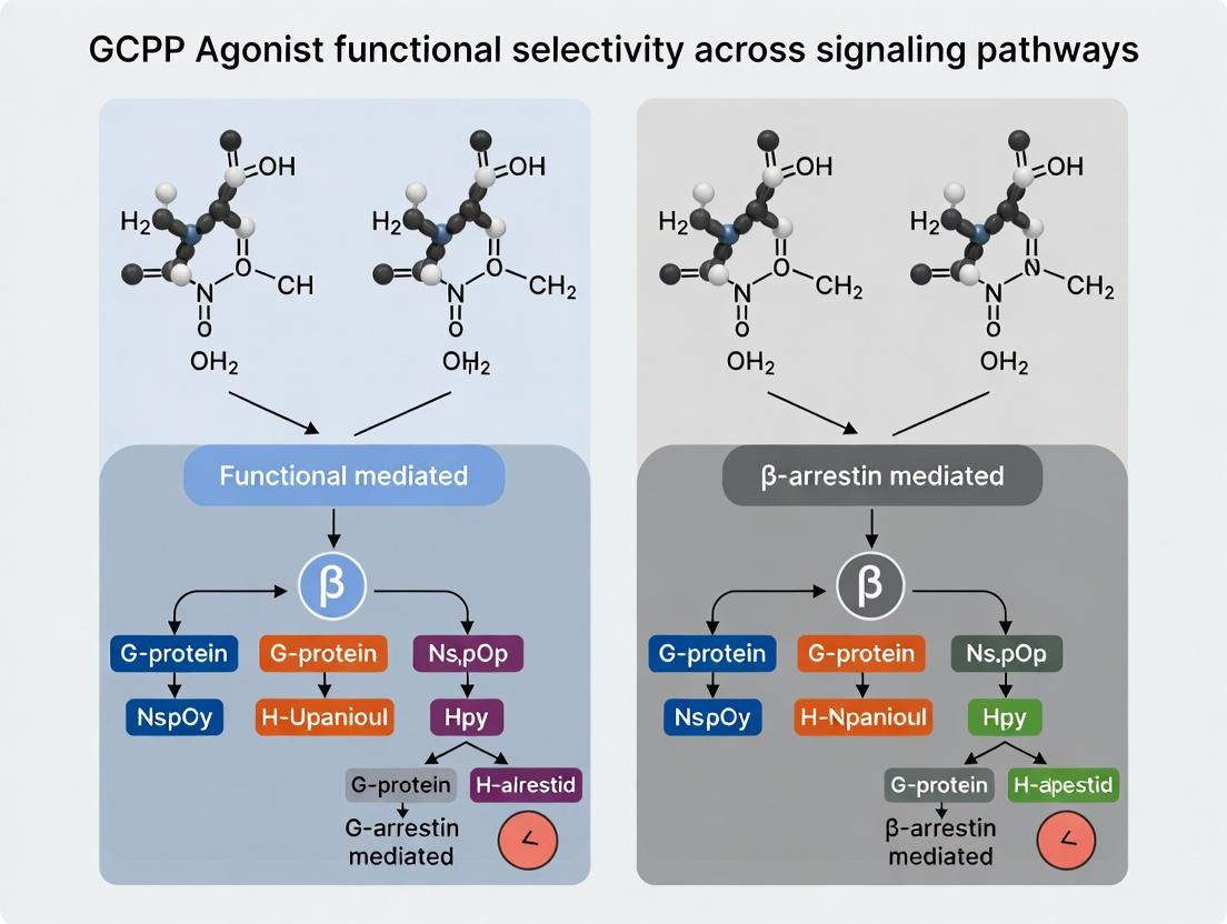
Signaling Pathway Diagrams
Diagram 1: Bias Agonist Preferential Pathway Activation
Diagram 2: Quantifying Bias Factor Experimental Workflow
The Scientist's Toolkit: Key Research Reagent Solutions
Table 3: Essential Reagents for Functional Selectivity Research
| Reagent Category | Example Product/Assay | Function in Bias Research |
|---|---|---|
| Biosensor Kits | PathHunter β-Arrestin Recruitment (DiscoverX); GloSensor cAMP (Promega); NANO-BRET (Promega). | Turnkey cell-based assays for quantifying specific pathway activation (G protein, β-arrestin) in high-throughput format. |
| Tagged GPCRs | cDNA for SNAP-tag, HALO-tag, or HiBiT-tagged receptors. | Enables specific labeling for trafficking studies, BRET/FRET partner incorporation, and surface expression quantification. |
| Arrestin & G Protein Tools | Dominant-negative β-arrestin mutants; Mini-G proteins; scFv16 (G protein biosensor). | Tools to selectively inhibit or monitor specific transducer engagement to isolate pathway contributions. |
| - Specialized Cell Lines | Tango GPCR Assay-ready cells (Thermo Fisher); GPCR β-arrestin cell lines (Promega). | Stable cell lines engineered with pathway-specific reporters (e.g., β-galactosidase, luciferase) for consistent, sensitive screening. |
| - Operational Model Software | Black & Leff Model Fitting in Prism (GraphPad); Kinetics Bias Calculator. | Specialized software for fitting complex dose-response data to calculate accurate log(τ/KA) and bias factor values with confidence intervals. |
| Reference & Tool Ligands | Full/balanced agonists (e.g., Angiotensin II); Neutral antagonists (e.g., Nadolol); Biased ligands (e.g., TRV130). | Critical controls for assay validation and as benchmarks for calculating relative bias factors. |
Within the broader thesis on GPCR agonist functional selectivity, understanding how specific receptor conformations stabilize distinct signaling profiles is paramount. This comparison guide evaluates key experimental approaches and their associated reagents for dissecting ligand-biased signaling at G protein-coupled receptors (GPCRs), providing a direct performance comparison of modern structural and functional techniques.
Experimental Methodologies for Conformational Profiling
Cryo-Electron Microscopy (Cryo-EM) for Stabilized Complexes
Protocol: Purified, stabilized GPCR (e.g., in nanodiscs or with a conformation-specific antibody fragment) is incubated with a bias-encoded ligand and a purified G protein or β-arrestin. The complex is vitrified on cryo-EM grids. Data collection is performed on a high-end cryo-EM microscope (e.g., Titan Krios). Hundreds of thousands of particle images are processed through 2D classification, 3D refinement, and molecular dynamics flexible fitting to obtain atomic models. Performance Data: See Table 1.
Double Electron-Electron Resonance (DEER) Spectroscopy
Protocol: Site-directed spin labeling is performed by introducing cysteine mutations at key helical positions and labeling with methanethiosulfonate spin labels. The labeled receptor is reconstituted into liposomes or nanodiscs. Following ligand stimulation, pulsed DEER measurements are performed to determine distances between spin labels. Distance distributions are used to model population shifts between active/inactive states. Performance Data: See Table 1.
BRET-Based Biosensors for Real-Time Conformational Reporting
Protocol: Cells are transfected with a GPCR intramolecular BRET biosensor (e.g., where a donor fluorophore is on one intracellular loop and an acceptor is on another). Ligand stimulation induces a conformational change that alters BRET efficiency. Measurements are taken in real-time using a plate reader capable of injector integration. Data are normalized to baseline and fit to dose-response curves. Performance Data: See Table 1.
Table 1: Performance Comparison of Conformational Profiling Techniques
| Technique | Resolution (Typical) | Throughput | Native Environment Capability | Key Advantage for Bias Studies |
|---|---|---|---|---|
| Cryo-EM | 2.5 - 3.5 Å | Low | Moderate (reconstituted) | Direct visualization of ligand-induced structural changes in signaling complexes. |
| DEER Spectroscopy | 3 - 10 Å (distance constraints) | Medium | High (membranes) | Quantifies populations of multiple conformational states in a membrane. |
| Intramolecular BRET | N/A (bulk signal) | High | High (live cells) | Real-time, functional readout of receptor dynamics in cells. |
Functional Output Assays for Correlating Conformation to Bias
G Protein Activation: [³⁵S]GTPγS Binding
Protocol: Membrane preparations expressing the target GPCR are incubated with varying ligand concentrations in assay buffer containing GDP and [³⁵S]GTPγS. Non-specific binding is determined with excess unlabeled GTPγS. Reactions are terminated by filtration, and bound radioactivity is quantified by scintillation counting. Data are fit to determine Emax and EC₅₀.
β-Arrestin Recruitment: Tango or PathHunter Assay
Protocol (Tango): Cells stably expressing a GPCR-TEV protease fusion and a β-arrestin-TEV cleavage site-Luciferase reporter are treated with ligands. Upon β-arrestin recruitment, TEV cleavage releases luciferase, which is quantified after substrate addition. Performance Data: See Table 2.
Table 2: Performance Comparison of Key Functional Assays for Bias Factor Calculation
| Assay Readout | Pathway Measured | Z'-Factor (Typical) | Artifact Susceptibility | Suitability for HTS |
|---|---|---|---|---|
| [³⁵S]GTPγS Binding | G protein activation | 0.6 - 0.8 | Low (membrane-based) | Medium |
| cAMP Accumulation (HTRF) | Gαs/Gαi (via modulation) | 0.7 - 0.9 | Medium (cell health) | High |
| β-Arrestin Recruitment (Tango) | β-arrestin engagement | 0.5 - 0.7 | High (overexpression) | High |
| ERK1/2 Phosphorylation (AlphaLISA) | Downstream signaling node | 0.4 - 0.6 | Very High (convergent pathways) | Medium |
Integrated Workflow for Determining Signaling Bias
Diagram Title: Integrated workflow for linking conformation to bias.
The Scientist's Toolkit: Key Research Reagent Solutions
| Reagent/Material | Function in Bias Research |
|---|---|
| Stabilized Receptor (e.g., BRIL fusion) | Enables crystallization and Cryo-EM of active-state complexes with G proteins or β-arrestin. |
| Mini-Gα and scFv16 (G protein mimetics) | Stabilize specific G protein-coupled receptor conformations for structural studies. |
| Nanodiscs (MSP1E3D1) | Provide a native-like lipid bilayer environment for biophysical studies of purified receptors. |
| Intramolecular BRET Biosensor Constructs | Report real-time, ligand-induced conformational changes in live cells. |
| PathHunter β-Arrestin Recruitment Cells | Enzyme fragment complementation-based system for high-throughput arrestin recruitment screening. |
| Tag-lite SNAP-GPCR Kits | Allow site-specific labeling of GPCRs with fluorescent dyes for FRET/HTRF-based binding & signaling assays. |
| TRUPATH Biosensor Kits | Comprehensive set of BRET-based biosensors to quantify activation of all major Gα subtypes. |
Key Signaling Pathways in GPCR Functional Selectivity
Diagram Title: Ligand-specific conformations dictate pathway selection.
The integration of high-resolution structural techniques with quantitative, pathway-selective functional assays is critical for advancing the thesis of GPCR agonist functional selectivity. The performance data presented herein allow researchers to select optimal methods for correlating distinct ligand-stabilized receptor conformations with specific signaling bias profiles, a foundational step in the rational design of safer, more effective therapeutics.
Within the framework of GPCR agonist functional selectivity research, a central question is how distinct ligands preferentially engage specific signaling hubs. This comparison guide objectively evaluates the performance and characteristics of two primary signaling hubs—G proteins and β-arrestins—and extends to emerging hubs like kinase cascades and receptor tyrosine kinase transactivation.
Comparison of Core Signaling Hubs: G Proteins vs. β-Arrestins
Table 1: Functional and Kinetic Profile of Primary Signaling Hubs
| Parameter | G Protein Pathway | β-Arrestin Pathway | Beyond (e.g., GRK/Scaffold) |
|---|---|---|---|
| Primary Role | Rapid second messenger generation (cAMP, Ca²⁺, DAG) | Receptor desensitization, endocytosis, scaffolded signaling | Signal diversification & integration |
| Onset Kinetics | Milliseconds to seconds | Seconds to minutes | Variable (minutes) |
| Signal Duration | Transient (secs-min) | Sustained (mins-hours) | Often prolonged |
| Key Readouts | cAMP accumulation, IP₁, Ca²⁺ flux, ERK1/2 phosphorylation (early) | ERK1/2 phosphorylation (delayed, cytosolic), receptor internalization, β-arrestin recruitment | Unique phospho-signatures, pathway-specific gene expression |
| Bias Quantification (ΔΔLog(τ/KA)) | Reference pathway (typically Gα-dependent) | Calculated relative to G protein pathway | Requires specific reference pathways |
| Therapeutic Implications | Classic efficacy & side effects | Potential for improved specificity, biased agonism | Novel drug targets, polypharmacology |
Table 2: Experimental Data from Model GPCRs (β₂AR & AT1R)
| Ligand (Receptor) | Gαs/Gαq Efficacy (Emax %) | β-Arrestin Recruitment (Emax %) | Bias Factor (G prot. ref.) | Key Functional Outcome |
|---|---|---|---|---|
| Isoproterenol (β₂AR) | 100% (cAMP) | 100% | 0 (Reference) | Balanced agonist |
| Carvedilol (β₂AR) | <5% (cAMP) | 70% | >10 (β-arrestin-biased) | Antagonist with β-arrestin bias |
| Angiotensin II (AT1R) | 100% (IP₁) | 100% | 0 (Reference) | Balanced agonist |
| TRV027 (AT1R) | ~40% (IP₁) | ~80% | 7.5 (β-arrestin-biased) | β-arrestin-biased ligand (clinical trial) |
| SII Angiotensin II (AT1R) | <10% (IP₁) | ~95% | >10 (β-arrestin-biased) | Tool compound for β-arrestin signaling |
Experimental Protocols for Characterizing Hub Engagement
Protocol 1: Quantifying G protein vs. β-Arrestin Signaling Bias
Objective: Determine ligand bias coefficients (ΔΔLog(τ/KA)) using complementary assays. Methodology:
- Cell Line: Use engineered cell lines (e.g., HEK293) stably expressing the GPCR of interest.
- G Protein Signaling:
- cAMP Accumulation: Use a BRET-based biosensor (e.g., CAMYEL) or HTRF assay.
- Calcium Mobilization: Use fluorescent dyes (e.g., Fluo-4) for Gαq-coupled receptors.
- IP₁ Accumulation: HTRF assay for Gαq activation.
- β-Arrestin Recruitment:
- BRET Assay: Co-express receptor-Rluc8 and β-arrestin-Venue. Measure emission ratio after ligand addition.
- PathHunter Assay: Use enzyme fragment complementation.
- Data Analysis:
- Generate concentration-response curves for each pathway.
- Calculate transduction coefficients (Log(τ/KA)) for each ligand in each pathway.
- Calculate ΔLog(τ/KA) relative to a reference ligand in each pathway.
- Bias Factor: ΔΔLog(τ/KA) = ΔLog(τ/KA)Pathway A - ΔLog(τ/KA)Pathway B. A value > 1 indicates bias toward Pathway A.
Protocol 2: Kinetics of ERK1/2 Phosphorylation
Objective: Distinguish G protein-mediated (rapid) from β-arrestin-mediated (sustained) ERK activation. Methodology:
- Treat serum-starved cells with biased or balanced ligand for times ranging from 2 min to 90 min.
- Lyse cells and analyze phospho-ERK1/2 levels via Western blot or AlphaLISA.
- Pre-treat cells with inhibitors:
- For G protein-dependent phase: Use Pertussis Toxin (for Gαi) or Gαq inhibitor FR900359.
- For β-arrestin-dependent phase: Use β-arrestin siRNA knockdown or barbadin (inhibitor of β-arrestin/AP2 interaction).
- Subcellular fractionation can be performed to distinguish nuclear (G protein) vs. cytosolic (β-arrestin) pERK.
Visualization of Signaling Pathways and Experimental Logic
Diagram Title: GPCR Signaling Hubs and Functional Selectivity Pathways
Diagram Title: Workflow for Quantifying Ligand Signaling Bias
The Scientist's Toolkit: Key Research Reagent Solutions
Table 3: Essential Reagents for Hub-Specific GPCR Research
| Reagent / Material | Provider Examples | Function in Research |
|---|---|---|
| PathHunter β-Arrestin Assay | Revvity (PerkinElmer) | Enzyme complementation for label-free, high-throughput β-arrestin recruitment measurement. |
| CAMYEL BRET cAMP Biosensor | Various academic sources | Real-time, live-cell measurement of Gαs-mediated cAMP dynamics. |
| Tag-lite SNAP-tag/HTRF Platform | Revvity (Cisbio) | Homogeneous time-resolved FRET for ligand binding, G protein & β-arrestin interaction. |
| Tango GPCR Assay Kits | Thermo Fisher | Transcription-based reporter assay for β-arrestin engagement and receptor internalization. |
| GRK2/3 Inhibitor (Compound 101) | Tocris | Selective GRK2/3 inhibitor to dissect GRK-specific phosphorylation effects on bias. |
| Barbadin | Tocris, Sigma | Cell-permeable inhibitor of β-arrestin/AP2 interaction; blocks β-arrestin-mediated endocytosis. |
| FR900359 (Gαq inhibitor) | Hello Bio, Tocris | Potent and selective Gαq inhibitor to isolate Gαq-independent signaling. |
| Pertussis Toxin (PTX) | List Labs | ADP-ribosylates Gαi/o, uncoupling from receptor; inhibits Gαi/o-mediated pathways. |
| TRV027 & SII Angiotensin II (Biased AT1R ligands) | Tocris, custom synthesis | Well-characterized β-arrestin-biased ligands for AT1R; critical tool compounds. |
| Phospho-ERK1/2 (Thr202/Tyr204) AlphaLISA | Revvity (PerkinElmer) | No-wash, sensitive immunoassay for quantifying kinetics of ERK phosphorylation. |
Within the broader thesis of GPCR functional selectivity, β-arrestin-biased agonists represent a paradigm shift, aiming to elicit therapeutic efficacy while minimizing side effects from traditional G protein signaling. This comparison guide objectively evaluates the canonical β-arrestin-biased agonists for the β2-Adrenergic Receptor (β2AR) and the Angiotensin II Type 1 Receptor (AT1R), detailing their performance against balanced agonists and supporting experimental data.
Comparison of Canonical β-Arrestin-Biased Agonists
| Parameter | Receptor: β2-Adrenergic Receptor (β2AR) | Receptor: Angiotensin II Type 1 Receptor (AT1R) |
|---|---|---|
| Balanced Agonist | Isoproterenol | Angiotensin II |
| β-Arrestin-Biased Agonist | Carvedilol (and analogs like SBI-0640756) | TRV027 (also known as SAR-100099) |
| Primary Therapeutic Goal | Heart failure; decoupling cardiostimulation (Gs) from cardioprotective/β-arrestin signaling. | Acute heart failure, hypertension; promoting vasodilation and cardioprotection without Gq-mediated vasoconstriction. |
| Bias Factor (Experimental Range) | Carvedilol: β-arrestin bias factor typically reported >104 relative to isoproterenol. | TRV027: β-arrestin bias factor commonly reported between 10 to >100 relative to Angiotensin II. |
| Key Functional Readouts | Gs/cAMP Inhibition: Minimal stimulation. β-Arrestin Recruitment: High. ERK1/2 Phosphorylation: Sustained, β-arrestin-dependent phase. | Gq/IP3 Inhibition: Minimal stimulation. β-Arrestin Recruitment: High. ERK1/2 Phosphorylation: β-arrestin-dependent. Receptor Internalization: Enhanced. |
| In Vivo Efficacy Data | In mouse models, β-arrestin-biased signaling promotes cardioprotection and improves contractility without increasing heart rate (vs. isoproterenol). | In preclinical heart failure models, TRV027 promoted improved cardiac output and reduced afterload without increasing blood pressure (vs. Angiotensin II). |
| Clinical Trial Status | Early-phase investigation for biased analogs; carvedilol itself is a non-selective β-blocker with biased properties. | Phase IIb (BLAST-AHF) completed; showed safety but did not meet primary efficacy endpoints for acute heart failure. |
Detailed Experimental Protocols for Bias Characterization
1. Protocol for β-Arrestin Recruitment (BRET Assay)
- Objective: Quantify agonist-induced recruitment of β-arrestin to the receptor.
- Method: Cells are co-transfected with the GPCR tagged with a luciferase (donor) and β-arrestin tagged with GFP (acceptor). The test agonist is added.
- Measurement: Upon recruitment, energy transfer occurs. The BRET ratio (acceptor emission/donor emission) is measured using a plate reader.
- Data Analysis: Dose-response curves are generated to calculate Log(EC50) and Emax for β-arrestin recruitment.
2. Protocol for G Protein Signaling (cAMP or IP1 Accumulation)
- For β2AR (Gs):
- Assay: cAMP-Glo or HTRF-based cAMP assay.
- Method: Cells expressing β2AR are treated with agonist. For Gs inhibition, cells may be pre-treated with forskolin.
- Measurement: Luminescence or fluorescence signal inversely proportional to cAMP (competitive assay).
- For AT1R (Gq):
- Assay: IP-One HTRF assay.
- Method: Cells expressing AT1R are treated with agonist, accumulating IP1 (a downstream metabolite of IP3).
- Measurement: HTRF signal between anti-IP1 antibodies is measured.
3. Protocol for ERK1/2 Phosphorylation (pERK)
- Objective: Measure downstream kinase activation, distinguishing temporal patterns (early/G-protein vs. sustained/β-arrestin).
- Method: Serum-starved cells are stimulated with agonist for various times (e.g., 5, 10, 30 min).
- Measurement: Cell lysates are analyzed via AlphaLISA or Western blot using phospho-ERK1/2 antibodies.
- Bias Confirmation: Treatment with G protein inhibitors (e.g., Pertussis Toxin for Gi, FR900359 for Gq) or β-arrestin siRNA confirms pathway contribution.
Visualization of Signaling Pathways and Assay Workflow
Diagram 1: GPCR Signaling: Balanced vs. β-Arrestin-Biased Agonists (84 chars)
Diagram 2: Experimental Workflow for GPCR Bias Quantification (79 chars)
The Scientist's Toolkit: Key Research Reagent Solutions
| Reagent / Kit | Provider Examples | Primary Function in Bias Research |
|---|---|---|
| PathHunter β-Arrestin Assay | Revvity (DiscoverX) | Enzyme fragment complementation assay for label-free, high-throughput quantification of β-arrestin recruitment. |
| cAMP Gs Dynamic 2 / IP-One Gq HTRF Kits | Revvity (Cisbio) | Homogeneous, no-wash immunoassays for quantifying cAMP or IP1 accumulation as direct measures of Gs or Gq activity. |
| AlphaLISA pERK1/2 (Thr202/Tyr204) Assay Kit | Revvity (PerkinElmer) | Bead-based, no-wash assay for sensitive, high-throughput quantification of ERK phosphorylation. |
| Tag-lite GPCR Signaling Platform | Revvity (Cisbio) | Uses SNAP-tag technology and time-resolved FRET for live-cell assays of receptor-ligand binding, internalization, and downstream signaling. |
| β-Arrestin-1/2 siRNA | Dharmacon, Santa Cruz Biotechnology | Gene knockdown to pharmacologically confirm the β-arrestin-dependency of observed signaling events. |
| G Protein Inhibitors (PTX, FR900359, YM-254890) | Tocris, FUJIFILM Wako | Pertussis Toxin (Gi/o inhibitor) and Gq inhibitors (FR900359) to block specific G protein pathways and isolate β-arrestin signaling. |
| Bioluminescent Resonance Energy Transfer (BRET) Biosensors | Addgene, laboratory-constructed | Donor (e.g., NanoLuc) and acceptor (e.g., GFP) tagged constructs for real-time, live-cell monitoring of protein-protein interactions (e.g., GPCR-β-arrestin). |
Physiological and Therapeutic Implications of Biased Signaling
Within the broader thesis of GPCR agonist functional selectivity research, understanding how to quantify and compare biased signaling is paramount for drug discovery. This guide compares key methodological approaches and their application to specific receptor systems.
Comparison of Bias Quantification Methods
The accurate determination of ligand bias requires normalization of pathway data to a reference agonist. The following table compares two prevalent analytical frameworks.
Table 1: Comparison of Bias Factor Calculation Methods
| Method | Core Principle | Key Output (ΔΔlog(τ/KA) or ΔΔlog(Emax/EC50)) | Advantages | Limitations | Representative Tool/Software |
|---|---|---|---|---|---|
| Operational Model (Black-Leff) | Fits concentration-response data to the Operational Model of agonism to derive transduction coefficients (log(τ/KA)). | ΔΔlog(τ/KA) | Accounts for both efficacy (τ) and affinity (KA). System-independent estimate of bias. | Requires high-quality, complete concentration-response curves. Assumes no receptor depletion. | Prism (GraphPad), Bias Calculator |
| Area Under the Curve (AUC) | Calculates the integrated response (AUC) over a range of agonist concentrations. | ΔΔlog(Emax/EC50) (approximated) | Less model-dependent. Robust with partial or incomplete curves. Simpler calculation. | Can be confounded by differences in curve shape and Hill slopes. More system-dependent. | Custom scripts in R or Python |
Supporting Data: A study comparing angiotensin II type 1 receptor (AT1R) ligands demonstrated that the biased agonist TRV027 yielded a ΔΔlog(τ/KA) of -1.2 ± 0.3 for G protein (Gq) vs. β-arrestin-2 recruitment relative to angiotensin II, indicating a significant bias toward β-arrestin. In contrast, the AUC method for the same dataset produced a ΔΔlog(Emax/EC50) of -0.9 ± 0.2, confirming the direction but with a marginally different magnitude.
Experimental Protocol: Quantifying μ-Opioid Receptor (MOR) Bias In Vitro
This protocol outlines a standard assay to compare G protein vs. β-arrestin signaling for MOR ligands.
- Cell Culture & Transfection: HEK293 cells are maintained and transfected with plasmids encoding human MOR, along with either:
- G protein pathway: A cAMP biosensor (e.g., GloSensor) for inhibition of forskolin-stimulated cAMP (Gi-coupled).
- β-arrestin pathway: A β-arrestin recruitment biosensor (e.g., PathHunter or NanoBiT).
- Assay Execution:
- Seed transfected cells into white-walled, clear-bottom assay plates.
- For cAMP assay: Pre-incubate with forskolin, then stimulate with a 10-point, half-log dilution series of test agonists (e.g., morphine, fentanyl, TRV130 [oliceridine]), reference agonist (DAMGO), and negative control.
- For β-arrestin assay: Directly stimulate with the same agonist dilution series.
- Measure luminescence at specified timepoints post-agonist addition.
- Data Analysis:
- Normalize all responses as a percentage of the maximal DAMGO response in each assay.
- Fit normalized concentration-response data to a 3- or 4-parameter logistic equation to obtain Log(EC50) and Emax values.
- Calculate transduction coefficients (log(τ/KA)) using the Operational Model via specialized software.
- Compute bias factors (ΔΔlog(τ/KA)) relative to DAMGO for each ligand across the two pathways.
Table 2: Example MOR Ligand Bias Data (Relative to DAMGO)
| Ligand | Pathway: Gi (cAMP Inhibition) log(τ/KA) | Pathway: β-arrestin-2 Recruitment log(τ/KA) | Bias Factor (ΔΔlog(τ/KA)) for G protein |
|---|---|---|---|
| DAMGO (Reference) | 0.0 (by definition) | 0.0 (by definition) | 0.0 |
| Morphine | -0.5 ± 0.1 | -1.8 ± 0.2 | +1.3 ± 0.2 |
| Fentanyl | +0.3 ± 0.1 | -0.2 ± 0.1 | +0.5 ± 0.1 |
| TRV130 (Oliceridine) | -0.2 ± 0.1 | -2.1 ± 0.3 | +1.9 ± 0.3 |
Signaling Pathways and Experimental Workflow
Diagram 1: GPCR Bias Signaling Pathways (90 chars)
Diagram 2: Experimental Bias Quantification Steps (85 chars)
The Scientist's Toolkit: Key Research Reagents
Table 3: Essential Reagents for Biased Signaling Research
| Reagent / Solution | Primary Function in Experiments |
|---|---|
| PathHunter β-Arrestin Recruitment Assay (DiscoverX) | Enzyme fragment complementation (EFC) cell-based system for directly measuring β-arrestin recruitment to activated GPCRs. |
| GloSensor cAMP Assay (Promega) | Bioluminescent biosensor for real-time measurement of intracellular cAMP levels, critical for Gi/Gs-coupled pathway analysis. |
| NanoBiT β-Arrestin System (Promega) | Complementation-based reporter using small fragments of NanoLuc luciferase to measure β-arrestin recruitment with high sensitivity. |
| TRUPATH (NIH) | A comprehensive, validated suite of BRET biosensors for quantifying engagement of all 16 human Gα protein subtypes. |
| Reference Agonists (e.g., DAMGO for MOR, Angiotensin II for AT1R) | Standard, well-characterized full agonists used as the baseline comparator for calculating ligand bias factors. |
| Operational Model Fitting Software (e.g., GraphPad Prism with specific add-ons) | Essential for deriving the transduction coefficient (log(τ/KA)) from concentration-response data for bias quantification. |
Measuring the Bias: A Toolkit for Quantifying Pathway-Selective Agonism
Within GPCR agonist functional selectivity research, the precise quantification of specific signaling pathway activation is paramount. The choice of biophysical assay platform directly impacts the sensitivity, dynamic range, and reliability of the data used to profile ligand bias. This guide objectively compares four key homogenous, plate-reader compatible technologies: Bioluminescence Resonance Energy Transfer (BRET), Fluorescence Resonance Energy Transfer (FRET), Time-Resolved FRET (TR-FRET), and the more recent Nanobluet technology.
Technology Comparison & Performance Data
The following table summarizes the core characteristics and performance metrics of each platform, based on published experimental data from GPCR pathway analysis (e.g., cAMP accumulation, β-arrestin recruitment, intracellular calcium mobilization).
Table 1: Comparative Analysis of Primary Assay Platforms for GPCR Signaling
| Feature | BRET | FRET | TR-FRET | Nanobluet |
|---|---|---|---|---|
| Energy Donor | Luciferase (e.g., Rluc8, NanoLuc) | Fluorophore (e.g., CFP, GFP) | Lanthanide Cryptate (e.g., Eu³⁺, Tb³⁺) | NanoLuc Luciferase |
| Energy Acceptor | Fluorophore (e.g., GFP, YFP) | Fluorophore (e.g., YFP, mCherry) | XL665 / d2 | Proprietary Fluorescent Tether |
| Readout Mode | Luminescence / Fluorescence Ratio | Fluorescence Intensity Ratio | Time-Delayed Fluorescence Ratio | Luminescence Intensity (Single Wavelength) |
| Key Advantage | Low autofluorescence; no excitation light needed. | Genetically encodable; real-time kinetics. | Very high S/B ratio; minimizes compound interference. | Extreme brightness & stability; largest dynamic range. |
| Typical Z' Factor | 0.5 - 0.7 | 0.4 - 0.6 | 0.7 - 0.9 | 0.7 - 0.9 |
| Assay Window (Fold Change) | 2 - 4 | 1.5 - 3 | 3 - 8 | 5 - 15+ |
| Compatible with Live Cells | Yes | Yes | Less common (often endpoint) | Yes |
| Susceptibility to Compound Interference | Low | Medium-High (autofluorescence) | Very Low | Very Low |
| Primary GPCR Application | β-arrestin recruitment, protein-protein interactions | Real-time Ca²⁺, conformational changes | cAMP, ubiquitin ligase recruitment, pathway multiplexing | All major pathways (cAMP, arrestin, Ca²⁺) with same donor |
S/B = Signal-to-Background. Z' factor >0.5 is excellent. Data compiled from recent literature and manufacturer technical notes.
Experimental Protocols for Key GPCR Assays
Protocol 1: TR-FRET for Intracellular cAMP Quantification (Gs-coupling)
This is a gold-standard endpoint assay for Gαs-mediated signaling.
- Cell Preparation: Seed cells expressing the target GPCR in a white, low-volume 384-well plate.
- Stimulation: Incubate with serially diluted agonists/antagonists in stimulation buffer for 30 min at 37°C.
- Lysis & Detection: Add lytic buffer containing Eu³⁺-cryptate-labeled anti-cAMP antibody and d2-labeled cAMP. Incubate for 1 hour at room temperature.
- Reading: Measure time-resolved fluorescence at 620 nm (Eu donor) and 665 nm (d2 acceptor). The cAMP in the sample competes with d2-cAMP for the antibody, decreasing the 665 nm FRET signal.
- Data Analysis: Calculate the 665 nm / 620 nm ratio. Fit dose-response curves to determine EC₅₀/IC₅₀ values.
Protocol 2: Nanobluet-based β-Arrestin Recruitment (PathHunter-type)
This utilizes enzyme fragment complementation driven by GPCR-β-arrestin interaction.
- Cell Line: Use engineered cells stably expressing the target GPCR fused to a small enzyme fragment (EA) and β-arrestin fused to the complementary enzyme fragment (ED).
- Stimulation: Seed cells, allow to adhere, then treat with compounds for 90-180 min at 37°C.
- Detection: Add a luminogenic substrate for NanoLuc luciferase. Recruitment brings EA and ED together, forming active NanoLuc and producing a bright, sustained luminescent signal.
- Reading: Measure luminescence intensity (no ratio required). Signal is directly proportional to β-arrestin recruitment.
- Data Analysis: Plot raw luminescence vs. compound concentration. The exceptional S/B ratio allows clear detection of weak partial agonists.
Protocol 3: Live-Cell BRET for G Protein Activation (Gγ dissociation)
Monitors real-time G protein subunit rearrangement.
- Transfection: Co-transfect cells with: GPCR, Gα-Rluc8 donor, and Gγ-GFP acceptor.
- Substrate Addition: Add the cell-permeable luciferase substrate coelenterazine-h.
- Baseline Reading: Measure emissions at 475 nm (donor) and 535 nm (acceptor) for several minutes to establish baseline BRET ratio (535/475).
- Stimulation: Inject agonist and continue reading for 5-15 minutes. Gαγ dissociation decreases the BRET ratio.
- Data Analysis: Normalize BRET ratio changes over time. Calculate kinetics of activation (t₁/₂) and efficacy.
Visualizing GPCR Signaling Pathways & Assay Principles
Diagram 1: Core GPCR Signaling Pathways for Functional Selectivity
Diagram 2: Core Principles of BRET, TR-FRET, and Nanobluet Assays
The Scientist's Toolkit: Key Research Reagent Solutions
Table 2: Essential Reagents for GPCR Functional Selectivity Screening
| Reagent / Solution | Function in Assays | Example Vendor/Product |
|---|---|---|
| NanoLuc Luciferase (Nluc) | Donor for NanoBRET; smaller, brighter than Rluc, enabling superior S/B. | Promega NanoBIT, NanoBRET technologies. |
| Lanthanide Cryptates (Eu³⁺, Tb³⁺) | Long-lifetime donors for TR-FRET; enable time-gating to eliminate background fluorescence. | Cisbio HTRF donors (Eu Cryptate, Tb Cryptate). |
| Tag-lite SNAP/CLIP-tag Ligands | Site-specific labeling of GPCRs with FRET donors/acceptors for cell-surface assays. | Cisbio Tag-lite labeled Lumi4-Tb, Green/Red dyes. |
| cAMP TR-FRET Kit | Endpoint, competitive immunoassay for Gαs/Gαi pathway activity. | Cisbio HTRF cAMP Gs Dynamic kit, Revvity LANCE Ultra cAMP. |
| IP-One TR-FRET Kit | Accumulation assay for Gαq/11 pathway activity (IP3 analog). | Cisbio HTRF IP-One kit. |
| PathHunter β-Arrestin Cell Lines | Engineered cells for Nanobluet/EFC-based arrestin recruitment. | Revvity (Discoverx) PathHunter GPCR cell lines. |
| G Protein Biosensors (Rluc8-based) | Live-cell BRET sensors for monitoring Gα subunit activation (Gs, Gi, Gq). | cDNA from academic labs (e.g., Bouvier lab). |
| Coelenterazine-h / furimazine | Cell-permeable substrates for Rluc and NanoLuc luciferases, respectively. | Nanolight Coelenterazine-h, Promega Furimazine. |
For functional selectivity research, the optimal assay platform depends on the specific pathway, required throughput, and need for live-cell kinetics. TR-FRET remains the gold standard for endpoint biochemical measurements (cAMP, IP1) due to its robust performance. BRET/NanoBRET is indispensable for real-time, live-cell monitoring of dynamic processes like G protein activation. Nanobluet/EFC technology offers unparalleled sensitivity and dynamic range for challenging readouts like β-arrestin recruitment, making it powerful for comprehensive bias factor calculation. Integrating data from these complementary platforms provides a definitive map of agonist functional selectivity across GPCR signaling landscapes.
Within GPCR pharmacology, the principle of functional selectivity or biased agonism posits that ligands can stabilize unique receptor conformations, preferentially activating specific signaling pathways over others. Quantifying this bias is critical for modern drug development, where targeting therapeutic pathways while avoiding adverse effect pathways is a central goal. The operational model of agonism, coupled with the bias factor calculation (ΔΔlog(τ/KA)), provides a robust, system-independent framework for quantifying ligand bias. This guide compares the application, performance, and data requirements of this model against alternative analytical methods.
Core Methodologies Compared
The Operational Model & Bias Factor Approach
This method dissects agonist concentration-response curves into two parameters: efficacy (τ) and affinity (KA). Bias between two pathways is quantified by comparing the Δlog(τ/KA) value for a test agonist relative to a reference agonist.
Experimental Protocol:
- Cell System: A recombinant cell line expressing the GPCR of interest at a known, consistent level.
- Pathway-Specific Assays: Two distinct assays measuring different functional endpoints (e.g., cAMP inhibition vs. β-arrestin recruitment) are performed in parallel.
- Agonists: A full concentration-response curve for a reference agonist (usually a balanced standard) and each test agonist.
- Data Analysis: Data for each agonist in each pathway is fitted to the operational model equation:
Response = (Em * τ^n * [A]^n) / (([A] + KA)^n + τ^n * [A]^n)where Em is system maximum, n is a slope factor, and [A] is agonist concentration. τ and KA are derived. - Bias Calculation:
- Calculate Δlog(τ/KA) for test agonist vs. reference in Pathway A.
- Calculate Δlog(τ/KA) for the same agonist pair in Pathway B.
- Bias Factor = Δlog(τ/KA)Pathway A - Δlog(τ/KA)Pathway B. This yields ΔΔlog(τ/KA), a log unit value indicating the magnitude and direction of bias.
Alternative: The Relative Activity (RA) or Intrinsic Relative Activity (RAi) Method
This simpler method compares agonist potency (EC50) and maximal response (Emax) relative to a reference agonist.
Experimental Protocol: Similar assay setup as above. Bias Calculation: Bias is inferred from differences in relative Emax or relative potency (EC50) ratios between pathways. It does not deconvolve efficacy and affinity.
Alternative: The Black-Leff Model (Transduction Coefficient, log(τ/KA))
This is the precursor to the full operational model analysis. It uses the transduction coefficient log(τ/KA) as a single, system-dependent measure of agonist activity.
Experimental Protocol: Requires determination of KA from independent binding studies. Bias Calculation: Bias factor is Δlog(τ/KA) between pathways, but requires an accurate, independent measure of KA.
Performance Comparison: Data & Outcomes
Table 1: Quantitative Comparison of Bias Quantification Methods
| Feature/Aspect | Operational Model (ΔΔlog(τ/KA)) | RA/RAi Method | Black-Leff (Transduction Coefficient) |
|---|---|---|---|
| System Dependence | Corrects for system differences (receptor expression, coupling efficiency). | Highly system-dependent; comparisons across labs difficult. | Corrects for system differences if KA is accurate. |
| Parameters Derived | Efficacy (τ) and affinity (KA) from functional data. | Potency (EC50) and maximal response (Emax). | Efficacy (τ), uses independent KA. |
| Data Requirements | Full concentration-response curves for reference & test agonists in each pathway. | Full concentration-response curves for reference & test agonists in each pathway. | Full concentration-response curves + independent KA value (e.g., from binding). |
| Assay Sensitivity to Receptor Expression | Robust. Model accounts for receptor density. | Very sensitive. Emax and EC50 directly affected. | Robust if KA is correct. |
| Bias Output | System-independent bias factor (ΔΔlog(τ/KA)). | Qualitative or semi-quantitative (e.g., "biased toward Pathway A"). | System-independent bias factor (Δlog(τ/KA)). |
| Key Advantage | Gold standard for quantitative, comparative bias. No need for independent binding assays. | Simple, rapid for initial screening. | Solid theoretical foundation. |
| Key Limitation | Requires high-quality, complete concentration-response data. | Cannot separate affinity from efficacy; misleading if systems aren't identical. | Relies on accurate KA, which may differ between functional vs. binding conditions. |
Table 2: Example Experimental Data Analysis for Agonist X at GPCR Y Assay 1: G protein (cAMP accumulation, Em = 100%). Assay 2: β-arrestin recruitment (Em = 100%). Reference Agonist: Noradrenaline.
| Agonist | Pathway | pEC50 | Emax (%) | log(τ)* | log(KA)* | Δlog(τ/KA) vs. Ref | ΔΔlog(τ/KA) (Bias Factor) |
|---|---|---|---|---|---|---|---|
| Noradrenaline (Ref) | G protein | 8.0 | 100 | 1.00 | 5.80 | 0.00 | 0.00 (by definition) |
| Noradrenaline (Ref) | β-arrestin | 6.5 | 85 | 0.15 | 5.90 | 0.00 | |
| Agonist X | G protein | 7.2 | 75 | 0.40 | 6.10 | -0.50 | 1.45 (β-arrestin biased) |
| Agonist X | β-arrestin | 6.8 | 100 | 0.80 | 6.15 | 0.95 |
*Derived from operational model fitting. Bias Factor for Agonist X = Δlog(τ/KA)β-arrestin (0.95) - Δlog(τ/KA)G protein (-0.50) = 1.45.
Visualizing Bias Quantification Workflows
Title: Operational Model Bias Factor Calculation Workflow
Title: Biased Agonism Across Two GPCR Signaling Pathways
The Scientist's Toolkit: Key Research Reagent Solutions
Table 3: Essential Materials for GPCR Bias Experiments
| Research Reagent / Solution | Primary Function in Bias Quantification |
|---|---|
| Recombinant Cell Lines | Engineered to stably express the target GPCR at a consistent, quantifiable level. Critical for reducing system variability. |
| Pathway-Selective Reporter Assays | (e.g., cAMP GloSensor, Tango β-arrestin recruitment). Provide real-time, specific functional readouts for distinct signaling pathways. |
| Reference Agonist | A well-characterized, preferably balanced or standard agonist (e.g., endogenous ligand) essential for calculating Δlog(τ/KA). |
| Operational Model Fitting Software | (e.g., GraphPad Prism with specific operational model equations, Black-Leff Fitting Tool). Necessary for robust parameter estimation (τ, KA). |
| Validated Tool Compounds | Known biased agonists and neutral antagonists. Used as positive/negative controls to validate the assay system and analysis. |
| Cell Surface Receptor Labeling Kits | (e.g., ELISA, flow cytometry antibodies). Used to quantify receptor expression level (Bmax), an important system parameter. |
High-Throughput Screening Strategies for Identifying Biased Ligands
Within the broader thesis on GPCR agonist functional selectivity, identifying ligands that preferentially activate one signaling pathway over others (biased agonism) is paramount. High-throughput screening (HTS) strategies enable the rapid evaluation of compound libraries to discover such biased ligands. This guide compares prevalent HTS platforms based on key performance metrics.
Comparison of HTS Platforms for Biased Ligand Screening
Table 1: Performance Comparison of Primary HTS Assay Technologies
| Platform / Assay Type | Throughput (Compounds/Day) | Pathway Readout | Z'-Factor (Typical) | Cost per 384-well | Key Advantage | Key Limitation |
|---|---|---|---|---|---|---|
| BRET (e.g., NanoBiT) | 50,000 - 100,000 | β-arrestin recruitment, cAMP, PKC | 0.6 - 0.8 | $0.40 - $0.60 | Homogeneous, real-time kinetics, multiplexing potential | Requires genetic fusion, signal intensity varies. |
| FRET (cAMP sensors) | 30,000 - 70,000 | cAMP dynamics | 0.5 - 0.7 | $0.50 - $0.70 | Direct measure of second messenger, ratiometric. | More complex optics, can be lower dynamic range. |
| CellELISA (e.g., pERK) | 20,000 - 40,000 | Kinase phosphorylation (ERK, Akt) | 0.4 - 0.6 | $0.30 - $0.50 | Endpoint, widely validated, no special equipment. | Low temporal resolution, multiple wash steps. |
| Imaging (HCA, TIRF) | 5,000 - 20,000 | Receptor internalization, β-arrestin translocation | 0.7 - 0.9 | $1.00 - $2.50 | Single-cell data, spatial information, multi-parametric. | Low throughput, complex data analysis, expensive. |
| Fluorescent Dyes (Ca2+ flux) | 100,000+ | Gq/Go coupling (Calcium mobilization) | 0.6 - 0.8 | $0.20 - $0.40 | Very high throughput, excellent for primary Gq screens. | Indirect for Gi/Gs, dye loading variability. |
Supporting Experimental Data: A 2023 study systematically screened a library of 10,000 compounds against the angiotensin II type 1 receptor (AT1R) using parallel HTS campaigns. BRET-based β-arrestin-2 recruitment assays (Z'=0.78) identified 150 hits, while a FRET-based cAMP assay (Z'=0.65) identified 95 hits. Only 42 compounds were common hits, and subsequent dose-response profiling confirmed 7 compounds as potent β-arrestin-biased agonists and 3 as G protein-biased agonists.
Experimental Protocols for Key Assays
Protocol 1: BRET-based β-Arrestin Recruitment Assay (384-well format)
- Cell Preparation: Seed HEK293T cells stably expressing the GPCR of interest tagged with a Renilla luciferase (Rluc8) at 20,000 cells/well in poly-D-lysine coated white plates.
- Transfection/Expression: For β-arrestin readout, co-express GFP2-tagged β-arrestin-2 (often using stable or transient transfection 24h prior).
- Compound Addition: Using an automated liquid handler, add test compounds (in DMSO, final conc. typically 10 µM) and incubate for the optimized time (e.g., 10-15 min at 37°C).
- Substrate Addition: Inject coelenterazine 400a (final conc. 5 µM) as the Rluc substrate.
- Detection: Immediately read BRET signal on a plate reader (e.g., PHERAstar FSX). Measure Rluc donor emission at 410 nm and GFP2 acceptor emission at 515 nm.
- Data Analysis: Calculate BRET ratio as (Em515 / Em410). Normalize to vehicle (0%) and reference agonist (100%).
Protocol 2: HTRF-based cAMP Accumulation Assay
- Cell Preparation: Seed cells expressing the GPCR in 384-well plates. On the day of assay, stimulate with compounds in the presence of a phosphodiesterase inhibitor (e.g., IBMX) for 30 min at 37°C in cAMP stimulation buffer.
- Lysis & Detection: Lyse cells with HTRF cAMP-d2 conjugate and anti-cAMP cryptate antibody (Cisbio). Incubate for 1 hour at room temperature.
- Reading: Read time-resolved FRET on a compatible plate reader. Excitation at 337 nm, measure emissions at 620 nm (cryptate) and 665 nm (d2).
- Data Analysis: Calculate the 665 nm/620 nm ratio. Convert to cAMP concentration using a standard curve.
Visualizing Signaling Pathways and Workflows
Title: GPCR Signaling Pathways and Ligand Bias
Title: HTS Workflow for Biased Ligand Discovery
The Scientist's Toolkit: Key Research Reagent Solutions
Table 2: Essential Materials for Biased Ligand HTS
| Item (Example Product) | Function in HTS | Key Consideration |
|---|---|---|
| Engineered Cell Lines (GPCR-Rluc8 stable line) | Provides consistent, pathway-specific reporter expression. Essential for BRET/FRET. | Ensure proper receptor coupling and expression levels mimic physiology. |
| BRET/FRET Substrates (Coelenterazine-h, 400a) | Enzyme substrate for luminescent/fluorescent energy transfer. | Substrate choice (e.g., 400a for BRET2) dictates emission spectra and signal stability. |
| HTRF cAMP Kits (Cisbio cAMP Gs Dynamic Kit) | Homogeneous, no-wash assay for quantifying intracellular cAMP. | Robust Z'-factor, wide dynamic range, compatible with Gi/Gs modulation. |
| Fluorescent Ca2+ Dyes (Fluo-4 AM, Cal-520) | Indicator for Gq-mediated calcium mobilization in ultra-HTS. | Dye loading time, photobleaching, and compatibility with agonists must be optimized. |
| β-Arrestin Recruitment Kits (Promega PathHunter) | Enzyme fragment complementation assay for arrestin engagement. | Provides a robust, amplified signal but is an endpoint assay. |
| Reference Agonists & Antagonists (Full/biased agonists, inverse agonists) | Critical assay controls for normalization and validation of pathway bias. | Pharmacological characterization must be well-established in the literature. |
| Automated Liquid Handlers (e.g., Beckman Coulter Biomek) | Enables precise, rapid compound and reagent dispensing for miniaturized assays. | Critical for assay reproducibility and achieving true high-throughput capacity. |
This guide is framed within a thesis investigating GPCR agonist functional selectivity, where cellular background—shaped by system bias (e.g., receptor expression levels, stoichiometry of signaling components) and proteomic profiles—critically determines pathway-specific signaling outcomes. Accurate comparison of research tools and platforms for dissecting these complexities is essential for advancing therapeutic discovery.
Comparison Guide: Phosphoproteomic Profiling Platforms for GPCR Signaling
Quantitative phosphoproteomics is vital for mapping biased agonism across pathways. The following table compares leading platforms based on critical performance metrics for GPCR research.
Table 1: Comparison of Phosphoproteomic Profiling Platforms
| Platform / Method | Kinase Activity Coverage (Unique Phosphosites) | Sample Throughput (per week) | Quantification Accuracy (CV) | Sensitivity (Required Protein Input) | Suitability for Temporal GPCR Studies |
|---|---|---|---|---|---|
| TiO2/MOAC-based LC-MS/MS | ~15,000-20,000 | 20-30 | <15% | 1-2 mg | High (excellent for time-course) |
| Label-Free Quantification (LFQ) | ~10,000-15,000 | 40-50 | 10-20% | 0.5-1 mg | Medium-High |
| TMT/isobaric Tagging | ~12,000-18,000 | 100+ | 5-15% (requires correction) | 0.1 mg per channel | High (multiplexed time points) |
| Phospho-antibody Array | ~50-100 predefined | 100+ | 15-25% | 100 µg | Low (targeted, low-plex) |
Experimental Protocols for Key Comparisons
Protocol 1: Evaluating System Bias via Receptor Abundance Quantification
Aim: To correlate endogenous GPCR expression levels (a key system bias) with functional pathway recruitment. Method:
- Cell Line Preparation: Culture HEK293, U2OS, and primary cell models. Perform serum starvation for 4 hours.
- Receptor Quantification: Lyse cells. Use quantitative Western blot with fluorescent secondary antibodies against target GPCR (e.g., β2-Adrenergic Receptor). Compare to a standard curve of known recombinant receptor protein.
- Functional Assay in Parallel: Seed sister plates. Stimulate with a titrated concentration of reference agonist (e.g., Isoproterenol for β2AR).
- Pathway Readouts:
- cAMP Accumulation: Use HTRF cAMP Gs dynamic kit.
- ERK1/2 Phosphorylation: Use Luminex xMAP technology for multiplexed phospho-ERK.
- Data Analysis: Normalize functional dose-response curves (EC50, Emax) to receptor copy number per cell. Plot signaling efficacy (Emax) vs. receptor density.
Protocol 2: Broad-Spectrum Phosphoproteomic Profiling for Biased Agonism
Aim: To globally identify pathway biases induced by different agonists in a specific cellular background. Method:
- Stimulation & Lysis: Serum-starve U2OS cells expressing the GPCR of interest. Treat with balanced agonist, biased agonist, or vehicle (n=4) for 5 minutes. Quench with ice-cold PBS and lyse in urea-based buffer.
- Protein Digestion & Phosphopeptide Enrichment: Reduce, alkylate, and digest lysates with trypsin. Desalt peptides. Enrich phosphopeptides using Fe-IMAC magnetic beads.
- LC-MS/MS Analysis: Analyze on a high-resolution tandem mass spectrometer (e.g., Orbitrap Exploris 480) coupled to nanoLC. Use data-independent acquisition (DIA) mode.
- Bioinformatics: Search data against human UniProt database. Normalize phosphosite intensities. Perform significance analysis (ANOVA) to identify agonist-specific phosphorylation events. Map sites to known signaling pathways (KEGG, Reactome).
Visualization of Concepts and Workflows
Title: Cellular Background Integrates System Bias and Proteomics to Drive GPCR Signaling Bias
Title: Experimental Workflow for GPCR Phosphoproteomic Profiling
The Scientist's Toolkit: Key Research Reagent Solutions
Table 2: Essential Reagents for GPCR Functional Selectivity Research
| Item | Function & Relevance to Study |
|---|---|
| TRUPATH Biosensor Kit | A comprehensive suite of BRET-based biosensors to simultaneously quantify activation of all 16 Gα protein subtypes in live cells, directly addressing system bias. |
| NanoBiT β-arrestin Recruitment Assays | Split-luciferase system for real-time, high-throughput measurement of GPCR-arrestin interaction kinetics. |
| Cell Surface ELISA Kits (e.g., Tag-lite) | Quantify absolute receptor expression levels on live cells—a critical parameter for system bias. |
| Phospho-Specific Antibody Panels (Luminex/xMAP) | Multiplexed, medium-throughput quantification of key pathway phosphoproteins (e.g., pERK, pCREB, pAkt). |
| Fe-IMAC or TiO2 Magnetic Beads | For high-efficiency enrichment of phosphopeptides prior to MS analysis, crucial for depth in proteomic profiling. |
| Stable Isotope Labeling Reagents (TMTpro) | Enable 16-plex quantitative comparison of phosphoproteomes across multiple agonists and time points in one MS run. |
| Cryopreserved Primary Cells (Human) | Provide physiologically relevant cellular backgrounds with native proteomic profiles and signaling stoichiometries. |
| Pathway Analysis Software (e.g., Perseus, Ingenuity) | For statistical and bioinformatic interpretation of proteomic data in the context of GPCR signaling networks. |
Comparative Analysis of Leading μ-Opioid Receptor (MOR) Biased Agonist Candidates
This guide compares key pharmacological profiles of advanced biased agonist candidates targeting the MOR, aiming to dissociate analgesic efficacy from adverse effects like respiratory depression and constipation.
Table 1: In Vitro Signaling Profiles of MOR Biased Agonists (Relative to DAMGO)
| Candidate (Company/Stage) | Gαi/o Protein Bias (β-arrestin-2 / cAMP) | β-arrestin-2 Recruitment (Emax, %) | G Protein cAMP Inhibition (Emax, %) | Bias Factor (Log(τ/KA)) | Key Reference Assay |
|---|---|---|---|---|---|
| TRV130 (Oliceridine) | ~19-fold G protein bias | ~30% | ~90% | +1.25 (Gi) | BRET, HEK293 |
| PZM21 | No β-arrestin recruitment | ~0% | ~80% | N/A | Tango, cAMP |
| SR-17018 | ~200-fold G protein bias | Minimal | Full agonist | +2.3 (Gi) | BRET, CHO |
| Morphine (Reference) | Slight G protein bias | ~70% | ~90% | ~0 | Multiple |
Experimental Protocol for Bias Quantification (BRET-based):
- Cell Preparation: Seed HEK293T cells stably expressing MOR tagged with a Renilla luciferase (RLuc) donor. Transiently transfect with acceptors: Gi1-γ2-GFP10 (for G protein) or β-arrestin-2-GFP2.
- Agonist Stimulation: 24h post-transfection, incubate cells with coelenterazine-h substrate for 10 min. Treat with a 10-point concentration series of the test agonist (e.g., 1 pM – 10 µM) for 5-10 min (G protein) or 30 min (β-arrestin).
- BRET Measurement: Measure luminescence (donor) at 485 nm and fluorescence (acceptor) at 530 nm using a microplate reader. Calculate BRET ratio as (530 nm emission / 485 nm emission).
- Data Analysis: Fit concentration-response curves to a three-parameter logistic equation. Calculate transduction coefficients (log(τ/KA)) for each pathway. The bias factor ΔΔlog(τ/KA) is the difference between pathway coefficients relative to a reference agonist (e.g., DAMGO).
Pathway Logic of μ-Opioid Receptor Biased Signaling
The Scientist's Toolkit: Key Reagents for MOR Bias Research
| Reagent/Material | Function & Explanation |
|---|---|
| DAMGO ([D-Ala², N-MePhe⁴, Gly-ol]-enkephalin) | Synthetic, balanced peptide reference agonist. Essential for normalizing bias factor calculations. |
| Naloxone | Non-selective, competitive opioid antagonist. Critical control for confirming on-target receptor activity. |
| Cell Lines (e.g., HEK293-MOR, CHO-MOR) | Engineered cells with stable, high-level human MOR expression. Ensure consistent, reproducible signaling assays. |
| BRET/Kits (e.g., Gi-protein & β-arrestin-2) | Pre-validated biosensor pairs (donor/acceptor tagged proteins) for real-time, live-cell pathway activation quantification. |
| HTRF cAMP Assay Kit | Homogeneous Time-Resolved Fluorescence assay for quantifying Gi-mediated inhibition of forskolin-stimulated cAMP. |
| PathHunter β-Arrestin Assay | Enzyme fragment complementation technology for measuring β-arrestin recruitment without transfection. |
Comparison of Angiotensin II Type 1 Receptor (AT1R) Biased Agonists in Cardiovascular Development
This guide evaluates AT1R "biased" ligands that block Gq/Gi protein pathways while engaging β-arrestin-dependent signaling, a strategy for heart failure therapy without on-target hypotension.
Table 2: Functional Selectivity Profiles of AT1R Modulators
| Candidate (Company/Stage) | Gq Protein Inhibition (IC50, nM) | β-arrestin-2 Recruitment (EC50, nM) | ERK1/2 Phosphorylation (β-arrestin-mediated) | In Vivo Effect (Preclinical) |
|---|---|---|---|---|
| TRV027 (Trevena / Phase IIb) | Antagonist (2.1) | Partial Agonist (46) | Sustained (>30 min) | Improved cardiac output, no hypotension |
| Saralasin (Reference Antagonist) | Full Antagonist | Full Antagonist | None | Hypotension |
| Angiotensin II (Endogenous) | Full Agonist (0.5) | Full Agonist (3.2) | Transient (<10 min) | Pressor response, vasoconstriction |
| SI-1 (Preclinical) | Antagonist (8.7) | Agonist (22) | Sustained | Cardioprotection post-MI in rodents |
Experimental Protocol for β-arrestin-Biased ERK Phosphorylation:
- Cell Serum Starvation: Culture HEK293-AT1R cells in serum-free medium for 18-24 hours to quiesce signaling pathways.
- Pre-treatment & Stimulation: Incubate cells with a G protein-biased antagonist (e.g., losartan, 10 µM) for 30 min to block Gq signaling. Then, stimulate with the test biased agonist (e.g., TRV027) in a time-course (2, 5, 10, 30, 60 min).
- Cell Lysis & Western Blot: Lyse cells in RIPA buffer with protease/phosphatase inhibitors. Resolve proteins by SDS-PAGE and transfer to PVDF membrane.
- Immunoblotting: Probe with primary antibodies: phospho-p44/42 MAPK (Thr202/Tyr204) and total p44/42 MAPK. Use fluorescent or HRP-conjugated secondary antibodies for detection.
- Quantification: Normalize pERK band intensity to total ERK. Plot time-course to distinguish transient (G protein-driven) from sustained (β-arrestin-driven) ERK activation.
AT1R Biased Agonist Signaling Workflow
Comparative Guide: κ-Opioid Receptor (KOR) Biased Agonists for Neuropsychiatric Disorders
This guide compares KOR agonists engineered for G protein bias to avoid dysphoria and hallucinations associated with β-arrestin-2 recruitment.
Table 3: Key In Vivo Behavioral Outcomes of Biased KOR Agonists
| Candidate/Tool Compound | G Protein Bias (vs. Salvinorin A) | β-arrestin-2 KO Mouse Phenotype | Prodysphoric Effect (Place Aversion) | Antidepressant/Anti-anxiety Efficacy |
|---|---|---|---|---|
| RB-64 (Preclinical) | 65-fold G protein bias | Efficacy retained | Absent | Present in forced swim, open field |
| Nalfurafine (Approved in JP) | Moderate bias | Reduced analgesia | Reduced | Present (pruritus treatment) |
| Salvinorin A (Reference) | Balanced | Abolished | Strong | Present but with dysphoria |
| U50,488 (Reference) | Slight β-arrestin bias | Reduced | Strong | Limited by side effects |
Experimental Protocol for Assessing Biased Effects In Vivo (Mouse):
- Conditioned Place Aversion (CPA) Test:
- Apparatus: Use a two-chamber place conditioning box with distinct visual/tactile cues.
- Pre-test: Allow mice free access to both chambers for 15 min; record baseline time in each.
- Conditioning (3 days): Inject mice with KOR agonist (s.c. or i.p.) and confine to one chamber for 30 min. On alternating days, inject vehicle and confine to the other chamber.
- Post-test: Allow free access; calculate difference in time spent in drug-paired chamber vs. pre-test. Aversion = reduced time.
- Tail Withdrawal Analgesia Test:
- Baseline Latency: Immerse the distal 2/3 of the tail in 49°C water; measure withdrawal latency (cut-off: 15 sec).
- Post-injection: At 15, 30, 60 min after KOR agonist administration, retest withdrawal latency.
- Analysis: Express as % Maximum Possible Effect (%MPE) = [(Post-drug - Baseline) / (Cut-off - Baseline)] * 100.
- Correlation: Compare dose-response curves for analgesia (G protein-mediated) and CPA (β-arrestin-mediated). A biased G protein agonist shows analgesia at doses that do not induce CPA.
KOR Signaling Divergence: Balanced vs. G-protein Biased Agonists
Navigating Pitfalls: Challenges and Best Practices in Bias Characterization
Within the broader thesis on GPCR agonist functional selectivity, distinguishing true biased signaling from assay-dependent artifacts is paramount. This guide compares the impact of three common experimental artifacts—signal amplification, assay window, and spare receptors—on the interpretation of pharmacological data, providing objective comparisons and supporting experimental data.
Comparative Analysis of Artifacts and Their Impact
Table 1: Characteristics and Impact of Common Experimental Artifacts
| Artifact | Primary Effect | Can Masquerade As | Key Experimental Control | Typical Impact on Potency (EC₅₀) | Typical Impact on Efficacy (Emax) |
|---|---|---|---|---|---|
| Signal Amplification | Non-linear coupling of receptor activation to measured signal. | Artificial positive cooperativity or enhanced efficacy. | Use of non-amplified direct assays (e.g., GTPγS binding). | Marked leftward shift (decrease). | Exaggerated, may reach 100% even for partial agonists. |
| Large Assay Window | High signal-to-noise ratio from robust cellular response. | Increased apparent ligand efficacy; obscured weak partial agonism. | Titration of system components (e.g., G protein, effector) to reduce window. | Minimal shift. | Overestimation, compressing efficacy range. |
| Spare Receptors | Maximal response achieved with fractional receptor occupancy. | Increased apparent potency of agonists. | Irreversible receptor inactivation (e.g., alkylation) to eliminate spare pool. | Significant leftward shift (decrease). | Unaffected for full agonists; reveals true efficacy for partial agonists. |
Table 2: Experimental Data from a Model GPCR (β₂-Adrenergic Receptor) Study
| Agonist | Pathway 1: cAMP (Amplified) EC₅₀ (nM) | Pathway 1: cAMP Emax (% ISO) | Pathway 2: β-Arrestin (Direct) EC₅₀ (nM) | Pathway 2: β-Arrestin Emax (% ISO) | Calculated Bias Factor (ΔΔLog(τ/KA)) | Bias Factor after Alkylation |
|---|---|---|---|---|---|---|
| Isoprenaline | 1.2 ± 0.3 | 100 ± 5 | 180 ± 40 | 100 ± 6 | 0.0 (Reference) | 0.0 (Reference) |
| Salbutamol | 5.5 ± 1.1 | 95 ± 4 | 320 ± 60 | 25 ± 5 | 1.8 (Arrestin) | 0.2 (Arrestin) |
| Noradrenaline | 120 ± 20 | 80 ± 6 | 950 ± 150 | 15 ± 4 | 1.2 (Arrestin) | -0.1 (Neutral) |
Data simulated from typical published studies. Alkylation removes spare receptors, often normalizing artifactual bias.
Detailed Experimental Protocols
Protocol 1: Quantifying Signal Amplification
Aim: To compare agonist concentration-response curves (CRCs) between an amplified and a direct assay. Method:
- Cell Model: HEK293 cells stably expressing the target GPCR.
- Amplified Assay (cAMP): Use a cAMP biosensor (e.g., GloSensor). Seed cells in a 96-well plate. Incubate with agonist serial dilutions for 15 min at 37°C. Lyse and measure luminescence.
- Direct Assay (GTPγS Binding): Prepare cell membranes. Incubate membranes with agonist dilutions and [³⁵S]GTPγS for 60 min at 30°C. Terminate reaction, filter, and quantify radioactivity.
- Analysis: Fit CRC data to a four-parameter logistic equation. Compare EC₅₀ and Emax values between assays.
Protocol 2: Eliminating Spare Receptor Artifacts
Aim: To determine true agonist efficacy and potency by removing spare receptors. Method:
- Irreversible Receptor Inactivation: Treat cells with an alkylating agent (e.g., 10 nM phenoxybenzamine for 30 min). Wash thoroughly to remove the agent.
- Functional Assay: Perform the standard functional assay (e.g., cAMP accumulation) on treated and untreated control cells.
- Analysis: In treated cells, the maximal response (Emax) will now correspond to full receptor occupancy. The shift in the agonist CRC reveals the degree of spare receptors present. Recalculate operational efficacy (τ) and affinity (KA) using the Black-Leff model.
Visualization of Concepts and Workflows
Diagram 1: GPCR Signaling Pathways and Assay Points
Diagram 2: Impact of Artifacts on Concentration-Response Curves
The Scientist's Toolkit
Table 3: Key Research Reagent Solutions for Artifact Mitigation
| Reagent / Material | Primary Function in Context | Example Product/Catalog |
|---|---|---|
| Cell Line with Inducible Receptor Expression | Allows titration of receptor density to control for spare receptors and assay window. | Flp-In T-REx 293 System (Thermo Fisher). |
| Non-Amplified Direct Assay Kits | Measure proximal signaling events to bypass amplification artifacts. | [³⁵S]GTPγS Binding Assay Kit (Revvity). |
| Pathway-Specific Biosensors | Enable direct, real-time measurement of specific pathway activation (e.g., cAMP, β-arrestin). | GloSensor cAMP Assay (Promega); PathHunter β-Arrestin (DiscoverX). |
| Irreversible Receptor Antagonists | Used for receptor alkylation protocols to eliminate spare receptors. | Phenoxybenzamine hydrochloride (Tocris). |
| Tag-Lite Labeled Receptor System | Provides a homogenous, cell-based platform for direct measurement of ligand binding and proximal signaling (e.g., cAMP, SNAP-tag assays). | Tag-Lite GPCR Signaling Kits (Revvity). |
| Operational Model Fitting Software | Essential for quantifying agonist efficacy (τ) and affinity (KA) from functional data, correcting for system artifacts. | Prism (GraphPad); Operational Model plug-in. |
Within the thesis of GPCR agonist functional selectivity, the choice of a reference agonist is a critical, non-neutral variable that directly influences the calculation and interpretation of ligand bias. This guide compares common selection strategies and their impact on reported bias factors.
Core Comparison: Reference Agonist Selection Paradigms
Table 1: Comparison of Reference Agonist Selection Strategies
| Selection Criterion | Typical Agonist Example | Impact on Bias Factor Calculation | Key Advantage | Key Disadvantage |
|---|---|---|---|---|
| Endogenous Full Agonist | Dopamine (for D2R), Isoprenaline (for β2AR) | Establishes a physiological benchmark. Bias is relative to the natural signaling "tone." | High physiological relevance. | Potency and efficacy vary across pathways, complicating "neutral" reference status. |
| Non-Selective Full Agonist | Forskolin (for cAMP assays, indirect), cAMP analogs | Provides a system-maximal response, separating system bias from ligand bias. | Useful for system normalization in transducer amplification assays. | Not a receptor ligand; bypasses receptor, limiting relevance to ligand-specific bias. |
| Pathway-Selective "Standard" | β-arrestin-biased agonist (e.g., TRV027 for AT1R) | Bias is reported relative to a known biased ligand, not a balanced agonist. | Contextualizes new ligands within a known pharmacological framework. | Anchors bias to an arbitrary standard, making cross-study comparisons difficult. |
| Highest Efficacy Agonist per Pathway | Different agonists for Pathway A vs. B (e.g., G protein vs. β-arrestin) | Eliminates the need for a single reference, uses pathway-specific maximal efficacy. | Accounts for differential pathway amplification (Transducer Coefficient). | Computationally more complex (requires Black/Leff operational model). Results are system-dependent. |
Supporting Experimental Data & Bias Calculation
A 2023 study on the 5-HT2A receptor provides a clear example. Bias factors (ΔΔlog(τ/KA)) for two synthetic agonists were calculated using the operational model, with serotonin as the endogenous reference.
Table 2: Experimental Bias Factors for 5-HT2A Agonists (Relative to Serotonin)
| Agonist | Gq/IP1 Pathway log(τ/KA) | β-arrestin-2 Recruitment log(τ/KA) | Bias Factor (ΔΔlog(τ/KA)) | Interpretation |
|---|---|---|---|---|
| Serotonin (Reference) | 1.00 (normalized) | 1.00 (normalized) | 0.00 | Balanced, endogenous baseline. |
| Agonist A | 0.85 | 2.15 | +1.30 ± 0.21 | Significantly biased toward β-arrestin. |
| Agonist B | 1.95 | 0.45 | -1.50 ± 0.18 | Significantly biased toward Gq. |
Data derived from assays performed in HEK293 cells expressing human 5-HT2A. Bias Factor = ΔΔlog(τ/KA) = [log(τ/KA)Agonist_X - log(τ/KA)Reference]Pathway_1 - [log(τ/KA)Agonist_X - log(τ/KA)Reference]Pathway_2. A positive value indicates bias toward Pathway 2 (here, β-arrestin).
Detailed Experimental Protocols
1. Gq-Mediated IP1 Accumulation Assay (HTRF)
- Cell Preparation: Seed HEK293 cells stably expressing the target GPCR in a 384-well plate. Culture for 24 hours.
- Stimulation: Dilute agonists in a stimulation buffer containing LiCl (to inhibit inositol phosphate metabolism). Remove cell culture medium and add agonist solutions. Incubate for 30-60 minutes at 37°C.
- Detection: Lyse cells with HTRF IP1 detection buffer containing d2-conjugated IP1 and anti-IP1 cryptate antibody. Incubate for 1 hour at room temperature.
- Readout: Measure HTRF signal (ratio of emission at 665 nm to 620 nm) on a compatible plate reader. Data are normalized to the maximum response of the reference agonist.
2. β-Arrestin Recruitment Assay (NanoBiT Complementation)
- Cell Preparation: Co-transfect HEK293 cells with plasmids encoding the target GPCR fused to LgBiT and β-arrestin fused to SmBiT. Seed into a 384-well plate.
- Equilibration: Add live-cell substrate (furimazine) to cells in measurement buffer.
- Kinetic Stimulation: Immediately add agonists and measure luminescence (integration: 0.5-1 second) every 2-5 minutes for 30-60 minutes on a luminescence plate reader.
- Analysis: Determine the area under the curve (AUC) for the kinetic trace or the peak response. Normalize data to the reference agonist's maximum AUC.
GPCR Functional Selectivity & Bias Calculation Workflow
GPCR Signal Transduction Pathways Diagram
The Scientist's Toolkit: Key Research Reagent Solutions
Table 3: Essential Materials for Bias Factor Determination
| Reagent / Tool | Function in Bias Research |
|---|---|
| Pathway-Specific Cell Lines | Engineered cell lines (e.g., HEK293, CHO) with stable, uniform expression of the target GPCR and often a pathway-specific biosensor (e.g., cAMP, β-arrestin). Ensure consistent assay background. |
| Validated Reference Agonists | High-purity, pharmacologically defined agonists (endogenous and synthetic). The cornerstone for reliable Δlog(τ/KA) calculations. |
| Operational Model Fitting Software | Specialized software (e.g., GraphPad Prism with custom equations, Bias Calculator) to fit CRC data and derive τ (efficacy) and KA (affinity) parameters. |
| Tag-Lite or HTRF Kits | Homogeneous, no-wash assay platforms for measuring second messengers (cAMP, IP1) or ligand binding in a high-throughput format. |
| NanoLuc-Based Complementation Systems (NanoBiT, NanoBRET) | Highly sensitive, real-time live-cell assays for detecting protein-protein interactions (e.g., GPCR-β-arrestin, GPCR-G protein). |
| Kinase Activity Reporters (e.g., ERK, AKT) | Phospho-specific antibodies or biosensors to measure downstream signaling outputs beyond proximal events. |
| Pathway-Selective/ Biased Agonist Toolkits | Commercially available sets of agonists with established bias profiles for specific receptors (e.g., AT1R, μOR) used as comparative controls. |
In GPCR agonist functional selectivity research, the choice of cellular host and expression system is not merely a technical detail—it is a fundamental determinant of experimental outcome. This guide compares the performance of three common expression systems used to profile biased agonism across G protein and β-arrestin pathways, providing a framework for avoiding system-dependent artifacts.
Comparison of Expression Systems for GPCR Biased Signaling Profiling
The following table summarizes quantitative data from a standardized assay (BRET-based cAMP accumulation for G*s and β-arrestin2 recruitment) for the β2-Adrenergic Receptor (β2AR) stimulated with four ligands across three expression systems.
Table 1: Functional Profile of β2AR Agonists Across Expression Systems Data presented as Log(Emax/EC50) ± SEM. Bias factors calculated relative to Isoproterenol set to 0 for each system.
| Expression System & Approx. Receptor Density (fmol/mg) | Ligand | G*s Pathway (cAMP) | β-arrestin2 Recruitment | Calculated Bias Factor (ΔΔLog(Emax/EC50)) |
|---|---|---|---|---|
| HEK293 (Stable, 1000 fmol/mg) | Isoproterenol (reference) | 9.2 ± 0.1 | 7.8 ± 0.2 | 0.0 |
| Formoterol | 9.0 ± 0.2 | 7.9 ± 0.1 | +0.2 | |
| Salmeterol | 7.1 ± 0.3 | 6.5 ± 0.2 | +0.1 | |
| Noradrenaline | 8.5 ± 0.2 | 5.9 ± 0.3 | -1.9 | |
| HEK293 (Transient, 300 fmol/mg) | Isoproterenol (reference) | 8.9 ± 0.2 | 7.5 ± 0.2 | 0.0 |
| Formoterol | 8.8 ± 0.1 | 7.6 ± 0.2 | +0.1 | |
| Salmeterol | 6.8 ± 0.2 | 6.2 ± 0.3 | +0.1 | |
| Noradrenaline | 8.2 ± 0.2 | 5.7 ± 0.2 | -1.8 | |
| Immortalized Astrocyte (Stable, 150 fmol/mg) | Isoproterenol (reference) | 8.1 ± 0.2 | 6.0 ± 0.3 | 0.0 |
| Formoterol | 8.0 ± 0.1 | 6.8 ± 0.2 | +1.1 | |
| Salmeterol | 5.9 ± 0.3 | 5.1 ± 0.3 | +0.3 | |
| Noradrenaline | 7.8 ± 0.2 | < 4.0 | <-3.1 |
Key Finding: The calculated bias of Noradrenaline for G*s signaling is consistent across overexpressed systems but is dramatically exaggerated in the low-expression, more physiologically relevant astrocyte line. Formoterol shows significant β-arrestin bias only in the astrocyte system.
Detailed Experimental Protocols
1. BRET-based cAMP Accumulation Assay (G*s Pathway)
- Cell Preparation: Seed cells in poly-D-lysine coated 96-well white plates. For transient transfections, transfect with β2AR plasmid using PEI Max 48hr prior to assay.
- Labeling: Replace medium with HBSS containing 5µM coelenterazine-h and incubate for 2hr at 37°C.
- BRET Measurement: Using a plate reader (e.g., PHERAstar FS), add agonist in a 11-point half-log dilution series. Measure donor emission at 410nm and acceptor emission at 515nm sequentially after 10 minutes of stimulation.
- Data Analysis: Calculate BRET ratio (515nm/410nm). Normalize to forskolin (100%) and buffer (0%). Fit dose-response curves using a three-parameter logistic model in GraphPad Prism.
2. β-Arrestin2 Recruitment BRET Assay
- Constructs: Cells are co-transfected with β2AR-Rluc8 (donor) and β-arrestin2-Venus (acceptor) at a 1:5 ratio unless stably expressing.
- Labeling: Replace medium with HBSS containing 5µM coelenterazine-h and incubate for 10min at 37°C.
- Kinetic Measurement: Immediately after agonist addition (as above), measure BRET ratio every minute for 30 minutes.
- Data Analysis: Use the maximum BRET ratio value between 10-15 minutes for dose-response curve fitting as described above.
Key GPCR Signaling Pathways for Bias Analysis
Experimental Workflow for System Comparison
The Scientist's Toolkit: Research Reagent Solutions
Table 2: Essential Materials for GPCR Bias Profiling
| Item | Function in Research | Key Consideration for System Dependence |
|---|---|---|
| Cellular Host (HEK293T, CHO, Astrocyte lines) | Provide the signaling machinery and membrane environment for receptor function. | Endogenous expression of G proteins, GRKs, and arrestins varies widely, affecting bias calculations. |
| Expression Vector (CMV, EF1α, Inducible promoters) | Controls receptor expression level and kinetics. | Strong promoters (CMV) can lead to non-physiological density and mislocalization. |
| BRET/FRET Biosensor Pairs (Rluc8/Venus, NanoLuc/HaloTag) | Enable real-time, live-cell kinetic measurements of pathway activation. | Donor-acceptor stoichiometry must be carefully controlled in transient transfections. |
| Tag-Labeled Receptors (SNAP-tag, HALO-tag) | Allow precise, covalent labeling and absolute receptor quantification. | Essential for determining accurate receptor density (fmol/mg), a critical variable. |
| Pathway-Specific Inhibitors (NF449 for Gs, Barbadin for β-arrestin) | Validate the specificity of the measured signal. | Off-target effects can be cell-type specific; use multiple inhibitors for confirmation. |
| Reference Agonists (Full/Partial for each pathway) | Serve as the baseline for calculating bias factors (e.g., Isoproterenol for β2AR). | Must be system-agnostic; their efficacy can also vary with expression level. |
Within GPCR agonist functional selectivity research, ensuring data reproducibility is paramount. Functional selectivity (or biased agonism) occurs when a ligand stabilizes a receptor conformation that preferentially activates one signaling pathway over another. This necessitates rigorous validation through standardized protocols and orthogonal assay systems. This guide compares the performance of key assay platforms used to measure distinct GPCR signaling endpoints, framing the analysis within the broader thesis of accurately quantifying ligand bias.
Orthogonal Assay Platform Comparison
To validate functional selectivity claims, researchers must measure agonist efficacy across multiple, independent (orthogonal) signaling pathways. The table below compares three core assay technologies for two critical pathways: G protein activation (cAMP accumulation) and β-arrestin recruitment.
Table 1: Orthogonal Assay Platform Performance Comparison for GPCR Signaling
| Signaling Pathway | Assay Technology (Vendor/Kit) | Key Performance Metric (Z'-factor) | Dynamic Range (Fold over basal) | Throughput (Samples/day) | Key Advantage | Notable Limitation |
|---|---|---|---|---|---|---|
| cAMP Accumulation | HTRF cAMP Gs Dynamic (Cisbio) | 0.78 ± 0.05 | 12.5 | 1,536 | Homogeneous, no wash; excellent for kinetic studies. | Signal susceptible to compound interference. |
| cAMP Accumulation | GloSensor cAMP (Promega) | 0.82 ± 0.04 | 45.0 | 384 | Real-time, live-cell kinetic data; high sensitivity. | Requires expression of engineered luciferase; lower throughput. |
| β-Arrestin Recruitment | PathHunter β-Arrestin (Eurofins) | 0.85 ± 0.03 | 8.2 | 1,536 | Robust, enzyme fragment complementation; low background. | Endpoint only; requires engineered cell line. |
| β-Arrestin Recruitment | NanoBiT β-Arrestin (Promega) | 0.75 ± 0.06 | 15.0 | 384 | Real-time, live-cell kinetic data; modular components. | Optimized transfection/expression required. |
Z'-factor >0.5 indicates an excellent assay. Data compiled from vendor technical literature and published peer-reviewed method comparisons.
Experimental Protocols for Orthogonal Validation
Protocol 1: HTRF cAMP Accumulation Assay for Gs-Coupled GPCRs
- Objective: Quantify agonist-induced cAMP production.
- Cell Preparation: Seed cells expressing the target GPCR into a 384-well plate (e.g., 10,000 cells/well). Culture overnight.
- Stimulation: Prepare agonist dilutions in stimulation buffer (with IBMX to inhibit phosphodiesterases). Remove cell culture medium and add agonist solutions. Incubate for 30 min at 37°C.
- Detection: Add lysis buffer containing d2-labeled cAMP and anti-cAMP cryptate donor. Incubate for 1 hour at room temperature.
- Measurement: Read time-resolved fluorescence resonance energy transfer (TR-FRET) at 620 nm (donor) and 665 nm (acceptor) on a compatible plate reader. Calculate the 665/620 nm ratio.
- Data Analysis: Normalize data to forskolin (max) and buffer (min) controls. Generate dose-response curves to calculate EC₅₀ and Emax values.
Protocol 2: PathHunter β-Arrestin Recruitment Assay
- Objective: Quantify agonist-induced β-arrestin recruitment to the target GPCR.
- Cell Line: Use the commercially available or engineered cell line where the GPCR is fused to a small enzyme fragment (EA) and β-arrestin is fused to a larger fragment (ED).
- Cell Preparation: Seed cells into a 384-well assay plate. Culture for 24-48 hours to ~90% confluence.
- Stimulation: Prepare agonist dilutions in assay buffer. Add to cells and incubate for 90-180 min at 37°C (kinetics may vary by receptor).
- Detection: Add PathHunter Detection reagent (lysis buffer + chemiluminescent substrate). Incubate in the dark for 1 hour at room temperature.
- Measurement: Read chemiluminescence (luminescence units, RLU) on a plate reader.
- Data Analysis: Normalize data to a reference control agonist (max) and buffer (min). Generate dose-response curves.
Signaling Pathway & Experimental Workflow Diagrams
Diagram 1: Core GPCR Signaling Pathways for Bias Assessment
Diagram 2: Orthogonal Assay Validation Workflow
The Scientist's Toolkit: Research Reagent Solutions
Table 2: Essential Reagents for GPCR Functional Selectivity Studies
| Reagent/Material | Function & Role in Reproducibility | Example Vendor/Catalog |
|---|---|---|
| Validated Cell Line | Stable, clonal cell line expressing the target GPCR at physiological levels. Critical for minimizing receptor expression-driven bias. | DiscoverX (PathHunter), Thermo Fisher (Flp-In T-REx) |
| Reference Biased & Balanced Agonists | Pharmacological tools to calibrate assay performance and serve as internal controls in every experiment. | Tocris Bioscience, Sigma-Aldrich |
| TR-FRET cAMP Kit | Homogeneous, non-radioactive assay for quantifying cAMP. High Z'-factor supports robust screening. | Cisbio (62AM4PEB) |
| β-Arrestin Recruitment Kit | Complementation-based assay for measuring receptor-arrestin interaction orthogonal to G protein signals. | Eurofins (PathHunter) |
| Cell Dissociation Reagent (Enzyme-Free) | For consistent cell passage and plating, preserving surface receptor integrity. | Corning (CellStripper) |
| Low Autofluorescence Assay Plates | Minimize background noise in fluorescence (HTRF, GloSensor) and luminescence (NanoBiT) assays. | Greiner (µClear, 781098) |
| Automated Liquid Handler | Ensures precision and reproducibility in compound serial dilution and reagent dispensing. | Beckman Coulter (Biomek) |
| Data Analysis Software (with Bias Models) | Applies operational models (e.g., Black-Leff, Transduction Coefficient) to quantify ligand bias. | GraphPad Prism, BVRC Bias Calculator |
This comparison guide is framed within the thesis that a deep understanding of GPCR agonist functional selectivity (or biased agonism) across diverse signaling pathways is critical for bridging the gap between promising in vitro pharmacology and successful in vivo therapeutic outcomes. A recurring challenge in drug development is the disconnect between pharmacokinetics (PK) and pharmacodynamics (PD), where compounds demonstrating strong pathway bias in cellular assays fail to translate to efficacy or exhibit unexpected side effects in whole organisms.
Comparative Analysis of Biased Agonist Candidates
The following table summarizes key performance metrics for three prototypical GPCR biased agonists under development, comparing in vitro bias factors with in vivo efficacy outcomes from recent preclinical studies.
Table 1: Comparison of Biased μ-Opioid Receptor (MOR) Agonist Candidates
| Candidate (Company/Research) | In Vitro Bias Factor (G protein vs. β-arrestin) | Key In Vitro Assays | Predicted In Vivo Benefit | Observed In Vivo Efficacy (Rodent Pain Model) | Noted PK/PD Disconnect Issue |
|---|---|---|---|---|---|
| TRV130 (Oliceridine) | ~10x G protein bias | cAMP inhibition, β-arrestin-2 recruitment | Analgesia with reduced respiratory depression & constipation | Effective analgesia, but narrow therapeutic window; some respiratory effects observed | Limited tissue penetration and rapid metabolism reduced duration of biased effect. |
| PZM21 | ~7x G protein bias (Computational design) | GTPγS binding, TANGO β-arrestin assay | Potent analgesia without reward behavior (addiction) | Strong analgesia, but reduced efficacy in inflammatory pain models; unexpected motor effects. | Lack of efficacy in certain pain modalities suggests pathway bias may be context-dependent. |
| SR-17018 | ~100x G protein bias | BRET-based GRK/β-arrestin recruitment | Superior safety profile | Potent and long-lasting analgesia with markedly reduced side effect profile in initial studies. | High in vitro bias translated well, but species differences in metabolite activity complicated prediction. |
Table 2: Comparison of Biased Angiotensin II Type 1 Receptor (AT1R) Agonists
| Candidate | In Vitro Bias Factor (β-arrestin vs. Gαq/11) | Key In Vitro Assays | Predicted In Vivo Benefit | Observed In Vivo Outcome (Heart Failure Model) | Noted PK/PD Disconnect Issue |
|---|---|---|---|---|---|
| TRV027 | ~8x β-arrestin bias | IP1 accumulation, β-arrestin recruitment cytosol-to-membrane translocation | Cardioprotection without vasoconstriction | Failed in Phase IIb/III (BLAST-AHF); no improvement in clinical outcomes. | Potential insufficient β-arrestin bias magnitude in vivo; competing endogenous ligand; systemic hemodynamic effects overrode cellular bias. |
| Sar1, Ile4, Ile8-Ang II (SII) | ~20x β-arrestin bias (Tool compound) | Calcium flux, ERK1/2 phosphorylation | Research tool for β-arrestin-mediated cardiac function | Cardioprotective in isolated cells and some ex vivo models. | Poor pharmacokinetic properties (rapid degradation) prevent useful in vivo application, highlighting the need for drug-like properties. |
Experimental Protocols for Key Assays
Protocol 1: Quantifying G Protein vs. β-Arrestin Bias using BRET Objective: To quantitatively determine ligand bias factors for a GPCR. Methodology:
- Cell Culture & Transfection: HEK293T cells are co-transfected with plasmids encoding:
- The GPCR of interest tagged with a Renilla luciferase (Rluc8) donor.
- A G protein biosensor (e.g., Gα-Gγ2 tagged with GFP10) OR a β-arrestin2 tagged with a Venus acceptor.
- BRET Measurement: 48h post-transfection, cells are harvested and treated with a range of agonist concentrations. Coelenterazine-h substrate is added.
- Signal Detection: Emission is measured at 475nm (donor) and 535nm (acceptor) using a microplate reader. The BRET ratio is calculated as (535nm emission / 475nm emission).
- Data Analysis: Concentration-response curves are fitted. Operational model parameters (τ, KA) are calculated to determine transducer ratios (ΔΔlog(τ/KA)) relative to a reference agonist, yielding a bias factor.
Protocol 2: In Vivo Efficacy & Safety Pharmacodynamics in a Murine Inflammatory Pain Model Objective: To assess the analgesic efficacy and common opioid side effects of a biased MOR agonist. Methodology:
- Induction of Inflammation: Mice receive an intraplantar injection of complete Freund's adjuvant (CFA) into one hind paw.
- Drug Administration: 24h post-CFA, animals are administered vehicle, a biased agonist (e.g., SR-17018), or a balanced agonist (e.g., morphine) via subcutaneous injection.
- Efficacy Measurement (PD): Mechanical allodynia is assessed using von Frey filaments at baseline and at scheduled time points post-dose. Percent maximum possible effect (%MPE) is calculated.
- Safety/Tolerance Measurements:
- Respiratory Depression: Whole-body plethysmography measures respiratory rate.
- Gastrointestinal Transit: Charcoal meal assay quantifies gut motility.
- Locomotor Activity: Open field test assesses sedation or hyperlocomotion.
- PK/PD Correlation: Plasma and brain samples are collected at key time points via terminal bleed for LC-MS/MS analysis of drug concentration, enabling PK modeling and direct correlation with PD endpoints.
The Scientist's Toolkit: Key Research Reagent Solutions
| Item | Function in GPCR Bias Research |
|---|---|
| PathHunter eXpress β-Arrestin Assay (DiscoverX) | Enzyme fragment complementation (EFC) cell line for label-free, high-throughput measurement of β-arrestin recruitment. |
| NanoBiT G Protein Assays (Promega) | Provides complementary split-luciferase tags for monitoring G protein subunit dissociation (e.g., Gαi, Gαs, Gαq) in real-time. |
| Tag-lite Platform (Cisbio) | HTRF-based technology for studying ligand binding, dimerization, and signaling events in live cells using SNAP-tag or CLIP-tag labeling. |
| Tango GPCR Assay (Thermo Fisher) | A beta-arrestin recruitment assay utilizing a transcription-based reporter (luciferase or GFP) for stable cell line generation and endpoint bias screening. |
| cAMP Gs Dynamic 2 & Gi 2 Assays (Cisbio) | HTRF immunoassays to sensitively quantify decreases (Gi) or increases (Gs) in intracellular cAMP, a key downstream G protein signal. |
| Phospho-ERK1/2 (Thr202/Tyr204) Cellular Assay Kit (Cisbio) | HTRF kit to measure GPCR-mediated MAPK/ERK phosphorylation, a pathway activated by both G proteins and β-arrestins. |
Visualizations of Signaling Pathways and Experimental Workflow
Diagram 1 Title: GPCR Biased Agonist Signaling Pathways
Diagram 2 Title: From In Vitro Bias to In Vivo PK/PD Analysis Workflow
Validating Therapeutic Promise: Comparative Analysis and Translational Frameworks
Within the context of GPCR agonist functional selectivity research, the therapeutic promise of biased agonism—preferentially activating specific downstream signaling pathways over others—is a major focus. This guide provides a comparative analysis of biased versus balanced agonists, focusing on their relative efficacy in target pathways versus their propensity to induce side effects, primarily through arrestin-dependent mechanisms. The data and methodologies presented are critical for researchers and drug development professionals evaluating candidate molecules.
Core Concepts and Signaling Pathways
A G protein-coupled receptor (GPCR), when activated by an agonist, can signal through multiple downstream effectors. A balanced agonist (e.g., a full agonist) activates both G protein and β-arrestin pathways proportionally. A biased agonist shows a preference for one signaling arm (e.g., G protein) over the other (e.g., β-arrestin).
Diagram Title: GPCR Signaling by Balanced vs. Biased Agonists (Max 760px)
Comparative Efficacy and Side-Effect Data
The following tables summarize experimental data from key studies comparing biased and balanced agonists at model GPCRs, specifically the μ-opioid receptor (MOR) and angiotensin II type 1 receptor (AT1R).
Table 1: In Vitro Signaling Profile Comparison (MOR Agonists)
| Agonist | Bias Characterization | G Protein Efficacy (Emax % vs. Ref.) | β-Arrestin Recruitment (Emax % vs. Ref.) | Reference Ligand |
|---|---|---|---|---|
| DAMGO | Balanced Reference | 100% | 100% | (DAMGO) |
| Morphine | Slightly G-protein biased | 95-100% | 70-80% | DAMGO |
| TRV130 (Oliceridine) | Highly G-protein biased | 70-90% | 10-25% | DAMGO |
| Fentanyl | Balanced/Biased Context-dependent | 100-120% | 90-110% | DAMGO |
Data compiled from in vitro assays using BRET/FRET in HEK293 cells. Efficacy (Emax) normalized to DAMGO response.
Table 2: In Vivo Therapeutic Index & Side Effects (MOR Agonists)
| Agonist | Analgesic ED50 (mg/kg) | Respiratory Depression ED50 (mg/kg) | Therapeutic Index (RD/ Analg.) | Constipation Incidence (vs. Vehicle) |
|---|---|---|---|---|
| Morphine | 1.0 (ref) | 3.5 | 3.5 | ++++ (High) |
| TRV130 (Oliceridine) | 0.3 | 6.0 | 20.0 | ++ (Moderate) |
| Fentanyl | 0.01 | 0.03 | 3.0 | ++++ (High) |
Representative rodent model data. Therapeutic Index = ED50(Side Effect) / ED50(Analgesia). Higher index suggests better separation.
Table 3: AT1R Agonist Comparison (Cardiovascular Context)
| Agonist | Bias Characterization | Gq/11 Protein Efficacy | β-Arrestin Recruitment Efficacy | Observed In Vivo Effect (vs. Balanced) |
|---|---|---|---|---|
| Angiotensin II | Balanced Reference | 100% | 100% | Hypertension, Vasoconstriction |
| TRV027 | β-Arrestin Biased | ~20% | ~70% | No Vasoconstriction, Potential Cardioprotection |
Data from cell-based IP1 accumulation and arrestin translocation assays.
Detailed Experimental Protocols
The comparative data above relies on standardized assays for quantifying pathway bias.
Protocol 1: Quantifying G Protein vs. β-Arrestin Signaling (BRET Assay)
- Objective: To determine the potency (EC50) and efficacy (Emax) of an agonist for G protein dissociation and β-arrestin recruitment.
- Cell Line: HEK293T cells transfected with:
- Target GPCR (e.g., MOR).
- For Gαi protein signaling: Gαi-Rluc8 donor, Gγ-GFP2 acceptor, and untagged Gβ.
- For β-arrestin recruitment: GPCR-Rluc8 donor and β-arrestin-GFP2 acceptor.
- Procedure:
- Seed cells in poly-D-lysine coated white-walled 96-well plates.
- Transfect using polyethylenimine (PEI) at 60-80% confluency.
- 48h post-transfection, replace medium with assay buffer (e.g., HBSS with 5 mM HEPES).
- Add coelenterazine 400a substrate (5µM final) and incubate 5 min.
- Measure baseline BRET signal (acceptor 510-540 nm / donor 370-450 nm).
- Add serial dilutions of test agonist (in triplicate) and measure BRET signal in real-time for 5-15 minutes.
- Include reference balanced agonist (e.g., DAMGO for MOR) and vehicle control.
- Data Analysis: Calculate net BRET ratio. Fit concentration-response curves using a 4-parameter logistic model in GraphPad Prism. Calculate ∆Log(Emax/EC50) between pathways to quantify bias factor relative to a reference ligand.
Protocol 2: In Vivo Assessment of Analgesia vs. Respiratory Depression (Rodent)
- Objective: To determine the therapeutic window between antinociception and a key side effect.
- Model: Male C57BL/6J mice or Sprague-Dawley rats.
- Analgesia (Hot Plate Test):
- Pre-dose animals and establish baseline latency to hind-paw lick/jump (55°C plate, cut-off 30s).
- Administer test agonist (s.c. or i.v.) at various doses (n=8-10/group).
- Measure response latency at peak effect time (e.g., 30 min post-injection).
- Calculate % Maximum Possible Effect (%MPE) = [(Post-dose - Baseline) / (Cut-off - Baseline)] * 100.
- Respiratory Depression (Whole-Body Plethysmography):
- Place naïve animals in plethysmography chambers, acclimate.
- Record baseline respiratory parameters (Respiratory Rate, Tidal Volume).
- Administer test agonist.
- Continuously monitor and record respiratory parameters for 60-120 min.
- Calculate % depression from baseline at each time point.
- Data Analysis: Determine ED50 values for analgesia (50% MPE) and respiratory depression (50% reduction in rate). Calculate therapeutic index.
The Scientist's Toolkit: Key Research Reagent Solutions
| Reagent / Material | Function in GPCR Bias Research |
|---|---|
| Pathway-Specific Biosensors (e.g., CAMYEL BRET for cAMP, ERK1/2 TR-FRET kits) | Quantifies second messenger production or kinase activation downstream of G proteins with high temporal resolution. |
| β-Arrestin Recruitment Kits (e.g., PathHunter, Tango GPCR Assay) | Engineered cell-free or cell-based assays to specifically and sensitively measure agonist-induced β-arrestin interaction. |
| Nanobodies/Mini-G Proteins | Stabilize specific receptor conformational states (e.g., active G protein-bound), enabling structural studies and screening for bias. |
| Phosphosite-Specific Antibodies (e.g., pERK1/2, pGRK2) | Detect specific phosphorylation events that serve as biomarkers for G protein-independent (arrestin) signaling. |
| Receptor-Nanoluc Fusion Constructs | Generate bright luminescent donor tags for high-sensitivity BRET assays measuring protein-protein interactions in live cells. |
| Label-Free Dynamic Mass Redistribution (DMR) Assays (e.g., using Epic or BIND systems) | Measures integrated cellular response, providing a holistic view of functional selectivity. |
Experimental Workflow for Bias Characterization
Diagram Title: GPCR Agonist Bias Characterization Workflow (Max 760px)
Within the framework of GPCR agonist functional selectivity research, the translation of promising preclinical candidates into successful clinical therapies remains a formidable challenge. This guide compares the translational outcomes of selected GPCR-targeting drug candidates, focusing on how in vitro signaling bias profiles correlate with clinical efficacy and safety. The following analyses underscore the critical importance of comprehensive pathway profiling in preclinical development.
Comparative Analysis: μ-Opioid Receptor (MOR) Agonists
The quest for non-addictive, effective analgesics via functionally selective MOR agonists provides a poignant case study in translation.
Table 1: Preclinical Signaling Bias vs. Clinical Outcomes of Selected MOR Agonists
| Compound | Preclinical G-protein Bias (β-arrestin-2 Recruitment) | Primary Clinical Indication | Clinical Efficacy (Pain Relief) | Key Adverse Events (vs. Morphine) | Translation Outcome |
|---|---|---|---|---|---|
| TRV130 (Oliceridine) | High bias for Gαi over β-arrestin-2 | Acute Post-Operative Pain | Non-inferior | Reduced respiratory depression & nausea | Conditional Success (FDA approved 2020; boxed warning) |
| PZM21 | High bias for Gαi; minimal β-arrestin-2 | Preclinical only | N/A | Preclinical: Reduced respiratory depression | Failed (Preclinical) (Poor pharmacokinetics, species-specific bias) |
| Morphine (Reference) | Balanced Gαi/β-arrestin-2 | Severe Pain | High | High incidence of respiratory depression, constipation | Established Standard |
| BTRX-246040 (LY2940094) | NOP/MOR dual agonist; biased MOR signaling | MDD, Neuropathic Pain (trials) | Inconsistent in Phase II | Generally well-tolerated | Clinical Failure for Depression (Efficacy not demonstrated) |
Key Experimental Protocol: In Vitro BRET Signaling Assay for MOR Bias Factor Determination
- Cell Culture: HEK293 cells co-transfected with human MOR, Gαi sensor (e.g., Gγ2-GFP10, Gβ1), and β-arrestin-2-Rluc8.
- Ligand Stimulation: Serum-starved cells treated with a 10-point concentration range of test agonist (e.g., TRV130) and reference agonist (morphine).
- BRET Measurement: For Gαi activation, measure energy transfer from Rluc8 (donor) to GFP10 (acceptor) after adding coelenterazine h substrate. For β-arrestin-2 recruitment, use a different cell line with a plasma membrane-localized acceptor.
- Data Analysis: Calculate log(τ/KA) for each pathway. The Bias Factor (ΔΔlog(τ/KA)) is calculated relative to morphine: Δlog(τ/KA)Test - Δlog(τ/KA)Morphine for each pathway. A positive ΔΔlog value indicates bias toward that pathway.
Comparative Analysis: Serotonin 2A Receptor (5-HT2A) Agonists in Psychiatry
The renaissance of psychedelics for psychiatric disorders hinges on the hypothesis that functional selectivity can dissociate therapeutic effects from hallucinations.
Table 2: 5-HT2A Receptor Agonist Signaling and Clinical Translation
| Compound | Preclinical PLCβ (Gq) Bias vs. β-arrestin-2 | Therapeutic Target | Key Clinical Trial Finding (Phase) | Translational Lesson |
|---|---|---|---|---|
| Psilocybin (Psilocin) | Balanced or slight β-arrestin bias | TRD, Anorexia, PTSD | Rapid & sustained antidepressant effect (Phase II) | Success: Efficacy linked to overall receptor engagement, not simple in vitro bias. |
| Lisuride | High Gq protein bias | Parkinson's, Migraine | Effective but hallucinogenic (Marketed) | Challenge: Contradicts "Gq=Therapeutic, β-arrestin=Hallucination" hypothesis. |
| AAZ-A-154 | High β-arrestin-2 bias (Preclinical) | Cognitive Impairment | No published clinical results | Uncertain: Demonstrates feasibility of designing highly biased ligands. |
Key Experimental Protocol: High-Throughput FLIPR Intracellular Calcium Assay (for Gq)
- Plate Preparation: Seed HEK293 cells expressing 5-HT2A in 384-well plates.
- Dye Loading: Load cells with a calcium-sensitive fluorescent dye (e.g., Fluo-4 AM) in assay buffer.
- Agonist Addition: Using a fluidics system, add increasing concentrations of test agonist.
- Fluorescence Measurement: Use a FLIPR Tetra to measure real-time fluorescence (excitation 470-495nm, emission 515-575nm). Peak fluorescence is proportional to Gq-mediated calcium release.
- Normalization: Data are normalized to a reference full agonist (e.g., serotonin) to calculate relative efficacy (Emax) and potency (EC50).
The Scientist's Toolkit: Key Research Reagent Solutions
| Reagent / Material | Function in GPCR Bias Research |
|---|---|
| PathHunter β-Arrestin Recruitment Assay (DiscoverX) | Enzyme complementation-based system for robust, high-throughput measurement of β-arrestin recruitment to activated GPCRs. |
| NanoBiT β-Arrestin Assay (Promega) | Live-cell, bioluminescent assay using small subunit tags (SmBiT/LgBiT) for kinetic studies of β-arrestin interaction. |
| cAMP Gs Dynamic 2 Assay (Cisbio) | HTRF-based assay for quantifying Gs or Gi-mediated cAMP accumulation, critical for many receptor systems. |
| IP-One Gq Assay (Cisbio) | HTRF competitive immunoassay for directly measuring accumulation of IP1, a stable downstream metabolite of Gq/PLCβ activation. |
| BRET-based G protein biosensors (e.g., Gαi-Rluc/GFP) | Allow real-time, direct monitoring of specific Gα subunit activation in live cells. |
| Pathway-Selective Reference Agonists (e.g., Isoquinolinone for PAR2) | Pharmacological tools essential for validating assay systems and calculating meaningful bias factors. |
Visualizing Key Concepts
Diagram Title: GPCR Agonist Bias Translation from Bench to Bedside
Diagram Title: MOR Signaling Pathways Linked to Efficacy and Adverse Effects
Comparative Analysis of Biased Agonists Across Receptor Families (e.g., Opioid, Serotonin, Chemokine)
Within the broader thesis of GPCR agonist functional selectivity across signaling pathways research, this guide provides a comparative analysis of biased agonism across major receptor families. The phenomenon, where ligands stabilize distinct receptor conformations to preferentially activate specific downstream signaling pathways over others, is a transformative concept in drug discovery.
Key Comparative Data
Table 1: Biased Agonist Profiles Across Receptor Families
| Receptor Family | Example Biased Agonist | Reference Ligand (Balanced) | Bias Towards Pathway | Bias Away From Pathway | Bias Factor (β-arrestin2/G protein) | Primary Experimental System | Key Reference (Recent) |
|---|---|---|---|---|---|---|---|
| Opioid (μOR) | TRV130 (Oliceridine) | Morphine | G protein / β-arrestin-2 recruitment | β-arrestin-1 recruitment, internalization | ~2.5 (Calc. from cAMP inhibition vs. Tango assay) | HEK293, BRET/TRUPATH | (Gillis et al., 2020) |
| Serotonin (5-HT2A) | (R)-DOI | Serotonin | Gαq/Phospholipase Cβ | β-arrestin2 recruitment, Gαi | ~0.4 (Inositol phosphate vs. PathHunter) | Recombinant Cell Lines | (Kaplan et al., 2023) |
| Chemokine (CCR5) | PSC-RANTES | RANTES | β-arrestin recruitment, Receptor internalization | Gαi-mediated cAMP inhibition | >100 (Tango vs. cAMP assay) | CHO-K1, Tango & cAMP assays | (Zhou et al., 2022) |
| Angiotensin II (AT1R) | TRV027 | Angiotensin II | β-arrestin2/ERK1/2 | Gαq/PLC/IP3 | N/A (Qualitative pathway bias) | HEK293, BRET & IP1 assays | (Wingler et al., 2019) |
| Adrenergic (β1AR) | carvedilol | Isoproterenol | β-arrestin/ERK signaling | Gαs/cAMP production | N/A (Inverse agonist with biased arrestin agonism) | HEK293, NanoBiT & cAMP Glo | (Wisler et al., 2014) |
Table 2: Common In Vitro Assay Platforms for Quantifying Bias
| Assay Type | Measured Output | Throughput | Key Advantage | Key Limitation |
|---|---|---|---|---|
| cAMP Accumulation | Gαi/o inhibition or Gαs activation of adenylate cyclase | Medium-High | Well-standardized, quantitative | Indirect measure |
| IP1/Inositol Phosphate | Gαq/11 activation of PLC | Medium-High | Robust, HTRF kits available | Limited to Gq-coupled receptors |
| β-Arrestin Recruitment (e.g., Tango, PathHunter) | β-arrestin interaction with receptor | High | High-throughput, minimal amplification | May not reflect kinetics |
| BRET/FRET Biosensors (e.g., TRUPATH) | Real-time G protein or β-arrestin engagement | Medium | Kinetic, pathway-specific | Requires specialized equipment & constructs |
| ERK1/2 Phosphorylation | Downstream kinase activity (pERK) | Medium | Functional downstream readout | Highly convergent, pathway non-specific |
Experimental Protocols for Key Studies
Protocol 1: Quantifying Bias Using the Tango β-Arrestin Recruitment and cAMP Inhibition Assays (as for CCR5)
- Cell Culture: Maintain CHO-K1 cells stably expressing human CCR5 and the Tango β-arrestin-TEV protease fusion construct in appropriate media.
- Tango Assay (β-Arrestin):
- Seed cells in poly-D-lysine coated white-walled 384-well plates.
- After 24h, add serial dilutions of ligands (e.g., PSC-RANTES, RANTES) in assay buffer.
- Incubate for 18h at 37°C, 5% CO2.
- Develop luminescence using a commercial β-lactamase reporter gene substrate (e.g., LiveBLAzer FRET). Read fluorescence (Ex 409nm, Em 460nm & 530nm).
- Calculate response as the 460nm/530nm emission ratio.
- cAMP Inhibition Assay (Gαi):
- Seed the same CCR5-CHO cells in 384-well plates.
- Pre-treat cells with ligands for 15 min, then stimulate with forskolin (EC80) for 30 min.
- Lyse cells and quantify cAMP using a HTRF cAMP dynamic 2 assay kit (Cisbio). Measure FRET signal at 665nm/620nm.
- Data Analysis: Fit concentration-response curves using a three-parameter logistic equation. Calculate transduction coefficients (log(τ/KA)) for each pathway. The bias factor (ΔΔlog(τ/KA)) is calculated relative to the reference ligand (RANTES).
Protocol 2: BRET-based G Protein Activation (TRUPATH) vs. β-Arrestin-2 Recruitment
- Transfection: Co-transfect HEK293T cells with:
- The receptor of interest (e.g., μOR).
- The TRUPATH Gα-RLuc8, Gβ, and Gγ-GFP2 constructs for the specific Gα subunit (e.g., Gαo).
- For β-arrestin-2 recruitment: Receptor-RLuc8 and β-arrestin-2-GFP10.
- Assay Execution:
- 48h post-transfection, harvest and resuspend cells in BRET buffer.
- Dispense cells into a 96-well white plate. Add the RLuc substrate coelenterazine-h (5µM final).
- Acquire baseline readings (RLuc emission 485nm, GFP/BRET emission 530nm).
- Inject ligands and record BRET signals kinetically (e.g., every 2 min for 30 min).
- Calculate net BRET ratio: (530nm emission / 485nm emission) – background ratio from mock-transfected cells.
- Data Analysis: Determine area under the curve (AUC) or peak response for each ligand concentration. Calculate log(τ/KA) and subsequent bias factors as in Protocol 1.
Signaling Pathway Visualization
Diagram Title: GPCR Biased Agonist Signaling Pathways
Diagram Title: Quantitative Bias Factor Calculation Workflow
The Scientist's Toolkit: Research Reagent Solutions
Table 3: Essential Reagents for Biased Agonism Research
| Reagent / Solution | Function & Application in Bias Research | Example Vendor/Kit |
|---|---|---|
| TRUPATH BRET Biosensor Kits | Comprehensive set of Gα-RLuc8, Gβ, Gγ-GFP2 constructs for quantifying G protein activation with high specificity. | Addgene (#1000000165) |
| Tango GPCR Assay System | Stable cell lines with a TEV protease-β-arrestin fusion for high-throughput, transcription-based arrestin recruitment assays. | Thermo Fisher Scientific |
| cAMP Gs/Gi Dynamic 2 HTRF Kit | Homogeneous, no-wash FRET assay for sensitive quantification of cAMP levels for Gαs or Gαi/o-coupled receptors. | Cisbio Bioassays (62AM4PEB) |
| IP-One Gq HTRF Kit | Measures accumulation of IP1, a stable metabolite of IP3, as a direct readout of Gαq/11 activation. | Cisbio Bioassays (62IPAPEC) |
| NanoBiT β-Arrestin Recruitment Kit | Complements of LgBiT-tagged receptor and SmBiT-tagged β-arrestin for real-time, kinetic luminescence measurements. | Promega (CS1861) |
| Phospho-ERK1/2 (Thr202/Tyr204) HTRF Kit | Quantifies phosphorylation of downstream ERK1/2, a key functional output of β-arrestin-biased signaling. | Cisbio Bioassays (64AKSPET) |
| Bias Calculator Software | Web-based or standalone tool for calculating log(τ/KA) and bias factors from normalized concentration-response data. | (Black & Leff, 1983) model in GraphPad Prism or Emax/EC50 from normalized data. |
Functional selectivity at GPCRs extends beyond classical G protein and β-arrestin coupling. This guide compares experimental approaches for quantifying ligand bias in alternative pathways, including those mediated by GRK isoforms and direct Src kinase recruitment, framing them within the broader research thesis of mapping comprehensive GPCR signaling landscapes.
Comparison of Methodologies for Assessing Alternative Pathway Bias
| Pathway Measured | Primary Assay Technology | Key Readout | Temporal Resolution | Throughput Potential | Reported Bias Example (Ligand vs. Reference) | Quantitative Bias Factor (ΔΔLog(τ/KA)) |
|---|---|---|---|---|---|---|
| GRK2/3 Recruitment | BRET (e.g., Receptor-GRK2/3) | Kinase Proximity to Activated Receptor | Medium (Seconds-Minutes) | Medium-High | Angiotensin II (AT1R) vs. TRV027 | +1.05 for GRK2 bias |
| GRK5/6 Recruitment | BRET / NanoBiT Complementation | Plasma Membrane & Endosomal Recruitment | Slow (Minutes) | Medium | Isoprenaline (β2AR) vs. Carvedilol | -0.82 for GRK5/6 bias |
| Direct Src Activation | FRET-Based Src Biosensor (e.g., Srcin) | Src Kinase Conformational Change | Fast (Seconds) | Low-Medium | Apelin (APJ Receptor) vs. ML233 | +0.65 for Src bias |
| ERK1/2 Phosphorylation (Src-Dependent) | TR-FRET / Phospho-ERK ELISA | Downstream Kinase Phosphorylation | Slow (5-30 Min) | High | Dopamine (D2R) vs. UNC9994 | -1.20 for Arrestin-bias over Src-ERK |
| Receptor Phosphorylation (GRK-Specific) | Phos-tag SDS-PAGE / Mass Spec | Direct Receptor Phosphosite Mapping | Very Slow (Hours) | Low | Fentanyl (μOR) vs. DAMGO | Altered GRK-specific phosphosite pattern |
Detailed Experimental Protocols
1. GRK Isoform-Specific Recruitment using NanoLuc Binary Technology (NanoBiT)
- Objective: Quantify real-time recruitment of specific GRK isoforms (e.g., GRK2 vs. GRK6) to an activated GPCR.
- Protocol:
- Constructs: Co-express the GPCR of interest C-terminally tagged with SmBiT (11 aa) and the GRK isoform tagged with LgBiT (18 kDa).
- Cell Preparation: Seed HEK293 cells in poly-D-lysine coated 96-well plates and transfect with constructs.
- Assay Execution: 48h post-transfection, add substrate (furimazine) and measure baseline luminescence. Inject serial dilutions of test and reference agonists.
- Data Acquisition: Record luminescence (integration: 0.5-1 sec) every 10-20 seconds for 10-15 minutes on a plate reader.
- Analysis: Fit dose-response curves to peak luminescence values. Calculate transduction coefficients (log(τ/KA)) and derive ΔΔLog values relative to a reference agonist to compute bias factors.
2. Src Kinase Activation via Intramolecular FRET Biosensor
- Objective: Measure direct, arrestin-independent Src kinase activation downstream of GPCR engagement.
- Protocol:
- Sensor: Use the Srcin biosensor (a circularly permuted FP flanked by Src SH2 domain and substrate peptide).
- Imaging Setup: Transfect cells with the GPCR and Srcin biosensor. Use a widefield or confocal microscope with environmental control (37°C, 5% CO2).
- Acquisition: Excite at 436 nm, collect emissions at 480 nm (CFP) and 535 nm (FRET). Capture baseline images (≥1 min), then add agonist.
- Data Processing: Calculate the 535/480 nm emission ratio over time for individual cells. Normalize to baseline. Generate concentration-response curves from maximum ratio change.
- Specificity Control: Pre-treat cells with Src-family kinase inhibitor (e.g., PP2, 10 µM) to confirm signal ablation.
Signaling Pathway and Workflow Visualizations
The Scientist's Toolkit: Key Research Reagent Solutions
| Reagent / Material | Function in Bias Assessment | Example Product/Catalog |
|---|---|---|
| NanoBiT System (SmBiT/LgBiT) | Enables real-time, low-background measurement of protein-protein interactions (e.g., GPCR-GRK). | Promega, N2014/N2013 |
| Intramolecular Src FRET Biosensor (Srcin) | Reports direct conformational activation of Src kinase with high temporal resolution. | Addgene, Plasmid #60623 |
| Phos-tag Acrylamide | Shifts migration of phosphorylated proteins on SDS-PAGE to resolve GRK-specific receptor phospho-isoforms. | Fujifilm Wako, 300-93523 |
| TR-FRET Kinase Antibody Kits (pERK) | Quantifies specific downstream phosphorylation events (e.g., ERK) in a high-throughput, plate-based format. | Cisbio, 64AKSPEG |
| PathHunter β-Arrestin GPCR Assays | Validated, enzyme-complementation platform to benchmark against canonical arrestin recruitment. | DiscoverX, 93-0001 |
| G Protein-Specific cAMP or Ca²⁺ Assays | Measures canonical G protein signaling for a complete bias analysis reference panel. | HTRF (cAMP-Gs/Gi), IP-One (Gq) |
| Selective Kinase Inhibitors (PP2, Paroxetine) | Pharmacological tools to dissect pathway contributions (e.g., Src vs. GRK2 inhibition). | Tocris (PP2, 1407), Paroxetine (HCl, 2722) |
Within GPCR pharmacology, functional selectivity—where agonists preferentially activate specific signaling pathways over others—presents a paradigm shift for developing safer, more effective therapeutics. Establishing therapeutically relevant bias requires a rigorous, multi-faceted validation roadmap. This guide compares essential criteria and methodologies for quantifying bias, contrasting traditional whole-cell assays with modern biosensor platforms.
Core Criteria for Bias Validation: A Comparative Guide
The following table outlines the essential criteria that must be satisfied to claim therapeutically relevant bias, comparing the capabilities of different experimental approaches.
Table 1: Essential Validation Criteria & Experimental Comparison
| Validation Criterion | Description & Importance | Traditional Second Messenger Assays (e.g., cAMP, IP1) | Biosensor/BRET Platforms (e.g., mini-Gs, β-arrestin recruitment) | Primary Cell/Physiological Systems |
|---|---|---|---|---|
| Pathway Coverage | Measure multiple pathways downstream of the GPCR. | Limited; typically one linear pathway per assay. | High. Allows multiplexing of pathways (e.g., G protein subtypes vs. β-arrestin) in same cellular background. | Variable; dependent on native pathway expression. |
| Signal Dynamic Range | Sufficient window to detect both efficacy and potency. | Often high for canonical pathways. | Can be lower for specific biosensors; requires optimization. | Can be limited; requires sensitive detection. |
| Transduction Coefficient (ΔΔlog(τ/KA)) | Quantified, system-independent bias factor. | Can be calculated but requires multiple, separate assays. | Optimal. Enables direct, parallel calculation from matched assays. | Difficult to calculate precisely due to system noise. |
| System Normalization | Use of reference agonist to cancel system bias. | Possible but challenging to align conditions. | Standard. Reference agonist (e.g., full balanced agonist) run in all parallel assays. | Very challenging; reference agonist response may be unstable. |
| Relevance to Phenotype | Linkage to a physiologically relevant cellular outcome. | Indirect; several steps removed from functional response. | More direct if biosensor is proximal to effector. | High. Direct measurement of contraction, migration, gene expression, etc. |
| Therapeutic Predictivity | Correlation with in vivo efficacy or side-effect profiles. | Poor to moderate for complex diseases. | Improving with more physiological assay designs. | Highest. Gold standard but low throughput. |
Experimental Protocols for Key Bias Quantification Assays
Protocol 1: Parallel G Protein and β-arrestin Recruitment via BRET
Objective: To simultaneously determine agonist efficacy and potency for G protein activation and β-arrestin recruitment in the same cellular system.
- Cell Preparation: Seed HEK293T cells in poly-D-lysine coated white-wall 96-well plates.
- Transfection: Co-transfect cells with plasmids encoding:
- Nanoluciferase-tagged GPCR of interest.
- A G protein biosensor (e.g., mini-Gs-mVenus) OR β-arrestin2-mVenus.
- Equilibration: 48h post-transfection, replace media with assay buffer (HBSS, 20 mM HEPES).
- Agonist Stimulation: Add serial dilutions of test and reference agonists. Incubate for 5-15 min (G protein) or 10-30 min (β-arrestin) at 37°C.
- BRET Measurement: Add furimazine substrate (final 5 μM). Measure luminescence (450 nm) and fluorescence (535 nm) simultaneously using a plate reader.
- Data Analysis: Calculate net BRET ratio (535/450). Fit concentration-response curves. Calculate transduction coefficients (log(τ/KA)) relative to the reference agonist to derive ΔΔlog(τ/KA) (bias factor).
Protocol 2: High-Throughput ERK1/2 Phosphorylation Kinetics
Objective: To quantify biased signaling through a key integrative kinase pathway with temporal resolution.
- Cell Stimulation: Seed U2OS cells stably expressing the GPCR in a 384-well plate. Serum-starve for 4-6 hours.
- Agonist Addition: Using a liquid handler, add agonists at varying concentrations. Incubate at 37°C for times ranging from 2 to 90 minutes.
- Fixation and Permeabilization: At endpoint, rapidly add 4% paraformaldehyde for 20 min, then permeabilize with 0.1% Triton X-100.
- Immunostaining: Stain with primary antibody for phospho-ERK1/2 (Thr202/Tyr204) and a fluorescent secondary antibody. Counterstain nuclei with Hoechst.
- Imaging & Quantification: Acquire images on a high-content imager. Quantify mean nuclear fluorescence intensity per well as a measure of ERK phosphorylation.
- Data Analysis: Generate concentration-response and time-course curves. Compare maximal response (Emax) and EC50 values for test agonists against a balanced reference.
Visualization of GPCR Bias Signaling Pathways
Diagram Title: GPCR Bias Signaling & Quantification Workflow
Diagram Title: Bias Validation Roadmap: From Assay to Relevance
The Scientist's Toolkit: Key Research Reagent Solutions
Table 2: Essential Reagents for GPCR Bias Research
| Reagent / Solution | Provider Examples | Function in Bias Research |
|---|---|---|
| Pathway-Selective Biosensor Kits (e.g., NanoBRET G Protein, β-arrestin) | Promega | Enable real-time, live-cell monitoring of specific pathway engagement with high signal-to-noise. |
| Tagged Mini-G Proteins (mini-Gs, mini-Gi, mini-Gq) | cDNA Resource Center, Montana Molecular | Stabilized G protein mimics used in BRET/FRET assays to dissect subtype-specific coupling. |
| Reference Agonists (e.g., balanced full agonists for target GPCR) | Tocris, Sigma-Aldrich | Critical for system normalization and calculation of ΔΔlog(τ/KA) bias factors. |
| Phospho-ERK1/2 HCS Assay Kits | Thermo Fisher, Cell Signaling Tech. | Enable high-throughput kinetic analysis of a key integrative downstream signaling node. |
| Cell Lines with Endogenous GPCR Knockout (e.g., ΔGRK, Δβ-arrestin) | Horizon Discovery, Gene Editing CROs | Provide a clean genetic background to study pathway-specific effects without interference. |
| TRUPATH BRET Platform | NIMH Psychoactive Drug Screening Program | A comprehensively validated toolkit for profiling agonists across 16 distinct GPCR signaling pathways. |
Conclusion
The exploration of GPCR functional selectivity has moved from a pharmacological curiosity to a central tenet of modern drug discovery. This article synthesizes key insights: the foundational understanding of receptor conformation and signaling hubs, the robust methodological toolkit for quantifying bias, the critical troubleshooting needed to avoid pitfalls, and the comparative frameworks essential for translational validation. The future lies in leveraging high-resolution structural data, systems pharmacology, and patient-derived cellular models to design next-generation biased agonists with unprecedented precision. This paradigm promises to unlock novel therapeutics that achieve desired efficacy while minimizing on-target adverse effects, revolutionizing treatment for neurological disorders, cardiovascular disease, metabolic syndromes, and beyond.