Mastering UPLC Method Development for Pharmaceutical Analysis: A Comprehensive Guide for Scientists
This article provides a systematic guide to UPLC (Ultra-High Performance Liquid Chromatography) method development for drug analysis, tailored for researchers and pharmaceutical professionals.
Mastering UPLC Method Development for Pharmaceutical Analysis: A Comprehensive Guide for Scientists
Abstract
This article provides a systematic guide to UPLC (Ultra-High Performance Liquid Chromatography) method development for drug analysis, tailored for researchers and pharmaceutical professionals. It covers foundational principles, from core UPLC advantages and column chemistry selection, through a step-by-step methodology for developing robust separation methods. The guide delves into practical troubleshooting for common issues like pressure spikes and peak distortion, and concludes with critical validation protocols and comparisons to HPLC. The aim is to equip scientists with the knowledge to create efficient, reliable, and compliant analytical methods for modern drug development pipelines.
UPLC Fundamentals: Core Principles and Advantages for Modern Drug Analysis
What is UPLC? Defining Ultra-High Performance Liquid Chromatography
Ultra-High Performance Liquid Chromatography (UPLC) is a specialized form of high-pressure liquid chromatography that utilizes columns packed with smaller particles (typically sub-2 µm) and operates at significantly higher pressures (up to 15,000 psi or 1000 bar) compared to traditional HPLC. Within drug analysis research, UPLC represents a foundational advancement for method development, offering superior resolution, speed, and sensitivity for the separation, identification, and quantification of drug compounds, their metabolites, and impurities.
Core Principles and Technical Specifications
The performance gains in UPLC are achieved through fundamental modifications to the chromatographic system, governed by the Van Deemter equation, which describes the relationship between linear velocity (flow rate) and plate height (HETP, a measure of efficiency). Smaller particles reduce the path length for mass transfer, leading to a flatter Van Deemter curve. This allows operation at higher optimal linear velocities without a loss of efficiency, enabling faster separations with maintained or improved resolution.
Key Quantitative Comparisons: UPLC vs. Traditional HPLC
Table 1: System and Performance Comparison
| Parameter | Traditional HPLC | UPLC | Impact on Drug Analysis |
|---|---|---|---|
| Typical Particle Size | 3-5 µm | 1.7-1.8 µm | Higher efficiency, sharper peaks. |
| Operating Pressure | < 400 bar (6,000 psi) | Up to 1000-1200 bar (15,000-18,000 psi) | Requires robust instrumentation. |
| Column Length | 50-150 mm | 50-100 mm | Faster equilibration and method gradients. |
| Analysis Time | Often 10-60 minutes | Can be 1-10 minutes | High-throughput screening of formulations. |
| Peak Capacity | Lower | Significantly Higher | Better resolution of complex mixtures (e.g., degradants). |
| Solvent Consumption | Higher | Reduced by up to 80% | Lower cost and environmental impact. |
| Detector Sampling Rate | Standard (e.g., 20 Hz) | High-speed (e.g., 40-100 Hz) | Accurate representation of narrow peaks. |
Table 2: Typical Method Parameters for Small Molecule Drug Analysis
| Component | Specification/Setting | Rationale |
|---|---|---|
| Column | C18, 2.1 x 50 mm, 1.7 µm | Standard for reverse-phase small molecule separation. |
| Flow Rate | 0.4 - 0.6 mL/min | Balances backpressure, speed, and efficiency. |
| Injection Volume | 1-5 µL | Minimizes extra-column band broadening. |
| Column Temperature | 40-50°C | Reduces viscosity, improves efficiency. |
| Mobile Phase A | Water + 0.1% Formic Acid | Aqueous phase for acidic compound ionization. |
| Mobile Phase B | Acetonitrile + 0.1% Formic Acid | Organic phase for elution; acid promotes [M+H]+ ions. |
| Gradient | 5% B to 95% B over 3-5 minutes | Fast, generic gradient for method scouting. |
| Detection | PDA (210-400 nm) and/or MS | Dual confirmation for identity and purity. |
Experimental Protocol: UPLC Method Development for API Purity Assay
This protocol outlines a systematic approach to developing a UPLC method for the purity analysis of an Active Pharmaceutical Ingredient (API).
Objective: To develop a robust, stability-indicating UPLC-PDA method for the quantification of an API and its related impurities.
Materials & Instrumentation:
- UPLC system equipped with binary pump, autosampler, column oven, and PDA detector.
- Data acquisition and processing software (e.g., Empower, Chromeleon).
- Analytical balance, pH meter, sonicator, vacuum filtration apparatus.
- Reference standards: API and known impurities.
- Chemicals: HPLC-grade water, acetonitrile, methanol, formic acid, phosphoric acid.
Procedure:
1. Initial Scouting and Column Selection:
- Prepare stock solutions of the API and available impurities (~1 mg/mL) in a suitable solvent (e.g., 50:50 water:acetonitrile).
- Test 2-3 different column chemistries (e.g., C18, phenyl, HILIC) under generic gradient conditions (e.g., 5-95% acetonitrile in water over 5 min, 0.1% formic acid in both phases).
- Evaluate based on peak shape, retention, and selectivity. Select the column providing the best overall separation.
2. Optimization of Mobile Phase and Gradient:
- With the selected column (e.g., C18, 50mm, 1.7µm), vary the organic modifier (acetonitrile vs. methanol), pH (e.g., 2.5-4.5 using formate or phosphate buffers), and gradient slope.
- Perform a Design of Experiments (DoE) study if multiple critical parameters are identified. Common factors: initial %B, gradient time, temperature, pH.
- Goal: Achieve baseline resolution (Rs > 1.5) between all critical peak pairs, especially the API and its closest eluting impurity.
3. Method Finalization and Validation:
- Finalize the chromatographic conditions. Example: Column: 50°C; Flow: 0.5 mL/min; Detection: 230 nm; Injection: 2 µL; Gradient: Time (min)/%B: 0/10, 3.0/50, 5.0/95, 5.5/95, 5.6/10, 7.0/10.
- Validate the method per ICH Q2(R1) guidelines for specificity, linearity, accuracy, precision (repeatability, intermediate precision), range, detection limit (LOD), and quantification limit (LOQ).
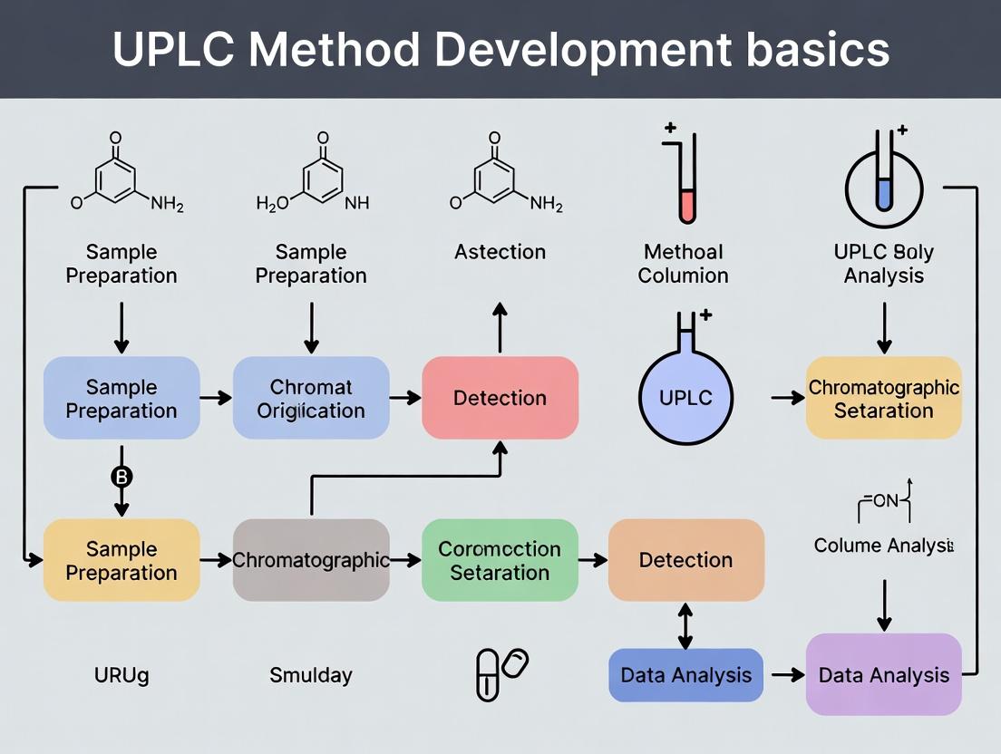
Visualization of UPLC Method Development Workflow
Title: UPLC Method Development Workflow for Drug Analysis
The Scientist's Toolkit: Essential Research Reagent Solutions
Table 3: Key Reagents and Materials for UPLC Method Development
| Item | Function & Specification | Critical Notes for UPLC |
|---|---|---|
| UPLC Columns | Stationary phase for separation. e.g., C18, 1.7µm, 2.1 x 50-100mm. | Must withstand high pressure. Sub-2µm particles are standard. |
| LC-MS Grade Solvents | Mobile phase components. Water, Acetonitrile, Methanol. | Low UV absorbance and minimal particulates to prevent background noise and clogging. |
| Buffer Salts & Additives | Modifies mobile phase pH/ionic strength. Ammonium formate/acetate, Formic Acid, TFA. | Use volatile additives for MS compatibility. Always filter (0.22µm) and degas. |
| Reference Standards | Primary standard for API; secondary for impurities. | High purity (>95%) is essential for accurate quantification and identification. |
| Vial & Cap Assemblies | Sample containers for autosampler. | Use low-volume vials with low-adsorption/pierceable caps to minimize sample loss. |
| Syringe Filters | For sample clarification pre-injection. PTFE or Nylon, 0.22µm. | Essential to protect the UPLC column from particulates. |
| Seal Wash Solvents | Prevents carryover in autosampler. | Typically a mix of water and a strong organic solvent (e.g., 90:10 Water:Isopropanol). |
| System Suitability Test Mix | A standard mixture to verify performance. | Contains compounds to test plate count, tailing factor, and resolution before sample runs. |
This technical guide details the core performance advantages of Ultra-Performance Liquid Chromatography (UPLC) over traditional High-Performance Liquid Chromatography (HPLC) within the critical framework of modern pharmaceutical method development. As drug analysis research demands greater efficiency, sensitivity, and data quality for complex matrices and high-throughput screening, UPLC has emerged as a foundational technology. This document provides an in-depth comparison of the two techniques, focusing on the quantitative parameters that directly impact method development for drug substances and products.
Core Comparative Analysis: UPLC vs. HPLC
The fundamental differences between UPLC and HPLC stem from instrument and particle design, which directly translate to performance gains. The following table summarizes the key technical specifications driving these advantages.
Table 1: Foundational Technical Specifications and Performance Outcomes
| Parameter | Traditional HPLC | UPLC | Direct Consequence for Method Development |
|---|---|---|---|
| Typical Particle Size | 3.5 - 5 µm | 1.7 - 1.8 µm | Reduced band broadening, higher peak capacity. |
| Operating Pressure | Up to 400 bar (6,000 psi) | Up to 1000-1500 bar (15,000-22,000 psi) | Enables use of smaller particles at optimal linear velocities. |
| System Dispersion (Extra-Column Volume) | Higher (≥ 10 µL) | Minimized (< 5 µL) | Preserves separation efficiency, especially for narrow peaks. |
| Detector Sampling Rate | Typically 10-40 Hz | Typically 40-100 Hz | Accurate digitization of very narrow peaks. |
| Recommended Column I.D. | 4.6 mm | 2.1 mm | Reduces solvent consumption, increases mass sensitivity. |
Quantitative Comparison of Speed, Sensitivity, and Resolution
Live search data and published literature consistently demonstrate the following performance improvements when migrating methods from HPLC to UPLC platforms under optimized conditions.
Table 2: Measurable Performance Advantages of UPLC
| Performance Metric | Typical Improvement Factor | Experimental Basis & Notes |
|---|---|---|
| Analysis Speed | 3x to 10x reduction in run time | Achieved via shorter columns with smaller particles and increased flow rates while maintaining efficiency. |
| Peak Capacity (Resolution) | 1.5x to 2x increase | Direct result of increased plate count (often > 100,000 plates/m). |
| Signal-to-Noise (Sensitivity) | 2x to 5x increase in S/N | Due to narrower peak widths (increased peak height) and reduced system noise. |
| Solvent Consumption | 70% to 90% reduction | Consequence of smaller column dimensions and shorter run times. |
| Mass Sensitivity | Up to 10x increase | Result of lower chromatographic dilution into a more concentrated peak. |
Detailed Experimental Protocols for Comparison
To empirically validate the advantages listed, the following protocols can be implemented.
Protocol 1: Direct Method Transfer and Comparison
Objective: To transfer an existing HPLC method for a small molecule drug to UPLC and compare key performance indicators (KPIs).
- HPLC Baseline: Execute the existing method (e.g., 150 x 4.6 mm, 5 µm C18 column, 1.0 mL/min, 10 µL injection).
- UPLC Method Translation: Calculate scaling factors. For a 2.1 mm I.D. UPLC column, flow rate scaling factor = (2.1² / 4.6²) ≈ 0.21. Initial UPLC flow: 1.0 mL/min * 0.21 ≈ 0.21 mL/min. Adjust gradient time proportionally to column void volume change.
- Optimization: Fine-tune gradient slope or flow rate to achieve equivalent or improved resolution.
- Data Collection: Inject identical sample concentrations on both systems. Record retention times, peak widths at half height (W0.5), plate count (N), signal-to-noise ratio (S/N) for a low-level impurity, and solvent volume used per run.
- Analysis: Calculate improvement factors for speed (run time reduction), sensitivity (S/N increase), and resolution (plate count or peak capacity increase).
Protocol 2: Determining Maximum Practical Plate Count
Objective: To compare the intrinsic efficiency of HPLC and UPLC columns.
- Isocratic Conditions: Select a low molecular weight, neutral test compound (e.g., uracil, alkylphenone).
- System Setup: Install a 150 mm or longer column for each platform (HPLC: 5 µm, UPLC: 1.7 µm). Use the optimal flow rate for each (determined via van Deemter experiment or manufacturer recommendation).
- Injection: Make a small, precise injection to minimize extra-column effects.
- Measurement: Record the peak, measure retention time (tR) and peak width at baseline (Wb) or half height (W0.5).
- Calculation: Compute plate count N = 16(tR/Wb)² or N = 5.54(tR/W0.5)². Normalize to plates per meter. The UPLC column will demonstrate significantly higher N/m.
Protocol 3: Sensitivity and Detection Limit Assessment
Objective: To compare the limits of detection (LOD) for a target analyte.
- Sample Preparation: Prepare a dilution series of the analyte (e.g., drug compound) in a suitable solvent, down to expected sub-ng/mL levels.
- Chromatography: Use the optimized methods from Protocol 1. Ensure detector settings (UV gain, sampling rate, time constant) are appropriately configured for each system.
- Injection: Triplicate injections at each concentration level.
- Calculation: Plot peak height vs. concentration for both systems. Determine LOD as concentration yielding S/N = 3. The UPLC system will typically show a lower LOD due to peak focusing.
Workflow and Logical Relationships
Diagram 1: Logical flow from UPLC technology to research outcomes.
Diagram 2: HPLC to UPLC method transfer and optimization workflow.
The Scientist's Toolkit: Essential Research Reagent Solutions
Table 3: Key Materials for UPLC Method Development in Drug Analysis
| Item | Function & Importance in UPLC |
|---|---|
| UPLC-Quality Solvents (HPLC-MS Grade) | Minimizes baseline noise and detector interference, crucial for high-sensitivity UV and MS detection. |
| High-Purity Buffers & Additives (e.g., Ammonium Formate, TFA) | Provides consistent ionization in MS interfaces and stable pH control. Must be filterable (0.22 µm) to prevent system clogging. |
| Certified UPLC Columns (1.7-1.8 µm particles) | The core component. Requires chemistries (C18, HILIC, etc.) stable at high pressures and compatible with intended mobile phases. |
| Low-Volume, Chemically Inert Vials & Caps | Reduces sample evaporation and minimizes adsorptive losses, especially critical for low-concentration drug metabolites. |
| Low-Dispersion, Pre-slit Screw Caps & Septa | Prevents coring and reduces extra-column peak broadening from the injection source. |
| Specialized UPLC Instrument Tuning & Calibration Kits | Contains test mixes for verifying system pressure, gradient delay volume, detector linearity, and MS mass accuracy. |
| Stable Isotope-Labeled Internal Standards (for LC-MS) | Essential for quantitative bioanalysis to correct for matrix effects and variability in sample preparation and ionization. |
The transition from HPLC to UPLC represents a paradigm shift in liquid chromatography, offering substantive, quantifiable improvements in speed, sensitivity, and resolution. For drug analysis research, these advantages translate directly into faster method development cycles, superior characterization of complex drug mixtures and degradants, and the ability to quantify compounds at lower concentrations—all while reducing solvent consumption and operational costs. A systematic understanding of the underlying principles and a careful approach to method translation, as outlined in this guide, are fundamental to leveraging UPLC technology for advanced pharmaceutical research.
Ultra-Performance Liquid Chromatography (UPLC) is a cornerstone technology in modern pharmaceutical research, enabling faster, higher-resolution separations with increased sensitivity compared to traditional HPLC. Within the context of UPLC method development for drug analysis, the selection and optimization of three core hardware components—the pump, autosampler, and detector—are critical for developing robust, reliable, and high-throughput analytical methods. This guide provides an in-depth technical examination of these essential subsystems, focusing on their impact on key method parameters such as resolution, linearity, precision, and sensitivity.
High-Pressure Binary and Quaternary Pump Systems
UPLC pumps must deliver precise, pulse-free mobile phase gradients at pressures up to 18,000-20,000 psi. Their performance directly influences retention time reproducibility, baseline stability, and method precision.
Core Technology and Performance Specifications
Modern UPLC systems employ binary high-pressure mixing pumps. Key operational metrics are summarized in Table 1.
Table 1: Quantitative Specifications of Modern UPLC Pump Systems
| Parameter | Typical Specification Range | Impact on Method Development |
|---|---|---|
| Maximum Pressure | 15,000 - 22,000 psi | Enables use of sub-2µm particles for high efficiency. |
| Flow Rate Range | 0.001 - 5.0 mL/min | Critical for scaling from analytical to semi-prep. |
| Flow Precision (RSD) | < 0.07% | Directly impacts retention time reproducibility. |
| Gradient Accuracy | ± 0.5% | Essential for robust, transferable gradient methods. |
| Gradient Precision (RSD) | < 0.15% | Affects peak area and retention time precision. |
| Delay Volume | 50 - 150 µL | Smaller volume enables faster gradient start and sharper peaks. |
| System Dispersion | < 15 µL² | Lower dispersion preserves peak shape and resolution. |
Experimental Protocol: Evaluating Pump Performance for Method Robustness
This protocol assesses the pump's contribution to system suitability.
Objective: Determine the retention time and peak area precision delivered by the UPLC pump under typical gradient conditions. Materials: UPLC system with binary pump, PDA or UV detector, validated column (e.g., C18, 2.1 x 50 mm, 1.7 µm), mobile phase A (0.1% Formic Acid in Water), mobile phase B (0.1% Formic Acid in Acetonitrile), test analyte (e.g., caffeine, paracetamol). Procedure:
- System Equilibration: Flush and purge pump lines. Set flow rate to 0.5 mL/min. Condition column with 5% B for 10 column volumes.
- Gradient Program Setup: Program a linear gradient: 5% B to 95% B over 3.0 minutes, hold at 95% B for 0.5 min, re-equilibrate at 5% B for 1.5 min. Total run time: 5.0 min.
- Sample Injection: Prepare a 10 µg/mL solution of the test analyte. Perform ten consecutive injections from the same vial using the autosampler.
- Data Analysis: For the primary analyte peak, calculate the %RSD for retention time and peak area across the ten injections. Acceptance criteria for a robust system are typically < 0.5% RSD for retention time and < 1.0% RSD for peak area.
- Pressure Analysis: Monitor the system pressure profile across all injections. The baseline pressure and pressure fluctuations should be stable (< 2% variation).
Title: Experimental Workflow for UPLC Pump Performance Evaluation
Low-Carryover Autosamplers with Temperature Control
The autosampler is pivotal for injection precision, sample integrity, and method throughput. Modern UPLC autosamplers feature low-dispersion, low-carryover designs with precise temperature control (4-40°C).
Key Performance Metrics
Table 2: Quantitative Specifications of UPLC Autosamplers
| Parameter | Typical Specification Range | Impact on Method Development |
|---|---|---|
| Injection Volume Precision (RSD) | < 0.5% for ≥ 1 µL | Directly affects quantitative accuracy and precision. |
| Injection Volume Range | 0.1 - 50 µL | Flexibility for sensitive or concentrated samples. |
| Carryover | < 0.002% - 0.05% | Critical for trace analysis and high-concentration samples. |
| Temperature Control Range | 4°C - 40°C (± 0.5°C) | Maintains stability of labile analytes. |
| Sample Capacity | 96-well plates or > 100 vials | Enables high-throughput screening. |
Experimental Protocol: Determining Injection Precision and Carryover
A standardized protocol to validate autosampler performance.
Objective: Quantify injection volume precision and percentage carryover for a UPLC method. Materials: UPLC system, autosampler, column, mobile phase, high-concentration sample (e.g., 1 mg/mL drug compound in suitable solvent), blank solvent (e.g., mobile phase A or water:acetonitrile 80:20). Procedure for Injection Precision:
- Sample Preparation: Prepare a single vial of a mid-range calibration standard (e.g., 100 µg/mL).
- Chromatographic Conditions: Use an isocratic or fast gradient method appropriate for the analyte.
- Injection Sequence: Perform six replicate injections from the same vial.
- Calculation: Determine the %RSD of the analyte peak area. Acceptance is typically < 1.0% RSD. Procedure for Carryover Assessment:
- Injection Sequence: Inject the high-concentration sample (1 mg/mL), followed by two consecutive injections of the blank solvent.
- Chromatogram Analysis: In the first blank injection following the high sample, identify any peak corresponding to the analyte.
- Calculation: Carryover (%) = (Peak Area in Blank / Peak Area of High Sample) x 100% Acceptance criteria is often < 0.05% for drug substance analysis and < 0.01% for bioanalysis.
The Scientist's Toolkit: Key Reagents & Materials for Autosampler Validation
| Item | Function & Rationale |
|---|---|
| Certified Volumetric Flasks & Pipettes | For accurate and precise preparation of test standard solutions. |
| LC-MS Grade Water & Acetonitrile | High-purity solvents minimize background interference in blanks. |
| Low-Adsorption, Certified Vials & Caps | Reduce nonspecific binding of analyte, especially for proteins or peptides. |
| High-Concentration Analytic Standard (e.g., 1 mg/mL) | Provides a stringent test for wash efficiency and carryover. |
| Weak Needle Wash Solvent (e.g., 5% Organic) | Removes polar components from the injection needle. |
| Strong Needle Wash Solvent (e.g., 50% Organic) | Removes non-polar components to prevent carryover. |
High-Sensitivity and High-Speed Detectors
UPLC detectors must have fast sampling rates (> 40 Hz) and low dispersion flow cells to accurately capture narrow peaks (1-2 sec wide at base) without losing resolution or sensitivity.
Detector Types and Comparative Performance
Table 3: Comparison of Primary UPLC Detector Technologies
| Detector Type | Key Principle | Typical Data Rate | Linear Dynamic Range | Key Application in Drug Analysis |
|---|---|---|---|---|
| Photodiode Array (PDA) | Full-spectrum UV-Vis | 80 - 160 Hz | Up to 5.0 AU | Method development, peak purity, identification. |
| UV/Vis (Variable Wavelength) | Single/dual wavelength | 40 - 80 Hz | Up to 2.5 AU | Routine quantification of APIs with strong chromophores. |
| Fluorescence (FLR) | Emission after excitation | 40 - 80 Hz | > 10^4 | Ultra-sensitive detection of native or derivatized compounds. |
| Charged Aerosol (CAD) | Universal, mass-sensitive | 40 - 80 Hz | 10^3 - 10^4 | Impurity profiling, excipients, compounds lacking UV chromophores. |
| Evaporative Light Scattering (ELSD) | Universal, mass-sensitive | 40 - 80 Hz | 10^2 - 10^3 | Lipids, sugars, polymers. Less sensitive than CAD. |
| Mass Spectrometry (MS) | Mass-to-charge ratio | > 10 Hz | 10^3 - 10^5 | Structural identification, metabolite profiling, bioanalysis. |
Experimental Protocol: Optimizing Detector Settings for Narrow UPLC Peaks
This protocol ensures detector settings are optimized to preserve the separation efficiency generated by the UPLC column.
Objective: Configure detector sampling rate and response time to accurately capture peak shape without introducing artificial broadening or noise. Materials: UPLC system with PDA or UV detector, column (C18, 2.1 x 50 mm, 1.7 µm), test mix (e.g., 3-5 related compounds with slight retention differences). Procedure:
- Initial Conditions: Set a flow rate of 0.6 mL/min and a fast gradient to generate narrow peaks (~2-3 sec peak width at base). Set detector wavelength as needed.
- Sampling Rate Test: Perform an injection with the detector data acquisition rate set to its maximum (e.g., 80 Hz or 12.5 pts/sec). Note the peak shape and the number of data points across the peak (aim for > 20 points).
- Response Time/Filter Constant Test: Many detectors use an electronic filter (response time) to reduce high-frequency noise. Start with the fastest setting (e.g., 0.1 s). Perform an injection and observe the signal-to-noise ratio (S/N). Gradually increase the response time (e.g., to 0.5 s, 1.0 s) and reinject.
- Optimization Balance: Find the slowest response time that does not visibly distort or broaden the peak shape. This setting provides the best S/N without sacrificing resolution. The optimal sampling rate is typically 2-3x the reciprocal of the peak width in seconds.
Title: Logic Flow for UPLC Detector Parameter Optimization
Integrated System Considerations for Method Development
The synergistic performance of the pump, autosampler, and detector defines the overall system dwell volume, dispersion, and sensitivity. For robust method development, the total system extra-column volume must be minimized to prevent peak broadening, especially when using small-volume (e.g., 50 mm) columns.
Protocol: Measuring System Dwell Volume
Dwell volume is the volume between the point where gradient solvents are mixed and the head of the column. It causes a delay between the programmed and actual gradient start.
Procedure:
- Setup: Disconnect the column and connect a union in its place. Place the outlet tubing into a vial containing water.
- Mobile Phase: Use Mobile Phase A: 0.1% Acetone in Water; Mobile Phase B: 0.1% Acetone in Acetonitrile. Set detector wavelength to 265 nm (λ_max for acetone).
- Run Gradient: Set a flow rate of 0.5 mL/min. Program a step gradient: 0% B for 2 min, then an immediate step to 100% B. Run for 10-15 minutes.
- Calculation: Plot the detector signal. The dwell volume is calculated as: Dwell Volume (mL) = Flow Rate (mL/min) x Time Delay (min), where the time delay is measured from the gradient step command to the point at 50% of the step transition at the detector.
Understanding this volume is critical for scaling and transferring gradient methods between different UPLC systems and is a foundational step in systematic method development for pharmaceutical analysis.
Within the thesis on UPLC method development basics for drug analysis, the selection of an appropriate stationary phase is the single most critical factor determining success. The column dictates selectivity, efficiency, and robustness. This guide provides an in-depth technical comparison of the three primary modes—Reversed-Phase (RP), Hydrophilic Interaction Liquid Chromatography (HILIC), and Ion-Exchange (IEX)—for the analysis of drug molecules and related impurities.
Core Principles and Selectivity Mechanisms
Reversed-Phase (RP)
RP chromatography is the workhorse of pharmaceutical analysis (>80% of methods). Separation is based on hydrophobicity, with analytes partitioning between a polar mobile phase (typically water/organic mixtures) and a non-polar stationary phase.
Common Phases:
- C18 (Octadecylsilane): Highest hydrophobicity; general-purpose.
- C8 (Octylsilane): Moderate hydrophobicity; for moderately to highly hydrophobic analytes.
- Phenyl/Alkyl-Phenyl: Offers π-π interactions with aromatic compounds.
- Polar-Embedded Groups (e.g., amide): Enhanced retention for polar compounds and improved wettability.
Hydrophilic Interaction Liquid Chromatography (HILIC)
HILIC is employed for polar and hydrophilic compounds that are poorly retained in RP. A hydrophobic stationary phase (often bare silica or derivatized silica with cyano, diol, or amino groups) is used with a mobile phase high in organic solvent (typically >70% acetonitrile). Separation involves partitioning into a water-enriched layer on the stationary surface and secondary interactions (hydrogen bonding, ion-exchange).
Ion-Exchange (IEX)
IEX separates ions or ionizable molecules based on electrostatic interactions with charged functional groups on the stationary phase.
- Cation-Exchange (SCX): Negatively charged surface (e.g., sulfonate) retains cations.
- Anion-Exchange (SAX): Positively charged surface (e.g., ammonium) retains anions. Retention is controlled by mobile phase pH, ionic strength, and ion type.
Quantitative Comparison of Column Phases
Table 1: Key Characteristics of Chromatographic Phases
| Parameter | Reversed-Phase (C18) | HILIC (Silica) | Ion-Exchange (SCX) |
|---|---|---|---|
| Primary Mechanism | Hydrophobic partitioning | Hydrophilic partitioning & H-bonding | Electrostatic attraction |
| Typical Mobile Phase | Water/Methanol or Acetonitrile | >70% ACN in aqueous buffer | Aqueous buffer with salt gradient |
| Optimal Analyte pKa/Log P | Log P > 0, neutral or ion-suppressed | Log P < 0, polar, basic | Ionizable (pKa within 2 units of pH) |
| Typical pH Range | 2-8 (for silica-based) | 3-8 (for bare silica) | 2-10 (polymer-based) |
| Key Strength | Broad applicability, robustness | Retention of polar metabolites | Separation of charges, biomolecules |
| Major Challenge | Poor retention of very polar analytes | Method development complexity, sensitivity to %water | Requires high salt, often incompatible with MS |
Table 2: Selection Guide Based on Analyte Properties
| Analyte Characteristic | Recommended Primary Mode | Alternative Mode | Rationale |
|---|---|---|---|
| Hydrophobic (Log P > 2) | RP (C8 or C18) | - | Excellent retention and peak shape. |
| Polar, Neutral | HILIC (Diol or Cyano) | RP with polar-embedded phase | RP may not retain; HILIC provides good retention. |
| Polar, Basic | RP (at low pH) or HILIC | IEX (if cationic) | RP with ion-pairing or HILIC for selectivity. |
| Polar, Acidic | RP (at high pH) or HILIC | IEX (if anionic) | Careful pH control needed; HILIC can be effective. |
| Charged Biomolecule (Protein, Peptide) | IEX or RP | HILIC | IEX for native state; RP for denatured/digested. |
| Ionic Metabolite | HILIC or IEX | RP with ion-pairing | HILIC-MS is often the preferred approach. |
Experimental Protocols for Phase Selection Screening
Protocol 1: Initial Mode Selection Scouting for a New Chemical Entity
Objective: Determine the most suitable chromatographic mode for an API and its related substances. Materials: UPLC system with PDA and MS detection, columns (e.g., C18, HILIC silica, SCX), analytical standards. Procedure:
- Prepare stock solutions of the API and known impurities in a suitable solvent (e.g., DMSO or water/ACN 50:50).
- RP Screening: Inject on a C18 column. Use a generic gradient: 5-95% acetonitrile in 10 mM ammonium formate (pH 3.0 or unadjusted) over 5 minutes. Note retention and peak shape.
- HILIC Screening: Inject on a bare silica column. Use a generic gradient: 95-50% acetonitrile in 10 mM ammonium formate (pH 3.0) over 5 minutes. Note retention and peak shape.
- IEX Screening (if ionic): Inject on an SCX column. Use an isocratic hold at 20 mM potassium phosphate (pH 3.0) for 2 min, then a 0-500 mM KCl gradient over 8 minutes.
- Evaluation: Select the mode where the API elutes between 2-10 minutes, all peaks are resolved, and peak shape is symmetric (asymmetry factor 0.9-1.2).
Protocol 2: HILIC Method Optimization (DoE Approach)
Objective: Optimize a HILIC method for a polar drug substance using a Design of Experiments (DoE) approach. Materials: UPLC-HILIC column, analytical standards. Procedure:
- Define Critical Process Parameters (CPPs): %ACN (X1), buffer concentration (X2), and pH (X3).
- Define Critical Quality Attributes (CQAs): Resolution of critical pair (Rs), retention time of API (k).
- Create a Central Composite Design (CCD) with 3 factors: Run 17 randomized experiments covering the space (e.g., %ACN: 75-90%; buffer: 5-50 mM; pH: 3.0-6.0).
- Perform all experiments, recording CQAs.
- Use statistical software to generate a response surface model. Identify the optimal conditions that maximize Rs while keeping k within 1-10.
Logical Workflow and Signaling Pathways
Diagram 1: Phase Selection Decision Tree for Drug Analysis
Diagram 2: UPLC Method Development Workflow Integrating Column Selection
The Scientist's Toolkit: Essential Research Reagent Solutions
Table 3: Key Reagents and Materials for Column Chemistry Studies
| Item | Function & Description | Key Consideration for Selection |
|---|---|---|
| UPLC Columns (1.7-2.6µm) | Core separation media. High-pressure stable columns for RP, HILIC, IEX phases. | Particle size (1.7µm for max efficiency), pore size (100-120Å for small molecules), pH stability range. |
| LC-MS Grade Water | Ultrapure water for mobile phase preparation. | Low TOC, resistivity >18 MΩ·cm to minimize MS background noise and column contamination. |
| LC-MS Grade Acetonitrile & Methanol | Primary organic modifiers for mobile phases. | Low UV cutoff, low acid/base/aldehyde content for minimal baseline drift and artifact peaks. |
| Ammonium Formate/Acetate | Volatile buffers for MS-compatible methods (RP & HILIC). | Concentration (5-50 mM) and pH control retention and ionization efficiency. |
| Trifluoroacetic Acid (TFA) | Ion-pairing agent and strong acidifier for RP peptide/protein analysis. | Can cause ion suppression in MS; use at low concentrations (0.05-0.1%). |
| Phosphate & Potassium Chloride | Non-volatile buffers and salts for IEX or UV-only methods. | Provides precise pH and ionic strength control. Not MS-compatible. |
| pH Standard Buffers | For accurate calibration of mobile phase pH. | Use buffers traceable to NIST standards. pH should be measured in the aqueous portion. |
| Column Regeneration Kits | Solvents for cleaning and restoring column performance. | Specific to phase chemistry (e.g., high-water for HILIC, high-organic for RP). |
Within the foundational thesis on UPLC (Ultra-Performance Liquid Chromatography) method development for drug analysis, the initial scouting phase is paramount. This phase, termed "Critical Method Scouting," involves the systematic characterization of two fundamental pillars: the intrinsic chemical and physical properties of the analyte(s) of interest, and the complete composition of the sample matrix. Success in this stage directly dictates the selection of chromatographic mode, column chemistry, and detection parameters, ensuring a robust, sensitive, and specific analytical method. This guide details the protocols and considerations for this critical first step in modern pharmaceutical research.
Defining Analyte Properties: Experimental Protocols
A comprehensive understanding of analyte properties is non-negotiable for rational method development.
Protocol for Determination of Acid Dissociation Constant (pKa)
Objective: To determine the pKa value(s) of the analyte, guiding selection of mobile phase pH. Materials: Analytical balance, pH meter, UV-Vis spectrophotometer or potentiometric titrator, standard buffer solutions, 0.1 M HCl, 0.1 M NaOH. Procedure:
- Prepare a stock solution of the analyte in a solvent of known ionic strength (e.g., 0.1 M KCl).
- Prepare a series of buffered solutions across a wide pH range (e.g., pH 2-12).
- Dilute the analyte stock into each buffer to achieve identical final concentration.
- Measure the UV-Vis spectrum or potentiometric potential of each solution.
- Plot the spectral shift or potential against pH. The pKa is the pH at the inflection point (midpoint) of the sigmoidal curve.
Protocol for Determination of LogP/LogD
Objective: To quantify the lipophilicity of the analyte, informing reversed-phase column and organic modifier selection. Materials: Shaker, centrifuge, HPLC system with UV detector, n-octanol, aqueous buffer (e.g., phosphate buffer pH 7.4), vials. Procedure (Shake-Flask Method):
- Pre-saturate n-octanol and buffer phase with each other by mixing and separating.
- Dissolve analyte in a known volume of one phase (e.g., buffer).
- Mix with an equal volume of the other phase (e.g., octanol) in a sealed vial.
- Shake vigorously for 1 hour to reach partitioning equilibrium.
- Centrifuge to separate phases completely.
- Quantify the analyte concentration in both phases using a calibrated HPLC-UV method.
- Calculate LogP (for neutral species) or LogD (for ionizable species at a specific pH): LogP/D = log10([Analyte]octanol / [Analyte]aqueous).
Protocol for UV-Vis Spectroscopic Scan
Objective: To identify optimal detection wavelengths and assess analyte chromophore. Materials: UV-Vis spectrophotometer, quartz cuvettes, appropriate solvent. Procedure:
- Prepare a dilute solution of the analyte in a solvent transparent in the scan range (e.g., methanol, mobile phase).
- Fill a quartz cuvette with the solvent blank and perform a baseline correction.
- Replace with the analyte solution and scan from 200 nm to 400 nm (or higher if needed).
- Identify wavelength(s) of maximum absorption (λmax) for potential use in PDA or UV detection.
Table 1: Core Analyte Properties and Their Impact on UPLC Method Development
| Property | Typical Experimental Method | Impact on UPLC Method Development | Target Range for Drug-like Molecules |
|---|---|---|---|
| pKa | Potentiometric titration, UV-pH titration | Dictates mobile phase pH for controlling ionization, retention, and peak shape. | Knowledge of all acidic/basic sites is required. |
| LogP / LogD | Shake-flask + HPLC, Chromatographic estimation (e.g., ChromlogD) | Predicts retention in Reversed-Phase (RP) mode. High LogP (>4) may require HILIC or normal phase. | LogP 1-4 is ideal for standard RP-UPLC. |
| λmax (UV) | UV-Vis Spectrophotometry | Sets primary detection wavelength for optimal sensitivity. | Typically 200-350 nm for most APIs. |
| Solubility | Equilibrium solubility shake-flask | Determines feasible injection solvent and potential for on-column precipitation. | Should be >1 mg/mL in initial mobile phase. |
| Chemical Stability | Stress testing (pH, thermal, oxidative light) | Informs sample handling, storage, and mobile phase compatibility requirements. | Should be stable for duration of analysis. |
Defining the Sample Matrix: A Comprehensive Analysis
The sample matrix is the vehicle containing the analyte and is a primary source of interference and method failure.
Protocol for Sample Matrix Component Mapping
Objective: To catalog all major and minor components in the sample to anticipate interferences. Materials: UPLC system with PDA and High-Resolution Mass Spectrometry (HRMS), sample preparation equipment. Procedure:
- Sample Pretreatment: Subject the neat matrix (e.g., plasma, tablet extract, formulation vehicle) to a generic protein precipitation or dilution.
- Chromatographic Separation: Inject the pretreated matrix onto a wide-scope analytical column (e.g., C18, 100 x 2.1 mm, 1.7 µm) using a generic gradient (e.g., 5-95% acetonitrile in water over 10 min).
- Detection: Use PDA (210-400 nm) and HRMS in full-scan mode (positive/negative electrospray ionization).
- Data Analysis: Deconvolute the resulting chromatograms to identify endogenous compounds, excipients, preservatives, degradation products, and potential metabolites. Compare against analyte retention and detection response.
Table 2: Common Drug Analysis Sample Matrices and Key Interferents
| Sample Matrix | Typical Composition | Primary Analytical Challenges | Recommended Scouting Cleanup |
|---|---|---|---|
| Human Plasma/Serum | Proteins (Albumin, Globulins), Lipids, Amino Acids, Salts, Metabolic Waste | Protein binding, phospholipid interference, ion suppression in MS, clogging of frits. | Protein Precipitation (ACN/MeOH), SPE (Phospholipid Removal), Derivatization. |
| Tablet Formulation | API, Fillers (Lactose, MCC), Binders, Disintegrants, Lubricants (Mg Stearate), Glidants, Coatings. | Co-elution of excipients with API, spectral interference, column contamination. | Simple dilution/filtration, Solid-Phase Extraction (SPE). |
| Cell Culture Media | Salts, Sugars, Amino Acids, Vitamins, Buffers, Phenol Red, Serum Proteins. | Complex background in UV, high salt content, multiple isobaric interferences in MS. | Deproteinization, Ultrafiltration, Online Sample Cleanup (2D-LC). |
| Topical Cream/Ointment | API, Emulsifiers, Thickeners, Oils, Waxes, Preservatives, Water. | Extreme sample heterogeneity, high viscosity, incompatible solvents, lipid matrix. | Liquid-Liquid Extraction (LLE), Heated Dilution/Sonication, Saponification. |
Visualization of the Critical Method Scouting Workflow
Title: Critical Method Scouting Decision Workflow
The Scientist's Toolkit: Essential Research Reagent Solutions
Table 3: Key Reagents and Materials for Method Scouting Experiments
| Item Name | Function & Purpose in Scouting |
|---|---|
| pH Buffers (Broad Range Kit) | For creating stable mobile phase conditions during pKa determination and initial chromatographic screening. |
| n-Octanol (HPLC Grade) | The organic phase for the classical shake-flask LogP/D determination experiment. |
| Phospholipid Removal SPE Plates | For efficient removal of phospholipids from biological matrices during sample prep optimization. |
| Hybrid SPELC (LC-MS) Certified Vials | To prevent leachables that cause background interference, especially critical in high-sensitivity MS detection scouting. |
| UPLC Column Scouting Kit | A set of columns (C18, Phenyl, HILIC, Charged Surface) for rapid empirical evaluation of retention and selectivity. |
| PDA and HRMS Compatible Mobile Phase Additives (e.g., FA, AA, NH4OAc) | High-purity additives to test ionization efficiency in MS and pH control without causing detector noise. |
| Chemical Stability Stress Kits (Oxidative, Acid/Base, Thermal) | Standardized reagents to systematically assess analyte degradation pathways and identify potential impurities. |
| Protein Precipitation Plates (96-well) | For high-throughput screening of deproteinization efficiency for bioanalytical matrix methods. |
Within the foundational framework of UPLC (Ultra-Performance Liquid Chromatography) method development for drug analysis, the initial setting of core performance goals is a critical determinant of success. This phase moves beyond theoretical ideals, establishing the practical, quantifiable targets that guide every subsequent experimental decision. Three interdependent parameters—Resolution (Rs), Runtime, and Detection Limits—form the cornerstone of this goal-setting exercise. This guide provides an in-depth technical examination of these targets, framed within the rigorous demands of modern pharmaceutical research.
Defining Core Performance Goals
Resolution (Rs)
Chromatographic resolution is the non-negotiable prerequisite for accurate quantification. It quantitatively measures the separation between two adjacent peaks.
Calculation: Rs = 2(tR2 - tR1) / (w1 + w2) where tR is retention time and w is peak width at baseline.
Goal-Setting Criteria:
- Rs ≥ 2.0: Required for baseline separation for precise quantitative analysis of known impurities and degradants.
- Rs ≥ 1.5: Often set as a system suitability requirement for main analyte separation from the closest eluting peak.
- Forced Degradation Studies: The method must demonstrate sufficient robustness to maintain Rs > 1.5 under mild stress conditions.
Runtime
Runtime directly impacts laboratory throughput and operational cost. The goal is the shortest runtime that meets all resolution and sensitivity requirements.
Key Considerations:
- Analytical Throughput: High-throughput stability-indicating methods may target runtimes of 5-10 minutes.
- Method Complexity: Methods for complex mixtures (e.g., peptide digests) may necessitate longer runs (20-60 minutes).
- Gradient Re-equilibration: Must be factored into total cycle time.
Detection Limits
These define the method's sensitivity, crucial for low-level impurity and degradant profiling.
- Limit of Detection (LOD): Signal-to-Noise (S/N) ≥ 3.
- Limit of Quantification (LOQ): S/N ≥ 10, with acceptable precision (typically %RSD < 20) and accuracy (80-120%).
- Goal-Setting Driver: Typically based on the reporting threshold (e.g., 0.05% or 0.1% of the drug substance concentration).
Table 1: Typical Initial Goal Ranges for UPLC Drug Analysis Methods
| Parameter | Typical Target Range | Regulatory/Scientific Basis | Key Influencing Factors |
|---|---|---|---|
| Resolution (Rs) | ≥ 1.5 (System Suitability) ≥ 2.0 (Critical Pair) | ICH Q2(R1), USP <621> | Column chemistry, gradient slope, temperature, mobile phase pH |
| Runtime | 5 - 15 minutes (standard) | Throughput requirements & method scope | Column dimensions (length, particle size), flow rate, gradient span |
| LOD (S/N) | ≥ 3 | ICH Q2(R1) definition | Detector sensitivity (e.g., DAD vs. MS), injection volume, peak shape |
| LOQ (S/N) | ≥ 10 | ICH Q2(R1) definition | As above, plus analyte-specific response |
| LOQ Level | 0.05% - 0.1% w/w | ICH Q3A/B Impurity Guidelines | Drug substance dosage strength |
Table 2: Effect of UPLC Parameters on Primary Goals
| Parameter Adjustment | Effect on Resolution | Effect on Runtime | Effect on Sensitivity |
|---|---|---|---|
| Decrease Particle Size | Increase | Decrease | Increase (sharper peaks) |
| Increase Column Length | Increase | Increase | Minor Decrease (broader peaks) |
| Reduce Flow Rate | Increase (to a point) | Increase | Increase (for MS, ESI) |
| Flatten Gradient Slope | Increase | Increase | Decrease (broader peaks) |
Experimental Protocols for Goal Determination
Protocol 1: Establishing Minimum Required Resolution
Objective: Determine the chromatographic conditions to achieve Rs ≥ 2.0 for the critical pair (API and closest known impurity).
Materials: See "Scientist's Toolkit" below. Method:
- Prepare standard solutions of the API and the known impurity at approximately 0.1% level relative to API.
- Inject the mixture onto the initial screening column (e.g., C18, 100mm x 2.1mm, 1.7µm).
- Employ a linear gradient (e.g., 5-95% organic modifier over 10 min) at 0.4 mL/min, 40°C.
- If Rs < 2.0, iteratively adjust:
- Primary: Modify gradient slope (e.g., 5-50% organic over 15 min).
- Secondary: Adjust column temperature (±10°C).
- Tertiary: Change column chemistry (e.g., to phenyl-hexyl or HILIC).
- Record the conditions where Rs meets the target. This forms the "separation core" of the method.
Protocol 2: Determining Limit of Quantification (LOQ)
Objective: Empirically establish the lowest concentration meeting S/N ≥ 10, with precision ≤20% RSD.
Method:
- Prepare a dilution series of the analyte from the target concentration down to ~0.01%.
- Perform six replicate injections of the solution at the estimated LOQ level.
- Calculate the S/N for each injection (typically via chromatographic software).
- Calculate the %RSD of the peak area response for the six replicates.
- Verify accuracy via spike/recovery at the LOQ level in placebo if required.
- The LOQ is the lowest concentration that fulfills both S/N ≥ 10 and %RSD ≤ 20.
Visualizing the Goal-Setting Workflow
Diagram 1: UPLC Method Goal-Setting & Conflict Resolution Workflow (76 chars)
The Scientist's Toolkit: Research Reagent Solutions
Table 3: Essential Materials for UPLC Goal-Setting Experiments
| Item | Function & Rationale |
|---|---|
| Acetonitrile (LC-MS Grade) | Low-UV-cutoff organic modifier; provides high elution strength and low backpressure. |
| Ammonium Formate/Acetate | Volatile buffers for MS-compatible methods; formate is preferred for negative-mode MS. |
| Trifluoroacetic Acid (TFA) | Ion-pairing agent for improving peak shape of basic analytes; use at low (0.1%) concentration. |
| Phosphoric Acid / K2HPO4 | Non-volatile buffer system for non-MS methods (e.g., pharmacopoeial assays). |
| 1.7µm UPLC Columns | Core separation media. C18 for general use; phenyl-hexyl for aromatics; HILIC for polar compounds. |
| Reference Standards | High-purity drug substance and known impurities for establishing resolution and sensitivity baselines. |
| Placebo/Excipient Blend | To assess interference and confirm specificity of detection at the target LOQ. |
| In-Vial Filters (0.2µm) | To prevent particulate matter from damaging the UPLC system and column. |
A Step-by-Step UPLC Method Development Protocol for Pharmaceuticals
In the systematic development of a UPLC (Ultra-Performance Liquid Chromatography) method for drug analysis, the selection and optimization of the mobile phase is the foundational step. This phase directly dictates critical chromatographic outcomes: resolution, peak shape, analysis time, and overall method robustness. Framed within the broader thesis of UPLC method development basics, this guide provides a technical deep-dive into the core principles and practical protocols for optimizing buffer systems, pH, and organic modifiers to achieve a reliable, stability-indicating assay for active pharmaceutical ingredients (APIs) and related substances.
Core Principles and Chemical Interactions
The mobile phase in reversed-phase UPLC (RP-UPLC) is a ternary system comprising water (aqueous component), a water-miscible organic solvent (modifier), and often a buffer or additive. Optimization aims to control two primary interaction mechanisms:
- Hydrophobic Interactions: Governed by the strength of the organic modifier. Increasing organic percentage decreases retention for all analytes.
- Ionogenic Interactions: Controlled by the mobile phase pH relative to the analyte's pKa. For ionizable compounds (≈80% of APIs), pH is the most powerful tool for modulating selectivity (elution order).
The effective pH of the aqueous buffer must be maintained between 2.0 and 8.0 for silica-based stationary phases to ensure long-term column stability. Modern hybrid or charged surface hybrid particles extend this range.
Systematic Optimization Strategy
Buffer Selection and Preparation
The buffer's role is to maintain a consistent pH, ensuring reproducible ionization states of the analyte and the silanol groups on the stationary phase. Capacity (β) is key; a minimum of 10 mM buffer concentration is recommended.
Table 1: Common Buffers for RP-UPLC Method Development
| Buffer Salt | Useful pH Range | UPLC Compatibility Notes | Typical Concentration |
|---|---|---|---|
| Ammonium Formate | 3.0-4.5 | Excellent MS compatibility, volatile. Can form formic acid at low pH. | 5-20 mM |
| Ammonium Acetate | 3.8-5.8 | Excellent MS compatibility, volatile. Limited buffering at pH 4.8. | 5-20 mM |
| Potassium Phosphate | 2.1-3.1; 6.2-8.2 | High buffering capacity. Non-volatile; not MS-compatible. Requires thorough flushing. | 10-25 mM |
| Sodium Phosphate | 2.1-3.1; 6.2-8.2 | High buffering capacity. Non-volatile; not MS-compatible. Bio-compatibility concerns. | 10-25 mM |
| Trifluoroacetic Acid (TFA) | ~1.5-2.5 | Ion-pairing agent, improves peak shape for bases. MS-compatible but can cause signal suppression. | 0.05-0.1% (v/v) |
| Formic Acid | ~2.0-4.0 | Common MS-compatible additive. Limited buffering capacity. | 0.1-0.5% (v/v) |
Protocol 1: Preparation of 20 mM Ammonium Acetate Buffer, pH 4.5
- Weigh 1.54 g of ammonium acetate (NH₄C₂H₃O₂) into a 1 L volumetric flask.
- Add approximately 900 mL of HPLC-grade water and dissolve completely.
- Adjust the pH to 4.50 using glacial acetic acid (to lower pH) or ammonium hydroxide (to raise pH). Use a calibrated pH meter.
- Dilute to volume with HPLC-grade water.
- Filter through a 0.22 μm nylon or PVDF membrane filter under vacuum.
- Degas by sonication or online sparging with helium.
pH Scouting
A preliminary pH scouting run identifies the pH of maximal selectivity change and optimal peak shape for ionizable analytes.
Protocol 2: pH Scouting Gradient Run
- Stationary Phase: Select a robust C18 column (e.g., 100 mm x 2.1 mm, 1.7-1.8 μm).
- Mobile Phase A: Prepare three separate aqueous buffers at pH 3.0, 4.5, and 7.0 (e.g., using ammonium formate for pH 3.0 & 4.5, ammonium phosphate for pH 7.0), each at 20 mM.
- Mobile Phase B: Acetonitrile (ACN) or Methanol (MeOH).
- Gradient: Use a linear gradient from 5% B to 95% B over 10 minutes. Hold initial and final conditions for 1 column volume each.
- Sample: Inject a solution containing the API and all known impurities/degradants.
- Analysis: Overlay chromatograms. Identify the pH where critical peak pair resolution is maximized and peak asymmetry is minimized (typically 1.0-1.5).
Diagram Title: pH Scouting Experimental Workflow (Max 100 char)
Organic Modifier Selection and Optimization
The choice between acetonitrile (ACN) and methanol (MeOH) significantly impacts selectivity, viscosity, and backpressure.
Table 2: Comparison of Organic Modifiers
| Property | Acetonitrile (ACN) | Methanol (MeOH) |
|---|---|---|
| Elution Strength | Higher (lowers retention more) | Lower |
| Viscosity | Lower (reduces backpressure) | Higher, especially with water |
| Selectivity | Different H-bonding & dipole interactions | Strong proton-donor character |
| UV Cutoff | ~190 nm | ~205 nm |
| Typical Use | First choice for most methods; sharp peaks. | Alternative for selectivity tuning; cheaper. |
Protocol 3: Modifier and Gradient Slope Scouting
- Fix the aqueous buffer at the optimal pH from Protocol 2.
- Perform two separate gradient runs (e.g., 5-95% B in 10 min), one with ACN and one with MeOH.
- Analyze for selectivity differences. If resolution remains inadequate, perform a gradient slope optimization.
- Gradient Slope Experiment: Using the chosen modifier, run gradients of varying steepness (e.g., 5-95% B in 5, 10, and 15 minutes).
- Plot log k vs. %B. An optimal gradient yields a resolution (Rs) > 2.0 for all critical pairs while minimizing run time.
The Scientist's Toolkit: Research Reagent Solutions
Table 3: Essential Materials for Mobile Phase Optimization
| Item / Reagent | Function & Technical Note |
|---|---|
| HPLC-Grade Water (≥18.2 MΩ·cm) | Aqueous component base. Minimizes baseline noise and impurity interference. |
| HPLC-Grade Acetonitrile & Methanol | Organic modifiers. Low UV absorbance and particulate matter are critical. |
| Buffer Salts (≥99% purity) | Provides pH control and ionic strength. Use high-purity to avoid ghost peaks. |
| pH Meter with ATC Probe | For accurate buffer preparation. Must be calibrated daily with ≥2 NIST-traceable buffers. |
| 0.22 μm Nylon or PVDF Filters | For mobile phase filtration to protect UPLC system and column from particulates. |
| 2 mL Glass Vials with Pre-Slit PTFE/Silicone Caps | For mobile phase and sample storage, ensuring compatibility and minimizing leachables. |
| Ultrasonic Bath or Online Degasser | Removes dissolved gases to prevent pump cavitation and baseline drift. |
| Vacuum Filtration Apparatus | For efficient filtration of large volumes of mobile phase. |
Integrated Optimization and Final Method Conditions
The final step is a fine-tuning experiment combining the optimal pH and organic modifier, often using a statistical Design of Experiments (DoE) approach for multi-factor efficiency.
Table 4: Example DoE Results for a Basic API (pKa ~9.0)
| Experiment | pH | %ACN Start | Gradient Time (min) | Resolution (Critical Pair) | Peak Asymmetry (API) | |
|---|---|---|---|---|---|---|
| 1 | 3.0 | 15 | 8 | 4.2 | 1.05 | |
| 2 | 3.0 | 15 | 12 | 4.8 | 1.08 | |
| 3 | 3.0 | 20 | 8 | 3.8 | 1.02 | |
| 4 | 3.0 | 20 | 12 | 4.3 | 1.04 | |
| 5 | 4.5 | 15 | 8 | 1.5 | 1.20 | |
| 6 | 4.5 | 15 | 12 | 1.8 | 1.22 | |
| Optimal | 7 | 4.0 | 15 | 10 | 5.1 | 1.03 |
| 8 | 4.0 | 20 | 12 | 4.1 | 1.01 |
Example Conclusion from Table 4: pH 4.0 provides superior resolution over pH 4.5 for this basic compound, with a moderate initial %ACN and longer gradient time yielding the best compromise between resolution and run time.
Diagram Title: Logical Relationship of Mobile Phase Optimization Goals (Max 100 char)
Phase 1 optimization is an iterative, science-driven process. Beginning with a systematic pH scouting study followed by organic modifier and gradient slope optimization provides a clear pathway to a robust, stability-indicating UPLC method. The selected conditions—typically a volatile buffer at a pH 1.5-2.0 units away from the analyte pKa, paired with acetonitrile under a finely tuned gradient—form the cornerstone of reliable drug analysis, setting the stage for subsequent phases of column selection and robustness testing in the overall method development thesis.
Within the systematic framework of UPLC method development for drug analysis, Phase 2 represents the critical empirical core. Following initial scouting (Phase 1), this phase involves the rigorous, structured evaluation of stationary phase chemistry and the pivotal thermodynamic variable—column temperature. The objective is to identify the optimal combination that provides baseline resolution of all critical pairs, including the active pharmaceutical ingredient (API), its synthetic intermediates, degradation products, and known impurities, while also ensuring method robustness and efficiency.
The Imperative of Systematic Column Screening
Column chemistry is the primary lever for manipulating selectivity in reversed-phase UPLC. A systematic approach moves beyond trial-and-error, employing a strategic selection of columns with complementary selectivity to maximize the probability of finding a suitable separation.
Theoretical Underpinning: The hydrophobic subtraction model (HSM) categorizes columns based on five interaction parameters: hydrophobicity (H), steric resistance (S'), hydrogen-bond acidity (A) and basicity (B), and ion-exchange capacity (C). Screening columns with divergent HSM profiles probes different interaction mechanisms with analyte functional groups.
Designing the Column Screening Set
A robust screening set typically includes 4-6 columns. The following table outlines a modern, recommended set based on recent literature and column technology.
Table 1: Systematic Column Screening Set for Small Molecule Drug Analysis
| Column Brand & Name | Stationary Phase Chemistry | Key Selectivity Characteristics (HSM Profile) | Primary Application Role |
|---|---|---|---|
| Waters ACQUITY UPLC HSS C18 SB | High-Strength Silica C18 | Low silanol activity, high hydrophobicity (H), minimal ionic interaction (low C). | Benchmark for hydrophobic retentivity. |
| Waters ACQUITY UPLC BEH C18 | Ethylene-Bridged Hybrid C18 | Superior pH stability (1-12), moderate steric resistance (S'), reduced silanol activity. | Robustness for methods requiring extreme pH. |
| Phenomenex Kinetex F5 | Core-Shell, Pentafluorophenyl Propyl | Unique shape selectivity (high S'), π-π interactions, H-bond basicity (B). | Separating isomers and planar/non-planar compounds. |
| Agilent ZORBAX Eclipse Plus C8 | Dense bonding C8 | Lower hydrophobicity than C18, often different selectivity for polar molecules. | Alternative retention for early eluters on C18. |
| Waters ACQUITY UPLC CSH Fluoro-Phenyl | Charged Surface Hybrid Fluoro-Phenyl | Mild positive surface charge, fluorine-specific interactions, H-bond acidity (A). | Separating acids and bases, exploiting dipole & charge interactions. |
| Thermo Scientific Accucore Phenyl-Hexyl | Core-Shell, Phenyl-Hexyl | Combined hydrophobic and π-π interactions, unique selectivity vs. alkyl phases. | Differentiating analytes with aromatic rings. |
Experimental Protocol: Systematic Column Screening
Objective: To evaluate the separation of the target API and its seven known related substances (Imp A-G) across the defined column set under standardized, unoptimized conditions.
Materials: Drug substance and impurity standards, columns from Table 1, UPLC system (e.g., Waters, Agilent, Thermo), volatile buffers (ammonium formate/acetic acid), acetonitrile (ACN) and methanol (MeOH) (LC-MS grade), water (LC-MS grade).
Method:
- Standard Solution: Prepare a system suitability solution containing the API and all impurities at approximately 0.1 mg/mL each.
- Mobile Phase: Use a generic, shallow gradient. Buffer: 10 mM ammonium formate, pH 3.0 (adjusted with formic acid). Gradient: 5-50% ACN over 15 minutes. Flow Rate: 0.4 mL/min. Injection Volume: 1 µL. Temperature: Hold constant at 30°C.
- Detection: UV-Vis PDA detector, acquire at 220 nm and λ-max for API.
- Procedure: Equilibrate each column with the starting mobile phase for 10 column volumes. Inject the standard solution in triplicate.
- Data Analysis: Record retention times, calculate resolution (Rs) between all adjacent peaks, particularly the critical pair. Note peak asymmetry (As) for each analyte.
Diagram 1: Systematic Column Screening Workflow
The Role of Column Temperature: Thermodynamic Effects
Temperature is a potent, often under-utilized optimization parameter. It directly affects mobile phase viscosity, analyte mass transfer, and the thermodynamic equilibrium of partitioning (ln k vs. 1/T – van't Hoff relationship).
Key Effects:
- Retention: Generally, retention in reversed-phase LC decreases with increasing temperature (exothermic process).
- Selectivity (α): Temperature can differentially alter the free energy of transfer (ΔΔG°) for analyte pairs, thereby changing selectivity. This is most pronounced for ionizable compounds or when specific secondary interactions (e.g., H-bonding) are present.
- Efficiency: Higher temperature reduces mobile phase viscosity, improving diffusion and column efficiency (lower C-term in van Deemter equation).
- Backpressure: Increased temperature reduces backpressure, allowing for higher flow rates or longer columns.
Experimental Protocol: Temperature Scouting
Objective: To characterize the effect of temperature on critical resolution and analysis time for the lead column(s) identified in Phase 2 screening.
Method:
- Column: Use the top 1-2 columns from initial screening.
- Mobile Phase: Use a slightly refined gradient based on initial results (e.g., 15-40% ACN over 10 mins).
- Temperature Scouting: Perform the separation at a minimum of four temperatures: 30°C, 40°C, 50°C, 60°C. Allow the column compartment to equilibrate for at least 10 minutes at each new temperature.
- Data Analysis: For the critical pair (lowest resolution), plot ln(k) vs. 1/T (K⁻¹) for both analytes. Calculate the enthalpy of transfer (ΔH°) from the slope (-ΔH°/R). Plot Resolution (Rs) vs. Temperature and Analysis Time vs. Temperature.
Table 2: Quantitative Data from Temperature Scouting on a Lead C18 Column
| Temperature (°C) | Critical Pair Resolution (Rs) | Analysis Time (min) | Backpressure (psi) | Peak Asymmetry (As, API) | Column Plate Count (N/m) |
|---|---|---|---|---|---|
| 30 | 1.5 | 12.5 | 10,200 | 1.15 | 185,000 |
| 40 | 1.8 | 10.8 | 8,500 | 1.08 | 195,000 |
| 50 | 2.2 | 9.5 | 7,100 | 1.05 | 205,000 |
| 60 | 1.9 | 8.6 | 6,000 | 1.02 | 210,000 |
Integrated Optimization and Final Selection
The optimal condition is found at the intersection of column selectivity and temperature response. The goal is Rs ≥ 2.0 for all critical pairs with minimal analysis time and robust operation.
Diagram 2: Decision Logic for Phase 2 Optimization
The Scientist's Toolkit: Key Research Reagent Solutions
Table 3: Essential Materials for Systematic Column & Temperature Studies
| Item/Category | Specific Example & Specification | Critical Function in Phase 2 |
|---|---|---|
| UPLC Columns (1.7-1.8 µm) | As per Table 1 (e.g., BEH C18, 2.1 x 100 mm) | The test substrates for evaluating selectivity; particle size ensures high efficiency. |
| Volatile Buffers | Ammonium formate & ammonium bicarbonate (≥99.0%, LC-MS grade). | Provides pH control and ionic strength; volatile for compatibility with MS detection. |
| pH Adjustment Agents | Formic acid, acetic acid, ammonium hydroxide (LC-MS grade). | Fine-tuning mobile phase pH, critical for ionizable analytes. |
| Organic Modifiers | Acetonitrile & Methanol (LC-MS grade, low UV cutoff). | Primary solvents for the organic mobile phase; choice affects selectivity and viscosity. |
| Column Heater/Chiller | Thermostatted column compartment (±0.5°C accuracy). | Precisely controls the column temperature for reproducible thermodynamic studies. |
| System Suitability Standards | Custom mix of API and all known related substances. | Benchmarks separation performance across different column/temperature conditions. |
| Data Analysis Software | Empower, Chromeleon, or equivalent with modeling tools. | Calculates resolution, efficiency, asymmetry; plots van't Hoff curves. |
Within the systematic framework of UPLC method development for drug analysis, Phase 3 represents a critical juncture. Following initial column screening (Phase 1) and isocratic/scouting gradient refinement (Phase 2), this phase focuses on the precise mathematical and empirical optimization of the gradient profile. The primary objective is to achieve baseline resolution of the active pharmaceutical ingredient (API) from complex matrices of synthetic impurities, degradation products, and excipients, while minimizing analysis time. This guide details the advanced strategies and experimental protocols essential for this stage.
Core Principles of Gradient Optimization
The resolution of two adjacent peaks in gradient elution is governed by a complex interplay of factors, described by the following fundamental relationship:
Rs ∝ (Δtg * F) / (w * (1 + k*))
Where:
- Rs: Resolution
- Δtg: Difference in retention time of the two solutes.
- F: Flow rate.
- w: Average peak width.
- k*: Average retention factor at the moment the solute elutes (typically k* ≈ 2-10 for optimal gradient performance).
The key to optimization lies in manipulating the gradient parameters—initial and final %B, gradient time (tG), and gradient shape—to maximize Δtg and control peak width (w) for critical peak pairs.
Quantitative Framework and Modeling
Modern optimization relies on predictive modeling based on a minimal set of initial experimental runs. The linear solvent strength (LSS) model is foundational, where log k is linearly related to solvent strength (%B).
log k = log kw - Sφ
Where:
- k: Retention factor at a specific %B.
- kw: Extrapolated retention factor in 100% water.
- S: Solvent strength parameter (compound-specific, typically 3-10 for small molecules).
- φ: Volume fraction of organic modifier (B).
From this model, gradient retention time can be predicted using the following equation:
tR = (t0 / b) * log(2.3 * k0 * b + 1) + t0 + tD
Where:
- t0: Column dead time.
- b: Gradient steepness parameter (b = (Δφ * Vm * S) / (tG * F)).
- k0: Retention factor at the start of the gradient.
- tD: System delay time.
Table 1: Key Gradient Parameters and Their Impact on Separation
| Parameter | Symbol | Typical Range | Primary Impact on Separation | Effect on Analysis Time |
|---|---|---|---|---|
| Initial %B | φ₀ | 5-25% | Controls retention/selectivity of early eluters. | High φ₀ reduces time; may compromise early peaks. |
| Final %B | φ_f | 70-95% | Controls elution of strongly retained compounds. | Low φ_f reduces time; may fail to elute all peaks. |
| Gradient Time | tG | 5-30 min | Main driver of resolution and peak capacity. | Directly proportional. |
| Flow Rate | F | 0.2-0.6 mL/min (for 2.1mm ID) | Affects pressure, efficiency (van Deemter). | Inversely proportional; optimal is column-dependent. |
| Gradient Shape | - | Linear, multi-step, curved | Fine-tunes selectivity for specific peak pairs. | Variable. |
Experimental Protocol: Automated Method Scouting and Optimization
This protocol uses a Design of Experiments (DoE) approach for efficient optimization.
1. Objective: Determine the optimal gradient profile (initial %B, final %B, gradient time) to resolve all known impurities (Imp A-H) from the API with Rs > 2.0.
2. Materials & Instrumentation:
- UPLC system with binary pump, auto-sampler, DAD or MS detector.
- Column: C18 (100 x 2.1 mm, 1.7 µm) maintained at 40°C.
- Mobile Phase A: 0.1% Formic acid in water.
- Mobile Phase B: 0.1% Formic acid in acetonitrile.
- Sample: API spiked with 0.5% (w/w) of each impurity.
3. Procedure:
- Step 1 – Initial Scouting Runs: Perform three fast linear gradients (e.g., 5-95% B in 5, 10, and 15 min). Use data to estimate initial S and kw values for each analyte via software (e.g., DryLab, ChromSword, or ACD Labs).
- Step 2 – DoE Design: Construct a full factorial or central composite design exploring three factors: Initial %B (5-15%), Final %B (70-90%), and Gradient Time (8-22 min). A minimum of 8-10 experimental runs is required.
- Step 3 – Execution & Modeling: Run the designed experiments in random order. Record tR for API and all impurities. Input data into modeling software to generate a resolution map and identify the robust operating region.
- Step 4 – Verification: Run the predicted optimal method (e.g., 10-85% B over 18 min). Validate resolution and system suitability.
Table 2: Example DoE Results and Predicted Resolution (Critical Pair: API / Imp D)
| Run | Initial %B | Final %B | tG (min) | Predicted Rs (API/Imp D) | Overall Peak Capacity |
|---|---|---|---|---|---|
| 1 | 5 | 70 | 8 | 1.2 | 120 |
| 2 | 15 | 70 | 8 | 0.8 | 98 |
| 3 | 5 | 90 | 8 | 1.5 | 115 |
| 4 | 15 | 90 | 8 | 1.1 | 105 |
| 5 | 5 | 70 | 22 | 2.5 | 185 |
| 6 | 15 | 70 | 22 | 1.9 | 165 |
| 7 | 5 | 90 | 22 | 2.8 | 180 |
| 8 | 15 | 90 | 22 | 2.3 | 172 |
| Optimal | 7 | 80 | 18 | 2.4 | 178 |
Advanced Strategies for Co-Eluting Impurities
When baseline resolution is unattainable via linear gradient optimization, advanced tactics are required.
1. Gradient Segmentation (Multi-Step Gradients): Introduce a shallow segment across the critical co-elution zone to increase Δtg. For example: 10-50% B in 10 min (4%/min), 50-55% B in 5 min (1%/min), 55-95% B in 2 min.
2. Temperature Coupling: Simultaneously optimize column temperature (T) and gradient time (tG). A higher T reduces viscosity and often increases selectivity differences. A combined DoE (tG vs. T) can be powerful.
3. pH Scouting in Gradients: For ionizable compounds, performing the gradient optimization at two different pH values (e.g., pH 2.7 and pH 7.0) can dramatically alter selectivity due to changes in ionization state.
4. Alternative Organic Modifiers: Replacing acetonitrile with methanol can lead to significant selectivity shifts for aromatic or heterocyclic impurities.
Visualization of Workflows and Relationships
UPLC Method Dev Phase 3 Workflow
Interaction of Critical Parameters
The Scientist's Toolkit: Key Reagent Solutions & Materials
Table 3: Essential Materials for Gradient Optimization
| Item | Function & Rationale |
|---|---|
| MS-Grade Water & Organic Solvents | Minimizes baseline noise and ghost peaks in sensitive UV/low wavelengths and MS detection, crucial for tracing low-level impurities. |
| High-Purity Buffering Agents (e.g., Ammonium formate, ammonium acetate) | Provides consistent pH control for ionizable analytes. Volatile salts are MS-compatible. |
| pH Standard Solutions (pH 4.0, 7.0, 10.0) | For accurate calibration of the mobile phase pH meter; critical for reproducibility. |
| Column Equilibration Solution | A mimic of the gradient starting conditions (e.g., 5% B) used for rapid column re-equilibration between runs in high-throughput screening. |
| Stability-Indicating Spike Mixture | A prepared blend of the API and all known process impurities and forced degradation products. The primary test sample for optimization. |
| System Suitability Test (SST) Solution | A standard mixture containing the API and key critical pairs at specified levels to confirm method performance before a sample batch. |
| Prediction & Modeling Software (e.g., DryLab, ChromSword) | Uses LSS model and DoE data to predict chromatographic outcomes, saving significant lab time and solvent. |
| Thermostatted Column Compartment | Maintains constant column temperature (±0.5°C), essential for reproducible retention times in gradient elution. |
Within the systematic framework of UPLC method development for drug analysis, Phase 4 represents the critical stage where detector optimization is performed. Following method scouting, selectivity optimization, and column screening, tuning detector parameters ensures maximum sensitivity, specificity, and data quality for the target analytes. This phase is essential for achieving reliable quantification, impurity profiling, and structural confirmation in pharmaceutical research. This guide focuses on the core principles and practical tuning of three primary detectors: Photodiode Array (PDA), Fluorescence (FLD), and Mass Spectrometry (MS).
Fundamental Principles and Tuning Parameters
Each detector type responds to different physicochemical properties of analytes, requiring unique tuning approaches.
Photodiode Array (PDA) Detection
The PDA detector measures absorbance across a spectrum of wavelengths. Key tuning parameters include wavelength selection, bandwidth, sampling rate, and spectral resolution.
- Optimal Wavelength (λ): Selected to maximize the signal-to-noise ratio (S/N) for the analyte, often at the absorption maximum (λmax). For method robustness, a wavelength on a plateau of the absorption spectrum is preferred over a sharp peak.
- Bandwidth: The width of the wavelength window monitored. A narrower bandwidth increases selectivity but may reduce light intensity and signal.
- Reference Wavelength: Used in dual-wavelength monitoring to compensate for baseline drift and background interference.
Fluorescence (FLD) Detection
FLD offers superior sensitivity and selectivity for inherently fluorescent compounds or those derivatized with a fluorescent tag. Key parameters are excitation (Ex) and emission (Em) wavelengths and detector gain.
- Excitation and Emission Wavelengths: Determined by scanning to find the optimal Ex/Em pair that yields the highest S/N. A large Stokes shift (difference between Ex and Em λ) is advantageous.
- PMT Voltage/Gain: Adjusted to amplify the signal without introducing excessive noise.
Mass Spectrometric (MS) Detection
MS provides unmatched selectivity and structural information. Tuning is complex and involves ion source, mass analyzer, and detector parameters.
- Ion Source Parameters (ESI/APCI): Capillary voltage, cone voltage, desolvation temperature, and gas flows.
- Mass Analyzer Tuning (Quadrupole, Q-TOF): Resolution, ion transmission settings.
- MRM Transitions (for Tandem MS): Optimization of precursor/product ion pairs, collision energies (CE), and cone voltages for each analyte.
Experimental Protocols for Parameter Optimization
Protocol: PDA Wavelength Selection and Bandwidth Optimization
- Preparation: Inject a standard solution of the target analyte (e.g., 10 µg/mL in mobile phase).
- Spectral Acquisition: Acquire a full UV-Vis spectrum (e.g., 200-400 nm) of the analyte peak.
- λmax Identification: From the spectrum, identify the wavelength of maximum absorbance (λmax).
- Bandwidth Test: Inject the standard at the chosen λmax using different bandwidths (e.g., 1, 4, 10 nm). Monitor the peak height and baseline noise.
- Selection Criterion: Choose the bandwidth that provides the best compromise between S/N and specificity. For multi-analyte methods, select a wavelength or set of wavelengths that adequately detects all compounds of interest.
- Preparation: Inject a standard solution of the fluorescent analyte.
- Excitation Scan: Set the emission monochromator to a broad window (e.g., λem = 350 nm) and perform an excitation wavelength scan (e.g., 200-350 nm). Plot signal intensity vs. Ex λ to find the optimum.
- Emission Scan: Set the excitation monochromator to the optimum Ex λ and perform an emission wavelength scan (e.g., Ex λ + 10 nm to 500 nm). Plot signal intensity vs. Em λ to find the optimum.
- Fine-Tuning: Slightly adjust the optimal pair to maximize S/N using a standard injection.
Protocol: MS/MS MRM Optimization (for Triple Quadrupole)
- Preparation: Continuously infuse a standard solution (e.g., 100 ng/mL) of the individual analyte into the MS via a syringe pump at 10 µL/min, combined with the LC mobile phase flow via a T-union.
- Precursor Ion Selection: In Q1 MS scan mode, identify the predominant precursor ion ([M+H]⁺, [M-H]⁻, etc.).
- Product Ion Scan: Select the precursor ion in Q1, introduce collision gas into Q2, and scan Q3 to generate a product ion spectrum. Select the 2-3 most abundant and characteristic product ions.
- Collision Energy (CE) Ramp: For each precursor → product ion transition, ramp the CE (e.g., from 5 to 50 eV) while monitoring the signal intensity of the product ion.
- Optimum CE: Determine the CE value that yields the maximum intensity for each transition. Repeat for all analytes and internal standards.
Table 1: Comparative Detector Characteristics and Typical Optimal Parameters
| Parameter | PDA | FLD | MS (ESI, MRM Mode) |
|---|---|---|---|
| Primary Use | General detection, purity check | Trace analysis of fluorescent compounds | Quantification, ID, metab. profiling |
| Typical LOD | 0.1-1 ng | 1-10 pg | 0.01-1 pg (matrix dependent) |
| Dynamic Range | 10³-10⁴ | 10³-10⁵ | 10⁴-10⁶ |
| Key Tuning Variable | Wavelength, Bandwidth | Ex/Em Wavelengths, PMT Gain | CE, Cone Voltage, Source Temp. |
| Optimal Signal Criterion | Max. S/N at λmax (or plateau) | Max. S/N at Ex/Em pair | Max. intensity of product ion |
| Selectivity | Moderate | High | Very High |
Table 2: Example Tuning Results for a Model Drug (Hypothetical Compound X)
| Detector | Optimized Parameter | Value | Result vs. Default Setting |
|---|---|---|---|
| PDA | Monitoring λ | 265 nm (vs. 254 nm) | S/N improved by 45% |
| PDA | Bandwidth | 4 nm (vs. 10 nm) | Selectivity ↑, S/N improved by 15% |
| FLD | Excitation λ | 285 nm | Signal increased 8-fold from 230 nm |
| FLD | Emission λ | 320 nm | Signal increased 3-fold from 350 nm |
| MS/MS | Precursor Ion | m/z 309.1 [M+H]⁺ | Base peak in Q1 scan |
| MS/MS | Product Ion | m/z 215.0 (Quantifier) | Most abundant fragment |
| MS/MS | Collision Energy | 22 eV | 40% higher intensity than at 15 eV |
Diagrams
PDA Wavelength and Bandwidth Optimization Workflow
FLD Excitation and Emission Wavelength Profiling
MS/MS MRM Transition Optimization Workflow
The Scientist's Toolkit: Essential Research Reagent Solutions
| Item | Function in Detector Tuning |
|---|---|
| High-Purity Analytical Standards | Certified reference materials of the target drug and related impurities are essential for accurate signal optimization and establishing detector response. |
| Stable Isotope-Labeled Internal Standards (e.g., ¹³C, ²H) | Critical for MS method development, correcting for matrix effects and ionization variability during optimization and quantitative analysis. |
| HPLC/UPLC Grade Solvents (Acetonitrile, Methanol, Water) | Minimize baseline noise and ghost peaks, especially critical for high-sensitivity FLD and MS detection. |
| Volatile Buffers & Additives (Ammonium Formate/Acetate, Formic Acid) | Essential for MS compatibility; facilitate droplet desolvation and efficient ionization. Concentration and pH are key tuning variables. |
| Derivatization Reagents (e.g., OPA, FMOC, Dansyl Chloride) | Used to introduce a fluorescent or chromophoric tag into non-absorbing/fluorescent analytes for PDA or FLD detection. |
| Tuning & Calibration Solutions for MS | Vendor-specific mixtures (e.g., sodium formate, ESI tuning mix) for mass accuracy calibration and automated optimization of ion optics voltages. |
| Infusion Syringe & T-Union | For direct infusion of standard solutions during MS parameter optimization (e.g., CE, cone voltage). |
Developing Methods for APIs, Impurities, Degradants, and Formulations
Within the framework of UPLC method development for drug analysis, creating robust, specific, and sensitive chromatographic methods for the Active Pharmaceutical Ingredient (API), its related impurities, degradants, and final formulations is a critical, multi-stage process. This guide details the systematic approach, grounded in Quality by Design (QbD) principles, required to develop validated Ultra-Performance Liquid Chromatography (UPLC) methods that meet regulatory standards (ICH Q2(R1), ICH Q3) and support the entire drug lifecycle from development to commercialization.
Method Development Strategy: A QbD Framework
The development follows a structured QbD approach, beginning with defining the Analytical Target Profile (ATP). The ATP states the method's purpose: to separate, identify, and quantify the API, all specified impurities, potential degradants, and excipients in formulations within a defined runtime. Critical Method Attributes (CMAs) include resolution of critical pair, tailing factor, and runtime. These are influenced by Critical Method Parameters (CMPs).
Title: QbD-Based UPLC Method Development Workflow
Core Method Development Phases
API and Impurity Profiling
The initial focus is on separating the API from its synthesis-related impurities. Knowledge of the synthetic pathway is essential to predict potential impurities.
Key Experiment: Column and Mobile Phase Screening
- Objective: Identify the best stationary phase and initial pH conditions.
- Protocol:
- Prepare stock solutions of API and available impurity standards.
- Select 3-4 UPLC columns with different chemistries (e.g., C18, phenyl, polar embedded).
- Test each column with 2-3 mobile phase buffers (e.g., pH 3.0 phosphate, pH 4.5 acetate, pH 7.0 phosphate) and a consistent organic modifier (acetonitrile or methanol).
- Run a fast, wide gradient (e.g., 5-95% organic in 10 min).
- Evaluate chromatograms for peak shape, retention, and early separation trends.
Table 1: Example Column Screening Results (Hypothetical API)
| Column Chemistry | pH 3.0 Resolution (API/Imp A) | pH 4.5 Resolution (API/Imp A) | API Tailing Factor (pH 4.5) |
|---|---|---|---|
| C18 (BEH) | 1.5 | 2.1 | 1.3 |
| Polar-Embedded C18 | 1.8 | 2.8 | 1.1 |
| Phenyl | 1.2 | 1.9 | 1.4 |
Forced Degradation Studies (Degradants)
Forced degradation studies stress the API under harsh conditions to generate degradants and prove method stability-indicating capability.
Key Experiment: Forced Degradation Protocol
- Objective: To validate the method can separate degradation products from the API and each other.
- Protocol:
- Subject the API to stress conditions: Acid (e.g., 1M HCl, 70°C), Base (e.g., 0.1M NaOH, 70°C), Oxidative (e.g., 3% H2O2, RT), Thermal (e.g., 105°C), and Photolytic.
- Neutralize acid/base samples immediately after stress period.
- Analyze stressed samples alongside untreated control using the draft UPLC method.
- Assess peak purity of the API peak using a photodiode array (PDA) detector.
- Aim for degradation of 5-20% to ensure formation of relevant degradants without over-degradation.
Title: Common API Forced Degradation Pathways
Formulation Analysis
Method development must account for excipients present in the drug product (e.g., tablets, capsules). Excipients should not interfere with the analysis.
Key Experiment: Placebo Interference Check and Recovery
- Objective: Confirm specificity of the method for the API/impurities in the presence of all formulation components.
- Protocol:
- Prepare a placebo sample containing all excipients at the nominal concentration found in the formulation.
- Prepare a spiked placebo sample by adding known amounts of API and impurities to the placebo mixture.
- Analyze the placebo, spiked placebo, and standard solutions.
- Verify no peak from the placebo co-elutes with the API or impurity peaks.
- Calculate percentage recovery of the API and impurities from the spiked placebo to assess accuracy in the formulation matrix.
Optimization via Design of Experiments (DoE)
After initial screening, a DoE approach is used to optimize multiple interacting parameters (e.g., gradient slope, temperature, pH) simultaneously to achieve optimal resolution within a minimal runtime.
Table 2: Example 2-Factor, 3-Level (Full Factorial) DoE Design for Gradient & Temperature Optimization
| Experiment Run | Factor A: Gradient Time (min) | Factor B: Column Temp (°C) | Response: Resolution (Critical Pair) | Response: Runtime (min) |
|---|---|---|---|---|
| 1 | 5 | 30 | 1.5 | 7.5 |
| 2 | 10 | 30 | 2.8 | 13.0 |
| 3 | 5 | 45 | 1.7 | 7.5 |
| 4 | 10 | 45 | 2.5 | 13.0 |
| 5 | 7.5 | 37.5 | 2.3 | 10.3 |
The Scientist's Toolkit: Key Research Reagent Solutions
Table 3: Essential Materials for UPLC Method Development
| Item | Function/Explanation |
|---|---|
| UPLC System | High-pressure capable system (e.g., Waters ACQUITY, Agilent 1290) for sub-2µm particle columns. Provides data acquisition and control. |
| BEH C18 Column | (e.g., Waters ACQUITY UPLC BEH C18). A versatile, high-strength silica-based column often used as a starting point for method development. |
| MS-Compatible Buffers | Ammonium formate & ammonium acetate. Volatile buffers for UPLC-MS methods to prevent source contamination. |
| Phosphoric Acid & Potassium Phosphate | For non-MS methods, provides broad pH control and high UV transparency at low wavelengths. |
| HPLC-Grade Acetonitrile & Methanol | Primary organic modifiers for mobile phase. Acetonitrile often preferred for lower backpressure and sharper peaks. |
| Forced Degradation Reagents | 1M HCl, 0.1M NaOH, 3% H2O2, for generating degradants and validating method specificity. |
| PDA Detector | Photodiode Array Detector. Essential for assessing peak purity and identifying co-elution during forced degradation studies. |
Method Validation
Once developed, the method is validated per ICH Q2(R1) guidelines. Key validation parameters include specificity, accuracy, precision (repeatability, intermediate precision), linearity, range, detection limit (LOD), quantification limit (LOQ), and robustness.
Table 4: Summary of Typical Validation Criteria for an Assay Method
| Parameter | Acceptance Criteria (Example) |
|---|---|
| Specificity | No interference from placebo, degradants, or impurities. Peak purity index > 0.999. |
| Accuracy (Recovery) | 98.0–102.0% for API at specification levels. |
| Precision (RSD) | Repeatability: NMT 1.0%. Intermediate Precision: NMT 2.0%. |
| Linearity (R²) | Correlation coefficient > 0.999 over specified range (e.g., 50-150% of target concentration). |
| LOD / LOQ | Signal-to-Noise ratio of ~3:1 for LOD and ~10:1 for LOQ. |
Developing a single, stability-indicating UPLC method for APIs, impurities, degradants, and formulations requires a systematic, science-based strategy. By integrating QbD principles—starting with a clear ATP, leveraging screening and DoE for optimization, and rigorously testing through forced degradation and placebo interference—analytical scientists can create efficient, robust, and compliant methods. These methods form the backbone of reliable drug analysis, ensuring product quality, safety, and efficacy from development through to the commercial shelf.
This case study is presented as a core chapter within a broader thesis elucidating the foundational principles of Ultra-Performance Liquid Chromatography (UPLC) method development for modern drug analysis. The thesis posits that a systematic, quality-by-design (QbD) approach is paramount for developing robust, stability-indicating methods critical for drug substance characterization and control. This study exemplifies the practical application of these UPLC fundamentals to a specific small-molecule Active Pharmaceutical Ingredient (API), detailing the sequential development, optimization, and validation of a method for separating the API from its related substances (process impurities and degradation products).
Method Development Strategy: A QbD Approach
The development followed the ICH Q8(R2) guideline on Pharmaceutical Development, incorporating QbD principles. The Analytical Target Profile (ATP) defined the method's purpose: to quantitatively separate and accurately quantify the API and all known related substances at a specification level of 0.10%.
Key Method Attributes (ATP):
- Separation: Resolution (Rs) ≥ 2.0 between all critical peak pairs.
- Runtime: ≤ 10 minutes.
- Detection: UV detection at an optimized wavelength.
Initial Scouting and Column Screening
An initial screening was performed to select a stationary phase and organic modifier.
Table 1: Column and Modifier Screening Results
| Stationary Phase | Organic Modifier | Critical Peak Pair Resolution (Rs) | Tailing Factor (API) |
|---|---|---|---|
| C18 (BEH) | Acetonitrile | 1.5 | 1.2 |
| C18 (BEH) | Methanol | 0.8 | 1.3 |
| Phenyl-Hexyl | Acetonitrile | 2.2 | 1.1 |
| Phenyl-Hexyl | Methanol | 1.1 | 1.4 |
BEH: Ethylene Bridged Hybrid; Conditions: Gradient 5-95% organic in 10 min, 0.1% Formic Acid in water, 40°C, 0.3 mL/min.
Protocol 1: Initial Scouting Gradient
- Column: Four different 50 x 2.1 mm, 1.7 µm columns (C18 BEH, Shield RP18, Phenyl-Hexyl, HILIC) were equilibrated at 5% organic solvent.
- Mobile Phase A: 0.1% (v/v) Formic Acid in Water.
- Mobile Phase B: 0.1% (v/v) Formic Acid in Acetonitrile or Methanol.
- Gradient: 5% to 95% B over 10 minutes.
- Detection: UV PDA scan from 210 to 400 nm.
- Analysis: The chromatogram was evaluated for peak capacity, shape, and the separation of the spiked impurity mixture.
Conclusion: The Phenyl-Hexyl column with Acetonitrile provided the best selectivity for the critical pair and optimal peak shape.
Systematic Optimization of Critical Parameters
A Design of Experiments (DoE) was employed to model the effects of critical method parameters (CMPs) on critical quality attributes (CQAs). A central composite design studied three factors: Gradient Time (TG), Column Temperature (T), and pH of the aqueous phase.
Table 2: DoE Results for Optimization (Partial Data Set)
| Run | pH | Temp (°C) | Gradient Time (min) | Min. Resolution (Rs) | API Tailing |
|---|---|---|---|---|---|
| 1 | 2.5 | 35 | 8 | 1.8 | 1.3 |
| 2 | 3.0 | 40 | 10 | 2.5 | 1.1 |
| 3 | 3.0 | 45 | 8 | 2.1 | 1.1 |
| 4 | 3.5 | 40 | 6 | 1.6 | 1.0 |
| 5 | 3.0 | 40 | 10 | 2.5 | 1.1 |
Protocol 2: DoE Execution for Method Optimization
- Design: A face-centered central composite design (CCD) with 3 factors (pH, Temperature, Gradient Time) and 3 center points.
- Sample: A mixture of the API and all known related substances at ~0.15% level relative to API (1.5 mg/mL).
- Procedure: For each run in the randomized design order, fresh mobile phases were prepared at the target pH (adjusted with formic acid or ammonium hydroxide). The column was equilibrated at the set temperature. The gradient program was executed with a final wash and re-equilibration.
- Analysis: Responses (resolution of all adjacent peak pairs, API retention time, tailing factor) were recorded. Response surface methodology (RSM) was used to generate predictive models and identify the design space.
The optimal conditions from the DoE were: pH 3.0, Temperature 40°C, and a 10-minute gradient from 10% to 50% Acetonitrile (0.1% Formic Acid).
Diagram Title: QbD Method Development Workflow
Forced Degradation Studies and Specificity
The optimized method was challenged with forced degradation samples to demonstrate specificity and stability-indicating capability.
Table 3: Forced Degradation Results Summary
| Stress Condition | API Degradation | Peak Purity (Angle) | New Degradants Detected | Mass Balance (%) |
|---|---|---|---|---|
| Acid (0.1M HCl, 60°C) | 15% | 0.201 | 2 | 98.5 |
| Base (0.1M NaOH, 60°C) | 22% | 0.198 | 3 | 97.8 |
| Oxidative (3% H₂O₂) | 12% | 0.189 | 2 | 99.1 |
| Thermal (105°C) | 5% | 0.172 | 1 | 99.5 |
| Photolytic (ICH) | <2% | 0.165 | 0 | 99.8 |
Protocol 3: Forced Degradation Study
- Acid/Base Hydrolysis: Dissolve API in 0.1 M HCl or 0.1 M NaOH. Heat at 60°C for 1 hour. Neutralize before injection.
- Oxidation: Expose API solution to 3% v/v hydrogen peroxide at room temperature for 6 hours.
- Thermal: Expose solid API to 105°C in an oven for 24 hours.
- Photolytic: Expose solid API to ICH Option 2 conditions (1.2 million lux hours, 200 W h/m² UV).
- Analysis: Analyze all samples against unstressed API and blank. Use a Photo-Diode Array (PDA) detector to assess peak purity and confirm homogeneity.
The final method was validated per ICH Q2(R1) guidelines. Key quantitative results are summarized below.
Table 4: Summary of Method Validation Parameters
| Validation Parameter | Result (API) | Result (Impurity I) | Acceptance Criteria |
|---|---|---|---|
| Accuracy (% Recovery) | 99.8% | 98.5-101.2% | 98-102% |
| Precision (%RSD) | |||
| - Repeatability | 0.3% | 1.8% | NMT 2.0% |
| - Intermediate Precision | 0.5% | 2.1% | NMT 3.0% |
| Specificity | Resolved all peaks | - | Resolution ≥ 2.0; Peak Purity Pass |
| Linearity (R²) | 0.9999 | 0.9995 | ≥ 0.999 |
| Range | 50-150% of test conc. | 0.05-0.5% | As per Accuracy, Linearity, Precision |
| LOD (μg/mL) | 0.15 | 0.05 | Signal-to-Noise ≥ 3 |
| LOQ (μg/mL) | 0.50 | 0.15 | Signal-to-Noise ≥ 10; %RSD ≤ 5.0 |
| Robustness (Variations) | Resolution remained >2.0 for all deliberate changes in pH (±0.1), Temp (±2°C), and organic proportion (±2%) |
Diagram Title: API Related Substance Formation Pathways
The Scientist's Toolkit: Key Research Reagent Solutions
Table 5: Essential Materials for UPLC Method Development
| Item / Reagent Solution | Function / Purpose |
|---|---|
| UPLC System (e.g., Acquity, InfinityLab) | Provides high-pressure capability for sub-2µm particles, low dwell volume for precise gradients, and low-dispersion flow paths for peak integrity. |
| Hybrid Particle Columns (e.g., BEH C18) | Provides high efficiency and stability across wide pH ranges (1-12), essential for method robustness and screening. |
| MS-Grade Solvents & Volatile Buffers | Ensure low UV background, prevent signal suppression in MS, and minimize system contamination. Examples: Optima LC/MS grade solvents. |
| Certified Reference Standards (API & Impurities) | Essential for accurate peak identification, method development, and validation. Must be of high, documented purity. |
| Photo-Diode Array (PDA) Detector | Enables peak purity assessment by collecting full UV spectra across each peak, confirming homogeneity in forced degradation studies. |
| pH-Calibrated Buffers & Standard Solutions | Critical for mobile phase preparation with accurate and reproducible pH, a key factor in retention and selectivity for ionizable compounds. |
| Column Heater/Oven | Provides precise and stable temperature control, a critical parameter for retention time reproducibility and selectivity tuning. |
| In-line Degasser | Removes dissolved gases from mobile phases to prevent baseline noise and drift, and ensure consistent pump operation. |
Solving Common UPLC Problems: Peak Shape, Pressure, and System Suitability
Thesis Context: Within the framework of UPLC method development fundamentals for drug analysis research, achieving optimal peak shape is paramount. It directly impacts method robustness, sensitivity, accuracy, and regulatory compliance. This guide provides an in-depth technical analysis of peak distortion mechanisms and their resolution.
Core Principles of Peak Shape in UPLC
Peak shape anomalies—tailing, fronting, and splitting—are manifestations of physicochemical non-idealities during the chromatographic process. In UPLC, with its high pressures and small particle sizes, these effects are accentuated, demanding precise diagnosis.
Quantitative Diagnosis: Peak Asymmetry and Tailing Factors
Acceptable peak shape is typically quantified using the asymmetry factor (As) or tailing factor (Tf). For a Gaussian peak, As = 1.0. Regulatory guidelines often require Tf ≤ 2.0.
Table 1: Quantitative Metrics for Peak Shape Diagnosis
| Metric | Formula (IUPAC) | Ideal Value | Acceptable Range (Drug Analysis) | Indication of Problem |
|---|---|---|---|---|
| Asymmetry Factor (As) | As = b / a (a= distance from front to peak max at 10% height; b= distance from peak max to tail at 10% height) | 1.0 | 0.9 - 1.5 | As > 1.2 indicates tailing; As < 0.8 indicates fronting |
| Tailing Factor (Tf) | Tf = W0.05 / 2f (W0.05= peak width at 5% height; f= distance from front to peak max at 5% height) | 1.0 | 1.0 - 2.0 | Tf > 2.0 indicates significant tailing |
| Plate Count (N) | N = 16 (tR/W)2 | >10,000 for UPLC | Method-dependent | A sudden drop versus expected indicates peak broadening/distortion |
Systematic Diagnosis and Resolution
Tailing Peaks
Primary Causes: Secondary interactions with active sites on the stationary phase, overloading, mismatch between sample solvent and mobile phase, or column degradation.
Experimental Protocol for Diagnosis:
- Inject a very small mass (e.g., 10 ng) of analyte. If tailing decreases, mass overload is the cause.
- Add a competitive modifier: Prepare a mobile phase with 0.1% trifluoroacetic acid (for basic compounds) or 0.1% triethylamine (for acidic compounds). Inject the analyte. Reduction in tailing confirms ionic interaction with residual silanols.
- Perform a column void volume test using a low-k' marker (e.g., thiourea). Asymmetry > 2 suggests column deterioration or blockage.
- Ensure sample solvent strength ≤ mobile phase strength. Prepare the sample in a 50:50 mixture of the starting mobile phase and a weaker solvent (e.g., water).
Fronting Peaks
Primary Causes: Column overload (volume or mass), sample solvent stronger than the mobile phase, or channeling in the column bed.
Experimental Protocol for Diagnosis:
- Reduce injection volume by 80%. If fronting is reduced, volume overload is implicated.
- Reduce injection concentration by a factor of 10. If fronting is reduced, mass overload is confirmed.
- Reconstitute sample in a solvent weaker than the starting mobile phase (e.g., use water if the MP starts at 5% organic). Compare peak shape.
Splitting or Shouldering Peaks
Primary Causes: Multiple binding sites/mechanisms, column inlet frit issues, or a void at the column head.
Experimental Protocol for Diagnosis:
- Check system connections for voids or leaks, particularly between the injector loop and column inlet.
- Reverse the column and perform an injection. If splitting disappears, the issue is a void or contaminated frit at the original inlet.
- Test the analyte on a different column chemistry (e.g., switch from C18 to phenyl-hexyl). If splitting disappears, it suggests conformational or mixed-mode interactions on the original phase.
Optimized Experimental Protocol for Peak Shape Investigation
Title: Systematic UPLC Method Troubleshooting for Peak Shape Anomalies.
Materials: UPLC system (e.g., ACQUITY, 1290 Infinity II), analytical column (e.g., 50-100mm x 2.1mm, 1.7-1.8µm), test analytes (drug compound + neutral marker like uracil), water (LC-MS grade), acetonitrile (LC-MS grade), formic acid, ammonium formate, ammonium hydroxide.
Procedure:
- System Performance Baseline: Install a new, certified column. Inject a standard test mix (e.g., caffeine, phenol, toluene). Confirm As and Tf are within specifications.
- Analyte-Specific Test: Inject the target drug compound using a generic gradient (e.g., 5-95% acetonitrile in water over 3 minutes). Record tR, As, Tf, and plate count (N).
- Modifier Screening: Prepare three separate mobile phase buffers:
- A: 0.1% Formic Acid (pH ~2.7)
- B: 10mM Ammonium Formate (pH ~3.8)
- C: 10mM Ammonium Bicarbonate with 0.1% NH4OH (pH ~9) Run the analyte isocratically at ~30% organic with each buffer. Compare As and Tf.
- Loading Study: Prepare a dilution series of the analyte (1, 10, 100, 1000 µg/mL). Inject constant volume and plot peak area vs. As/Tf. Identify the onset of overload.
- Column Health Test: Inject the original system performance test mix. Compare plate count and asymmetry to the baseline. A >20% loss in N indicates column failure.
Table 2: Summary of Causes and Solutions for Poor Peak Shape
| Peak Anomaly | Root Cause | Diagnostic Experiment | Primary Fix | Secondary Fix |
|---|---|---|---|---|
| Tailing | Silanol activity (for bases) | Add acidic modifier; if tailing reduces, silanols are active. | Use low-pH mobile phase (<3) or a charged surface hybrid (CSH) column. | Add a competing amine (e.g., 25mM triethylamine phosphate). |
| Tailing | Metal impurities in column | Inject a chelating compound (e.g., citric acid); observe peak shape. | Use metal-free (e.g., high-purity silica) columns. | Add EDTA to mobile phase (Caution: not MS-compatible). |
| Fronting | Mass overload | Reduce injection concentration; observe peak shape. | Reduce sample load or increase column diameter/phase loading. | Switch to a stationary phase with higher capacity. |
| Fronting | Sample solvent mismatch | Inject sample in starting mobile phase vs. strong solvent. | Reconstitute sample in a solvent weaker than the starting MP. | Use a weaker injection solvent or smaller injection volume. |
| Splitting | Column void/inlet damage | Reverse column; if splitting disappears, inlet is damaged. | Replace column inlet frit or the entire column. | Ensure system is plumbed correctly without void volumes. |
| Splitting | Multiple analyte forms/conformations | Perform analysis at different temperatures (e.g., 25°C vs. 60°C). | Increase column temperature to >50°C. | Change organic modifier (ACN to MeOH) or stationary phase. |
Visual Guide: UPLC Peak Shape Troubleshooting Workflow
Title: UPLC Peak Shape Diagnostic & Resolution Workflow
The Scientist's Toolkit: Essential Research Reagent Solutions
Table 3: Key Reagents and Materials for UPLC Peak Shape Optimization
| Item | Function & Rationale | Example Product/Chemical |
|---|---|---|
| High-Purity Silica C18 Column | Baseline column for method development; ensures minimal metal impurities and silanol activity. | Waters ACQUITY BEH C18, 1.7µm; Agilent ZORBAX RRHD Eclipse Plus C18, 1.8µm |
| Charged Surface Hybrid (CSH) Column | Minimizes tailing of basic compounds at low pH via embedded charged groups. | Waters ACQUITY UPLC CSH C18, 1.7µm |
| Perfluorinated Phenyl (PFP) Phase | Provides orthogonal selectivity via π-π and dipole-dipole interactions; resolves splitting from mixed modes. | Phenomenex Kinetex F5, 1.7µm |
| LC-MS Grade Water/ACN/MeOH | Minimizes baseline noise and prevents column contamination from impurities. | Fisher Optima LC/MS Grade, Honeywell Burdick & Jackson LC/MS Grade |
| Volatile Buffers & Modifiers | Provides pH control and masks silanols without interfering with MS detection. | Ammonium Formate, Ammonium Acetate, Formic Acid, Trifluoroacetic Acid |
| Competitive Silanol Blockers | Reduces tailing by saturating active sites; used for diagnostic purposes. | Triethylamine, Dimethyloctylamine |
| Neutral & Acidic/Base Test Mixes | For column performance validation and diagnosing system/column issues. | USP Column Performance Test Mix, Waters ACQUITY Column Check Kit |
| In-Line 0.1µm Filter | Protects column from particulate matter in samples or mobile phases. | Stainless steel or PEEK filter, placed between injector and column |
Within the context of Ultra-Performance Liquid Chromatography (UPLC) method development for drug analysis, managing system pressure is a critical operational parameter. Elevated or unstable pressure directly impacts chromatographic performance, method reproducibility, and column longevity, ultimately threatening the integrity of quantitative data for pharmacokinetic, bioequivalence, and impurity profiling studies. This guide provides a technical examination of pressure dynamics in UPLC systems, outlining root causes, diagnostic protocols, and a structured preventive maintenance regimen.
Understanding Pressure in UPLC Systems
UPLC systems operate at significantly higher pressures (typically up to 15,000-18,000 psi) than traditional HPLC to achieve superior resolution and speed using sub-2-µm particle columns. Optimal pressure is system- and method-dependent, but deviations from the established baseline are key indicators of underlying issues.
Primary Causes of High System Pressure
The causes can be categorized into modules within the flow path.
Table 1: Root Causes of High Pressure in UPLC Systems
| Category | Specific Cause | Typical Manifestation |
|---|---|---|
| Mobile Phase | Microbial growth/buffer precipitation | Gradual pressure increase, particularly with aqueous buffers. |
| Inadequate filtration (particles >0.2 µm) | Sudden pressure spikes; clogged frits. | |
| Incompatible solvents | Pressure fluctuations or crystallization. | |
| Sample | Particulate matter | Sharp pressure rise post-injection. |
| Proteinaceous or viscous samples | Gradual buildup over multiple injections. | |
| Strong solvent mismatch with MP | Peak distortion alongside pressure changes. | |
| Column | Frit blockage (top) | Steady, high pressure. |
| Particle bed collapse/channeling | High pressure with loss of efficiency/tailing. | |
| Chemical degradation of stationary phase | Pressure changes coupled with selectivity shift. | |
| Hardware | Blocked inlet capillary/filter | High pressure from system startup. |
| Faulty or misaligned check valves | Erratic, pulsating pressure. | |
| Worn pump seals | Gradual pressure drop or instability. |
Diagnostic Experimental Protocols
Protocol 1: Systematic Flow Path Isolation
Objective: To isolate the component causing excessive backpressure. Materials: UPLC system, blank connectors, pressure sensor, tools for capillary disconnection. Methodology:
- Baseline Pressure: Disconnect the column and connect a union or blank capillary in its place. Prime the system with the intended mobile phase. Record the pressure at the method's flow rate. This is the system pressure baseline (typically <1000 psi).
- Column Test: Reconnect the column. Record the operating pressure. Subtract the baseline pressure to determine the column backpressure.
- Pre-column Isolation: If column pressure is excessive, disconnect the column from the injector. Use a tool to carefully remove the inlet frit. Flush the column in reverse direction with appropriate strong solvent (e.g., 100% acetonitrile for reversed-phase) at 50% of normal flow rate for 10-15 minutes into waste. Re-install frit, reconnect, and test pressure.
- Inlet Filter Check: If baseline pressure is high, locate and inspect the system inlet solvent filter (often in the degasser or pump modules). Replace if discolored or clogged.
- Detector Cell Check: Isolate the detector flow cell by disconnecting it post-column. If system pressure drops significantly, the cell may be contaminated and require cleaning with a sequence of 6M nitric acid (caution), water, and methanol.
Protocol 2: Particulate Contamination Quantification from Sample Matrices
Objective: To assess the particulate load of a biological sample (e.g., plasma) prior to UPLC-MS/MS analysis. Materials: Microfuge tubes, centrifuge, 0.2 µm centrifugal filters (preferably polypropylene), vortex mixer, simulated mobile phase. Methodology:
- Aliquot 100 µL of prepared plasma sample (post-protein precipitation) into a microfuge tube.
- Centrifuge at 16,000 × g for 10 minutes at 4°C.
- Carefully transfer 50 µL of supernatant, avoiding the pellet, to a 0.2 µm centrifugal filter unit.
- Centrifuge the filter unit at 10,000 × g for 5 minutes.
- Visually inspect the filter membrane under a microscope. A visible residue indicates a high particulate load requiring additional cleanup steps (e.g., solid-phase extraction) to prevent column frit blockage.
Preventive Maintenance Schedule
A proactive schedule is essential for robust method execution in long-term studies.
Table 2: Recommended Preventive Maintenance for UPLC in Drug Analysis
| Component | Frequency | Action |
|---|---|---|
| Solvent & Lines | Daily | Use HPLC-grade solvents, filtered (0.2 µm). Flush buffers daily with 10% water/organic. |
| Pump Seals & Check Valves | Quarterly or every 5k injections | Replace per manufacturer guidelines. Sonicate check valves in isopropanol. |
| Injector | Monthly (high load) | Clean needle wash port and seal wash. Replace rotor seal as needed. |
| Column | Per batch | Use in-line 0.2 µm pre-column filter. Store per manufacturer instructions. |
| Detector Flow Cell | Biannually or if noise increases | Flush with 6M nitric acid (if applicable), water, then methanol. |
| System-wide | Post-method or weekly | Perform a full flush with a strong solvent (e.g., 90:10 Water:ACN to 10:90). |
The Scientist's Toolkit: Key Research Reagent Solutions
Table 3: Essential Materials for UPLC Pressure Management & Method Development
| Item | Function & Rationale |
|---|---|
| 0.2 µm Nylon/PVDF Membrane Filters | For filtering all aqueous and organic mobile phases to prevent particulate introduction. |
| 0.2 µm In-line Pre-column Filters (e.g., 0.1" fittings) | Placed between injector and column to trap particulates from sample or system, protecting the costly analytical column. |
| HPLC-Grade Solvents & High-Purity Salts | Minimize baseline noise and prevent salt crystallization or microbial growth in lines and pump. |
| Seal Wash Solution (10% Isopropanol) | Continuously lubricates pump seals, preventing buffer crystallization and extending seal life. |
| Needle Wash Solvent | A strong solvent compatible with the sample and mobile phase to minimize carryover and prevent sample precipitation on the injector needle. |
| Column Storage Solution (e.g., 10% Methanol in Water for C18) | Prevents bacterial growth and stationary phase dehydration during column storage, maintaining efficiency. |
Visualizing the Diagnostic Workflow
Diagram Title: UPLC High Pressure Diagnostic Decision Tree
Effective pressure management is non-negotiable for developing robust, transferable, and reliable UPLC methods in pharmaceutical research. By integrating an understanding of pressure etiology with systematic diagnostic protocols and a rigorous preventive maintenance culture, scientists can ensure data integrity, maximize instrument uptime, and accelerate the drug development pipeline. This approach forms a fundamental pillar of sound analytical practice within the broader thesis of UPLC method development fundamentals.
Optimizing Injection Volume and Solvent Effects for Better Sensitivity
Within the framework of UPLC (Ultra-Performance Liquid Chromatography) method development for drug analysis, sensitivity is a paramount objective. This technical guide provides an in-depth examination of two critical, interrelated parameters: injection volume and sample solvent composition. Optimizing these factors is essential to prevent chromatographic distortions, maintain peak efficiency, and maximize detection sensitivity for trace-level analytes in pharmaceutical research.
The fundamental goal in UPLC method development is to achieve a robust, sensitive, and specific analytical method. Sensitivity directly impacts the ability to quantify low-concentration analytes, such as impurities and degradants, or to perform pharmacokinetic studies. While detector selection and column chemistry are vital, the initial introduction of the sample onto the column—governed by injection volume and solvent effects—often dictates method performance. A mismatch between the sample solvent and the mobile phase can lead to significant peak broadening, splitting, or fronting, thereby degrading sensitivity and resolution.
Theoretical Foundations: The Basis of Band Focusing
Upon injection, the sample forms an initial band at the head of the column. For optimal sensitivity and peak shape, this band must be "focused" before the chromatographic separation begins. Two primary focusing mechanisms are:
- Solvent-Focusing: When the sample solvent is weaker (less elution strength) than the mobile phase, the analyte is retained and concentrated at the column inlet.
- Delta-Pressure Focusing (in UPLC): In high-pressure systems, a mismatch in viscosity between the sample solvent and mobile phase can create a pressure wave that affects band shape.
An incompatible injection solvent (stronger than the mobile phase) leads to band broadening as the analyte migrates through the column before effective focusing occurs, reducing peak height and sensitivity.
Experimental Optimization of Injection Volume
Protocol: Determining Maximum Injection Volume without Peak Distortion
Objective: To establish the largest possible injection volume that does not cause significant loss of efficiency (>20% increase in peak width) or resolution. Materials: UPLC system, analytical column (e.g., 2.1 x 50 mm, 1.7 µm), test analyte solution, mobile phase. Procedure:
- Set mobile phase to the intended starting gradient conditions (e.g., 5% organic).
- Prepare the analyte dissolved in the mobile phase (or a weaker solvent) at a concentration that gives a clear signal.
- Perform injections of increasing volume (e.g., 0.5 µL, 1 µL, 2 µL, 5 µL, 10 µL) under isocratic conditions (at the starting % organic).
- Record peak width (W₀.₅ or Wᵦ), peak height, and asymmetry factor for each injection.
- Plot peak height/asymmetry vs. injection volume. The maximum injection volume is identified just before a significant deviation in peak shape or a plateau in peak height.
Key Data & Considerations
Table 1: Effect of Injection Volume on Peak Parameters (Hypothetical Data for a 2.1mm ID Column)
| Injection Volume (µL) | Peak Height (mAU) | Width at Half Height (min) | Asymmetry Factor (As) | Observed Effect on Sensitivity |
|---|---|---|---|---|
| 0.5 | 125 | 0.021 | 1.05 | Baseline |
| 1.0 | 248 | 0.022 | 1.08 | Linear increase |
| 2.0 | 495 | 0.023 | 1.10 | Near-optimal volume |
| 5.0 | 505 | 0.035 | 1.35 | Broadening, sensitivity loss |
| 10.0 | 510 | 0.062 | 1.82 | Severe fronting, poor sensitivity |
Critical Factor: The maximum permissible volume depends on column dimensions (internal diameter, length), particle size, and analyte retention (k'). A general rule is to keep the injection volume less than 10% of the peak volume at half height for isocratic methods, and typically 1-5 µL for 2.1 mm ID UPLC columns under gradient conditions.
Systematic Investigation of Solvent Effects
Protocol: Screening Sample Solvent Composition
Objective: To identify the sample solvent that provides the sharpest peaks and highest sensitivity by matching or strategically mismatching elution strength relative to the initial mobile phase. Materials: UPLC system, analytical column, stock solution of analyte in a universal solvent (e.g., DMSO). Various diluents: Water, 0.1% Formic Acid, Acetonitrile, Methanol, and mixtures (e.g., 10%, 50%, 90% organic in water). Procedure:
- Dilute the stock solution with each test diluent to the same final analyte concentration.
- Using a fixed, moderate injection volume (e.g., 2 µL), inject each sample solution in triplicate under the intended gradient method.
- Hold the initial mobile phase composition constant for the first minute to observe focusing.
- Measure and compare key parameters: peak area (for recovery), peak height, and asymmetry factor.
Key Data & Interpretation
Table 2: Impact of Sample Solvent Strength on Chromatographic Performance
| Sample Solvent (Diluent) | Approx. Elution Strength vs. MP* | Peak Height (mAU) | % Area Recovery | Asymmetry Factor | Resultant Sensitivity |
|---|---|---|---|---|---|
| 90% Acetonitrile | Much Stronger | 85 | 98% | 2.50 (Fronting) | Poor |
| 50% Acetonitrile | Stronger | 210 | 99% | 1.65 (Fronting) | Moderate |
| 10% Acetonitrile | Slightly Weaker | 495 | 100% | 1.05 | Optimal |
| 5% Acetonitrile / 0.1% FA | Weaker | 500 | 102% | 1.02 | Optimal |
| 100% Water / 0.1% FA | Much Weaker | 505 | 95% | 0.95 (Tailing) | Good (Potential for recovery issues) |
*MP = Initial Mobile Phase (e.g., 5% Acetonitrile/95% Water)
Interpretation: The data demonstrate that a sample solvent slightly weaker than the initial mobile phase provides the best compromise of peak focusing (excellent asymmetry), full analyte recovery (100% area), and maximum peak height (sensitivity). An overly strong solvent destroys focusing, while an excessively weak solvent can cause tardive elution or precipitation for hydrophobic compounds.
Integrated Workflow for Simultaneous Optimization
The following diagram outlines a decision-making workflow for concurrently optimizing injection volume and solvent composition during UPLC method development.
Diagram Title: Workflow for Optimizing Injection Volume & Solvent
The Scientist's Toolkit: Essential Research Reagent Solutions
Table 3: Key Materials for Injection Volume & Solvent Optimization Studies
| Item | Function & Rationale |
|---|---|
| LC-MS Grade Water | High-purity water to minimize background noise, essential for preparing aqueous components of sample diluents and mobile phases. |
| LC-MS Grade Acetonitrile & Methanol | High-purity organic modifiers with low UV absorbance; used to create solvent strength gradients in both mobile phases and sample diluents. |
| Volatile Additives (e.g., Formic Acid, Ammonium Formate) | Used to control pH and ionic strength in both sample solvent and mobile phase, improving ionization (for MS) and peak shape. |
| DMSO (Dimethyl Sulfoxide) | A universal solvent for preparing stock solutions of poorly soluble drug compounds; used at low percentage (<5%) in final diluent. |
| Autosampler Vials & Low-Volume Inserts | Chemically inert vials with inserts (e.g., 250 µL) to allow for precise injection of small volumes without excessive headspace. |
| UPLC Analytical Column (e.g., 2.1 x 50 mm, 1.7 µm) | The core separation device; small particle size and column dimensions are central to UPLC performance and volume/solvent tolerances. |
| Standard Reference Compound | A well-characterized analyte (e.g., drug candidate or related impurity) used to systematically test method parameters. |
In the context of UPLC method development for drug analysis, sensitivity optimization cannot be divorced from the careful management of injection parameters. As demonstrated, an iterative, experimental approach to simultaneously tailor injection volume and sample solvent strength is critical. The optimal condition is typically achieved with the largest possible injection volume of a sample dissolved in a solvent that is slightly weaker than the initial mobile phase. This strategy ensures effective on-column focusing, yielding narrow, symmetrical peaks that maximize detector response and enable the reliable quantification of trace-level analytes essential to modern pharmaceutical research.
This whitepaper, framed within a broader thesis on UPLC method development basics for drug analysis research, details a systematic approach for robustness testing through deliberate parameter variations. For drug development professionals, establishing a robust Ultra-Performance Liquid Chromatography (UPLC) method is critical for ensuring reliable, reproducible, and regulatory-compliant analytical data. Robustness assesses a method's capacity to remain unaffected by small, intentional variations in its operational parameters, providing a clear understanding of its suitable operating space.
The Imperative of Robustness Testing in Method Development
Robustness is a formal requirement per ICH Q2(R2) guidelines. It is evaluated after method optimization and before formal validation. While validation proves method performance under standard conditions, robustness testing anticipates and mitigates the impact of minor fluctuations inevitable in routine laboratory settings—such as mobile phase pH shifts, column temperature variations, or flow rate inconsistencies. This process de-risks method transfer between instruments, analysts, and laboratories.
Core Experimental Protocol: A Step-by-Step Guide
Selecting Critical Method Parameters (CMPs)
Identify parameters most likely to influence critical method attributes (e.g., retention time, resolution, peak area, tailing factor). For a typical reversed-phase UPLC method for small-molecule drug analysis, these often include:
- Mobile Phase pH (± 0.1-0.2 units)
- Buffer Concentration (± 2-5%)
- Organic Modifier Percentage (± 1-2% absolute)
- Column Temperature (± 2-3°C)
- Flow Rate (± 0.05 mL/min)
- Wavelength Detection (± 2 nm)
Designing the Variation Experiment
A structured approach like Design of Experiments (DoE) is superior to the traditional "one-factor-at-a-time" (OFAT) study. A fractional factorial design efficiently evaluates multiple parameters and their interactions with a minimal number of experimental runs.
Example Protocol for a DoE-Based Robustness Study:
- Define Objective: Assess impact of 5 CMPs on Resolution (Rs) between two critical analytes and retention time (tR) of the main peak.
- Select Design: A Resolution V fractional factorial design (e.g., 2^(5-1)) requiring 16 experimental runs, plus center point replicates for precision estimation.
- Set High/Low Levels: Define realistic variations based on anticipated operational control limits (see Table 1).
- Sample Preparation: Prepare a system suitability sample containing the drug substance and known critical impurities at specification levels (e.g., 100% for API, 0.5% for each impurity). Use a single, homogenous batch for all runs.
- Randomized Execution: Run the 16+ experiments in a randomized order to avoid bias from instrument drift.
- Data Collection: For each run, record tR, peak area, %RSD of replicate injections, tailing factor, and resolution between all critical peak pairs.
Data Analysis and Interpretation
- Statistical Analysis: Use multivariate analysis (e.g., ANOVA, Pareto charts, response surface models) to identify which parameter variations cause statistically significant effects (p-value < 0.05) on the responses.
- Establishment of System Suitability Test (SST) Limits: Data from robustness studies directly inform scientifically justified, tighter SST limits to ensure the method remains in control during routine use.
Summarized Quantitative Data from Representative Studies
Table 1: Example Parameter Levels and Effects on Critical Responses
| Parameter | Nominal Value | Low Level (-) | High Level (+) | Effect on Main Peak tR (sec)* | Effect on Critical Resolution (Rs)* | p-value (for Rs) |
|---|---|---|---|---|---|---|
| Mobile Phase pH | 3.10 | 2.95 | 3.25 | +4.2 | -0.35 | 0.002 |
| % Acetonitrile (B) | 65.0% | 63.5% | 66.5% | -12.5 | +0.41 | 0.001 |
| Column Temp. | 40°C | 37°C | 43°C | -3.1 | +0.12 | 0.045 |
| Flow Rate | 0.40 mL/min | 0.38 mL/min | 0.42 mL/min | +15.8 | -0.08 | 0.120 |
| Buffer Concentration | 20 mM | 19 mM | 21 mM | +0.7 | -0.02 | 0.550 |
Note: *Effects calculated from DoE model. tR effect is change in seconds from nominal. Rs effect is change in resolution units.
Table 2: Observed System Suitability Criteria Across All Robustness Runs
| Response Criteria | Acceptance Criteria (from Validation) | Observed Min (Robustness) | Observed Max (Robustness) | Pass/Fail |
|---|---|---|---|---|
| Retention Time (Main) | N/A (Monitored for consistency) | 4.52 min | 5.21 min | N/A |
| Plate Count (Main) | > 5000 | 6210 | 8450 | Pass |
| Tailing Factor (Main) | ≤ 1.5 | 1.05 | 1.38 | Pass |
| Critical Resolution | ≥ 2.0 | 2.15 | 2.91 | Pass |
| %RSD (Area, n=6) | ≤ 1.0% | 0.22% | 0.87% | Pass |
Visualization of Workflows and Relationships
Robustness Assessment in Method Development Workflow
Key Parameter Effects on Chromatographic Responses
The Scientist's Toolkit: Essential Research Reagent Solutions
Table 3: Key Materials for UPLC Robustness Testing
| Item / Reagent Solution | Function & Rationale |
|---|---|
| UPLC-Quality Water & Acetonitrile/Methanol | High-purity, LC-MS grade solvents are essential to minimize baseline noise, ghost peaks, and system pressure fluctuations during sensitive gradients. |
| Ammonium Formate/Acetate Buffers | Volatile buffers compatible with mass spectrometry. Precise preparation and pH adjustment are critical for reproducible ionization and retention. |
| Phosphoric Acid/Trifluoroacetic Acid (TFA) | Common ion-pairing or pH-modifying agents. Small variations in concentration can significantly impact peak shape and retention of ionizable compounds. |
| Characterized C18/UPLC Column Lot | The specific column chemistry and lot are critical. Robustness testing should use the intended routine column lot to assess real-world performance. |
| Stable System Suitability Reference Standard | A mixture of API and key impurities at defined levels, used in every run to monitor system performance and validate data from deliberate variations. |
| Certified pH Meter with Temperature Probe | Essential for accurate mobile phase pH adjustment (±0.02 units). Temperature compensation ensures consistency. |
| Precision Volumetric Glassware & Balances | For accurate preparation of mobile phases and samples, ensuring that introduced variations are controlled and intentional. |
Within the rigorous framework of Ultra-Performance Liquid Chromatography (UPLC) method development for drug analysis, the establishment of robust, reliable, and transferable analytical methods is paramount. This whitepaper positions System Suitability Testing (SST) not as a mere regulatory checkbox, but as the fundamental keystone ensuring the reliability of chromatographic data. The broader thesis contends that a method's validation is only as credible as the system suitability criteria that guard its daily execution. SST provides the statistical and operational evidence that the instrument, analyst, reagents, and environment, as an integrated system, are capable of producing data that meets the method's intended purpose at the time of analysis.
Core Principles and Regulatory Foundation
System Suitability Tests are a set of quantifiable parameters assessed prior to, and sometimes during, a batch analysis to verify that the chromatographic system is performing adequately. Their authority is enshrined in major pharmacopoeias (USP <621>, Ph. Eur. 2.2.46, ICH Q2(R1)) and ICH guidelines, which mandate their application in validated methods.
The core principle is fitness-for-purpose: an SST failure indicates the system is not controlled, and any sample data generated is unreliable and must be rejected. For UPLC methods, characterized by high pressure, small particle sizes, and rapid separations, SST parameters are particularly sensitive to subtle changes in conditions.
Key SST Parameters: Definitions, Calculations, and Acceptance Criteria
The following table summarizes the critical SST parameters, their calculations, and typical acceptance criteria for a stability-indicating UPLC assay method in drug analysis.
Table 1: Core System Suitability Parameters for UPLC Assay Methods
| Parameter | Definition & Calculation | Typical Acceptance Criteria (Example) | Significance in UPLC Method |
|---|---|---|---|
| Theoretical Plates (N) | A measure of column efficiency. N = 16 (tR/W)2 or 5.54 (tR/W1/2)2 Where tR = retention time, W = peak width at baseline, W1/2 = width at half height. | > 2000 for main peak | Confirms column performance and proper system setup. High efficiency is a hallmark of UPLC. |
| Tailing Factor (Ts) | Measure of peak symmetry. T = W0.05 / 2f Where W0.05 = width at 5% height, f = distance from peak front to apex at 5% height. | ≤ 2.0 for main peak | Indicates proper column conditioning and absence of active sites. Critical for accurate integration. |
| Resolution (Rs) | Degree of separation between two adjacent peaks. Rs = 2(tR2 - tR1) / (W1 + W2) | > 1.5 between critical pair | Ensures baseline separation of analyte from nearest eluting impurity/degradant, a core method requirement. |
| Relative Standard Deviation (RSD%) of Retention Time | Precision of analyte migration time. RSD% = (Standard Deviation of tR / Mean tR) * 100 | ≤ 1.0% for n ≥ 5 injections | Confirms system stability, consistent flow rate, and proper column temperature control. |
| RSD% of Peak Area | Precision of detector response for replicate injections of a standard. | ≤ 1.0% for n ≥ 5 injections (for assay) ≤ 2.0% for impurity methods | Verifies injection precision, detector stability, and absence of carryover. |
| Signal-to-Noise Ratio (S/N) | Ratio of analyte signal to background noise for trace-level analysis. S/N = 2H / h Where H = peak height, h = peak-to-peak noise. | ≥ 10 for quantitation limit (QL) | Demonstrates detector sensitivity, essential for low-level impurity/degradant detection. |
Experimental Protocols for SST Execution
Protocol 4.1: Standard SST Solution Preparation
- Purpose: To prepare the solution used for all SST parameter measurements.
- Materials: See "The Scientist's Toolkit" section.
- Procedure:
- Accurately weigh the reference standard of the Active Pharmaceutical Ingredient (API).
- Transfer quantitatively to a volumetric flask using an appropriate solvent (often the mobile phase).
- Dissolve and sonicate to ensure complete dissolution.
- Dilute to volume to prepare a stock solution at a concentration near the method's working level (e.g., 0.5 mg/mL for assay).
- For resolution testing, a system suitability solution is prepared by spiking the API with known specified impurities or degradation products (e.g., from stress studies) at appropriate levels.
Protocol 4.2: SST Injection Sequence and Evaluation
- Purpose: To execute the chromatographic sequence and calculate SST parameters.
- Procedure:
- System Equilibration: Prime and purge the UPLC system. Install the specified column and equilibrate with the starting mobile phase at the method-specified flow rate until a stable baseline is achieved (typically 15-30 column volumes).
- Blank Injection: Inject the sample diluent to confirm no interfering peaks at the retention times of interest.
- SST Solution Injections: Perform a minimum of five replicate injections of the SST solution.
- Data Acquisition: Record chromatograms for all injections.
- Parameter Calculation: Using the UPLC system's data processing software, calculate the parameters in Table 1 from the appropriate peaks in the final SST injection chromatogram or from the statistical analysis of the replicate injections (for RSD%).
- Acceptance: Compare calculated values against the pre-defined acceptance criteria established during method validation. All criteria must be met for the system to be deemed suitable.
Protocol 4.3: SST for Gradient UPLC Methods
- Purpose: To assess system performance under gradient elution conditions, common in impurity profiling.
- Procedure: Follow Protocol 4.2, with additional emphasis on:
- Gradient Delay Volume: Ensure the system's dwell volume is known and accounted for in method transfer.
- Blank Gradient Run: A blank injection should be performed to identify artifacts from solvent impurities or mixing.
- Retention Time Stability: Critical in gradients; RSD% of tR may be set tighter (e.g., ≤ 0.5%).
Logical Workflow for SST Implementation
Diagram Title: SST Implementation Decision Workflow in UPLC Analysis
The Scientist's Toolkit: Key Research Reagent Solutions
Table 2: Essential Materials for UPLC System Suitability Testing
| Item | Function & Rationale |
|---|---|
| Pharmaceutical Grade Reference Standard | Certified, high-purity material of the analyte used to prepare the SST solution. It is the benchmark for identity, retention time, and response. |
| UPLC-Quality Mobile Phase Solvents | HPLC-MS grade or equivalent acetonitrile, methanol, and water to ensure low UV absorbance, low particulates, and minimal interference. |
| Ultra-Pure Buffering Agents & Additives | High-purity salts (e.g., potassium phosphate) and ion-pairing agents (e.g., trifluoroacetic acid) for consistent mobile phase pH and ionic strength. |
| Certified Volumetric Glassware | Class A pipettes and flasks for accurate and precise preparation of standards and mobile phases, directly impacting RSD%. |
| Specified UPLC Column | The exact column (brand, chemistry, dimensions, particle size, lot) validated in the method. Column performance is central to N, T, and Rs. |
| Performance Check Standard/ Solution | Sometimes used in addition to SST; a separate test mix (e.g., of drug-like probes) to monitor long-term system performance trends. |
| Certified Vials & Caps | Chemically inert vials and pre-slit caps to prevent leachables and ensure consistent sample introduction without adsorption or contamination. |
Advanced Considerations in SST for UPLC
- Carryover Assessment: An explicit test for carryover (injection of blank after a high-concentration standard) should be part of SST for sensitive methods.
- Wavelength Verification (for PDA detectors): Confirming the accuracy of the detector's wavelength scale using a holmium oxide or caffeine standard solution.
- Pressure Profiles: Monitoring system pressure and its stability is a key diagnostic tool in UPLC. A significant deviation from the validated pressure range can indicate column blockage, mobile phase issues, or hardware faults.
- Mass Accuracy & Resolution (for LC-MS): In hyphenated systems, SST includes checks on mass accuracy (using an internal calibrant) and mass resolution to ensure the mass spectrometer's performance.
System Suitability Testing is the critical gatekeeper of reliability in UPLC method execution for drug analysis. By rigorously defining, executing, and evaluating SST criteria—theoretical plates, tailing, resolution, and precision parameters—researchers and analysts provide objective evidence that the entire chromatographic system is operating within a state of control. This practice, embedded within the method development and validation lifecycle, upholds data integrity, ensures compliance, and ultimately safeguards the quality and safety of pharmaceutical products. Establishing scientifically sound, method-specific SST criteria is non-negotiable for generating trustworthy analytical results.
UPLC Method Validation per ICH Guidelines and Comparative Analysis with HPLC
The development of a robust Ultra-Performance Liquid Chromatography (UPLC) method for drug analysis is a cornerstone of modern pharmaceutical research. The International Council for Harmonisation (ICH) guideline Q2(R2) on "Validation of Analytical Procedures" provides the definitive framework for ensuring that developed methods are suitable for their intended purpose. Within the UPLC workflow, three interconnected validation parameters—Specificity, Linearity, and Range—form a critical triad that establishes the method's ability to accurately measure the analyte of interest amidst complex matrices, its proportional response, and the interval over which this performance is consistent. This guide delves into the technical application of these parameters specifically within the context of UPLC method development for drug substances and products.
Specificity: Demonstrating Unambiguous Measurement
Definition: Specificity is the ability to assess unequivocally the analyte in the presence of components that may be expected to be present, such as impurities, degradation products, and matrix components. In UPLC, this is primarily demonstrated through resolution and peak purity.
Key Experimental Protocols:
Forced Degradation Studies (Stress Testing): The drug substance is subjected to stress conditions (acid, base, oxidation, heat, and light) to generate potential degradation products. The chromatogram of the stressed sample is compared to that of a control sample.
- Acid/Base Hydrolysis: Treat analyte solution with 0.1M HCl or 0.1M NaOH at 60°C for 1-8 hours, then neutralize.
- Oxidative Stress: Treat with 3% or 10% hydrogen peroxide at room temperature for 24 hours.
- Thermal Stress: Solid state: Expose to dry heat at 70-80°C for 1-2 weeks. Solution state: Heat at 60°C for 1-7 days.
- Photolytic Stress: Expose solid and/or solution to ICH Q1B-specified light conditions (e.g., 1.2 million lux hours of visible light and 200 watt-hours/square meter of UV).
- Acceptance: The analyte peak should be pure (via diode array or mass spectrometry detection) and resolved from all degradation peaks. Peak purity tools should confirm no co-elution.
Analysis of Placebo/Blank Matrix: Inject blank formulation excipients (placebo) and biological matrices (e.g., plasma) to demonstrate the absence of interfering peaks at the retention time of the analyte and internal standard.
- Resolution from Known Impurities: Chromatograph a mixture of the analyte and all available synthetic impurities or related substances. Demonstrate baseline resolution (R > 2.0 typically).
Table 1: Specificity Acceptance Criteria for a UPLC Assay Method
| Interfering Component | Requirement | Typical Acceptance Criterion |
|---|---|---|
| Placebo/Blank Matrix | No Interference | No peak at analyte RT > reporting threshold (e.g., 0.05% of analyte) |
| Degradation Products | Resolution | Resolution (R) ≥ 2.0 between analyte and nearest degradation peak |
| Known Impurities | Resolution | Resolution (R) ≥ 2.0 between all specified impurities |
| Peak Purity (DAD/MS) | Purity | Purity angle < purity threshold; no co-eluting mass ions detected |
Linearity: Establishing Proportional Response
Definition: Linearity is the ability of the method to elicit test results that are directly, or through a well-defined mathematical transformation, proportional to the concentration of analyte in the sample within a given range.
Experimental Protocol:
- Preparation: Prepare a minimum of five concentration levels of the analyte, typically spanning 50% to 150% of the target assay concentration (e.g., 50%, 80%, 100%, 120%, 150%). For impurity methods, linearity is often demonstrated from the reporting threshold to 120-150% of the specification.
- Analysis: Inject each concentration level in triplicate. The order should be randomized to minimize the effect of instrumental drift.
- Data Analysis: Plot the mean response (peak area) against the concentration. Perform a linear regression analysis (y = mx + c). The correlation coefficient (r), y-intercept, slope, and residual sum of squares are calculated.
- Statistical Evaluation: Evaluate the residual plot for randomness. The %y-intercept relative to the response at 100% concentration is a critical measure.
Table 2: Linearity Data Example for a Drug Substance Assay (Target Conc. 1.0 mg/mL)
| Concentration (mg/mL) | Mean Peak Area (n=3) | Residual |
|---|---|---|
| 0.50 | 502,145 | +2,100 |
| 0.80 | 803,780 | -1,050 |
| 1.00 | 1,004,550 | +550 |
| 1.20 | 1,205,900 | -800 |
| 1.50 | 1,508,250 | +1,200 |
| Regression Output | Value | |
| Slope | 1,003,400 | |
| Y-Intercept | 1,250 | |
| Correlation Coeff. (r) | 0.99998 | |
| %Y-Intercept (vs 100%) | 0.12% |
Typical Acceptance Criteria: Correlation coefficient r ≥ 0.999; %y-intercept ≤ 2.0%; residuals are randomly scattered.
Range: The Interval of Valid Performance
Definition: The range is the interval between the upper and lower concentration of analyte for which it has been demonstrated that the analytical procedure has a suitable level of precision, accuracy, and linearity. It is derived from the linearity and accuracy/precision data.
Determination: The validated range is established by confirming that the method meets all acceptance criteria for accuracy, precision, and linearity across the specified interval.
- For Assay of Drug Substance/Product: Typically 80-120% of the test concentration.
- For Content Uniformity: Extended to 70-130% of the test concentration, unless a wider range is justified (e.g., for potent drugs).
- For Impurity Quantification: From the reporting threshold (e.g., 0.05%) to 120% of the impurity specification.
Table 3: Typical Ranges for Different UPLC Test Procedures
| Analytical Procedure | Minimum Specified Range (as per ICH Q2(R2)) |
|---|---|
| Assay (Drug Product) | 80% to 120% of the target concentration |
| Content Uniformity | 70% to 130% of the test concentration |
| Impurity Quantitation | Reporting level to 120% of the specification |
| Dissolution Testing | ±20% over the specified range (e.g., Q=80%: 60-100%) |
Integrated Workflow in UPLC Method Development
Diagram Title: Interdependence of ICH Q2(R2) Parameters in UPLC Validation
The Scientist's Toolkit: Key Reagent Solutions & Materials
Table 4: Essential Materials for Specificity, Linearity, and Range Studies
| Item | Function & Specification in Validation Studies |
|---|---|
| Reference Standard (Drug Substance) | High-purity, well-characterized material used to prepare linearity and accuracy standards. Serves as the primary benchmark for quantification. |
| Forced Degradation Reagents | 0.1-1.0M HCl/NaOH (for hydrolysis), 3-30% H₂O₂ (for oxidation). Must be of suitable grade (e.g., ACS or HPLC) to avoid introducing interfering impurities. |
| Placebo/Blank Matrix | A mixture of all formulation excipients (for drug product) or biological fluid (e.g., human plasma for bioanalysis) without the active ingredient. Critical for specificity assessment. |
| Volumetric Glassware (Class A) | Used for precise preparation of linearity stock and working standard solutions. Pipettes, flasks, and cylinders ensure accurate dilution series. |
| UPLC-MS Grade Solvents (Acetonitrile, Methanol, Water) | Essential for mobile phase preparation to ensure low UV background, minimal particulates, and absence of ions that suppress/enhance MS signal (for MS detection). |
| Chromatographic Column (e.g., C18, 1.7-2.6µm particle size) | The core separation device. Column chemistry (e.g., BEH Shield RP18) is selected during development to achieve specificity. A dedicated column is often reserved for validation. |
| Mass Spectrometer (if applicable) | Used as a detector for definitive peak purity assessment and identification of degradation products in specificity studies. Provides spectral confirmation. |
| Diode Array Detector (DAD) | Standard tool for assessing peak purity and homogeneity by comparing spectra across the peak, confirming specificity against co-eluting impurities. |
Accuracy, Precision (Repeatability & Intermediate Precision), and LOD/LOQ
Within the framework of UPLC (Ultra-Performance Liquid Chromatography) method development for drug analysis, the validation of analytical procedures is a critical milestone. This guide provides an in-depth technical examination of three fundamental validation parameters: Accuracy, Precision (including Repeatability and Intermediate Precision), and the Limits of Detection (LOD) and Quantitation (LOQ). These parameters collectively ensure that a UPLC method is reliable, reproducible, and fit for its intended purpose in pharmaceutical research and quality control.
Core Concepts in Method Validation
Accuracy
Accuracy refers to the closeness of agreement between a measured value and an accepted reference value (true value). In UPLC method development for drug analysis, accuracy demonstrates the method's ability to correctly quantify the analyte of interest in the presence of sample matrix components.
Experimental Protocol for Accuracy Determination (Recovery Study):
- Prepare a blank matrix (e.g., placebo formulation or biological fluid).
- Spike the matrix with known quantities of the analyte at three concentration levels (e.g., 50%, 100%, and 150% of the target concentration), with a minimum of three replicates per level.
- Analyze the spiked samples using the developed UPLC method.
- Calculate the percentage recovery for each spike level: Recovery (%) = (Measured Concentration / Spiked Concentration) × 100.
- Report the mean recovery and relative standard deviation (RSD) at each level.
Precision
Precision describes the closeness of agreement between a series of measurements obtained from multiple sampling of the same homogeneous sample under specified conditions. It is typically subdivided into repeatability and intermediate precision.
Repeatability (Intra-assay Precision)
Repeatability expresses the precision under the same operating conditions over a short interval of time (same analyst, same instrument, same day).
Experimental Protocol for Repeatability:
- Prepare a homogeneous sample at 100% of the test concentration (e.g., from a drug formulation).
- Inject this preparation a minimum of six times into the UPLC system.
- Calculate the mean, standard deviation (SD), and relative standard deviation (RSD) of the analyte peak area or concentration.
- The RSD is reported as the measure of repeatability.
Intermediate Precision
Intermediate precision expresses within-laboratories variations, such as different days, different analysts, or different equipment.
Experimental Protocol for Intermediate Precision:
- Perform the repeatability experiment as described above.
- Repeat the entire procedure on a different day, with a different analyst, and/or using a different UPLC system within the same laboratory.
- Analyze results from both sets (e.g., 6+6 injections). The combined data's RSD, or the comparison of means via a statistical test (e.g., F-test, t-test), indicates the method's intermediate precision.
Limits of Detection (LOD) and Quantitation (LOQ)
- LOD: The lowest amount of analyte in a sample that can be detected, but not necessarily quantified, under the stated experimental conditions. It represents a signal-to-noise (S/N) ratio of approximately 3:1.
- LOQ: The lowest amount of analyte in a sample that can be quantitatively determined with suitable precision and accuracy. It represents an S/N ratio of approximately 10:1.
Experimental Protocols for LOD/LOQ Determination:
A. Signal-to-Noise Ratio Method (Most common for chromatographic methods):
- Prepare a series of dilute analyte solutions near the expected detection/quantitation limits.
- Inject each solution and measure the peak height (H) and the baseline noise (N) from a blank or a sample-free region of the chromatogram.
- LOD Concentration: The concentration yielding H/N ≈ 3.
- LOQ Concentration: The concentration yielding H/N ≈ 10. This level must then be validated by performing precision (RSD ≤ 20% for LOD, ≤ 10% for LOQ) and accuracy (80-120% recovery) studies.
B. Standard Deviation of the Response and Slope Method:
- Analyze a minimum of 5-7 blank matrix samples.
- Calculate the standard deviation (SD) of the response (peak area) of the blanks.
- Establish a calibration curve using low-concentration standards.
- Determine the slope (S) of the calibration curve.
- Calculate: LOD = 3.3 × (SD / S) LOQ = 10 × (SD / S)
Data Presentation
Table 1: Summary of Typical Acceptance Criteria in UPLC Method Validation for Drug Analysis
| Validation Parameter | Typical Acceptance Criteria | Comment/Application in UPLC |
|---|---|---|
| Accuracy (Recovery %) | 98.0% - 102.0% for API in dosage forms. May be wider for complex matrices. | Evaluated at minimum three concentration levels with triplicates. |
| Precision - Repeatability (RSD) | RSD ≤ 1.0% for API assay. | Based on minimum six replicates of a homogeneous sample. |
| Precision - Intermediate Precision (RSD) | RSD comparable to or slightly higher than repeatability. No significant statistical difference between sets. | Assesses method robustness against minor operational changes. |
| LOD (Signal-to-Noise) | S/N ≥ 3:1 | Used for impurity identification or trace analysis. |
| LOQ (Signal-to-Noise) | S/N ≥ 10:1, with precision RSD ≤ 10% and accuracy 80-120%. | Lowest point on the validated calibration curve for quantification. |
Table 2: Example Experimental Data from a UPLC Method Validation for Drug 'X' (10 mg/mL)
| Parameter | Level | Mean Result | SD | RSD (%) | Acceptance Met? |
|---|---|---|---|---|---|
| Accuracy (n=3) | 50% (5 mg/mL) | 99.5% Recovery | 0.4 | 0.4 | Yes |
| 100% (10 mg/mL) | 100.2% Recovery | 0.3 | 0.3 | Yes | |
| 150% (15 mg/mL) | 99.8% Recovery | 0.5 | 0.5 | Yes | |
| Repeatability (n=6) | 100% (10 mg/mL) | 10.05 mg/mL | 0.05 | 0.50 | Yes |
| Intermediate Precision (n=12) | 100% (10 mg/mL) | 10.04 mg/mL | 0.07 | 0.70 | Yes |
| LOD | - | 0.1 µg/mL (S/N=3.5) | - | - | Yes |
| LOQ | - | 0.3 µg/mL (S/N=12) | 0.02 | 6.7 (Precision) | Yes |
Visualizing Relationships and Workflows
Diagram 1: Method Validation Parameter Relationships
Diagram 2: LOD & LOQ Determination via S/N Workflow
The Scientist's Toolkit: UPLC Method Validation Essentials
Table 3: Key Research Reagent Solutions & Materials for Validation Experiments
| Item | Function in Validation | Key Considerations for UPLC |
|---|---|---|
| Reference Standard (Drug Substance) | Provides the known, pure analyte to prepare calibration standards and spike recovery samples. | Must be of high and certified purity (e.g., USP, EP). Essential for accuracy and LOQ studies. |
| Placebo Matrix | The formulation or sample matrix without the active ingredient. Used to prepare spiked samples for accuracy (recovery) and specificity studies. | Must be representative of the final sample to assess matrix effects. |
| Chromatographically Pure Solvents (ACN, MeOH) | Mobile phase components for UPLC separation. | Must be LC-MS or HPLC grade to minimize baseline noise and ghost peaks, critical for LOD/LOQ. |
| Volumetric Glassware & Micropipettes | For precise preparation of stock solutions, serial dilutions, and spiking operations. | High-accuracy Class A glassware and calibrated pipettes are mandatory for accurate concentration preparation. |
| Stable, Homogeneous Test Sample | A single, well-mixed batch of the drug product (e.g., tablet powder blend, injection solution) used for precision studies. | Homogeneity is critical to ensure variations are from the method, not the sample. |
| UPLC System with Suitable Detector | The primary analytical instrument. A UV/VIS PDA or Mass Spectrometer detector is typically used. | Detector sensitivity and stability directly impact precision and LOD/LOQ results. System suitability must be established first. |
| Validated Data Acquisition Software | To acquire, integrate, and calculate chromatographic data (peak area, height, noise). | Consistent and correct integration parameters are vital for precise and accurate results across all validation tests. |
Demonstrating Robustness and Solution Stability.
1. Introduction
Within the foundational thesis on UPLC (Ultra-Performance Liquid Chromatography) method development for drug analysis, the demonstration of robustness and solution stability is not merely a validation requirement but a cornerstone of analytical reliability. A method's robustness evaluates its capacity to remain unaffected by small, deliberate variations in method parameters, indicating its suitability for routine use. Solution stability assesses the integrity of analytes in their prepared states (stock, working, and autosampler conditions) over time, a critical factor for multi-sample analysis sequences. This guide provides a technical framework for conducting these essential studies, ensuring data integrity throughout the drug development pipeline.
2. Robustness Testing: Experimental Design and Protocol
Robustness is tested using a structured, multi-factor experimental design. A Plackett-Burman design is highly efficient for screening the influence of multiple parameters with a minimal number of experimental runs.
2.1. Key Variable Selection Common critical parameters in UPLC method development include:
- Mobile Phase pH: Variation ±0.1 or ±0.2 units.
- Organic Modifier Composition: Variation ±1-2% absolute.
- Column Temperature: Variation ±2-3°C.
- Flow Rate: Variation ±0.05 mL/min.
- Detection Wavelength: Variation ±2-3 nm (for UV/VIS).
2.2. Detailed Experimental Protocol
- Experimental Matrix: Generate an experimental run order table based on the Plackett-Burman design, defining the high (+) and low (-) levels for each selected parameter against the nominal method conditions (0).
- Sample Preparation: Prepare a system suitability test mixture containing the drug analyte and its key potential impurities at specification level (e.g., 0.1% w/w).
- Chromatographic Runs: Execute the UPLC runs according to the experimental matrix. Maintain all other parameters as per the nominal method.
- Data Collection: Record critical method attributes for each run: retention time (Rt), peak area, tailing factor (Tf), and theoretical plates (N) for the main peak and critical pairs.
- Statistical Analysis: Calculate the relative standard deviation (RSD%) or effects plot for each attribute across the parameter variations. The method is considered robust if all key system suitability criteria remain met across all runs.
2.3. Robustness Test Data Summary Table 1: Summary of Robustness Testing Results for Hypothetical Drug 'X' (n=12 runs per varied condition).
| Varied Parameter | Level | Retention Time (min) RSD% | Peak Area RSD% | Tailing Factor | Resolution (Critical Pair) |
|---|---|---|---|---|---|
| Mobile Phase pH | -0.1 | 0.15 | 0.78 | 1.08 | 4.52 |
| Nominal (4.80) | 0.12 | 0.65 | 1.05 | 4.75 | |
| +0.1 | 0.18 | 0.81 | 1.12 | 4.35 | |
| Organic % | -1.0% | 0.89 | 0.72 | 1.06 | 4.95 |
| Nominal (32%) | 0.12 | 0.65 | 1.05 | 4.75 | |
| +1.0% | 0.95 | 0.70 | 1.04 | 4.60 | |
| Column Temp. | -2°C | 0.45 | 0.68 | 1.05 | 4.82 |
| Nominal (40°C) | 0.12 | 0.65 | 1.05 | 4.75 | |
| +2°C | 0.48 | 0.69 | 1.05 | 4.71 | |
| Flow Rate | -0.05 mL/min | 1.22 | 0.71 | 1.07 | 5.10 |
| Nominal (0.5 mL/min) | 0.12 | 0.65 | 1.05 | 4.75 | |
| +0.05 mL/min | 1.30 | 0.69 | 1.03 | 4.41 | |
| Acceptance Criteria | ≤2.0% | ≤2.0% | ≤2.0 | ≥2.0 |
3. Solution Stability Assessment: Experimental Design and Protocol
Solution stability determines the storage conditions and timeframes for which sample and standard solutions remain chemically and physically stable.
3.1. Detailed Experimental Protocol
- Solution Preparation: Prepare separate stock solutions of the drug substance and its impurities in the appropriate solvent (e.g., methanol, diluent). From these, prepare working solutions at the target concentration (e.g., 100% for assay, 0.1% for impurities).
- Storage Conditions: Aliquot the working solutions into appropriate vials and store under defined conditions:
- Benchtop: Room temperature, exposed to laboratory light.
- Refrigerated: 2-8°C in the dark.
- Autosampler: Set to the method's run temperature (e.g., 10°C).
- Time Points: Analyze the stored solutions against a freshly prepared reference standard at specified intervals (e.g., 0, 6, 12, 24, 48 hours).
- Evaluation Criteria: Monitor for changes in:
- Assay: % Recovery relative to the initial value (should be 98.0-102.0%).
- Purity: Appearance of new degradation peaks in chromatograms.
- Impurity Levels: % change in known impurities (increase should be within acceptable limits).
3.2. Solution Stability Data Summary Table 2: Solution Stability Results for Drug 'X' Working Standard (100 µg/mL) in Diluent (n=3).
| Storage Condition | Time Point (h) | Assay (% Recovery) | Total Impurities (% Change from T0) | Observation |
|---|---|---|---|---|
| Autosampler (10°C) | 0 | 100.0 | 0.00 | Baseline |
| 24 | 99.8 | +0.02 | Stable | |
| 48 | 99.5 | +0.05 | Stable | |
| Benchtop, Light | 0 | 100.0 | 0.00 | Baseline |
| 24 | 98.2 | +0.15 | Stable | |
| 48 | 95.1 | +0.48 | Degradation Observed | |
| Refrigerated (5°C) | 0 | 100.0 | 0.00 | Baseline |
| 24 | 99.9 | +0.01 | Stable | |
| 168 (1 week) | 99.7 | +0.03 | Stable |
4. The Scientist's Toolkit: Key Research Reagent Solutions
Table 3: Essential Materials for UPLC Robustness & Stability Studies.
| Item | Function & Importance |
|---|---|
| UPLC-Quality Solvents & Buffers | High-purity, LC-MS grade solvents and buffers ensure low baseline noise, prevent system pressure issues, and avoid artifact peaks. Critical for reproducibility. |
| pH-Standardized Buffer Solutions | Certified reference buffers (pH 4.01, 7.00, 10.01) for accurate mobile phase pH adjustment. Small pH variations significantly impact robustness. |
| Stable Reference Standard | Well-characterized, high-purity drug substance with known potency. The absolute benchmark for all quantitative calculations in stability and robustness tests. |
| Impurity Standards | Isolated or synthesized degradation products and process-related impurities. Essential for identifying peaks in stability samples and verifying resolution during robustness. |
| Chemically Inert Vials & Inserts | Vials made of glass with low-adsorption, deactivated glass inserts and caps with PTFE/silicone septa. Prevent analyte loss due to adsorption or leaching. |
| Temperature-Controlled Autosampler | Precisely maintains sample temperature (±1°C) during sequence runs. Vital for assessing short-term solution stability and ensuring injection precision. |
| Validated Column Oven | Provides precise, uniform column temperature control (±0.5°C). A key variable in robustness testing and critical for retention time reproducibility. |
5. Visualizing the Workflow: An Integrated Approach
Title: Integrated Workflow for Robustness and Stability Testing
6. Decision Logic for Out-of-Specification (OOS) Results
Title: Decision Logic for OOS in Robustness and Stability Studies
Within the foundational thesis of UPLC (Ultra-Performance Liquid Chromatography) method development for modern drug analysis, a critical step is understanding the quantifiable performance advantages it holds over traditional High-Performance Liquid Chromatography (HPLC). This guide provides a technical, data-driven comparison of core performance metrics, framing UPLC not merely as an incremental improvement but as a paradigm shift enabling faster, more sensitive, and more sustainable analytical methods in pharmaceutical research and quality control.
Core Performance Metrics: Quantitative Comparison
The fundamental differences in particle size (typically <2 µm for UPLC vs. 3-5 µm for HPLC) and system pressure design (>15,000 psi for UPLC vs. ~6,000 psi for HPLC) translate into distinct, measurable outcomes.
Table 1: Comparison of Key Performance Metrics for UPLC and HPLC
| Performance Metric | Typical HPLC (5µm) | Typical UPLC (1.7µm) | Improvement Factor | Primary Driver |
|---|---|---|---|---|
| Analytical Run Time | 10-60 minutes | 2-10 minutes | 3-9x faster | Reduced diffusion, increased optimal linear velocity. |
| Peak Capacity | 100-300 | 200-500 | ~2x higher | Increased efficiency (theoretical plates). |
| Theoretical Plates (N/m) | ~80,000 - 100,000 | ~150,000 - 250,000 | ~2-3x higher | Reduced eddy diffusion and longitudinal diffusion. |
| Detection Sensitivity (Signal-to-Noise) | Baseline | 2-3x increase | 2-3x higher | Sharper peaks, less band broadening. |
| Solvent Consumption per Run | 5-10 mL | 1-3 mL | 3-5x lower | Shorter runs and higher flow rates for smaller columns. |
| Injection Precision (RSD) | <1.0% | <0.5% | Improved | Advanced injection system design. |
| Resolution (Rs) | Variable; method-dependent | Often increased for early eluters | Improved for closely eluting pairs | Higher efficiency and peak capacity. |
| Column Dimensions (Typical) | 150 x 4.6 mm | 50-100 x 2.1 mm | Smaller footprint | Smaller particle packing. |
| Operating Pressure | 2,000 - 6,000 psi | 8,000 - 15,000+ psi | 2-5x higher | Resistance to flow through smaller particles. |
Detailed Experimental Protocols for Comparison
To generate the data typified in Table 1, a standardized method transfer and comparison protocol is employed.
Protocol 1: Direct Method Transfer for Speed and Efficiency Gain
- Objective: To demonstrate reductions in analysis time and solvent use while maintaining or improving resolution.
- Materials: Identical analyte mixture (e.g., pharmaceutical active and 3 related impurities), HPLC system, UPLC system, compatible detector (e.g., PDA), solvents.
- HPLC Method: Column: 150 mm x 4.6 mm, 5 µm C18. Flow: 1.0 mL/min. Gradient: 20-80% B in 20 min (A=Water/0.1% TFA, B=Acetonitrile/0.1% TFA). Temp: 30°C.
- UPLC Method (Scaled): Column: 75 mm x 2.1 mm, 1.7 µm C18. Flow: 0.5 mL/min. Gradient: 20-80% B in 5 min (maintaining identical gradient slope). Temp: 30°C.
- Procedure:
- Execute HPLC method, recording chromatogram, noting retention time of last peak, resolution between critical pair, and peak width.
- Calculate method scaling factor (L/HPLC / L/UPLC * dp,UPLC² / dp,HPLC²) to determine appropriate UPLC flow rate and gradient time.
- Execute scaled UPLC method.
- Compare run time, solvent volume, and calculate theoretical plates (N = 16*(t_R/w)^2) for a target peak.
Protocol 2: Sensitivity and Peak Capacity Comparison
- Objective: To quantify gains in signal-to-noise ratio and number of peaks resolvable per unit time.
- Materials: Low-concentration analyte solution, systems and columns as in Protocol 1.
- Procedure:
- Inject a low-concentration standard (e.g., at limit of quantification) using both the original HPLC method and the scaled UPLC method.
- Measure the peak height (H) and baseline noise (N) for the target peak in a blank region. Calculate S/N (H/N).
- For peak capacity (nc): Inject a complex sample (e.g., forced degradation mixture). Apply the formula nc = 1 + (√N/4) * ln(tR2/tR1), where N is the average plate count, and tR1 and tR2 are the retention times of the first and last peaks of interest.
Visualizing Method Development and Selection Logic
The decision to use UPLC or HPLC is governed by specific project requirements and constraints. The following diagram outlines the logical workflow for selection.
Title: Analytical Platform Selection Logic for Drug Analysis
The Scientist's Toolkit: Essential Research Reagent Solutions
Table 2: Key Materials for UPLC/HPLC Method Development & Comparison
| Item | Function & Description | Critical for UPLC? |
|---|---|---|
| Sub-2µm UPLC Columns (e.g., C18, HILIC, Charged Surface Hybrid) | Stationary phase packed with particles <2 µm diameter. Enables high efficiency and resolution at high pressures. | Yes - Core component. Must be rated for >15,000 psi. |
| LC-MS Grade Solvents (Acetonitrile, Methanol, Water) | Ultra-pure solvents with minimal UV absorbance and low volatility for consistent baselines and MS compatibility. | Highly Recommended - Essential for sensitivity and system longevity. |
| High-Quality Mobile Phase Additives (e.g., Formic Acid, Ammonium Formate, TFA) | Purified additives to control pH, ion pairing, and improve analyte ionization, especially for MS detection. | Yes - Impurities can cause high background and signal suppression. |
| Low-Volume, Low-Dispersion Vials & Inserts | Vials with inserts as low as 100 µL to minimize sample volume and prevent autosampler needle issues with narrow-bore systems. | Yes - Reduces waste and improves injection precision. |
| In-Line Filters & Pre-column Filters (0.2 µm) | Protects the analytical column from particulates that can clog frits and increase backpressure. | Critical - Smaller particle columns are more susceptible to clogging. |
| System Suitability Test Mix | A standardized mixture of compounds (e.g., uracil, nitrobenzene, alkylphenones) to evaluate column efficiency, asymmetry, and retention. | Yes - Validates performance of both HPLC and UPLC systems. |
| Mass Spectrometer Compatible with UPLC flow rates (Q-TOF, Triple Quad) | Detector for definitive identification, trace analysis, and complex mixture characterization. Often paired with UPLC. | Common Pairing - UPLC's sharp peaks are ideal for MS detection. |
Within the foundational thesis of UPLC method development for drug analysis, a critical and practical step is the transfer of existing High-Performance Liquid Chromatography (HPLC) methods to Ultra-Performance Liquid Chromatody (UPLC) platforms. This transition is driven by the core advantages of UPLC: enhanced resolution, increased speed, and reduced solvent consumption. This guide details a systematic strategy for successful method transfer, underpinned by current technical principles and quantitative data.
Core Principles and Scaling Strategy
The transfer is governed by the need to maintain equivalent chromatographic selectivity and resolution. The primary scaling factor is the Column Geometry Conversion, ensuring the Linear Velocity and Volumetric Flow Rate are adjusted to preserve the Flow-to-Particle-Size Ratio. For isocratic methods, scaling is straightforward. For gradient methods, the Gradient Steepness (ΔΦ/t_G) must be maintained constant by adjusting both gradient time and flow rate proportionally.
The standard scaling equation is:
F_UPLC = F_HPLC × (d_p,UPLC² / d_p,HPLC²) × (L_UPLC / L_HPLC) × (D_UPLC² / D_HPLC²)
Where F = flow rate, d_p = particle size, L = column length, D = column internal diameter.
Table 1: Typical Column Parameters and Scaled Flow Rates for Method Transfer
| Parameter | Typical HPLC Column | Typical UPLC Column | Scaling Factor |
|---|---|---|---|
| Particle Size (μm) | 5 | 1.7-1.8 | ~0.12 |
| Column Length (mm) | 150 | 50-100 | 0.33-0.67 |
| Internal Diameter (mm) | 4.6 | 2.1 | 0.21 |
| Recommended Flow Rate (mL/min) | 1.0 | 0.2 - 0.4 | 0.2 - 0.4 |
Table 2: Quantitative Benefits of a Successful HPLC to UPLC Transfer (Representative Data)
| Metric | HPLC (5 μm, 150 x 4.6 mm) | UPLC (1.7 μm, 50 x 2.1 mm) | Improvement |
|---|---|---|---|
| Analysis Time | 10.0 min | 2.5 min | 75% reduction |
| Peak Capacity | 100 | 150 | 50% increase |
| Solvent Consumption per Run | 10 mL | 0.75 mL | 92.5% reduction |
| Theoretical Plates | 12,000 | 18,000 | 50% increase |
Detailed Transfer Protocol
1. Initial Method Assessment & System Suitability
- Objective: Ensure the original HPLC method is robust and identify critical peaks (e.g., active pharmaceutical ingredient, key impurities).
- Protocol: Run the original HPLC method with system suitability standards. Record retention times, resolution (Rs) of critical pair, peak asymmetry, and signal-to-noise ratio for a low-level impurity.
2. Column Selection & Instrument Setup
- Objective: Select an appropriate UPLC column with similar chemistry and configure the UPLC system.
- Protocol:
a. Choose a UPLC column with identical ligand chemistry (e.g., C18, phenyl) but with sub-2-μm particles.
b. Calculate the initial scaled flow rate and gradient time using the equations above.
c. Calculate the scaled injection volume:
V_inj,UPLC = V_inj,HPLC × (D_UPLC² × L_UPLC) / (D_HPLC² × L_HPLC). d. Adjust the detector sampling rate (≥ 20 points per peak) and cell volume settings for UPLC.
3. Initial Scaled Run & Optimization
- Objective: Achieve a chromatogram with comparable selectivity to HPLC.
- Protocol: Perform the initial scaled run. If selectivity is maintained but resolution is excessive, consider further reducing run time by increasing flow rate within pressure limits. If resolution is lost, fine-tune gradient steepness or temperature.
4. Method Validation
- Objective: Confirm the transferred UPLC method meets analytical performance criteria.
- Protocol: Perform a partial validation assessing specificity, linearity, accuracy, precision (repeatability), sensitivity (LOD/LOQ), and robustness to deliberate changes in flow (±0.05 mL/min) and temperature (±2°C). Compare results directly with HPLC validation data.
5. System Suitability for UPLC
- Objective: Establish new, appropriate system suitability criteria for the UPLC method.
- Protocol: Define acceptance criteria for resolution, tailing factor, theoretical plates, and retention time reproducibility specific to the UPLC operation.
Visualized Workflow and Relationships
Title: HPLC to UPLC Method Transfer Workflow
Title: Core Scaling Logic for Method Transfer
The Scientist's Toolkit: Key Research Reagent Solutions
Table 3: Essential Materials for HPLC/UPLC Method Transfer
| Item | Function & Importance in Transfer |
|---|---|
| UPLC-Quality Solvents & Buffers | High-purity, LC-MS grade solvents and volatile buffers (e.g., ammonium formate) are critical to prevent system pressure spikes and detector baseline noise on high-sensitivity UPLC systems. |
| Sub-2μm UPLC Columns | Columns packed with 1.7-1.8 μm particles (e.g., Acquity UPLC BEH C18, Kinetex XB-C18) provide the efficiency and pressure resistance needed for UPLC performance gains. |
| Certified Reference Standards | Well-characterized API and impurity standards are essential for accurate retention time matching, resolution assessment, and validation of the transferred method. |
| Vial Inserts with Minimal Dead Volume | Low-volume inserts (e.g., 250 μL) in autosampler vials reduce sample waste and minimize peak broadening due to unnecessary diffusion. |
| In-Line Filters & Guard Columns | 0.2 μm in-line filters and dedicated UPLC guard columns protect the expensive analytical column from particulate matter and matrix components, extending its life. |
| System Suitability Test Mix | A mixture of compounds known to probe efficiency, selectivity, and peak asymmetry, used to verify instrument and column performance before and during transfer experiments. |
The strategic transfer of HPLC methods to UPLC is a cornerstone activity in modern drug analysis research. By applying rigorous scaling equations, following a structured experimental protocol, and utilizing appropriate materials, researchers can reliably achieve faster, higher-resolution, and more sustainable analytical methods. This process not only enhances laboratory productivity but also aligns with the core thesis of UPLC method development: leveraging technological advancement to generate superior chemical data for drug development.
Regulatory Compliance and Documentation for Submission
Within the thesis on UPLC method development basics for drug analysis research, regulatory compliance and documentation form the critical bridge between scientific innovation and market approval. This guide details the stringent requirements for submitting analytical procedures, validation data, and associated documentation to agencies like the FDA and EMA, ensuring that developed UPLC methods meet the standards for drug identity, strength, quality, purity, and potency.
Core Regulatory Guidelines and Data Requirements
Adherence to current guidelines is paramount. The following table summarizes key quantitative and qualitative requirements from major regulatory bodies for analytical method submission.
Table 1: Core Regulatory Guidelines for Analytical Method Submission
| Regulatory Body | Guideline Reference | Key Focus Area for UPLC Methods | Required Validation Parameters (ICH Q2(R1)) | Typical Acceptance Criteria (e.g., Assay) |
|---|---|---|---|---|
| International | ICH Q2(R1) Validation of Analytical Procedures | All validation parameters for identification, assay, impurity testing. | Specificity, Linearity, Range, Accuracy, Precision (Repeatability, Intermediate Precision), Detection Limit (DL), Quantitation Limit (QL), Robustness. | Accuracy: 98-102% Recovery; Precision: RSD ≤2.0%. |
| U.S. FDA | FDA Guidance for Industry: Analytical Procedures and Methods Validation for Drugs and Biologics | Method development reports, robustness testing, system suitability criteria, change control. | As per ICH, with emphasis on robustness and system suitability. | System Suitability: Resolution ≥1.5, Tailing Factor ≤2.0, RSD for replicate injections ≤2.0%. |
| European EMA | ICH Q2(R1) adopted. EMA Guidance on validation of bioanalytical methods may be referenced for related studies. | Submission of complete validation data, justification of the chromatographic system (UPLC vs. HPLC). | As per ICH. | Similar to FDA, with particular focus on justification of methodology. |
| Pharmacopoeias | USP <621> Chromatography, EP 2.2.46 Chromatographic separation techniques | Detailed system suitability requirements, allowed modifications to compendial methods. | System Suitability parameters are critical for ongoing verification. | Varies by monograph; often references resolution, tailing, and precision. |
Essential Documentation Structure for Submission
A comprehensive submission dossier for a UPLC method must include specific, well-structured documents.
Table 2: Essential Documentation for Regulatory Submission of a UPLC Method
| Document | Purpose & Content | Key Elements for UPLC Methods |
|---|---|---|
| Method Development Report | Justifies the selection of the final chromatographic conditions. | Screenshots of chromatographic optimization (column, gradient, temperature, pH); justification for UPLC over HPLC (e.g., speed, resolution); Design of Experiments (DoE) data if used. |
| Analytical Procedure | Step-by-step instruction for performing the test. | Detailed UPLC instrument conditions (column details, mobile phase composition, gradient profile, flow rate, temperature, injection volume, detection wavelength), sample and standard preparation. |
| Method Validation Protocol & Report | Provides evidence the method is fit for purpose. | Tabulated results for all ICH Q2(R1) parameters; statistical analysis (e.g., linear regression data, ANOVA for precision); representative chromatograms for specificity (placebo, stressed samples). |
| System Suitability Test (SST) Protocol | Defines tests to ensure the system is functioning correctly at the time of analysis. | Clear, numerical criteria for parameters like plate count, tailing factor, resolution, and repeatability, derived from validation data. |
| Transfer Protocol/Report (if applicable) | Documents successful transfer to a quality control or other laboratory. | Comparative data (e.g., intermediate precision) between sending and receiving labs using the same UPLC method. |
| Stability Indicating Method Report | Demonstrates method specificity to detect degradants. | Forced degradation studies data (acid, base, oxidation, thermal, photolytic) showing separation of degradants from the main peak and mass balance. |
Experimental Protocols for Key Validation Experiments
Protocol: Specificity and Forced Degradation Studies
Objective: To demonstrate the UPLC method's ability to unequivocally assess the analyte in the presence of potential interferents (degradants, excipients).
- Sample Preparation: Prepare solutions of: a) Drug substance (active pharmaceutical ingredient, API), b) Placebo (formulation without API), c) Finished drug product, d) Stressed samples.
- Stress Conditions: Subject the API and product to forced degradation: Acid/Base hydrolysis (e.g., 0.1M HCl/NaOH, room temp, 1-24h), Oxidation (e.g., 3% H2O2, room temp, 1h), Thermal (e.g., 70°C, solid, 1 week), Photolytic (per ICH Q1B). Neutralize hydrolyzed samples.
- Chromatographic Analysis: Inject all samples (including unstressed controls) onto the UPLC system using the finalized method.
- Data Analysis: Examine chromatograms for peak purity of the main analyte (using a photodiode array detector) and the resolution between the analyte peak and the nearest degradant peak. Calculate mass balance (% of product found relative to control).
Protocol: Determination of Linearity and Range
Objective: To establish a proportional relationship between analyte concentration and detector response over the specified range.
- Standard Preparation: Prepare a minimum of five concentration levels of the analyte, typically spanning 50-150% of the target test concentration (e.g., 50%, 80%, 100%, 120%, 150%).
- Analysis: Inject each level in triplicate using the UPLC method.
- Data Analysis: Plot mean peak area (or height) vs. concentration. Perform linear regression analysis. Report the correlation coefficient (r), slope, y-intercept, and residual sum of squares. The y-intercept should not be statistically different from zero.
Protocol: Robustness Testing (Using a Design of Experiments Approach)
Objective: To measure the method's capacity to remain unaffected by small, deliberate variations in method parameters.
- Identify Critical Parameters: Select factors likely to influence results (e.g., mobile phase pH (±0.1 units), column temperature (±2°C), flow rate (±5%), gradient slope (minor changes)).
- Design Experiment: Utilize a fractional factorial design (e.g., Plackett-Burman) to efficiently study multiple factors.
- Execution: Perform the UPLC analysis using the method conditions altered according to the experimental design matrix.
- Response Measurement: Record responses such as retention time, resolution of a critical pair, tailing factor, and peak area.
- Analysis: Use statistical software to determine which factors have a significant effect on the responses. Establish system suitability limits that encompass the variations proving non-critical.
Visualization of Key Processes
Regulatory Submission Workflow for UPLC Methods
Regulatory Influence on Method Documentation
The Scientist's Toolkit: Research Reagent Solutions
Table 3: Essential Materials for UPLC Method Development & Validation
| Item | Function/Explanation | Key Considerations for Compliance |
|---|---|---|
| UPLC-Quality Solvents (Acetonitrile, Methanol, Water) | Mobile phase components. Low UV-cutoff, high purity, and minimal particulate matter are critical for baseline stability and detector performance. | Use HPLC/UPLC-grade solvents. Certificates of Analysis (CoA) should be archived. |
| Buffering Salts & Additives (e.g., Potassium phosphate, Ammonium formate, Trifluoroacetic acid) | Used to adjust mobile phase pH and ion strength to control selectivity, peak shape, and retention. | USP/EP-grade where applicable. pH meter calibration must be documented. |
| Certified Reference Standards | Highly characterized substance of known purity used to prepare the primary standard for quantification. | Source must be qualified (e.g., USP Reference Standard, supplier with CoA traceable to a recognized body). |
| UPLC Columns (e.g., C18, phenyl, HILIC) | Stationary phase for separation. Sub-2µm particles enable high efficiency and resolution. | Column specification (dimensions, particle size, lot number) is part of the analytical procedure. Column performance must be tracked. |
| Vial/Plate Certified for UPLC | Low-volume, low-recovery vials or plates for autosampler. | Must be inert and compatible with sample solvent to prevent adsorption or leachables. |
| Volumetric Glassware & Micropipettes | For accurate preparation of standard and sample solutions. | Must be Class A or calibrated with certification. Calibration records are part of GLP. |
| Stability Chambers (Light, Humidity, Temperature) | For conducting forced degradation and formal stability studies. | Must be qualified (IQ/OQ/PQ) and have continuous monitoring/data logging. |
Conclusion
Mastering UPLC method development is crucial for enhancing the efficiency and analytical power of pharmaceutical research. By understanding the foundational principles, following a systematic development protocol, proactively troubleshooting issues, and rigorously validating methods, scientists can create superior analytical tools. These UPLC methods enable faster development cycles, more sensitive detection of impurities and degradants, and greener chemistry through reduced solvent consumption. The future of drug analysis lies in leveraging UPLC's capabilities, potentially integrated with advanced detection like high-resolution mass spectrometry, to support emerging modalities such as complex biologics, gene therapies, and personalized medicine, ultimately ensuring safer and more effective medicines for patients.