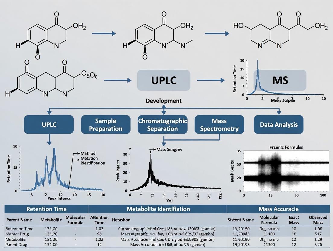
Mastering UPLC-MS/MS Method Development for Drug Metabolite Analysis: A Comprehensive Guide for Pharmaceutical Researchers
This comprehensive article provides a strategic framework for developing robust Ultra-High Performance Liquid Chromatography coupled with Tandem Mass Spectrometry (UPLC-MS/MS) methods specifically tailored for the identification and quantification of drug...
Mastering UPLC-MS/MS Method Development for Drug Metabolite Analysis: A Comprehensive Guide for Pharmaceutical Researchers
Abstract
This comprehensive article provides a strategic framework for developing robust Ultra-High Performance Liquid Chromatography coupled with Tandem Mass Spectrometry (UPLC-MS/MS) methods specifically tailored for the identification and quantification of drug metabolites. Targeting drug development scientists and analytical researchers, it systematically addresses the entire workflow. The scope covers the foundational principles of metabolite analysis, detailed methodology for UPLC and MS optimization, troubleshooting for complex matrices and challenging analytes, and the critical process of method validation and comparison with alternative techniques. The guide synthesizes current best practices to enable the creation of sensitive, selective, and reliable analytical methods crucial for pharmacokinetics, toxicology, and regulatory submissions.
Why Metabolite Analysis Matters: Core Principles and Strategic Goals for UPLC-MS/MS
The Critical Role of Metabolite Profiling in Drug Discovery and Development
Metabolite profiling, integral to a broader thesis on UPLC-MS method development, is a cornerstone of modern drug discovery and development. It provides critical data on the biotransformation of drug candidates, informing safety, efficacy, and pharmacokinetic profiles. This document outlines application notes and detailed protocols for employing Ultra-Performance Liquid Chromatography-Mass Spectrometry (UPLC-MS) in metabolite identification and quantification.
Application Note 1: Early-Stage Metabolite Identification
Objective: To identify major and minor metabolites of a new chemical entity (NCE) following in vitro incubation with hepatocytes.
Quantitative Data Summary: Table 1: Typical Metabolite Formation in Human Hepatocyte Incubations (10 µM NCE, 2 hr)
| Metabolite ID | Retention Time (min) | [M+H]+ (m/z) | Relative Abundance (%) | Metabolic Reaction |
|---|---|---|---|---|
| NCE (Parent) | 8.7 | 345.1567 | 100.0 | - |
| M1 | 5.2 | 361.1516 | 45.3 | Hydroxylation |
| M2 | 6.1 | 319.1461 | 22.1 | N-Dealkylation |
| M3 | 4.8 | 477.1790 | 8.7 | Glucuronidation |
| M4 | 7.3 | 329.1255 | 3.2 | Oxidative Deamination |
Protocol 1.1: Hepatocyte Incubation and Sample Preparation
- Thaw and Viability Check: Rapidly thaw cryopreserved human hepatocytes. Assess viability via trypan blue exclusion (>80% required).
- Incubation: Suspend hepatocytes (1 million cells/mL) in Williams' E medium. Pre-incubate at 37°C, 5% CO₂ for 15 min. Add NCE from 10 mM DMSO stock for a final concentration of 10 µM (0.1% DMSO v/v). Incubate for 0, 30, 60, 120 min.
- Reaction Termination: At each time point, transfer 100 µL aliquot to 300 µL of ice-cold acetonitrile containing internal standard (e.g., Tolbutamide-d9).
- Sample Processing: Vortex for 5 min, centrifuge at 14,000 g for 15 min (4°C). Transfer supernatant to a fresh tube, evaporate to dryness under nitrogen stream.
- Reconstitution: Reconstitute dry residue in 100 µL of initial mobile phase (95:5 Water:Acetonitrile + 0.1% Formic acid), vortex, centrifuge. Transfer to LC vial for analysis.
Protocol 1.2: UPLC-HRMS Analysis for Metabolite Identification
- System: UPLC coupled to high-resolution mass spectrometer (e.g., Q-TOF or Orbitrap).
- Column: C18 column (e.g., 2.1 x 100 mm, 1.7 µm).
- Mobile Phase: A: 0.1% Formic acid in Water; B: 0.1% Formic acid in Acetonitrile.
- Gradient: 5% B to 95% B over 12 min, hold 2 min, re-equilibrate.
- MS Parameters: ESI+ mode. Data Dependent Acquisition (DDA): Full scan (m/z 100-1000) at 70,000 resolution, followed by MS/MS scans on top 5 ions at 17,500 resolution. Collision energy: stepped 20, 40 eV.
- Data Analysis: Use software (e.g., Compound Discoverer, Metabolynx) to find metabolites via mass defect filter, product ion filtering, and comparison to control samples.
Diagram Title: In Vitro Metabolite ID Workflow
Application Note 2: Quantitative Metabolite Profiling in Preclinical Studies
Objective: To quantify circulating metabolites in plasma from a rat pharmacokinetic study to assess systemic exposure.
Quantitative Data Summary: Table 2: Pharmacokinetic Parameters for NCE and Major Metabolite M1 in Rat (10 mg/kg oral dose, n=3)
| Analyte | Cₘₐₓ (ng/mL) | Tₘₐₓ (h) | AUC₀–₂₄ (ng·h/mL) | Half-life (h) | % of Parent AUC |
|---|---|---|---|---|---|
| NCE | 520 ± 45 | 1.5 | 2450 ± 310 | 3.2 | 100 |
| M1 | 185 ± 22 | 2.0 | 1120 ± 150 | 4.8 | 45.7 |
Protocol 2.1: Bioanalytical Method for Plasma Quantification
- Calibrators & QCs: Prepare spiked plasma calibration standards (1-1000 ng/mL) and quality controls (Low, Mid, High).
- Sample Extraction: To 50 µL of rat plasma, add 10 µL of internal standard working solution (structural analog in ACN). Precipitate proteins with 150 µL of acetonitrile. Vortex 10 min, centrifuge at 14,000 g for 15 min.
- Analysis: Inject supernatant (diluted 1:1 with water) onto UPLC-MS/MS.
- UPLC: Similar gradient as Protocol 1.2, but total run time of 5 min.
- MS/MS: Operate in MRM mode. Optimized transitions: NCE: 345.2→128.1; M1: 361.2→144.1; IS: 350.2→133.1.
- Quantification: Use linear regression (1/x² weighting) of peak area ratios (analyte/IS) vs. concentration.
Diagram Title: Preclinical PK Study Pathway
The Scientist's Toolkit: Key Research Reagent Solutions
Table 3: Essential Materials for Metabolite Profiling Studies
| Item | Function & Rationale |
|---|---|
| Cryopreserved Hepatocytes (Human/Rat) | In vitro metabolic system representing phase I/II enzymes. Preferred over microsomes for comprehensive profiling. |
| Stable Isotope-Labeled Drug (¹³C, ²H) | Internal standard for quantification; aids in tracking metabolite origins in complex matrices. |
| β-Glucuronidase / Arylsulfatase | Enzymes for hydrolysis of conjugate metabolites (glucuronides/sulfates) to confirm identity. |
| Chemical Inhibitors (e.g., 1-Aminobenzotriazole) | To probe specific enzyme contributions (e.g., CYP450) to metabolite formation. |
| Authentic Metabolite Standards (when available) | For definitive confirmation of identity and for generating calibration curves for quantification. |
| Stable LC Columns (e.g., BEH C18) | Provides high-resolution, reproducible separation of complex metabolite mixtures. |
| Mobile Phase Additives (Formic Acid, Ammonium Acetate) | To optimize ionization in positive and negative ESI modes, respectively. |
Application Note 3: Reactive Metabolite Screening
Objective: To screen for the formation of reactive, potentially toxic metabolites via glutathione (GSH) trapping assays.
Protocol 3.1: Microsomal Incubation with Trapping Agents
- Incubation Mix: Combine liver microsomes (1 mg/mL), NCE (10 µM), NADPH (1 mM), and GSH (5 mM) in phosphate buffer (pH 7.4).
- Control: Include a control without NADPH.
- Incubate: 37°C for 60 min. Terminate with cold acetonitrile.
- Analysis: Use neutral loss scan of 129 Da (pyroglutamic acid) or precursor ion scan of m/z 272 (GSH moiety) on a triple quadrupole MS, or use HRMS to look for accurate masses of GSH adducts (+305.0682 Da).
Diagram Title: Reactive Metabolite Screening Pathway
Integrated UPLC-MS-based metabolite profiling protocols are non-negotiable for de-risking drug development. They enable a systematic transition from qualitative identification in discovery to robust quantitative assays in development, directly supporting the thesis that advanced method development is critical for understanding the complete metabolic fate of drug candidates.
Application Notes: The Scope of the Challenge
The comprehensive characterization of drug metabolites is a critical pillar of modern drug development, directly impacting safety and efficacy assessments. The core analytical challenge stems from the vast physicochemical diversity introduced by metabolic transformations, which UPLC-MS must reliably capture within a single analytical method.
Table 1: Key Biotransformations and Their Impact on Analyte Properties
| Biotransformation Type | Example | Common Effect on LogP | Common Effect on MS Response (ESI+) | Key Analytical Implication |
|---|---|---|---|---|
| Phase I: Functionalization | Aliphatic/ Aromatic Hydroxylation | Decrease (~0.5-1.0) | Variable; may increase [M+H]+ | Co-elution with parent; requires high-resolution MS/MS. |
| N-/O-Dealkylation | Decrease (if polar group exposed) | Often similar to parent | Mass shift diagnostic; may yield same product ion. | |
| Oxidation to Carboxylic Acid | Significant Decrease (~2.0+) | Often suppressed in (+); enhanced in (-) | Requires negative ion mode screening; poor retention on C18. | |
| Phase II: Conjugation | Glucuronidation | Significant Decrease (~3.0+) | Often good [M+H]+ & [M+NH4]+; may in-source fragment | Chromatographic tailing; thermally labile; check for acyl glucuronides. |
| Sulfation | Significant Decrease (~2.5+) | Better in (-) mode; may cleave in-source | Can be isobaric with glucuronides; requires MS/MS for distinction. | |
| Glutathione (GSH) Conjugation | Decrease (variable) | Characteristic neutral losses (129, 275 Da) | Often early eluting; requires specialized MS scans (NL, PI). |
Table 2: Quantitative Impact of Metabolite Polarity on UPLC Retention (C18 Column)
| Metabolite Class | Approximate ΔtR (vs. Parent Drug)* | Recommended LC Modifier | Rationale |
|---|---|---|---|
| Parent Drug (LogP ~3-5) | Reference | Formic Acid (0.1%) | Standard for positive ion mode, good ionization. |
| Hydroxylated Metabolites | -0.5 to -1.5 min | Formic Acid or Ammonium Formate (5-10 mM) | Maintains retention and peak shape for moderate polarity. |
| Carboxylic Acids | -2.0 to -4.0 min | Ammonium Formate/Acetate Buffer (pH ~5) | Suppresses ionization, enhancing retention on C18. |
| Glucuronides/Sulfates | -1.5 to -3.5 min | Ammonium Acetate/Carbonate (pH ~8 for glucuronides) | Can improve peak shape and sensitivity for anions. |
*ΔtR is illustrative; actual shift depends on specific chemistry and gradient.
Experimental Protocols
Protocol 1: Generic UPLC-MS Method for Broad Metabolite Screening
Objective: To develop a robust, high-resolution UPLC-HRMS method for untargeted detection of diverse drug metabolites in biological matrices (e.g., hepatocyte incubations, plasma).
I. Materials & Equipment (Research Reagent Solutions)
- UPLC System: Equipped with binary pump, autosampler (maintained at 4°C), and column oven.
- Mass Spectrometer: High-resolution accurate mass (HRAM) instrument (e.g., Q-TOF, Orbitrap) with electrospray ionization (ESI).
- Analytical Column: Acquity UPLC HSS T3 (2.1 x 100 mm, 1.8 µm) or equivalent. Function: Provides retention for polar compounds.
- Mobile Phase A: 10 mM Ammonium Formate in Water, pH 3.0 (adjusted with formic acid). Function: Aqueous buffer for pH control and ion pairing.
- Mobile Phase B: 10 mM Ammonium Formate in 90:10 Acetonitrile:Water, pH 3.0. Function: Organic buffer to maintain consistent ionization.
- Precipitation Solvent: Acetonitrile with 0.1% Formic Acid (3:1 v/v vs. sample). Function: Protein precipitation and metabolite extraction.
- Reference Lockmass Solution: Leucine Enkephalin (250 pg/µL in 50:50 ACN:H2O, 0.1% FA) or equivalent. Function: Real-time mass axis calibration.
II. Detailed Procedure
A. Sample Preparation:
- Thaw biological matrix (e.g., hepatocyte incubation supernatant) on ice.
- Vortex thoroughly for 30 seconds.
- Aliquot 100 µL of sample into a microcentrifuge tube.
- Add 300 µL of ice-cold Precipitation Solvent.
- Vortex vigorously for 2 minutes.
- Centrifuge at 14,000 x g for 15 minutes at 4°C.
- Transfer 200 µL of the clear supernatant to a fresh LC vial with insert.
B. UPLC Conditions:
- Column Temperature: 45°C
- Injection Volume: 5-10 µL
- Flow Rate: 0.45 mL/min
- Gradient Program:
Time (min) %A %B 0.0 99 1 1.0 99 1 12.0 40 60 13.0 5 95 15.0 5 95 15.1 99 1 18.0 99 1
C. MS Conditions (ESI Positive/Negative Switching):
- Source Temperature: 150°C
- Desolvation Temperature: 500°C
- Capillary Voltage: ±0.8 kV (positive/negative)
- Cone Voltage: 40 V (ramp 20-50 V for in-source fragmentation insight)
- Desolvation Gas Flow: 800 L/hr
- Scan Range: m/z 100-1000
- Scan Time: 0.25 sec
- Lockmass Spray: Continuously infused via reference probe at 10 µL/min.
- Data Acquisition: MSE or DIA mode (low/high collision energy).
III. Data Analysis Workflow:
- Use software (e.g., UNIFI, Compound Discoverer, XCMS) for peak picking, alignment, and background subtraction.
- Apply mass defect filter (±50 mDa from parent drug).
- Search for predicted biotransformations (common Phase I/II).
- Review extracted ion chromatograms (EICs) for potential metabolites.
- Confirm identities via MS/MS fragmentation (comparison to parent drug fragmentation patterns).
Protocol 2: Targeted Analysis for Reactive Metabolite Screening (GSH Adducts)
Objective: To selectively detect and characterize glutathione (GSH) conjugates as markers for reactive metabolite formation.
I. Materials:
- All items from Protocol 1, plus:
- GSH Trapping Agent: 5 mM Glutathione in incubation buffer. Function: Nucleophilic trap for electrophilic reactive metabolites.
- Neutral Loss Scanning (NLS) Solution: Custom tune mix for optimizing NLS parameters. Function: Method development for diagnostic scans.
II. Modified Procedure:
- Prepare hepatocyte incubations containing the test compound and 5 mM GSH.
- Follow Protocol 1, Step A for sample preparation.
- UPLC: Use gradient from Protocol 1. GSH conjugates typically elute early (2-6 min).
- MS Analysis (Positive Ion Mode):
- Perform Precursor Ion (PI) Scan of m/z 129 (pyroglutamic acid moiety).
- Perform Neutral Loss (NL) Scan of 129 Da (loss of pyroglutamic acid).
- Alternatively, in HRAM MS/MS, use an inclusion list of potential GSH adduct masses ([M+H]^+ = Drug + 307 Da (GSH - 2H) or +129 Da (GSH - Glu-Gly)).
Mandatory Visualization
Title: Drug Metabolism Pathways & Property Changes
Title: Generic Metabolite Screening UPLC-MS Workflow
Within the broader thesis on UPLC-MS method development for drug metabolism research, this document details specific applications and protocols that leverage the core advantages of Ultra-Performance Liquid Chromatography coupled with Tandem Mass Spectrometry (UPLC-MS/MS). The platform's superior speed, sensitivity, and resolution are critical for identifying and quantifying low-abundance metabolites in complex biological matrices.
Application Notes
The integration of UPLC with MS/MS provides transformative capabilities in metabolite identification and quantification.
Note 1: High-Throughput Pharmacokinetic Screening UPLC-MS/MS enables the rapid analysis of drug metabolites from hundreds of plasma samples per day. The use of sub-2µm particle columns reduces chromatographic run times to 2-5 minutes without compromising separation, accelerating critical decisions in lead optimization.
Note 2: Identification of Low-Abundance Reactive Metabolites The exceptional sensitivity of modern triple quadrupole and high-resolution MS/MS detectors allows for the detection of reactive, potentially toxic metabolites present at picogram-per-milliliter levels. This is paramount for comprehensive safety assessment.
Note 3: Resolving Isobaric and Structural Isomers The high chromatographic resolution of UPLC is essential for separating isobaric metabolites that have identical mass but different structures. This prevents misidentification and ensures accurate metabolic pathway elucidation.
Quantitative Performance Data
Table 1: Comparative Performance of UPLC-MS/MS vs. HPLC-MS/MS for Metabolite Analysis
| Performance Metric | Typical UPLC-MS/MS | Typical HPLC-MS/MS |
|---|---|---|
| Analysis Time | 2-5 min | 15-30 min |
| Peak Capacity | 200-400 | 50-150 |
| Theoretical Plates | >200,000/m | ~100,000/m |
| Typical LOQ (in matrix) | 0.1-1 pg/mL | 1-10 pg/mL |
| Sample Consumption | 1-5 µL | 10-50 µL |
Table 2: Key MS/MS Scan Modes for Metabolite Research
| Scan Mode | Primary Function | Key Application in Metabolite ID |
|---|---|---|
| Full Scan (HRMS) | Accurate mass measurement | Untargeted screening, elemental composition |
| Product Ion Scan | Fragmentation pattern | Structural elucidation of metabolites |
| Neutral Loss Scan | Detection of specific mass loss | Class-specific metabolite finding (e.g., glucuronides) |
| Precursor Ion Scan | Detection of specific fragment | Finding all metabolites yielding a common fragment |
| Multiple Reaction Monitoring (MRM) | Quantification of target analytes | High-sensitivity PK bioanalysis |
Experimental Protocols
Protocol 1: Generic UPLC-MS/MS Method for Untargeted Metabolite Profiling in Plasma
Objective: To separate and detect a wide range of phase I and phase II drug metabolites.
Materials & Reagents:
- UPLC System: Equipped with a binary pump, cooled autosampler, and column oven.
- MS Detector: Quadrupole time-of-flight (Q-TOF) or high-resolution Q-Orbitrap system.
- Column: C18, 1.7µm, 2.1 x 100 mm.
- Mobile Phase A: 0.1% Formic acid in water.
- Mobile Phase B: 0.1% Formic acid in acetonitrile.
- Sample: Plasma processed via protein precipitation (3:1 acetonitrile:plasma).
Procedure:
- Sample Preparation: Centrifuge protein-precipitated plasma at 14,000 x g for 10 min. Transfer supernatant for analysis.
- Chromatography:
- Flow Rate: 0.4 mL/min.
- Column Temp: 45°C.
- Injection Volume: 5 µL.
- Gradient:
- 0-1 min: 5% B
- 1-10 min: 5% to 95% B (linear)
- 10-12 min: Hold at 95% B
- 12-12.1 min: 95% to 5% B
- 12.1-15 min: Re-equilibrate at 5% B.
- Mass Spectrometry (ESI Positive/Negative Switching):
- Capillary Voltage: 3.0 kV (ESI+), 2.5 kV (ESI-)
- Source Temp: 150°C
- Desolvation Temp: 500°C
- Cone Gas Flow: 50 L/hr
- Desolvation Gas Flow: 800 L/hr
- Scan Range: m/z 50-1200
- Collision Energy: Ramped from 20 to 40 eV for MS/MS.
- Data Analysis: Use metabolomics software for peak picking, alignment, and comparison against control samples and metabolite databases.
Protocol 2: Targeted Quantification of a Specific Metabolite Panel via MRM
Objective: To achieve high-sensitivity, reproducible quantification of known metabolites.
Procedure:
- Method Development:
- Optimize compound-dependent MS parameters (CE, CV) for each metabolite and its stable-isotope-labeled internal standard.
- Establish chromatographic separation to minimize matrix suppression.
- Calibration Standards: Prepare in blank matrix (e.g., plasma). Typical range: 1 pg/mL to 1000 ng/mL.
- UPLC-MRM Method:
- Use a faster, optimized gradient (total run time ~3-5 min).
- Operate the triple quadrupole MS in scheduled MRM mode with a 30-45 sec detection window.
- Dwell times: 10-50 msec per transition.
- Validation: Perform intra- and inter-day accuracy and precision assessments per regulatory guidelines (e.g., FDA bioanalytical method validation).
The Scientist's Toolkit
Table 3: Essential Research Reagent Solutions for UPLC-MS/MS Metabolite Analysis
| Item | Function & Importance |
|---|---|
| Stable Isotope-Labeled Internal Standards (SIL-IS) | Corrects for matrix effects and variability in extraction/ionization; essential for accurate quantification. |
| β-Glucuronidase/Arylsulfatase Enzyme | Enzymatically hydrolyzes phase II conjugates (glucuronides/sulfates) to assess total metabolite levels. |
| Solid Phase Extraction (SPE) Plates (e.g., HLB) | For automated sample clean-up, removing phospholipids and salts to reduce matrix effects. |
| LC-MS Grade Solvents & Additives | Minimizes background noise and system contamination, ensuring sensitivity and reproducibility. |
| Pooled Control Matrix (e.g., Human Plasma) | Used for preparing calibration standards and quality controls in a biologically relevant medium. |
Visualized Workflows and Pathways
Title: UPLC-MS/MS Analytical Workflow for Metabolites
Title: Common Drug Metabolism Pathways
Within the framework of a thesis on UPLC-MS method development for drug metabolism studies, the initial and most critical step is the explicit definition of methodological objectives. The analytical strategy, from sample preparation to data processing, is fundamentally dictated by the choice between targeted quantification and untargeted identification. This document provides detailed application notes and protocols for both approaches, enabling informed decision-making in drug development research.
Targeted Quantification: Application Notes & Protocol
Objective: Precisely measure the concentrations of a predefined set of known analytes (e.g., parent drug and its major metabolites).
Core Principle: Method development focuses on maximizing sensitivity, specificity, and reproducibility for specific targets.
Detailed Protocol: Targeted UPLC-MS/MS Quantification of Drug Metabolites
A. Sample Preparation (Protein Precipitation)
- Thaw biofluid samples (plasma, urine) on ice.
- Aliquot 50 µL of sample into a 1.5 mL microcentrifuge tube.
- Add 150 µL of ice-cold acetonitrile (containing internal standards, e.g., stable isotope-labeled analogs of target analytes).
- Vortex vigorously for 1 minute.
- Centrifuge at 14,000 × g for 10 minutes at 4°C.
- Transfer 150 µL of the supernatant to a clean vial with insert for UPLC-MS/MS analysis.
B. UPLC Conditions (Example)
- Column: C18 (e.g., 2.1 x 50 mm, 1.7 µm).
- Mobile Phase A: 0.1% Formic acid in water.
- Mobile Phase B: 0.1% Formic acid in acetonitrile.
- Gradient: 5% B to 95% B over 3.5 minutes, hold for 1 minute.
- Flow Rate: 0.4 mL/min.
- Column Temp: 40°C.
- Injection Volume: 5 µL.
C. MS/MS Detection (Triple Quadrupole)
- Ionization: Electrospray Ionization (ESI), positive/negative mode as required.
- Data Acquisition: Multiple Reaction Monitoring (MRM).
- Step 1: For each target analyte, infuse standard to optimize precursor ion ([M+H]+ or [M-H]-).
- Step 2: Perform product ion scans to select 2-3 characteristic fragment ions.
- Step 3: Define MRM transition: Precursor Ion > Product Ion (e.g., m/z 322 > 202).
- Step 4: Optimize collision energy (CE) and cone voltage for each MRM transition for maximum signal.
- Step 5: Acquire data using scheduled MRM within specific retention time windows.
D. Data Analysis
- Integrate peak areas for each analyte and its corresponding internal standard.
- Generate a calibration curve using spiked matrix standards (typically 1-1000 ng/mL).
- Calculate analyte concentration using the internal standard method (peak area ratio vs. concentration).
Table 1: Example MRM Transitions and Parameters for a Hypothetical Drug (XYZ-123) and Metabolites
| Analyte | Precursor Ion (m/z) | Product Ion (m/z) | Collision Energy (V) | Retention Time (min) |
|---|---|---|---|---|
| XYZ-123 (Parent) | 322.2 | 202.1* / 134.0 | 22 / 30 | 3.1 |
| XYZ-123-Glucuronide | 498.2 | 322.2 / 202.1 | 18 / 25 | 2.4 |
| XYZ-123-OH (M1) | 338.2 | 218.1 / 165.0 | 20 / 28 | 2.8 |
| Internal Standard | ||||
| XYZ-123-d4 | 326.2 | 206.1 | 22 | 3.1 |
*Quantifier ion
Untargeted Metabolite Identification: Application Notes & Protocol
Objective: Comprehensively detect and identify both expected and novel metabolites without prior knowledge.
Core Principle: Method development focuses on maximizing chromatographic resolution, full-scan sensitivity, and informative fragmentation.
Detailed Protocol: Untargeted UPLC-HRMS for Metabolite Profiling
A. Sample Preparation (Supported Liquid Extraction)
- Load 100 µL of plasma onto a pre-conditioned (methanol, water) SLE plate.
- Allow sample to absorb onto sorbent for 5 minutes.
- Elute metabolites with 2 x 600 µL of methyl tert-butyl ether (MTBE):ethyl acetate (1:1).
- Evaporate eluent to dryness under a gentle nitrogen stream at 40°C.
- Reconstitute dried extract in 100 µL of 10% acetonitrile in water for analysis.
B. UPLC Conditions (High-Resolution)
- Column: HSS T3 or similar (2.1 x 100 mm, 1.8 µm) for broad metabolite polarity.
- Mobile Phase A: 10 mM Ammonium formate, pH 3.0 in water.
- Mobile Phase B: 10 mM Ammonium formate in acetonitrile:isopropanol (9:1).
- Gradient: 1% B to 99% B over 18 minutes.
- Flow Rate: 0.45 mL/min.
- Column Temp: 55°C.
- Injection Volume: 10 µL.
C. HRMS Detection (Q-TOF or Orbitrap)
- Ionization: ESI, alternating positive and negative modes in same run.
- Full Scan Acquisition: m/z 50-1200, resolution > 35,000 (FWHM).
- Data-Dependent Acquisition (DDA/MS²):
- Select top 5 most intense ions per cycle for fragmentation.
- Use stepped normalized collision energy (e.g., 20, 40, 60 eV).
- Dynamic exclusion: 15 seconds.
- Mass Accuracy: Calibrate daily; ensure < 3 ppm error.
D. Data Processing & Identification Workflow
- Use vendor-neutral software (e.g., MS-DIAL, Compound Discoverer) for peak picking, alignment, and deconvolution.
- Generate a list of features (m/z, RT) present in dosed samples but absent/less abundant in controls.
- Predict potential biotransformations (e.g., +O, +Glucuronide, -CH₂) on the parent drug structure.
- Annotate metabolites by matching accurate mass (< 5 ppm) of predicted metabolites.
- Confirm identities by interpreting MS/MS fragmentation patterns against the parent drug spectrum.
Table 2: Comparison of Targeted vs. Untargeted Method Objectives
| Aspect | Targeted Quantification | Untargeted Identification |
|---|---|---|
| Primary Goal | Accurate concentration measurement | Comprehensive metabolite discovery |
| Analytical Focus | Sensitivity, precision, linear range | Broad detection, mass accuracy, fragmentation |
| MS Acquisition | MRM (Triple Quadrupole) | Full Scan + MS² (Q-TOF, Orbitrap) |
| Data Output | Concentrations (ng/mL) | List of annotated features & putative IDs |
| Throughput | High | Moderate |
| Key Validation Parameters | LLOD, LLOQ, Accuracy, Precision | Mass accuracy, isotopic pattern fidelity |
The Scientist's Toolkit: Research Reagent Solutions
| Item | Function in Experiment |
|---|---|
| Stable Isotope-Labeled Internal Standards | Corrects for matrix effects & losses in targeted quantification. |
| Hybrid SPE-MPPT 96-well Plates | Efficient phospholipid removal for cleaner plasma extracts in both methods. |
| HSS T3 UPLC Column | Retains polar metabolites, critical for untargeted profiling. |
| Ammonium Formate (LC-MS Grade) | Provides buffering for stable mobile phase pH, improving peak shape. |
| Leucine Enkephalin (for ESI-TOF) | Provides lock mass correction for sustained high mass accuracy in HRMS. |
| All-in-One Tuning & Calibration Solution | Contains compounds for MS calibration across a broad m/z range (e.g., for Q-TOF). |
| Pooled Control Matrix | Essential for generating quality control samples and blank extracts for background subtraction. |
Method Selection Workflow & Data Analysis Pathways
Diagram 1: Decision Flow for Method Objective Selection
Diagram 2: Untargeted Data Analysis Workflow
Within the comprehensive thesis "Advanced UPLC-MS Method Development for Comprehensive Drug Metabolite Profiling," the initial pre-development phase is paramount. This phase systematically de-risks and informs the subsequent analytical development by leveraging existing knowledge and predictive computational tools. A rigorous literature review establishes the chemical and biological context, while in-silico metabolite prediction provides a targeted list of potential biotransformations to guide mass spectrometric method development. This document details the application notes and protocols for executing this foundational work.
Literature Review: Protocol and Application Notes
Protocol 1.1: Structured Literature Mining for Metabolic Pathways
Objective: To systematically identify and collate known metabolic pathways, enzymes involved, and analytical conditions for the parent drug and its structural analogues.
Methodology:
- Database Selection & Search String Formulation:
- Utilize primary databases: PubMed/MEDLINE, Scopus, Web of Science, and Google Scholar.
- Search strings combine: Drug Name (AND) [Metabolite* OR Metabolism OR Biotransformation OR "in vivo" OR "in vitro"] (AND) [LC-MS OR UPLC-MS OR Mass Spectrometry].
- Include Chemical Abstracts Service (CAS) registry number for precise identification.
- Search for key metabolizing enzymes (e.g., CYP450 isoforms like CYP3A4, CYP2D6; UGTs).
Data Extraction and Synthesis:
- Use reference management software (e.g., EndNote, Zotero) to deduplicate and organize results.
- Extract data into a structured template. Key fields include: metabolite structure, biotransformation type (e.g., oxidation, glucuronidation), enzyme system responsible, biological matrix, analytical technique used, and cited reference.
- Prioritize recent publications (last 10 years) for analytical techniques.
Critical Analysis and Gap Identification:
- Compare metabolic pathways across species (human, rat, dog) to anticipate in-vitro/in-vivo study relevance.
- Note any discrepancies in reported major vs. minor metabolites.
- Identify gaps where metabolism for the specific drug or analogue is poorly characterized.
Table 1: Summarized Literature Data on Metabolic Pathways for [Hypothetical Drug: "Xenobiol"]
| Metabolite ID | Biotransformation | Primary Enzyme Responsible | Reported Abundance (Species) | Key Analytical Reference (Method) |
|---|---|---|---|---|
| M1 | Aliphatic Hydroxylation | CYP3A4 | Major (Human, Rat) | Smith et al. (2022), Anal. Chem., UPLC-QTOF-MS |
| M2 | N-Dealkylation | CYP2C19 | Minor (Human) | Jones et al. (2020), J. Chrom. B, HPLC-MS/MS |
| M3 | Direct Glucuronidation | UGT1A9 | Major (Human) | Chen et al. (2023), Drug Metab. Dispos., HILIC-MS/MS |
| M4 | Oxidative Deamination | MAO-A / Aldehyde Oxidase | Trace (Rat) | Patel et al. (2021), Xenobiotica, HRMS |
Title: Workflow for Literature Review to Guide Hypothesis Generation
In-Silico Metabolite Prediction: Protocol and Application Notes
Protocol 2.1: Multi-Software Metabolite Prediction and Consensus Analysis
Objective: To generate a comprehensive and ranked list of probable and plausible metabolites using computational tools.
Methodology:
- Software Suite Selection:
- Employ a minimum of two prediction engines based on different algorithms (e.g., rule-based, machine learning).
- Recommended Tools: Schrödinger's BioLuminate (Meteor Nexus), GLORYx (rule-based/ML hybrid), ADMET Predictor (Simulations Plus).
Input Preparation and Prediction Execution:
- Prepare the parent drug structure in SMILES or SDF format. Ensure correct tautomeric and protonation states.
- Run Parameters: Set parameters to predict Phase I (oxidations, reductions, hydrolyses) and Phase II (conjugations) metabolites.
- Define constraints: likelihood threshold (e.g., "probable" and above), maximum number of transformation steps (e.g., 2).
Data Consolidation and Ranking:
- Export results from all software (Metabolite ID, Transformation, Predicted Likelihood/Score).
- Create a consensus list. Metabolites predicted by multiple tools are given higher priority.
- Rank metabolites by combining software scores and literature prevalence (from Protocol 1.1).
Table 2: Consolidated In-Silico Prediction Results for "Xenobiol"
| Predicted Metabolite | Biotransformation | Software A Score (Meteor) | Software B Score (GLORYx) | Consensus Priority | Corroborated in Literature? |
|---|---|---|---|---|---|
| M1 | Aliphatic C-Hydroxylation | Probable (78%) | Very Likely (0.89) | High (1) | Yes |
| M5 | Aromatic Hydroxylation | Probable (65%) | Likely (0.76) | High (2) | No (Novel Prediction) |
| M2 | N-Dealkylation | Probable (72%) | Likely (0.71) | Medium (3) | Yes |
| M6 | Sulfation | Plausible (45%) | Possible (0.41) | Low (4) | No |
Title: Consensus Approach for In-Silico Metabolite Prediction
The Scientist's Toolkit: Research Reagent Solutions & Essential Materials
Table 3: Essential Resources for Pre-Development Work
| Item / Solution | Function / Application | Example Vendor/Resource |
|---|---|---|
| Chemical Database | Provides accurate parent drug and known metabolite structures, physicochemical properties. | PubChem, ChemSpider |
| Reference Management Software | Organizes literature citations, PDFs, and enables de-duplication. | EndNote, Zotero, Mendeley |
| Metabolite Prediction Software | Computationally generates potential metabolite structures based on biotransformation rules. | Meteor Nexus (Lhasa), GLORYx, ADMET Predictor |
| Metabolism-Oriented Database | Curated knowledge on enzyme-specific metabolism, drug interactions, and pathways. | BioCyc, Human Metabolome Database (HMDB) |
| Chemical Structure Drawing/Editing Tool | Creates, edits, and exports chemical structures in standard formats (SDF, SMILES). | ChemDraw, MarvinSketch |
| Liver Microsomes / Hepatocytes (Human & Preclinical) | In-vitro systems to validate predictions; used in the next phase after pre-development. | Corning, Thermo Fisher, BioIVT |
| Mass Spectrometry Fragment Prediction Tool | Assists in predicting MS/MS fragmentation patterns for predicted metabolites (post-prediction). | Mass Frontier, CFM-ID |
Building Your Method: A Step-by-Step UPLC-MS/MS Workflow for Metabolites
In the development of UPLC-MS methods for drug metabolite research, sample preparation is the critical first step that dictates the success of downstream analysis. The complexity of biological matrices like plasma, urine, and tissue presents significant challenges, including matrix effects, endogenous interferences, and the wide dynamic range of analyte concentrations. This application note details contemporary, robust strategies for preparing these matrices to ensure optimal recovery, reproducibility, and MS compatibility for metabolite identification and quantification.
Plasma/Serum Preparation Strategies
Plasma and serum are central matrices for pharmacokinetic studies. Key challenges include high protein content and phospholipid-induced matrix effects.
Protein Precipitation (PPT)
A rapid, high-throughput method for removing proteins.
- Protocol: To 100 µL of plasma, add 300 µL of ice-cold acetonitrile (with 0.1% formic acid for basic analytes or 0.1% ammonia for acidic analytes). Vortex vigorously for 1 minute, centrifuge at 15,000 x g for 10 minutes at 4°C. Transfer the supernatant to a new tube for evaporation or direct injection. Optionally, perform a double precipitation for cleaner extracts.
- Considerations: Simple but can co-precipitate analytes and leaves phospholipids.
Liquid-Liquid Extraction (LLE)
Effective for broad analyte classes and offers clean extracts.
- Protocol: Acidify 200 µL of plasma with 20 µL of 1M HCl. Add 1 mL of ethyl acetate:methyl tert-butyl ether (1:1, v/v). Vortex for 5 minutes, centrifuge at 5000 x g for 5 minutes. Snap-freeze the aqueous layer in a dry ice/acetone bath and decant the organic layer. Evaporate under nitrogen and reconstitute in mobile phase.
Solid-Phase Extraction (SPE)
Provides selective cleanup and concentration. Mixed-mode sorbents (e.g., Oasis MCX, WCX) are ideal for metabolites.
- Protocol (Mixed-Mode Cation Exchange, MCX):
- Condition: 1 mL methanol, then 1 mL water.
- Load: Acidified plasma sample.
- Wash 1: 1 mL 0.1M HCl.
- Wash 2: 1 mL methanol.
- Elute: 2 x 1 mL of 5% ammonia in methanol. Evaporate eluent and reconstitute.
Quantitative Comparison of Plasma Prep Methods
Table 1: Comparison of Key Plasma Preparation Techniques
| Method | Protein Removal Efficiency | Phospholipid Removal Efficiency | Typical Recovery Range (%) | Throughput | Best For |
|---|---|---|---|---|---|
| Protein Precipitation | >95% | Low (<20%) | 60-90 | Very High | Rapid screening, high-throughput assays |
| Liquid-Liquid Extraction | High (>98%) | Moderate-High (70-90%) | 70-100 | Moderate | Lipophilic metabolites, reduced matrix effects |
| Solid-Phase Extraction | High (>99%) | High (>90% with selective sorbents) | 80-105 | Low-Moderate | Targeted metabolite profiling, complex samples |
Title: Strategic Selection of Plasma Sample Prep Methods
Urine Preparation Strategies
Urine contains fewer proteins but has high salt and urea content, and analytes are often conjugated (glucuronides, sulfates).
Dilution and Shoot
Applicable for high-abundance metabolites.
- Protocol: Dilute urine 1:5 or 1:10 with a water:methanol (90:10) mixture containing 0.1% formic acid. Vortex, centrifuge at 15,000 x g for 5 minutes, and inject the supernatant.
Enzymatic Hydrolysis
Crucial for quantifying total (free + conjugated) metabolite levels.
- Protocol (β-Glucuronidase/Arylsulfatase): Adjust 500 µL of urine to pH 5.0 with 0.2M acetate buffer. Add 10 µL of Helix pomatia enzyme preparation. Incubate at 37°C for 2 hours (or overnight for complete hydrolysis). Stop reaction by placing on ice, then proceed with SPE or LLE.
Supported Liquid Extraction (SLE)
A modern alternative to LLE, offering high recovery with minimal emulsion formation.
- Protocol: Load diluted/acidified urine onto diatomaceous earth SLE columns. Allow 5-10 minutes for adsorption. Elute analytes with 2 x 1 mL of an organic solvent (e.g., MTBE:ethyl acetate). Evaporate and reconstitute.
Tissue Preparation Strategies
Tissue analysis requires homogenization and often more exhaustive extraction to release intracellular metabolites.
Bead-Based Homogenization
The gold standard for efficient tissue disruption.
- Protocol: Weigh ~50 mg of snap-frozen tissue into a tube with ceramic beads. Add 500 µL of a cold methanol:water (80:20) extraction solvent. Homogenize in a bead mill at 4°C for two 45-second cycles. Centrifuge at 14,000 x g for 15 minutes at 4°C. Collect supernatant. The pellet can be re-extracted for comprehensive recovery.
Quenching and Extraction for Metabolomics
Aims to preserve the in vivo metabolic profile.
- Protocol (Two-Phase Methanol/Chloroform/Water Extraction):
- Add 400 µL of ice-cold methanol and 85 µL of water to homogenized tissue. Vortex.
- Add 200 µL of chloroform, vortex for 2 minutes.
- Add 200 µL of chloroform, then 200 µL of water, vortexing after each addition.
- Centrifuge at 14,000 x g for 15 minutes at 4°C. The upper aqueous phase (polar metabolites) and lower organic phase (lipids) are collected separately.
Workflow for Complex Tissue Analysis
Title: Tissue Metabolite Extraction and Prep Workflow
The Scientist's Toolkit: Essential Research Reagent Solutions
Table 2: Key Reagents and Materials for Sample Preparation
| Item | Primary Function & Rationale |
|---|---|
| Acetonitrile (LC-MS Grade) | Primary solvent for protein precipitation. High volatility and MS purity minimize background interference. |
| Methanol (LC-MS Grade) | Extraction solvent for metabolites; used in SPE conditioning and LLE. |
| Methyl tert-Butyl Ether (MTBE) | Organic solvent for LLE/SLE; excellent for lipid removal and recovery of mid-to-non-polar metabolites. |
| Oasis HLB/MCX/WCX SPE Cartridges | Mixed-mode sorbents for selective retention of acidic, basic, or neutral metabolites, providing superior cleanup. |
| β-Glucuronidase/Arylsulfatase (H. pomatia) | Enzyme cocktail for hydrolyzing Phase II glucuronide and sulfate conjugates to measure total metabolite levels. |
| Ceramic Beads (1.4mm, 2.8mm) | Inert, durable beads for mechanical tissue disruption in bead mill homogenizers. |
| Formic Acid / Ammonium Hydroxide (LC-MS Grade) | Used to adjust pH for analyte stability, improve ionization efficiency, and control SPE retention/elution. |
| Phospholipid Removal Plates (e.g., HybridSPE-PPT) | Specialized plates that selectively bind phospholipids post-PPT, drastically reducing matrix effects in plasma. |
General Protocol: Integrated SPE Workflow for Plasma Metabolite Profiling
This protocol is optimized for recovering a wide range of drug metabolites from plasma.
Materials: Oasis HLB µElution Plate (30 µm), vacuum manifold, appropriate solvents.
- Conditioning: Add 200 µL methanol to each well. Apply gentle vacuum (~5 in Hg) to draw through. Add 200 µL water, draw through. Do not let wells run dry.
- Sample Load: Acidify 100 µL of plasma with 100 µL of 0.1% formic acid in water. Load entire volume onto the conditioned well. Draw through slowly (~1-2 in Hg).
- Wash: Wash with 200 µL of 5% methanol in water. Draw through completely.
- Elution: Place a clean collection plate underneath. Elute metabolites with 2 x 25 µL of 80:20 acetonitrile:methanol (with 0.1% formic acid). Apply vacuum gently for the first elution, then use positive pressure (syringe) for the second to maximize recovery.
- Reconstitution: Add 50 µL of water to the collected eluent (final volume ~100 µL). Vortex gently. The sample is ready for UPLC-MS injection.
Selecting the optimal sample preparation strategy is contingent on the matrix, the physicochemical properties of the target drug metabolites, and the analytical goals of the study. While PPT offers speed, and LLE provides cleanliness, modern SPE and SLE techniques deliver the selectivity and sensitivity required for robust UPLC-MS method development in drug metabolism research. Integrating these Phase 1 strategies effectively minimizes matrix effects and is foundational for generating high-quality, reproducible metabolite data.
Application Notes
Within the broader thesis on UPLC-MS method development for drug metabolite research, Phase 2 focuses on optimizing chromatographic resolution and peak capacity. This is critical for separating structurally similar phase I and phase II metabolites, which is a prerequisite for accurate mass spectrometric identification and quantification. The core optimization triad consists of column chemistry selection, mobile phase composition, and gradient elution profile.
1. Column Chemistry Selection: The choice of stationary phase dictates the primary interaction mechanism with analytes. For metabolites, which range from polar hydroxylated species to non-polar parent drugs, reversed-phase chemistry is standard. The sub-2µm particle size in UPLC provides high efficiency, but the surface chemistry must be tailored.
- C18: The workhorse for most moderate to non-polar compounds.
- Phenyl-Hexyl or Biphenyl: Offers π-π interactions beneficial for separating aromatic metabolites and isomers.
- HILIC (Hydrophilic Interaction Liquid Chromatography): Essential for retaining highly polar metabolites (e.g., glucuronides, N-oxides) that elute near the void volume on reversed-phase columns.
- Chiral Columns: Necessary if researching stereoselective metabolism.
2. Mobile Phase Optimization: The aqueous and organic solvents, along with additives, control selectivity, efficiency, and MS compatibility.
- pH: A critical parameter. Modifying pH (typically between 2.5 and 4.0 for acidic analytes, or 6.0-8.0 for basic analytes using compatible buffers) can dramatically alter the ionization state of metabolites and their interaction with the stationary phase, improving resolution.
- Buffer Strength: 5-20 mM concentrations of ammonium formate or acetate are standard for MS compatibility, providing adequate buffering capacity without causing ion suppression or source contamination.
- Organic Modifier: Acetonitrile is preferred over methanol for UPLC due to its lower viscosity, resulting in lower backpressure and higher efficiency.
3. Gradient Elution Design: A well-designed gradient is paramount for separating a complex metabolite mixture with a wide polarity range.
- Initial %B: Should be low enough to retain early-eluting polar metabolites.
- Gradient Steepness (%B/min): Optimized to balance resolution and run time. Shallower gradients around expected critical metabolite pairs enhance resolution.
- Final %B and Equilibration: Sufficient column re-equilibration (3-5 column volumes) is non-negotiable for retention time stability in a sequence.
Table 1: Impact of Stationary Phase on Key Metabolite Pair Resolution (Rs)
| Metabolite Pair | C18 | Phenyl-Hexyl | HILIC | Notes |
|---|---|---|---|---|
| Hydroxyl-Parent / Parent Drug | 1.5 | 2.1 | N/A | Phenyl phase improves Rs via π-π interaction. |
| Glucuronide / Sulfate | 0.8 (co-elution) | 1.0 | 2.5 | HILIC is essential for resolving these polar conjugates. |
| Diastereomer A / B | 0.9 | 1.8 | N/A | Aromatic interactions on phenyl phase separate isomers. |
Table 2: Effect of Mobile Phase pH on Retention Time (tR) and Peak Shape (Asymmetry Factor, As)
| Metabolite (pKa) | pH 2.7 | pH 3.5 | pH 6.8 | Optimal Condition |
|---|---|---|---|---|
| Acidic Metabolite (4.2) | tR: 5.2 min, As: 1.0 | tR: 8.1 min, As: 1.0 | tR: 3.1 min, As: 1.8 | pH 3.5 (protonated, better retention) |
| Basic Metabolite (9.5) | tR: 3.0 min, As: 1.9 | tR: 3.5 min, As: 1.5 | tR: 9.5 min, As: 1.1 | pH 6.8 (deionized, better peak shape) |
Table 3: Gradient Optimization for a Complex Metabolite Profile
| Gradient Parameter | Method A | Method B (Optimized) | Impact |
|---|---|---|---|
| Initial %B (Acetonitrile) | 5% | 2% | Improved retention of early polar metabolites. |
| Gradient Time | 10 min | 15 min | Increased peak capacity, Rs improved by >30% for mid-eluting cluster. |
| Gradient Shape | Linear | Linear with 2 min isocratic hold at 15% B | Resolved critical pair (Rs from 1.0 to 1.8). |
| Equilibration Time | 1.0 min | 2.5 min | Retention time stability (RSD < 0.1%). |
Experimental Protocols
Protocol 1: Systematic Screening of Column Chemistries Objective: To identify the stationary phase providing the best resolution for target metabolite pairs. Materials: See "Scientist's Toolkit" below. Procedure:
- Prepare a standard mixture of the parent drug and its known metabolites in an appropriate solvent (e.g., 50:50 water:acetonitrile).
- Install the first column (e.g., C18) on the UPLC-MS system. Equilibrate with 95% Mobile Phase A (0.1% Formic acid in water), 5% Mobile Phase B (0.1% Formic acid in acetonitrile).
- Inject the standard mixture. Run a generic gradient from 5% to 95% B over 10 minutes at 0.4 mL/min. Column temperature: 40°C.
- Record retention times, peak widths, and calculate resolution (Rs) between all adjacent peaks of interest.
- Repeat steps 2-4 with different column chemistries (e.g., Phenyl-Hexyl, HILIC). For HILIC, equilibrate with 95% B and run a gradient from 95% to 50% B.
- Tabulate results as in Table 1 and select the best-performing column for further optimization.
Protocol 2: Optimization of Mobile Phase pH and Buffer Strength Objective: To fine-tune selectivity and peak shape for ionizable metabolites. Materials: Ammonium formate (or acetate), formic acid, ammonium hydroxide, UPLC-MS compatible vials. Procedure:
- Prepare three sets of Mobile Phase A buffers: i) 10 mM Ammonium Formate, pH 2.7 (adjusted with formic acid), ii) 10 mM Ammonium Formate, pH 3.5, iii) 10 mM Ammonium Formate, pH 6.8 (adjusted with ammonium hydroxide). Use acetonitrile with 0.1% formic acid as Mobile Phase B.
- Using the selected column from Protocol 1, perform three consecutive injections of the metabolite standard with the generic gradient, using each pH condition.
- Analyze the chromatograms. Note the shift in retention order, changes in resolution, and measure peak asymmetry (As) at 10% peak height. Record data as in Table 2.
- (Optional) Repeat with a different buffer strength (e.g., 5 mM and 20 mM) at the optimal pH to assess impact on signal intensity and peak shape.
Protocol 3: Design and Refinement of Gradient Elution Profile Objective: To achieve baseline resolution of all critical metabolite pairs in the shortest possible runtime. Materials: UPLC system with method development software, metabolite standard. Procedure:
- Using the optimal column and mobile phase from Protocols 1 & 2, run a very shallow, broad gradient (e.g., 2% to 95% B over 30 minutes). This "scouting" run identifies the elution window for all components.
- Identify poorly resolved (critical) pairs. Design an initial gradient (Method A) that starts 2-5% B below the earliest eluting peak and ends 5% B above the last peak.
- Run Method A. Calculate resolution (Rs) for all adjacent peaks.
- For any pair with Rs < 1.5, adjust the gradient. Introduce a shallower segment (reduced %B/min) around their elution window or add a short isocratic hold. This becomes Method B.
- Validate Method B. Ensure the final %B and a 2.5-3 minute re-equilibration step are included. Confirm retention time reproducibility over 5-10 injections.
Visualizations
Title: UPLC Separation Optimization Decision Workflow
Title: Key Research Reagent Solutions for UPLC Optimization
Within the comprehensive framework of UPLC-MS method development for drug metabolite identification and quantification, Phase 3 represents a critical juncture. Following chromatographic separation and initial MS detection, this phase focuses on optimizing the tandem mass spectrometry (MS/MS) system to generate rich, high-quality structural information. The performance of the ion source, the efficiency and selectivity of fragmentation, and the chosen data acquisition strategy directly dictate the depth of metabolite characterization, impacting the ability to elucidate biotransformation pathways and assess pharmacokinetic and safety profiles.
Optimizing the Ion Source for Metabolite Ionization
The ion source is the interface where gas-phase ions are produced from the liquid chromatographic effluent. For drug metabolites, which often exhibit diverse polarities and chemical structures compared to the parent drug, source parameter tuning is paramount for sensitivity and reproducibility.
Key Ion Source Parameters
For electrospray ionization (ESI), the most common technique in metabolite research, the following parameters require systematic optimization:
- Source Temperature: Afforts desolvation. Too low leads to inefficient droplet drying; too high can cause thermal degradation of labile metabolites.
- Nebulizer Gas Flow: Governs the initial pneumatic nebulization of the LC eluent into fine droplets.
- Drying Gas Flow and Temperature: Facilitates the evaporation of solvent from charged droplets, leading to ion release.
- Capillary Voltage (or Needle Voltage): Applied potential that induces droplet charging and affects ionization efficiency in both positive and negative modes.
- Cone Voltage (or Fragmentor Voltage): Voltage in the source region that can induce in-source collision-induced dissociation (CID), providing structural clues but risking the loss of the molecular ion for fragile metabolites.
Table 1: Typical Optimization Range for ESI Source Parameters in Metabolite Analysis
| Parameter | Typical Optimization Range | Impact on Signal |
|---|---|---|
| Capillary Voltage | 0.5 - 3.5 kV (mode-dependent) | Governs initial droplet charging and ionization efficiency. |
| Source Temperature | 100°C - 350°C | Improves desolvation; excessive heat degrades thermolabile species. |
| Nebulizer Gas (e.g., N₂) | 20 - 60 psi | Creates initial aerosol; optimal flow is solvent and flow-rate dependent. |
| Drying Gas Flow & Temp | 5 - 15 L/min, 150°C - 350°C | Removes residual solvent, critical for sensitivity at high LC flow rates. |
| Cone/Fragmentor Voltage | 10 - 150 V | Can induce in-source fragmentation; lower values preserve molecular ions. |
Protocol: Systematic Ion Source Tuning Using a Model Metabolite Mixture
Objective: To determine the optimal ESI source parameters for a set of phase I and phase II drug metabolites. Materials: See "The Scientist's Toolkit" below. Procedure:
- Prepare a standard mixture containing the parent drug and representative metabolites (e.g., hydroxylated, glucuronidated) at ~100 ng/mL in starting mobile phase.
- Infuse the mixture via a syringe pump connected pre-column at a flow rate matching the LC method (e.g., 0.3 mL/min).
- Using the instrument's tuning software, create a parameter set varying one key parameter at a time (e.g., capillary voltage from 1.0 to 3.0 kV in 0.5 kV steps).
- For each step, monitor the extracted ion chromatogram (EIC) signal intensity for the [M+H]⁺ or [M-H]⁻ ion of each analyte.
- Record the signal-to-noise ratio (S/N) for each compound at each setting.
- Plot parameter value vs. S/N for each analyte to find a compromise "sweet spot" that provides robust signal across diverse metabolites.
- Repeat the univariate optimization for drying gas temperature and flow, nebulizer pressure, and cone voltage.
- Confirm final settings with a chromatographic run of the standard mixture.
Fragmentation: Techniques and Optimization
Fragmentation generates product ions, providing the structural fingerprints necessary for metabolite identification.
Common Fragmentation Techniques
- Collision-Induced Dissociation (CID): The most prevalent method. Ions are accelerated and collide with neutral gas molecules (e.g., N₂, Ar), converting kinetic energy to internal energy, leading to bond cleavages.
- Higher-Energy C-trap Dissociation (HCD): A variant specific to Orbitrap platforms, using a higher-energy collision cell, often producing a different and complementary product ion spectrum compared to low-energy CID.
- Electron-Transfer/Higher-Energy Collision Dissociation (EThcD): Combines electron-transfer dissociation (ETD) with HCD, particularly useful for labile post-translational modifications but also applicable to certain drug conjugates (e.g., glutathione adducts).
Optimizing Collision Energy
Collision energy (CE) is the most critical parameter for CID and HCD. Optimal CE is compound-dependent and must balance sufficient fragmentation for structural elucidation against complete destruction of the precursor ion.
Table 2: Impact of Collision Energy on Fragmentation Patterns
| Collision Energy Setting | Typical Outcome for Small Molecules | Utility in Metabolite ID |
|---|---|---|
| Low (e.g., 5-15 eV) | Minimal fragmentation; precursor ion dominant. | Confirming molecular ion presence. |
| Medium/Optimal (e.g., 15-40 eV) | Balanced spectrum with diagnostic product ions. | Structural elucidation, defining fragmentation pathways. |
| High (e.g., >40 eV) | Extensive fragmentation; small, non-diagnostic ions. | Can be useful for specific bond cleavages. |
Protocol: Ramping Collision Energy for Comprehensive Fragmentation
Objective: To establish a collision energy ramp that generates informative product ion spectra across a range of metabolite masses and stability. Procedure:
- Select the precursor ion of interest from the full scan MS data.
- In the MS/MS method setup, select a collision energy ramp (e.g., 10, 20, 30, 40 eV) for the isolated precursor.
- Acquire the product ion spectrum using the ramp.
- Alternatively, use stepped normalized collision energy, a feature on many instruments that sums spectra across a defined CE range (e.g., 20 eV ± 10 eV) in a single acquisition.
- Analyze the composite spectrum for diagnostic product ions. This approach ensures capture of fragments from bonds with different activation energies, which is crucial for unknown metabolites whose optimal CE is not known a priori.
Data Acquisition Modes for Metabolite Screening
The choice of acquisition mode dictates the breadth and depth of data collected, balancing comprehensiveness against sensitivity and data file size.
Common MS/MS Acquisition Modes
Table 3: Comparison of Key MS/MS Acquisition Modes in Metabolite Research
| Acquisition Mode | How It Works | Key Advantages | Key Limitations | Best For |
|---|---|---|---|---|
| Data-Dependent Acquisition (DDA) | Full scan MS triggers MS/MS on the most intense ions. | Automated, captures spectra for abundant ions. | May miss low-abundance metabolites; can be biased towards co-eluting matrix. | Targeted metabolite profiling with known, expected metabolites. |
| Data-Independent Acquisition (DIA) | Fragments all ions within sequential, wide m/z isolation windows (e.g., SWATH). | Comprehensive, unbiased recording of all fragment ions. | Complex data deconvolution; requires specialized software. | Untargeted screening and retrospective analysis. |
| Multiple Reaction Monitoring (MRM) | Monitors specific precursor → product ion transitions. | Extremely sensitive and selective; high quantitative precision. | Requires prior knowledge of analyte and its fragmentation. | Validated, quantitative assays for known metabolites. |
| Neutral Loss / Precursor Ion Scanning | Scans for a specific mass loss or product ion common to a class of metabolites (e.g., 176 Da for glucuronides). | Excellent for class-specific metabolite screening. | Lower specificity than MRM; not universally applicable. | Screening for specific biotransformation types (e.g., glutathione conjugates). |
Protocol: Setting Up a DDA Method for Untargeted Metabolite Identification
Objective: To configure a DDA method that efficiently triggers MS/MS on potential drug-related ions. Procedure:
- Full Scan Parameters: Set a full scan MS range (e.g., m/z 100-1000) with a resolution adequate for expected metabolites (e.g., 70,000 FWHM for accurate mass).
- MS/MS Trigger Criteria:
- Intensity Threshold: Set above baseline chemical noise (e.g., 5,000 counts).
- Charge State: Exclude ions with charge states >1 for small molecules.
- Dynamic Exclusion: Exclude previously fragmented ions for 15-30 seconds to allow sampling of co-eluting, lower-abundance ions.
- Isolation Window: Set precursor isolation width (e.g., 1.2-1.5 m/z) to ensure selective fragmentation.
- Fragmentation: Apply a stepped collision energy ramp (see Protocol 3.3) or a normalized CE value (e.g., 30 eV) with a spread (e.g., ± 15 eV).
- MS/MS Scan Speed & Resolution: Balance acquisition speed (to get enough points across the LC peak) with resolution for product ion accurate mass.
Protocol: Implementing a DIA (SWATH) Method for Comprehensive Screening
Objective: To acquire fragment ion data for all ions across the mass range without bias. Procedure:
- Define a full scan MS survey (e.g., m/z 100-900) at high resolution.
- Define a set of consecutive, variable-sized isolation windows that cover the entire mass range (e.g., 25 Da windows from 100-900 m/z). Modern software can optimize window placement based on precursor density.
- Set a collision energy formula that scales with m/z (e.g., CE = (Slope * (m/z)/100) + Offset). A common strategy is to use a collision energy spread (e.g., 25 eV ± 15 eV) for each window.
- Cycle through all windows sequentially, fragmenting all ions within each window, and recording all product ions in a composite spectrum per window.
Visualizing Workflows and Relationships
MS/MS Detector Tuning & Acquisition Workflow
MS/MS Parameter Optimization Goals & Impacts
The Scientist's Toolkit: Key Research Reagent Solutions
Table 4: Essential Materials for MS/MS Detector Tuning in Metabolite Research
| Item | Function in Tuning & Optimization | Example/Note |
|---|---|---|
| Tuning/Calibration Solution | Provides a known mass spectrum for mass accuracy calibration and instrument performance verification. | Sodium formate cluster ions; proprietary mixes (e.g., from Agilent, Thermo Fisher). |
| Model Metabolite Standard Mix | A set of chemically diverse metabolites (phase I/II) for systematic optimization of source, fragmentation, and acquisition parameters. | Commercially available or synthesized in-house from parent drug. |
| Stable Isotope-Labeled Internal Standards (SIL-IS) | Used to assess ionization efficiency, matrix effects, and optimize MRM transitions quantitatively. | Deuterated or ¹³C-labeled versions of the parent drug and key metabolites. |
| High-Purity Mobile Phase Additives | Essential for consistent ionization. Choice affects adduct formation and sensitivity. | LC-MS grade ammonium acetate, formic acid, acetic acid. |
| Infusion Syringe Pump | Allows direct introduction of tuning solutions or standards into the ion source for parameter optimization independent of the LC system. | Essential for source tuning protocols. |
| Collision Gas | Inert gas used in the collision cell for CID. Purity impacts fragmentation reproducibility. | Ultra-high-purity (UHP) nitrogen or argon. |
| Data Processing & Analysis Software | Critical for interpreting complex DDA/DIA data, performing metabolite identification, and visualizing fragmentation trees. | Vendor-specific (e.g., MassHunter, Xcalibur, SCIEX OS) and third-party (e.g., MZmine, MS-DIAL, Skyline). |
Within the framework of UPLC-MS method development for drug metabolism research, selecting the appropriate data acquisition mode is paramount to achieving comprehensive metabolite coverage. The goal is to balance selectivity, sensitivity, and the breadth of data captured. Multiple Reaction Monitoring (MRM), Parallel Reaction Monitoring (PRM), and Data-Independent Acquisition (DIA) represent a continuum from highly targeted to untargeted approaches, each with distinct advantages for profiling drug metabolites.
- MRM (Multiple Reaction Monitoring): A targeted, triple quadrupole MS technique. It monitors predefined precursor ion → product ion transitions. It offers the highest sensitivity and quantitative precision for known metabolites but requires prior knowledge and method development for each transition.
- PRM (Parallel Reaction Monitoring): A targeted, high-resolution MS technique (typically on an Orbitrap). It isolates a predefined precursor ion and records all its fragment ions in parallel. It provides high selectivity and confirmatory data without sacrificing sensitivity for the targets.
- DIA (Data-Independent Acquisition): An untargeted technique that fragments all ions within sequential, wide isolation windows (e.g., SWATH-MS). It generates a comprehensive, permanent digital map of all detectable analytes, enabling retrospective analysis without pre-defining targets.
The choice of mode directly impacts the ability to identify phase I/II metabolites, assess metabolic soft spots, and generate robust quantitative data for pharmacokinetic studies.
Table 1: Comparative Analysis of MRM, PRM, and DIA for Metabolite Profiling
| Feature | MRM | PRM | DIA (e.g., SWATH) |
|---|---|---|---|
| Acquisition Type | Targeted | Targeted | Untargeted / Data-Independent |
| Instrument | Triple Quadrupole | Q-Orbitrap / Q-TOF | Q-TOF / Q-Orbitrap |
| Selectivity | High (two stages) | Very High (HRMS & MS2) | Moderate (wide windows) |
| Sensitivity | Highest | Very High | Lower than targeted modes |
| Quantitative Performance | Excellent (broad linear range) | Excellent | Good (requires deconvolution) |
| Metabolite Coverage | Limited to predefined list | Limited to predefined list | Comprehensive, hypothesis-free |
| Retrospective Analysis | Not possible | Limited to recorded precursors | Yes (full data record) |
| Ideal Application | Validated bioanalysis of known metabolites | Targeted screening with confirmation | Discovery-phase metabolite ID & profiling |
Detailed Experimental Protocols
Protocol 1: MRM Method Development for Metabolite Quantitation
Objective: To develop a sensitive and specific UPLC-MRM/MS method for the quantitative analysis of a drug and its key known metabolites in plasma.
Materials & Workflow:
- Standard Preparation: Prepare calibration and quality control (QC) samples of the parent drug and metabolite standards in blank plasma.
- Chromatography (UPLC):
- Column: C18 column (e.g., 2.1 x 100 mm, 1.7 µm).
- Mobile Phase: A: 0.1% Formic acid in water; B: 0.1% Formic acid in acetonitrile.
- Gradient: 5% B to 95% B over 5-10 minutes.
- Flow Rate: 0.4 mL/min.
- MS Method Development (Triple Quadrupole):
- Perform direct infusion of each standard to optimize precursor ion (typically [M+H]+ or [M-H]-).
- Optimize collision energy (CE) for each precursor to generate 2-3 abundant product ions.
- Select the most intense transition for quantification and a second for confirmation.
- Method Validation: Assess linearity, accuracy, precision, limit of quantification (LOQ), and matrix effects per regulatory guidelines (e.g., FDA bioanalytical method validation).
Protocol 2: PRM for Targeted Metabolite Screening and Confirmation
Objective: To screen for a panel of predicted metabolites with high-resolution, accurate-mass confirmation.
Materials & Workflow:
- In Silico Prediction: Use software (e.g., Meteor, StarDrop) to predict likely biotransformations (oxidations, conjugations).
- Inclusion List Creation: Generate a list of exact m/z values for predicted precursor ions ([M+H]+) of metabolites, including the parent drug.
- UPLC-PRM/MS Analysis (Q-Orbitrap):
- Chromatography: Similar to Protocol 1.
- MS Acquisition:
- Full MS scan (e.g., R=60,000) for survey.
- PRM events triggered by the inclusion list.
- Isolation window: 1.2 m/z.
- HCD fragmentation at stepped normalized collision energy (e.g., 20, 35, 50 eV).
- MS2 resolution: 15,000-30,000.
- Data Analysis: Process data using software (e.g., Compound Discoverer, Skyline). Confirm metabolites by matching the retention time, precursor mass accuracy (<5 ppm), and fragment ion pattern to standards or in-silico spectral libraries.
Protocol 3: DIA (SWATH-MS) for Comprehensive Metabolite Profiling
Objective: To acquire a complete dataset for untargeted identification of both predicted and unexpected metabolites.
Materials & Workflow:
- Sample Preparation: Process control and drug-dosed biological samples (plasma, urine, microsomal incubations).
- UPLC-DIA/MS Analysis (Q-TOF or Q-Orbitrap):
- Chromatography: As in Protocol 1, but with consideration for longer gradients for complex samples.
- MS Acquisition (SWATH):
- A full TOF-MS or Orbitrap scan (high resolution) is acquired.
- Sequentially cycle through a series of wide precursor isolation windows (e.g., 25 m/z windows covering 50-1000 m/z).
- Each window is subjected to collision-induced dissociation, and all product ions are recorded.
- This cycle repeats throughout the LC run, fragmenting all eluting ions.
- Data Processing & Mining:
- Use specialized DIA data processing tools (e.g., MS-DIAL, DIA-NN, or vendor-specific software).
- Deconvolute chimeric fragment ion spectra by aligning precursor and product ions using retention time and chromatographic co-elution.
- Search against digital spectral libraries of drug metabolites or perform neutral loss/diagnostic fragment filtering to identify metabolite classes.
Visualizations
Diagram 1: MRM acquisition workflow on a triple quadrupole.
Diagram 2: PRM logic flow on a Q-Orbitrap instrument.
Diagram 3: DIA (SWATH) acquisition cycling scheme.
The Scientist's Toolkit
Table 2: Key Research Reagent Solutions for Metabolite Profiling Studies
| Item | Function & Rationale |
|---|---|
| Pooled Human Liver Microsomes (HLM) | Contains cytochrome P450 enzymes and other drug-metabolizing enzymes for in vitro metabolite generation studies. |
| β-Glucuronidase / Arylsulfatase | Enzymes used for hydrolyzing phase II glucuronide and sulfate conjugates in biological samples to confirm and quantify deconjugated metabolites. |
| Stable Isotope-Labeled Internal Standards (SIL-IS) | Chemically identical to analytes but labeled with (e.g., ¹³C, ²H). Added to samples to correct for matrix effects and variability in extraction and ionization. Essential for robust quantification. |
| Phosphate Buffered Saline (PBS) & NADPH Regenerating System | Provides physiological pH and ionic strength for in vitro incubations. NADPH is the essential cofactor for CYP450 reactions. |
| Protein Precipitation Solvents (ACN, MeOH) | Acetonitrile and methanol are used to deproteinize plasma/serum samples prior to LC-MS analysis, improving column longevity and reducing ion suppression. |
| Solid Phase Extraction (SPE) Cartridges (e.g., Oasis HLB) | Used for sample clean-up and analyte concentration. Mixed-mode polymers are effective for extracting a wide range of acidic, basic, and neutral metabolites. |
1. Introduction and Thesis Context Within the broader thesis on UPLC-MS method development for drug metabolism research, a robust analytical method is paramount for the simultaneous identification and quantification of both Phase I and Phase II metabolites. This case study details the application notes and protocols for developing such a method using a model compound, diclofenac, to profile its oxidative (Phase I) and conjugative (Phase II) metabolites.
2. Research Reagent Solutions and Essential Materials
| Item | Function |
|---|---|
| UPLC-MS System | Ultra-Performance Liquid Chromatography coupled with a high-resolution mass spectrometer (e.g., Q-TOF or Orbitrap) for high-speed separation and accurate mass detection. |
| C18 Reverse-Phase Column (e.g., 2.1 x 100 mm, 1.7 µm) | Core separation media providing hydrophobic interaction-based resolution of metabolites. |
| Ammonium Acetate / Ammonium Formate | Volatile buffer salts for mobile phase to control pH and improve ionization efficiency in MS. |
| Acetonitrile (LC-MS Grade) | Organic modifier for the mobile phase to elute analytes from the column. |
| Human Liver Microsomes (HLM) | Enzyme system containing CYPs and UGTs for in vitro generation of Phase I and II metabolites. |
| NADPH Regenerating System | Cofactor required for CYP-mediated Phase I oxidation reactions. |
| UDP-Glucuronic Acid (UDPGA) | Cofactor for UGT-mediated Phase II glucuronidation reactions. |
| Diclofenac Sodium (Substrate) | Model non-steroidal anti-inflammatory drug (NSAID) with well-characterized metabolism. |
| Stable Isotope-Labeled Internal Standards | For ensuring quantification accuracy by correcting for matrix effects and ionization variability. |
3. Experimental Protocol for In Vitro Metabolite Generation
3.1 HLM Incubation
- Prepare incubation buffer (0.1 M phosphate buffer, pH 7.4).
- In a pre-warmed (37°C) tube, mix: 0.5 mg/mL HLM, diclofenac (10 µM), and MgCl₂ (5 mM) in buffer.
- Pre-incubate for 5 minutes at 37°C.
- Initiate Phase I reaction by adding NADPH regenerating system (1.3 mM NADP⁺, 3.3 mM Glucose-6-phosphate, 0.4 U/mL G6P dehydrogenase).
- Incubate at 37°C for 30 minutes. For combined Phase I/II, also add UDPGA (5 mM) at this step.
- Terminate the reaction by adding 2 volumes of ice-cold acetonitrile containing 0.1% formic acid and internal standard.
- Vortex, centrifuge at 14,000 x g for 10 minutes (4°C).
- Transfer supernatant and evaporate to dryness under a gentle nitrogen stream.
- Reconstitute the residue in 100 µL of initial mobile phase for UPLC-MS analysis.
4. UPLC-MS Method Development Protocol
4.1 Liquid Chromatography (UPLC) Conditions
- Column: Acquity UPLC BEH C18 (2.1 x 100 mm, 1.7 µm).
- Temperature: 45°C.
- Flow Rate: 0.4 mL/min.
- Mobile Phase A: 0.1% Formic acid in water.
- Mobile Phase B: 0.1% Formic acid in acetonitrile.
- Gradient: 5% B to 95% B over 12 minutes; hold at 95% B for 2 minutes; re-equilibrate to 5% B for 3 minutes.
- Injection Volume: 5 µL.
4.2 Mass Spectrometry (MS) Conditions
- Ionization: Electrospray Ionization (ESI), positive mode.
- Source Temperature: 150°C.
- Desolvation Temperature: 500°C.
- Cone Gas Flow: 50 L/hr.
- Desolvation Gas Flow: 800 L/hr.
- Capillary Voltage: 1.0 kV.
- Scan Mode: Data-Dependent Acquisition (DDA). Full scan (m/z 100-1000) at 0.2 secs, followed by MS/MS scans of top 3 most intense ions at 0.1 secs.
- Collision Energy: Ramped from 15 to 35 eV.
5. Data Analysis and Key Metabolite Profiles Quantitative data for major diclofenac metabolites detected under optimized conditions.
| Metabolite | Phase | Theoretical [M+H]⁺ | Observed m/z | Retention Time (min) | Key MS/MS Fragments (m/z) |
|---|---|---|---|---|---|
| Diclofenac | Parent | 296.0244 | 296.0241 | 8.2 | 250, 214, 178 |
| 4'-Hydroxydiclofenac | I | 312.0193 | 312.0190 | 6.5 | 266, 230 |
| 5-Hydroxydiclofenac | I | 312.0193 | 312.0188 | 6.8 | 294, 268 |
| Diclofenac Acyl Glucuronide | II | 472.0667 | 472.0660 | 5.9 | 296, 250, 214 |
| 4'-OH Diclofenac Glucuronide | I & II | 488.0616 | 488.0612 | 5.1 | 312, 266 |
6. Visualized Workflows and Pathways
Experimental Workflow for Metabolite Analysis
Key Pathways in Drug Metabolism
Solving Common Pitfalls: Advanced Troubleshooting and Optimization Strategies
Addressing Ion Suppression and Enhancement in Complex Matrices
In the development of Ultra-Performance Liquid Chromatography-Mass Spectrometry (UPLC-MS) methods for drug metabolite identification and quantification, matrix effects pose a significant analytical challenge. Ion suppression or enhancement (ISE) occurs when co-eluting matrix components from complex biological samples (e.g., plasma, bile, urine, tissue homogenates) alter the ionization efficiency of target analytes in the electrospray ionization (ESI) source. This compromises data accuracy, precision, and sensitivity, leading to erroneous pharmacokinetic and metabolic fate conclusions. Addressing ISE is not an optional step but a cornerstone of robust quantitative bioanalysis and reliable qualitative metabolite profiling within the broader thesis of rigorous UPLC-MS method development.
Core Mechanisms and Quantitative Assessment of Matrix Effects
Matrix effects are primarily evaluated by comparing the response of an analyte spiked into a post-extraction blank matrix to the response of the same analyte in a neat solution. The Matrix Effect (ME) is expressed as a percentage:
ME (%) = (Peak Area in Post−extraction Spiked Matrix / Peak Area in Neat Solution) × 100
A value of 100% indicates no effect, <100% indicates suppression, and >100% indicates enhancement. The extent of ISE varies with the sample matrix, sample preparation, chromatographic conditions, and the analyte itself.
Table 1: Common Sources and Impact of Matrix Effects in Bioanalysis
| Source Category | Specific Components | Typical Impact on Ionization | Affected MS Stage |
|---|---|---|---|
| Endogenous Biomolecules | Phospholipids, salts (Na+, K+), fatty acids, bile acids, urea, carbohydrates | High (Suppression) | ESI Droplet Formation & Evaporation |
| Sample Prep Reagents | Ion-pairing agents (e.g., TFA, HFBA), polymers, excess buffer salts | Medium-High (Suppression) | ESI Plume Gas-Phase Chemistry |
| Co-administered Drugs | Parent drug, isobaric metabolites, formulation excipients (PEG, polysorbate) | Variable (Suppression/Enhancement) | Gas-Phase Proton Transfer |
| Mobile Phase Additives | High-concentration non-volatile buffers, inappropriate pH modifiers | High (Suppression) | ESI Droplet Charge Capacity |
Table 2: Quantitative Assessment of Matrix Effects for a Model Drug & Metabolites Experiment: Post-extraction addition of analytes to 6 different lots of human plasma. Neat solution = analyte in 50:50 Water:Acetonitrile. LC conditions: BEH C18 column, 2.1x100 mm, 1.7 µm. Gradient elution with 0.1% Formic Acid.
| Analyte | Retention Time (min) | Mean ME (%) | RSD of ME (%) | Interpretation |
|---|---|---|---|---|
| Parent Drug | 5.2 | 65 | 15 | Significant Suppression |
| Hydroxyl Metabolite (M1) | 4.1 | 45 | 22 | Severe Suppression |
| Glucuronide Metabolite (M2) | 3.8 | 120 | 18 | Significant Enhancement |
| Stable Isotope Labeled Internal Standard (IS) | 5.2 | 67 | 14 | Matches Parent (Good) |
Detailed Experimental Protocols for Mitigating ISE
Protocol 1: Post-Column Infusion for Visualizing Matrix Effects
Objective: To identify chromatographic regions affected by ion suppression/enhancement. Materials: UPLC system, MS detector, syringe pump, analytical column, blank matrix extract. Procedure:
- Prepare a neat solution of the analyte (e.g., 1 µg/mL) in the starting mobile phase.
- Inject a blank matrix extract (prepared using your chosen sample prep method) onto the UPLC column.
- At the moment of injection, initiate a post-column infusion of the analyte neat solution via a T-connector using a syringe pump at a constant flow rate (e.g., 10 µL/min).
- Monitor the MS signal for the analyte's selected ion(s) in real-time.
- A stable signal indicates no matrix effect. A dip in the signal indicates ion suppression; a peak indicates enhancement. The retention time of the dip/peak corresponds to the elution region of the interfering matrix components.
Protocol 2: Systematic Method Optimization to Minimize ISE
Objective: To develop a UPLC-MS/MS method with minimized matrix effects. Materials: Multiple lots of control matrix, analytical standards, UPLC-MS/MS system. Procedure:
- Sample Preparation Optimization:
- Compare various techniques: Protein Precipitation (PPT), Liquid-Liquid Extraction (LLE), and Solid-Phase Extraction (SPE).
- For PPT: Test different organic solvents (Acetonitrile, Methanol, Acetone) at varying ratios. Acetonitrile often provides superior phospholipid removal.
- For SPE: Use selective sorbents (e.g., HybridSPE-PPT, Ostro Pass-through) designed for phospholipid removal.
- Chromatographic Optimization:
- Column Chemistry: Test different stationary phases (C18, phenyl-hexyl, HILIC) to alter analyte retention and separate it from early-eluting interferences.
- Gradient Elution: Optimize gradient steepness to shift analyte retention away from the void volume (0.9-2.0 min), where most phospholipids and salts elute. Aim for analyte retention factor (k) > 2.
- Mobile Phase Modifiers: Use volatile additives (Formic Acid, Ammonium Acetate, Ammonium Formate) at low concentrations (<10 mM). Avoid non-volatile buffers.
- MS Source Optimization:
- Adjust source parameters (desolvation temperature, gas flows) to promote efficient droplet evaporation.
- Consider using alternative ionization modes (APCI, APPI) which are generally less susceptible to certain matrix effects than ESI.
Protocol 3: Validation of ISE Compensation using Internal Standards
Objective: To confirm that the chosen internal standard adequately corrects for residual matrix effects. Materials: At least 6 individual lots of matrix, calibration standards, quality control samples. Procedure:
- Prepare calibration standards and QCs by spiking analytes and Internal Standard (IS) into each of the 6+ individual matrix lots.
- Process samples and analyze via the optimized UPLC-MS/MS method.
- For each lot, construct a calibration curve (Analyte/IS Peak Area Ratio vs. Nominal Concentration).
- Calculate the accuracy and precision of QC samples for each lot.
- The IS is deemed effective if the mean accuracy and precision across all lots meet validation criteria (typically ±15%). A stable isotope-labeled analog of the analyte (co-eluting) is the gold standard.
Diagrams and Visual Workflows
Title: Strategy for Diagnosing and Solving Ion Suppression/Enhancement
Title: Mechanisms of Ion Suppression and Enhancement in ESI
The Scientist's Toolkit: Key Research Reagent Solutions
Table 3: Essential Materials for Addressing Matrix Effects
| Item | Function & Rationale |
|---|---|
| HybridSPE-Phospholipid Cartridges (e.g., Supelco) | Pass-through SPE sorbent specifically designed to bind phospholipids via zirconia-coated silica, dramatically reducing a major source of suppression. |
| Ostro 96-Well Plates (Waters) | A sample preparation plate that utilizes a patented chemistry to remove phospholipids and proteins in a single step via pass-through filtration. |
| Stable Isotope-Labeled Internal Standards (e.g., ²H, ¹³C, ¹⁵N) | The ideal IS. Co-elutes with the analyte, has nearly identical physicochemical properties, and experiences identical matrix effects, providing perfect compensation. |
| Diversified Control Matrix Lots (≥6 individuals) | Essential for statistically assessing the variability and magnitude of matrix effects across a population. Pooled matrix can mask inter-individual variability. |
| BEH C18 or CSH Phenyl-Hexyl UPLC Columns (Waters) | Robust, high-resolution columns with different selectivities to help shift analyte retention away from early-eluting matrix interferences. |
| LC-MS Grade Ammonium Formate/Acetate & Formic Acid | High-purity, volatile mobile phase additives that promote ionization without causing source contamination or persistent suppression. |
| Polypropylene Vials & Low-Volume Inserts | Minimize leaching of polymeric surfactants (e.g., PEG) from plasticware, which can cause significant background noise and ion suppression. |
Improving Sensitivity for Low-Abundance and Labile Metabolites
Within the broader thesis on UPLC-MS method development for drug metabolite research, a critical challenge is the detection and quantification of low-abundance and chemically labile metabolites. These species, often pivotal for understanding bioactivation, toxicity, and clearance pathways, are frequently present at picomolar to low nanomolar concentrations and can degrade during sample preparation or analysis. This application note details integrated strategies to enhance analytical sensitivity and stability, focusing on advanced UPLC-MS/MS platforms, optimized sample handling, and derivatization chemistries.
Key Strategies for Sensitivity Improvement
The following table summarizes the quantitative impact of various strategies on signal-to-noise (S/N) ratio for model labile metabolites (e.g., acyl glucuronides, N-oxides, glutathione conjugates).
Table 1: Impact of Sensitivity Improvement Strategies on Model Labile Metabolites
| Strategy | Target Metabolite Class | Reported S/N Improvement | Key Parameter Optimized |
|---|---|---|---|
| Micro/Nano-LC (< 300 µm ID) | General low-abundance metabolites | 3- to 10-fold (vs. 2.1 mm ID) | Reduced flow rate (1-10 µL/min), increased ionization efficiency |
| Cold ESI Ion Source | Thermolabile metabolites (e.g., N-oxides) | ~2-fold | Source temperature reduced to 50-100°C |
| Chemical Derivatization (e.g., with DMIS) | Carboxylic acids, alcohols | 10- to 50-fold | Enhanced ionization efficiency and LC retention |
| In-Source Fragmentation Suppression | Acyl glucuronides | ~5-fold (reduced in-source loss) | Lowered source CID/declustering potential |
| SPE with Stabilizing Buffers | Acyl glucuronides | Recovery >90% (vs. <60% with standard) | pH-controlled extraction (pH 4-5), rapid processing |
| Immediate Acidification & Cold Chain | Labile Phase II conjugates | Degradation <10% (vs. >50%) | Sample pH adjusted to ~3 post-collection, storage at -80°C |
Detailed Experimental Protocols
Protocol for Stabilized Plasma Sample Preparation for Acyl Glucuronides
Objective: Extract and stabilize pharmacologically relevant acyl glucuronide metabolites from plasma with minimal degradation.
- Immediate Post-Collection Stabilization: Add 20 µL of 2M citric acid to each 1 mL of plasma immediately upon collection. Mix and place on wet ice.
- Solid-Phase Extraction (SPE): a. Condition Oasis HLB cartridge (30 mg) with 1 mL methanol, then 1 mL 0.1% formic acid in water (4°C). b. Load acidified plasma sample. c. Wash with 1 mL of 5% methanol in cold 0.1% formic acid water (pH ~4). d. Elute with 0.5 mL of cold acetonitrile:water (80:20, v/v) with 1% ammonium hydroxide.
- Evaporation & Reconstitution: Evaporate eluent under a gentle nitrogen stream at 4°C. Reconstitute in 50 µL of initial LC mobile phase (see 3.2). Keep at 4°C in autosampler until injection (<12h).
Protocol for UPLC-MS/MS Method with Cold ESI
Objective: Achieve high-sensitivity separation and detection with minimized in-source degradation.
- Chromatography (Acquity UPLC H-Class):
- Column: ACQUITY UPLC BEH C18 (1.7 µm, 2.1 x 100 mm) maintained at 10°C.
- Mobile Phase A: 0.1% Formic acid in water.
- Mobile Phase B: 0.1% Formic acid in acetonitrile.
- Flow Rate: 0.4 mL/min.
- Gradient: 2% B to 40% B over 8 min, then to 98% B in 1 min, hold for 1.5 min, re-equilibrate.
- Autosampler Temperature: 6°C.
- Mass Spectrometry (Sciex Triple Quad 6500+ with 'Cooled' ESI Source):
- Ion Source Temperature: 50°C (Cooled ESI).
- Ionization Mode: Negative ESI for glucuronides/GSH conjugates; Positive for N-oxides.
- Ion Spray Voltage: -4500 V (Negative), 5500 V (Positive).
- Source/Gas Parameters: Curtain Gas (CUR): 35 psi; GS1: 50 psi; GS2: 60 psi.
- Declustering Potential (DP): Optimize to lowest value that maintains precursor ion intensity (e.g., -40 V for glucuronides) to reduce in-source fragmentation.
- MRM Transitions: Define specific transitions for parent drug and predicted metabolites.
Protocol for Dimethylaminoethyl (DMAE) Derivatization of Carboxylic Acids
Objective: Enhance ionization efficiency and detection sensitivity of low-abundance carboxylic acid metabolites.
- Derivatization Reaction: a. Dry 100 µL of purified sample extract under nitrogen. b. Reconstitute in 50 µL of acetonitrile. c. Add 10 µL of N,N-dimethylaminoethylamine (DMIS) reagent and 10 µL of 1-ethyl-3-(3-dimethylaminopropyl)carbodiimide (EDC) solution (10 mg/mL in acetonitrile). d. Vortex and incubate at 40°C for 30 minutes.
- Reaction Quenching: Add 10 µL of 1% acetic acid in water to stop the reaction.
- Analysis: Inject 5-10 µL directly onto the UPLC-MS/MS system (use method 3.2, typically in positive ESI mode, as derivatization adds a readily ionizable tertiary amine group).
Visualizations
Diagram 1: Integrated Workflow for Labile Metabolite Analysis
Diagram 2: Ion Source Pathways for Labile Metabolite Detection
The Scientist's Toolkit
Table 2: Essential Research Reagent Solutions for Sensitive Metabolite Analysis
| Item | Function & Rationale |
|---|---|
| Oasis HLB SPE Cartridges | Mixed-mode reversed-phase polymer for broad metabolite retention; allows use of stabilizing acidic wash buffers. |
| Cryogenic Cooler for Autosampler | Maintains samples at 4-6°C to inhibit enzymatic & chemical degradation during queue. |
| Cooled ESI Source | Lowers ion source temperature (to 50-100°C) to prevent thermal decomposition of labile adducts before gas-phase detection. |
| Dimethylaminoethylamine (DMIS) | Derivatization reagent for carboxylic acids; adds a charged tertiary amine group to dramatically boost ionization efficiency in positive mode. |
| EDC (1-Ethyl-3-(3-dimethylaminopropyl)carbodiimide) | Coupling agent used with DMIS to catalyze amide bond formation with carboxyl groups. |
| pH-Stabilized Collection Tubes | Pre-treated with citric acid or formate to instantly lower pH of biological samples, stabilizing pH-sensitive conjugates. |
| Sub-2µm UPLC Columns (e.g., BEH C18) | Provides high chromatographic resolution, separating metabolites from matrix isobars and concentrating peaks for higher S/N. |
| High-Purity Ammonium Formate | Used in mobile phases for improved adduct formation and cleaner baseline in negative ion mode, crucial for anions. |
1. Introduction
Within UPLC-MS method development for drug metabolite identification and quantification, achieving robust and reproducible chromatography is paramount. Three prevalent challenges—peak tailing, co-elution, and retention time shifts—directly compromise data quality, leading to inaccurate quantification, misidentification, and reduced method sensitivity. This application note provides targeted protocols and solutions to diagnose, troubleshoot, and mitigate these issues, ensuring data integrity in metabolite research.
2. Quantitative Impact of Chromatographic Issues
Table 1: Impact of Common Chromatographic Challenges on UPLC-MS Data Quality
| Challenge | Typical Effect on Peak Asymmetry (As) | Impact on Quantification (RSD Increase) | Effect on Mass Spectrometry Detection |
|---|---|---|---|
| Peak Tailing | As > 1.5 | Up to 15-20% | Signal suppression, inaccurate integration. |
| Co-elution | N/A (Peak Capacity Loss) | >25% (Ion Suppression) | MS/MS spectral contamination, false ID. |
| RT Shifts | N/A | 10-30% (Misintegration) | Failed peak assignment in multiplexed assays. |
3. Experimental Protocols & Solutions
Protocol 3.1: Diagnosis and Mitigation of Peak Tailing
Objective: To identify the source of peak tailing and apply corrective measures to achieve peak symmetry (As) between 0.9 and 1.2.
Materials & Workflow:
- Inject a test mix of acidic, basic, and neutral analytes (e.g., uracil, caffeine, procainamide).
- Analyze using the initial gradient method.
- Calculate Asymmetry (As) for each peak at 10% peak height.
As = B/A, where B is the back half and A is the front half of the peak. - If tailing is observed:
- For basic compounds: Suspendicuous secondary interaction with acidic silanols. Action: (a) Increase mobile phase pH (if column stable) to suppress silanol ionization. (b) Use a stronger buffer (e.g., 25 mM ammonium formate vs. 5 mM). (c) Switch to a dedicated low-pH, charged surface hybrid (CSH) or bridged ethyl hybrid (BEH) column with reduced silanol activity.
- For all compounds: Check for column voiding or contamination. Action: (a) Reverse and flush column. (b) Use a guard column. (c) Dilute sample in mobile phase.
Protocol 3.2: Resolving Co-elution via Method Scouting
Objective: To achieve baseline resolution (Rs > 1.5) for critical metabolite pairs.
Materials & Workflow:
- Perform a scouting gradient (e.g., 5-95% organic in 10 min) on 2-3 different stationary phases (e.g., C18, phenyl, HILIC).
- Identify the column providing the best apparent separation.
- Optimize gradient conditions: Adjust initial and final %B, gradient time (tG), and temperature. Use modeling software if available.
- Fine-tune with pH: For ionizable metabolites, adjust pH (within column stability limits) to alter selectivity. A shift of ±0.5 pH units can significantly change elution order.
- Validate resolution with the formula:
Rs = (2*(tR2 - tR1)) / (w1 + w2), where tR is retention time and w is peak width at baseline.
Protocol 3.3: Stabilizing Retention Time
Objective: To minimize inter-run retention time drift (< 0.1 min) and shifts.
Materials & Workflow:
- Ensure mobile phase consistency: Use HPLC-grade solvents, fresh high-purity buffers (±0.05 pH unit tolerance), and prepare >5L batches for long studies.
- Implement column temperature control: Maintain column oven at ±1°C.
- Equilibrate thoroughly: After gradient runs, allow for ≥5 column volumes of initial conditions before next injection.
- Use a retention time marker: Spik a consistent, non-interfering compound into all samples and calibrate RTs post-acquisition.
- Monitor system pressure: A steady increase indicates column fouling; implement a robust cleaning-in-place (CIP) protocol with weekly flushing.
4. Visualized Workflows
Title: Systematic Troubleshooting Workflow for UPLC Issues
Title: UPLC-MS Method Development & Optimization Strategy
5. The Scientist's Toolkit: Essential Research Reagents & Materials
Table 2: Key Reagents and Materials for Mitigating Chromatographic Challenges
| Item | Function & Rationale |
|---|---|
| Charged Surface Hybrid (CSH) C18 Column | Minimizes peak tailing for basic metabolites via electrostatic shielding of surface silanols. |
| High-Purity MS-Grade Ammonium Formate/ Acetate | Provides consistent buffering capacity for pH control; volatile for MS compatibility. |
| Column Scouting Kit (C18, Phenyl, HILIC) | Enables rapid empirical screening of different selectivities to resolve co-elution. |
| In-line Mobile Phase Filter (0.1 µm) | Prevents particulate introduction, protecting column frits and reducing backpressure drift. |
| Pre-column Filter or Guard Column | Extends analytical column lifetime by trapping particulate and strongly retained contaminants. |
| Retention Time Marker Compound | A non-interfering, stable compound used for post-acquisition alignment and RT shift correction. |
| Thermostatted Column Oven | Maintains constant temperature (±0.5°C) to ensure reproducible retention times. |
| Test Mix for LC Performance | Contains probe solutes (acidic, basic, neutral) to diagnose column integrity and peak shape issues. |
Thesis Context: Within the framework of UPLC-MS method development for drug metabolism research, the unambiguous identification of isomeric and isobaric metabolites remains a critical challenge. This document provides detailed protocols and application notes focused on optimizing mass spectrometer parameters to enhance separation in the gas phase and detection specificity, thereby facilitating the differentiation of these structurally similar compounds.
Key Mass Spectrometry Parameters for Differentiation
The differentiation of isomeric/isobaric species relies on manipulating ion behavior in the gas phase. Key tunable parameters are summarized below.
Table 1: Key MS Parameters for Isomeric/Isobaric Differentiation
| Parameter | Typical Range for Optimization | Impact on Ion & Application for Differentiation |
|---|---|---|
| Collision Energy (CE) | 10-60 eV (unit resolution), 5-100 eV (high-res) | Controls fragmentation extent. Optimizing CE can yield distinct product ion spectra for isomers. |
| Trap/Transfer CE | 5-40 eV | Used in stepped or ramped modes to capture low and high-energy fragments in a single experiment. |
| Ion Mobility (CCS) | Drift Tube: 150-250 Td; Traveling Wave: Varies | Provides Collision Cross Section (CCS), a physiochemical property for separation based on ion shape. |
| In-Source CID | 0-80 eV | Induces fragmentation before MS1, can differentiate labile isomers based on in-source fragments. |
| Mass Resolution | 30,000 to >1,000,000 (FWHM) | Resolves isobaric interferences (e.g., metabolites from endogenous compounds) by accurate mass. |
| Electrospray Polarity | Positive, Negative, Switching | Some isomers show markedly different ionization efficiencies in different polarities. |
Table 2: Comparative Performance of Techniques
| Technique | Primary Metric | Isomeric Differentiation Power | Isobaric Differentiation Power | Throughput |
|---|---|---|---|---|
| Tandem MS (MS/MS) | Product Ion Spectrum | High (if fragments differ) | Low (same m/z precursor) | High |
| High-Resolution MS | Accurate Mass (< 5 ppm) | Low | High (resolves mass defects) | High |
| Ion Mobility-MS | Collision Cross Section (CCS) | High (shape-dependent) | Moderate (if CCS differs) | Medium-High |
| MSⁿ (Multistage MS) | Fragmentation Tree | Very High | Low | Low |
Detailed Experimental Protocols
Protocol A: Stepped Collision Energy Method for MS/MS Spectral Libraries
Objective: Generate comprehensive, reproducible MS/MS spectra for isomeric metabolites to populate searchable libraries.
- LC Separation: Use a reversed-phase UPLC column (e.g., C18, 1.7µm, 2.1x100mm). Employ a shallow, aqueous/organic gradient (e.g., 5-95% methanol over 15 min) with 0.1% formic acid to maximize chromatographic separation of isomers.
- MS Setup (Q-TOF/Q-Orbitrap):
- Polarity: Positive/Negative ESI, as appropriate.
- Mass Range: 50-1000 m/z.
- Resolution: >30,000 FWHM (MS1).
- Stepped CE Data Acquisition:
- Isolate precursor ion with a 1.5-2.0 m/z window.
- Acquire MS/MS spectra at three collision energies (e.g., low: 10 eV, medium: 25 eV, high: 40 eV).
- Alternative: Use a collision energy ramp (e.g., 20-50 eV over the scan).
- Merge spectra post-acquisition to create a composite spectrum containing low and high-energy fragments.
- Data Analysis: Use software (e.g., MS-DIAL, Progenesis QI) to align peaks, perform background subtraction, and generate a library entry with accurate mass precursor, retention time, and composite MS/MS spectrum.
Protocol B: Collision Cross Section (CCS) Measurement via Ion Mobility
Objective: Obtain a reproducible CCS value as an additional orthogonal identifier for isomeric metabolites.
- System Calibration: Perform daily ion mobility calibration using a mixture of known calibrants (e.g., Agilent Tune Mix, poly-DL-alanine) covering a broad m/z and CCS range.
- Sample Analysis:
- Use LC conditions from Protocol A.
- Enable the ion mobility cell (DTIMS, TWIMS, or TIMS).
- Set appropriate drift gas (N₂) flow and wave/field parameters per manufacturer guidelines.
- Acquire HDMS⁴ or similar data (high-definition MS with ion mobility separation).
- CCS Calculation & Database Matching:
- Process data using vendor or open-source software (e.g., DriftTube, LIQUID).
- Extract CCS values for precursor and fragment ions.
- Compare experimental CCS values (± 1-2% tolerance) against public databases (e.g., MetCCS, AllCCS).
Visualization of Workflows
Diagram 1: Orthogonal Separation & Identification Workflow (76 chars)
Diagram 2: Parameter Optimization & Differentiation Logic (71 chars)
The Scientist's Toolkit
Table 3: Essential Research Reagent Solutions & Materials
| Item | Function in Differentiation Experiments |
|---|---|
| HPLC-MS Grade Solvents (Methanol, Acetonitrile, Water) | Minimize background noise and ion suppression for high-sensitivity detection of trace-level metabolites. |
| Ammonium Acetate / Formate (LC-MS Grade) | Provides volatile buffer systems for stable mobile phase pH, crucial for reproducible chromatographic separation of isomers. |
| Ion Mobility Calibrant Kits (e.g., Agilent Tune Mix, Waters Major Mix) | Essential for daily calibration of the ion mobility cell to ensure accurate, reproducible CCS measurements. |
| Stable Isotope-Labeled Internal Standards (¹³C, ²H, ¹⁵N) | Used for retention time alignment, signal normalization, and as references to distinguish endogenous isobars from drug metabolites. |
| Chemical Derivatization Reagents (e.g., Girard's Reagent T, Dansyl Chloride) | Can be used to selectively tag functional groups (e.g., carbonyls, amines) on isomers, altering their mass, fragmentation, and CCS for easier differentiation. |
| High-Performance UPLC Columns (e.g., C18, HILIC, Charged Surface Hybrid) | Provide the primary chromatographic separation to reduce the number of isomers co-eluting into the MS source. |
| Commercial & In-House MS/MS Libraries | Contain curated spectra and CCS values for known metabolites, enabling rapid matching and identification of unknown isomers. |
Strategies for Method Robustness and Transferability Between Instruments.
1. Introduction
Within the broader thesis on UPLC-MS/MS method development for drug metabolite identification and quantification, ensuring method robustness and transferability is paramount. A method developed on a single instrument must perform reliably over time and be seamlessly transferred to other instruments in different laboratories. This application note details key strategies and protocols to achieve these critical objectives.
2. Core Strategies for Robustness and Transferability
2.1 System Suitability Testing (SST) as a Cornerstone A comprehensive, multi-parameter SST protocol is the primary tool for monitoring method performance. It must be executed at the start of each analytical batch.
Table 1: Quantitative SST Criteria for UPLC-MS/MS Metabolite Analysis
| Parameter | Acceptance Criterion | Measurement Protocol |
|---|---|---|
| Retention Time (RT) Shift | ≤ ± 0.1 min | Average RT of analyte in 6 replicate injections. |
| Peak Area RSD | ≤ 15% | RSD of peak areas for analyte in 6 replicates. |
| Peak Width (W₀.₅) | ≤ 0.2 min at baseline | Measured at 50% height for a representative peak. |
| Signal-to-Noise (S/N) | ≥ 10 for LLOQ | Calculated per EMA guidelines for the Lower Limit of Quantification. |
| Mass Accuracy | ≤ 5 ppm | For lockmass or reference compound. |
| Column Backpressure | Within ± 10% of initial method pressure | Monitor system pressure trace. |
2.2 Harmonization of Instrumental Parameters Focus on parameters that most significantly impact analyte response and separation. The goal is to define performance-equivalent settings, not necessarily identical hardware settings.
Table 2: Key Transfer Parameters for UPLC-MS/MS
| UPLC Module | Critical Parameters for Alignment | Transfer Protocol |
|---|---|---|
| Solvent Delivery | Gradient Delay Volume (GDV), System Dispersion | Measure GDV via step gradient; adjust initial isocratic hold to compensate. |
| Autosampler | Injection Volume Precision, Carryover | Perform ≤1% RSD test for volume; measure carryover with blank after high-conc sample. |
| Column Oven | Temperature Accuracy (± 2°C) | Calibrate with independent probe; monitor effect on critical pair resolution. |
| MS Detector | Ion Source Parameters, Collision Energy (CE) | Optimize source gas flows/temps for maximum response; create compound-specific CE curves for transfer. |
2.3 Protocol: Measuring and Compensating for Gradient Delay Volume (GDV) Objective: To determine the volumetric difference between the programmed gradient and the gradient arriving at the column head, and to adjust the method accordingly. Materials: UPLC system, UV detector (or MS in TIC mode), 0.1% acetone in water, water (weak solvent), acetonitrile (strong solvent). Procedure:
- Replace column with a zero-dead-volume union.
- Set flow rate to 0.5 mL/min. Mobile Phase A: 0.1% acetone in water. Mobile Phase B: Acetonitrile.
- Program a step gradient: 0% B for 5 min, then an immediate step to 100% B.
- Monitor UV at 280 nm (for acetone) or MS TIC.
- The GDV is calculated as: GDV = (t₂ - t₁) × Flow Rate, where t₁ is the programmed gradient start time (5.00 min) and t₂ is the time at 50% of the step response at the detector.
- On the receiving instrument, add an isocratic hold at the initial conditions equal to the difference in GDV between the two systems divided by the flow rate.
2.4 Protocol: Developing a Transferable Collision Energy (CE) Model Objective: To create a compound-independent equation for predicting optimal CE on any triple quadrupole MS. Materials: Reference standard of analyte and its metabolite(s), tuning solution (e.g., polyalanine), 5 concentration levels for each compound. Procedure:
- On the source instrument, for each compound, infuse a standard and perform a CE ramp (e.g., 10-50 eV) while monitoring the top 2-3 product ions.
- Plot normalized response vs. CE. Determine the optimal CE (CE_opt) for each compound.
- Plot the CE_opt for all compounds against their optimized Source-Declustering Potential (or similar compound-dependent voltage). Perform linear regression.
- The resulting equation (CE_opt = m × DP + b) is the instrument-specific model.
- On the receiving instrument, repeat steps 1-3 for a subset of compounds (≥3).
- Compare the slopes (m) of the two linear models. Apply a correction factor (if needed) to the CE values from the original method when programming the receiving instrument.
3. The Scientist's Toolkit: Essential Research Reagent Solutions
Table 3: Key Reagents for Robust Metabolite Method Development
| Reagent/Material | Function & Rationale |
|---|---|
| Stable Isotope-Labeled Internal Standards (SIL-IS) | Corrects for matrix effects, ionization variability, and sample preparation losses, crucial for reproducibility. |
| Universal QC Matrix (e.g., stripped plasma) | Provides a consistent, analyte-free background for preparing calibration standards and QCs across labs. |
| Mobile Phase Additives (e.g., Ammonium Formate) | Volatile buffers provide consistent pH control for reproducible ionization and chromatographic retention. |
| System Suitability Test Mix | A cocktail of relevant metabolites and internal standards to holistically assess chromatographic and MS performance. |
| Carryover Test Solution | A high-concentration sample of analyte to quantify and mitigate carryover, a key robustness factor. |
| Needle Wash Solvent | A solvent mixture (e.g., water:acetonitrile:isopropanol) stronger than the mobile phase to minimize carryover. |
4. Visualization of Workflows
Title: Method Development & Transfer Workflow
Title: Focus Areas for Method Transfer
Ensuring Reliability: Method Validation, Benchmarking, and Regulatory Considerations
Applying ICH M10 and FDA Guidelines for Bioanalytical Method Validation
This application note provides a detailed protocol for bioanalytical method validation, integrating the requirements of ICH M10 (Bioanalytical Method Validation and Study Sample Analysis) and the US FDA’s 2018 Bioanalytical Method Validation Guidance for Industry. The content is framed within the context of a broader thesis focusing on UPLC-MS/MS method development for the identification and quantification of drug metabolites in pharmacokinetic studies. Adherence to these guidelines is essential for generating reliable data that supports regulatory submissions.
Key Validation Parameters: ICH M10 vs. FDA Guidelines
A harmonized approach is recommended. The following table summarizes the acceptance criteria for key validation parameters for small molecules (e.g., drug metabolites).
Table 1: Summary of Key Validation Parameters and Acceptance Criteria
| Parameter | ICH M10 Recommendation | FDA Guidance Recommendation | Harmonized Protocol Acceptance Criteria |
|---|---|---|---|
| Selectivity | No interference ≥ 20% of LLOQ for analyte; ≥ 5% for IS | No interference ≥ 20% of LLOQ for analyte; ≥ 5% for IS | Meets the 20%/5% criterion in ≥ 6 individual matrix lots. |
| Carry-over | Should be minimized; ≤ 20% of LLOQ in blank sample following ULOQ. | Should be ≤ 20% of LLOQ. | ≤ 20% of LLOQ in blank after ULOQ injection. |
| Calibration Curve | Minimum of 6 non-zero levels. Use simplest suitable model. | Minimum of 6 non-zero levels. Typically linear or quadratic. | 6-8 levels. Correlation coefficient (r) ≥ 0.99. Back-calculated standards ±15% (±20% at LLOQ). |
| Accuracy & Precision | Within-run & Between-run. 5 replicates per QC level (LLOQ, L, M, H) over 3 runs. | Within-run & Between-run. Minimum 5 replicates per QC level at 4 concentrations. | Accuracy: 85-115% (80-120% at LLOQ). Precision: CV ≤15% (≤20% at LLOQ). |
| Matrix Effect | Assessed via matrix factor. IS-normalized MF should have CV ≤15%. | Recommended, especially for MS-based methods. CV ≤15% for IS-normalized MF. | IS-normalized Matrix Factor CV ≤15% across ≥ 6 lots. |
| Recovery | Not required to be 100%, but should be consistent and reproducible. | Not required to be 100%. | Document consistent recovery. Not a validation criterion per se. |
| Stability | Assess in matrix under various conditions (bench-top, frozen, freeze-thaw, processed). | Assess in matrix and processed samples under relevant conditions. | Stability established if mean concentration is within ±15% of nominal. |
Detailed Experimental Protocols
Protocol 1: Selectivity and Specificity Assessment
Objective: To demonstrate that the method can unequivocally differentiate and quantify the analyte(s) in the presence of matrix components.
- Prepare blank samples (no analyte, no IS) from at least 6 individual sources of matrix (e.g., human plasma).
- Inject and analyze these blanks.
- Prepare and analyze LLOQ samples from the same 6 individual sources.
- Acceptance: At each source, response in the blank at analyte/IS retention times must be <20% of LLOQ response for analyte and <5% for IS.
Protocol 2: Calibration Curve and Linearity
Objective: To establish the relationship between analyte concentration and instrument response.
- Prepare a minimum of six non-zero calibration standards in matrix, covering the expected range (e.g., LLOQ to ULOQ).
- Process and analyze in duplicate in a single run.
- Fit the data using a weighted (1/x or 1/x²) linear or quadratic regression model.
- Back-calculate concentrations. Acceptance: ≥75% of standards, including LLOQ and ULOQ, must be within ±15% of nominal (±20% at LLOQ).
Protocol 3: Intra- and Inter-Run Accuracy and Precision
Objective: To assess the method's reliability and reproducibility.
- Prepare QC samples at four concentrations: LLOQ, Low (3x LLOQ), Mid (~50% of range), High (~75-85% of range). Use a minimum of 5 replicates per level.
- For within-run assessment, analyze all QCs (5 reps x 4 levels) in a single analytical run.
- For between-run assessment, analyze one set of QCs (5 reps x 4 levels) in three separate runs on different days.
- Calculate mean accuracy (% of nominal) and precision (%CV). Acceptance: Accuracy 85-115% (80-120% at LLOQ). Precision CV ≤15% (≤20% at LLOQ).
Protocol 4: Matrix Effect and Recovery
Objective: To evaluate ionization suppression/enhancement and extraction efficiency.
- Prepare post-extraction spiked samples (Set A): Extract blank matrix, then spike analyte/IS into the cleaned extract.
- Prepare neat solution samples (Set B): Spike analyte/IS into mobile phase at same concentrations as Set A.
- Prepare pre-extraction spiked samples (Set C): Spike analyte/IS into matrix, then extract normally.
- Matrix Factor (MF): Calculate as Peak Response (Set A) / Peak Response (Set B). IS-normalized MF = MF(analyte) / MF(IS). Report %CV.
- Recovery: Calculate as Peak Response (Set C) / Peak Response (Set A) x 100%.
Protocol 5: Stability Assessments
Objective: To ensure analyte integrity throughout sample handling and storage.
- Bench-top Stability: Analyze QC samples left at room temperature for the expected sample preparation period (e.g., 24h).
- Freeze-Thaw Stability: Analyze QC samples after ≥3 complete freeze (-70°C/-80°C) and thaw (room temperature) cycles.
- Long-term Stability: Analyze QC samples stored at the intended storage temperature (e.g., -70°C) for a period equal to or exceeding the study sample storage time.
- Processed Sample Stability: Analyze processed QC samples stored in the autosampler (e.g., 4-10°C) for the maximum anticipated run time.
- Acceptance: Mean concentration of stability samples must be within ±15% of nominal.
The Scientist's Toolkit
Table 2: Essential Research Reagent Solutions for UPLC-MS/MS Metabolite Method Validation
| Item | Function in Validation |
|---|---|
| Authentic Metabolite Standards | Critical for unambiguous identification and as primary reference material for calibration and QC preparation. |
| Stable Isotope-Labeled Internal Standards (SIL-IS) | Corrects for variability in sample preparation, matrix effects, and ionization efficiency; essential for robust quantitative LC-MS. |
| Control Matrix (e.g., Human Plasma) | The biological fluid from the target species, used as the diluent for calibration standards and QCs. Must be screened for absence of interference. |
| Protein Precipitation Solvents (e.g., ACN, MeOH) | For rapid sample cleanup in 96-well plate formats, removing proteins that can foul the LC-MS system. |
| Solid Phase Extraction (SPE) Plates/Cartridges | Provides selective cleanup and enrichment of analytes from complex matrices, improving sensitivity and reducing matrix effects. |
| LC-MS Grade Solvents & Additives | High-purity mobile phase components (Water, ACN, MeOH) and additives (Formic Acid, Ammonium Acetate) minimize background noise and enhance sensitivity. |
| Matrix from Alternative Species (e.g., Rat, Monkey) | Required for cross-species validation in preclinical metabolite safety assessment (MIST) studies. |
Visualized Workflows
Validation Workflow for Bioanalytical Methods
Rapid UPLC-MS/MS Sample Analysis Workflow
Within the context of UPLC-MS method development for drug metabolite research, the rigorous validation of bioanalytical assays is a foundational requirement. Accurate quantification of metabolites is critical for pharmacokinetic, toxicokinetic, and metabolism studies, directly influencing drug development decisions. This document details the key validation parameters—sensitivity, specificity, accuracy, and precision—outlining their definitions, experimental protocols for determination, and their role in ensuring data reliability for regulatory submission.
Parameter Definitions & Quantitative Benchmarks
The following table summarizes the core validation parameters, their definitions, and typical acceptance criteria as per FDA and EMA bioanalytical method validation guidelines.
Table 1: Key Validation Parameters and Acceptance Criteria for Metabolite Assays
| Parameter | Definition | Typical Acceptance Criterion (Quantitative Assay) |
|---|---|---|
| Sensitivity | The lowest concentration of an analyte that can be reliably measured. | LLOQ: Signal-to-Noise ≥ 5, Accuracy & Precision within ±20%. |
| Specificity | The ability to unequivocally assess the analyte in the presence of matrix components (e.g., isobars, endogenous compounds). | No significant interference (>20% of LLOQ analyte response) at analyte & IS retention times. |
| Accuracy | The closeness of mean test results to the true value (concentration) of the analyte. | Within ±15% of nominal value (±20% at LLOQ). |
| Precision | The closeness of agreement among a series of measurements. | Intra-run & Inter-run: CV ≤ 15% (≤20% at LLOQ). |
Experimental Protocols for Parameter Determination
Protocol 2.1: Determination of Sensitivity (Lower Limit of Quantification, LLOQ)
Objective: To establish the lowest concentration of the metabolite that can be quantified with acceptable accuracy and precision. Materials: Blank biological matrix, analyte stock solution, internal standard (IS) stock solution. Procedure:
- Prepare a calibration curve standard at the presumed LLOQ (e.g., 0.1-1 ng/mL) and at least four higher concentrations by spiking analyte into blank matrix.
- Prepare six independent replicates of the LLOQ standard.
- Process all samples (LLOQ replicates and calibration curve) using the full UPLC-MS/MS method, including extraction.
- Inject processed samples and analyze chromatograms. Data Analysis:
- Calculate the signal-to-noise ratio (S/N) for the LLOQ analyte peak. S/N must be ≥ 5:1.
- Calculate the measured concentration for each of the six LLOQ replicates from the calibration curve.
- Determine the mean accuracy (% of nominal concentration) and precision (%CV). Both must be within ±20%.
- The LLOQ is validated if all criteria are met.
Protocol 2.2: Assessment of Specificity and Selectivity
Objective: To demonstrate that the assay response is specific for the target metabolite and free from matrix interference. Materials: Blank matrix from at least six individual sources, potential interfering substances (e.g., parent drug, structurally similar metabolites, common medications), LLOQ standard. Procedure:
- Inject processed samples of blank matrix from each of the six sources.
- Inject processed samples of blank matrix spiked with only the internal standard.
- Inject processed samples containing potential interfering substances at expected high concentrations.
- Inject the LLOQ standard. Data Analysis:
- Inspect chromatograms from Step 1 & 2. The response at the retention times of the analyte and IS must be < 20% of the response of the LLOQ standard (Step 4).
- Inspect chromatograms from Step 3. No co-eluting peak should cause a significant bias (>20%) in the quantitation of the analyte or IS.
Protocol 2.3: Determination of Accuracy and Precision
Objective: To evaluate the reliability and reproducibility of the assay across the calibration range. Materials: Blank matrix, QC samples at four concentrations: LLOQ, Low (3x LLOQ), Mid (mid-range), High (high-range). Procedure:
- Prepare calibration curve standards and three sets of QC samples at each concentration (n=6 per concentration per set).
- Analyze one complete run (Set 1) on Day 1. Process and analyze Set 2 and Set 3 on two separate days.
- For each run, calculate the concentration of QC samples from that day's calibration curve. Data Analysis:
- Intra-run Precision: Calculate the %CV for the six replicates at each QC level within a single run (Set 1).
- Inter-run Precision: Pool the data from all three runs (n=18 per QC level) and calculate the overall %CV.
- Intra-run Accuracy: Calculate the mean % of nominal concentration for each QC level within a single run.
- Inter-run Accuracy: Calculate the overall mean % of nominal concentration across all three runs.
- Acceptance: Accuracy within ±15%, Precision ≤15% CV for all QC levels except LLOQ (±20%/≤20%).
Visualizing the Validation Workflow
Title: Bioanalytical Method Validation Workflow
The Scientist's Toolkit: Essential Research Reagent Solutions
Table 2: Key Materials for UPLC-MS Metabolite Assay Validation
| Item | Function & Rationale |
|---|---|
| Stable Isotope-Labeled Internal Standard (SIL-IS) | Corrects for matrix effects and variability in extraction/ionization. Ideally, deuterated or 13C-labeled analog of the metabolite. |
| Matrix-Free (Neat) Solvent Calibrators | Used to assess absolute instrument sensitivity and background signal without matrix complications. |
| Pooled Blank Biological Matrix | Sourced from multiple donors. Used for preparing calibration standards and QC samples to mimic real samples. |
| Certified Reference Standard (Analyte) | High-purity, well-characterized metabolite for preparing stock solutions. Essential for defining true concentration (accuracy). |
| Mass-Defect Filters (Software) | Post-acquisition tool to filter MS data for metabolites based on predictable mass shifts from the parent drug, aiding specificity. |
| Protein Precipitation / SPE Plates | For high-throughput sample preparation. SPE plates (e.g., mixed-mode) offer cleaner extracts, improving sensitivity and specificity. |
| QC Sample Pools (Long-Term Stability) | Large-volume aliquots of Low, Mid, High QC samples stored at -80°C. Monitor assay performance over time (inter-run precision). |
This application note, framed within a thesis on UPLC-MS method development for drug metabolite research, provides a comparative analysis of three core liquid chromatography-mass spectrometry platforms: Ultra-Performance Liquid Chromatography-Tandem Mass Spectrometry (UPLC-MS/MS), High-Performance Liquid Chromatography-Mass Spectrometry (HPLC-MS), and High-Resolution Mass Spectrometry (HRMS). The focus is on their application in metabolite identification, profiling, and quantification in drug development.
Comparative Performance Data
Table 1: Chromatographic and System Performance Comparison
| Parameter | UPLC-MS/MS | HPLC-MS | HRMS (e.g., Q-TOF, Orbitrap) |
|---|---|---|---|
| Typical Pressure Range | 6,000 - 15,000 psi | 2,000 - 6,000 psi | Compatible with both HPLC/UPLC |
| Particle Size | 1.7 - 1.8 µm | 3 - 5 µm | 1.7 - 5 µm |
| Typical Analysis Time | 5-10 min | 15-30 min | 5-30 min |
| Peak Capacity (for 10 min run) | ~200-300 | ~50-100 | ~100-300 (when coupled to UPLC) |
| Solvent Consumption per Run | ~1-2 mL | ~5-10 mL | ~1-10 mL (depends on LC) |
| Mass Resolution | Unit Resolution (e.g., 0.7 Da FWHM) | Unit Resolution (e.g., 0.7 Da FWHM) | 25,000 - 500,000+ FWHM |
| Mass Accuracy | > 100 ppm | > 100 ppm | < 5 ppm (routinely < 2 ppm) |
| Dynamic Range | 10^4 - 10^6 | 10^3 - 10^5 | 10^3 - 10^4 (in full-scan mode) |
| Primary Metabolite Application | High-throughput quantitation (pk studies), targeted screening | Robust quantitation, metabolite profiling | Untargeted metabolite ID, structural elucidation, unknown screening |
Table 2: Suitability for Key Metabolite Study Tasks
| Study Task | Recommended Platform | Key Rationale |
|---|---|---|
| High-Throughput Pharmacokinetic (PK) Quantitation | UPLC-MS/MS | Speed, sensitivity, and selectivity for multiple analytes. |
| Targeted Metabolite Screening (e.g., CYP panels) | UPLC-MS/MS | Fast MRM transitions with high specificity. |
| Untargeted Metabolite Profiling & Discovery | HRMS (with UPLC) | Accurate mass, isotope pattern, and full-scan data for structure elucidation. |
| Stable Isotope Tracing Studies | HRMS | Ability to resolve isotopologue distributions accurately. |
| Low-Throughput, Robust GLP Quantitation | HPLC-MS/MS | Established, robust methods with lower backpressure. |
| Metabolite Structural Confirmation | HRMS (plus MSⁿ) | High-resolution fragmentation for elemental composition. |
Detailed Experimental Protocols
Protocol 1: Generic UPLC-MS/MS Method for Targeted Metabolite Quantitation
This protocol is designed for the quantitative analysis of a parent drug and its key phase I/II metabolites in plasma.
I. Sample Preparation (Protein Precipitation)
- Thaw frozen plasma samples on ice.
- Aliquot 50 µL of plasma into a 1.5 mL microcentrifuge tube.
- Add 150 µL of ice-cold acetonitrile (containing internal standards, e.g., stable isotope-labeled analogs) to precipitate proteins.
- Vortex vigorously for 2 minutes.
- Centrifuge at 16,000 × g for 10 minutes at 4°C.
- Transfer 100 µL of the clear supernatant to a low-volume autosampler vial with insert.
- Inject 2-5 µL onto the UPLC-MS/MS system.
II. UPLC Conditions (e.g., Waters ACQUITY UPLC HSS T3 Column, 2.1 x 100 mm, 1.8 µm)
- Mobile Phase A: 0.1% Formic acid in water.
- Mobile Phase B: 0.1% Formic acid in acetonitrile.
- Flow Rate: 0.4 mL/min.
- Column Temperature: 45°C.
- Autosampler Temperature: 10°C.
- Gradient:
Time (min) %A %B 0.0 95 5 1.0 95 5 8.0 5 95 9.0 5 95 9.1 95 5 11.0 95 5
III. MS/MS Conditions (e.g., Triple Quadrupole Mass Spectrometer)
- Ionization Mode: Electrospray Ionization (ESI), positive or negative mode as optimized.
- Source Temperature: 150°C.
- Desolvation Temperature: 500°C.
- Cone Gas Flow: 150 L/hr.
- Desolvation Gas Flow: 1000 L/hr.
- Data Acquisition: Multiple Reaction Monitoring (MRM). Optimize cone voltage and collision energy for each analyte and internal standard.
Protocol 2: Untargeted Metabolite Identification Using UPLC-HRMS
This protocol is for the discovery and identification of unknown metabolites in hepatocyte incubations or biological fluids.
I. Sample Preparation (Solid Phase Extraction for Clean-up)
- Reconstitute dried sample or dilute biofluid in 1% formic acid in water.
- Condition an Oasis HLB µElution Plate (30 µm) with 200 µL methanol, then 200 µL water.
- Load sample.
- Wash with 200 µL 5% methanol in water.
- Elute metabolites with 2 x 25 µL of 80% methanol in water.
- Dilute eluent with water if necessary and inject onto UPLC-HRMS.
II. UPLC Conditions (e.g., BEH C18 Column, 2.1 x 100 mm, 1.7 µm)
- Mobile Phase A: 5 mM Ammonium formate in water, pH 3.5.
- Mobile Phase B: Acetonitrile.
- Flow Rate: 0.35 mL/min.
- Gradient:
Time (min) %A %B 0.0 99 1 1.0 99 1 15.0 1 99 18.0 1 99 18.1 99 1 20.0 99 1
III. HRMS Conditions (e.g., Quadrupole-Time of Flight Mass Spectrometer)
- Ionization Mode: ESI positive and negative modes, separate runs.
- Mass Range: 50-1200 m/z.
- Scan Time: 0.2 s.
- Reference Mass: Use lock mass (e.g., leucine enkephalin) for real-time calibration.
- Collision Energy: Data-Independent Acquisition (MSE or bbCID): Low energy (4 eV) and high energy ramping (20-50 eV).
- Mass Resolution: > 25,000 FWHM.
IV. Data Processing
- Process raw data using informatics software (e.g., UNIFI, Compound Discoverer, XCMS).
- Use mass defect filter, isotope pattern matching, and background subtraction.
- Generate a list of potential metabolites based on common biotransformations (e.g., +O, -H2, +Glucuronide).
- Interpret high-energy MS/MS spectra for structural elucidation.
Visualizations
Title: Metabolite Analysis Platform Decision Workflow
Title: Common Drug Metabolite Formation Pathways
The Scientist's Toolkit: Key Research Reagent Solutions
Table 3: Essential Materials for Metabolite Studies by LC-MS
| Item | Function | Example/Description |
|---|---|---|
| Stable Isotope-Labeled Internal Standards (SIL-IS) | Critical for accurate quantification by MS/MS; corrects for matrix effects and recovery variability. | Deuterated (d3, d5) or 13C-labeled analogs of the parent drug and key metabolites. |
| Hybrid SPE-PPT Plates | Combine protein precipitation and solid-phase extraction for efficient sample clean-up prior to UPLC, minimizing ion suppression. | 96-well plates with hydrophilic-lipophilic balanced (HLB) sorbent. |
| LC-MS Grade Solvents & Additives | Essential for maintaining system performance, preventing background noise, and ensuring reproducible ionization. | Acetonitrile, methanol, water, formic acid, ammonium acetate/formate of LC-MS grade. |
| Quality Control Matrices | Used to prepare calibration standards and QCs for validating and monitoring assay performance. | Drug-free human plasma, urine, or specific biological matrices (e.g., hepatocyte incubation buffer). |
| Metabolite Standards (When Available) | Used for method development, optimizing MRM transitions, and confirming retention times/fragmentation patterns. | Synthesized or purchased authentic standards of suspected metabolites. |
| Well-Characterized In Vitro Systems | For metabolite generation and identification studies. | Cryopreserved human hepatocytes, liver microsomes (HLM), recombinant CYP enzymes. |
| Data Processing Software Suites | For data acquisition, quantification (MRM), and complex data mining for metabolite identification (HRMS). | MassLynx/UNIFI, SCIEX OS/MPP, Thermo Compound Discoverer, XCMS Online, MZmine. |
Within the context of UPLC-MS method development for drug metabolite research, stable isotope-labeled internal standards (SIL-IS) are non-negotiable for achieving precise, accurate, and reliable quantification. Unlike structural analogs, SIL-IS are chemically and physically identical to the target analyte except for isotopic mass, co-eluting during chromatography and exhibiting nearly identical ionization efficiency and matrix effects in the mass spectrometer. This intrinsic similarity allows them to correct for losses during sample preparation, variability in instrument response, and, most critically, significant ion suppression or enhancement caused by complex biological matrices. The implementation of SIL-IS is now considered a gold standard in regulated bioanalysis (e.g., FDA, EMA guidelines) and is fundamental for generating pharmacokinetic (PK) and metabolism data that informs drug development decisions.
Table 1: Impact of SIL-IS on Quantitative Method Performance in Metabolite Analysis
| Performance Parameter | Without SIL-IS (Structural Analog IS) | With SIL-IS (e.g., 13C, 15N, D-labeled) |
|---|---|---|
| Accuracy (% Bias) | Typically ±15-25% in biological matrices | Consistently within ±10-15%, often <±5% |
| Precision (% CV) | >10-15% at lower limit of quantitation (LLOQ) | <10-15% across calibration range, <20% at LLOQ |
| Matrix Effect Correction | Partial; cannot correct for analyte-specific ion suppression | Excellent; corrects for >90% of analyte-co-eluting matrix effects |
| Recoitation Efficiency Compensation | Approximate; assumes similar behavior to analyte | Near-perfect; tracks analyte losses throughout sample prep |
| Assay Robustness | Susceptible to batch-to-batch variability in extraction | High; reliable across different matrix lots and operators |
| Typical LLOQ Improvement | Limited by signal noise and matrix interference | Can lower LLOQ by 2-5 fold due to improved signal normalization |
Detailed Experimental Protocols
Protocol 1: Development and Validation of a UPLC-MS/MS Method with SIL-IS for a Phase I Metabolite Objective: To quantify M1, the oxidative metabolite of Drug X, in human plasma.
Materials & Workflow:
- Standards & Reagents: Authentic standard of metabolite M1. Stable isotope-labeled internal standard (M1-13C6). Blank human plasma. Precipitation reagents (e.g., acetonitrile with 0.1% formic acid). Mobile phases (Water and Acetonitrile, both with 0.1% formic acid).
- Calibration Standards & QCs: Prepare stock solutions of M1 and M1-13C6 separately. Spike M1 into blank plasma to create 8-point calibration curve (e.g., 1-1000 nM) and quality control (QC) samples at low, mid, and high concentrations. Add a fixed amount of M1-13C6 (e.g., 50 nM) to all samples, blanks, calibrators, and QCs.
- Sample Preparation: Aliquot 50 µL of plasma sample. Add 100 µL of SIL-IS working solution in precipitation solvent. Vortex mix vigorously for 1 minute. Centrifuge at 15,000 x g, 4°C for 10 minutes. Transfer 120 µL of supernatant to a fresh vial for UPLC-MS/MS analysis.
- UPLC Conditions:
- Column: C18 (e.g., 2.1 x 50 mm, 1.7 µm).
- Flow Rate: 0.4 mL/min.
- Gradient: 5% B to 95% B over 3.5 min, hold 0.5 min, re-equilibrate.
- Column Temp: 40°C.
- Injection Volume: 5 µL.
- MS/MS Conditions (ESI+):
- Monitor specific MRM transitions: m/z 312.1→195.0 for M1 and m/z 318.1→201.0 for M1-13C6.
- Optimize collision energies and source parameters for each transition.
- Data Analysis: Plot peak area ratio (Analyte / SIL-IS) vs. nominal concentration of the calibration standards. Apply a linear regression model with 1/x² weighting. Use the resulting equation to back-calculate QC and study sample concentrations.
Protocol 2: Use of SIL-IS for Correcting Variable Matrix Effects in Tissue Homogenate Analysis Objective: To quantify a drug metabolite in rat liver homogenate where phospholipid content causes significant ion suppression.
Procedure:
- Prepare post-extraction spiked samples: Extract blank liver homogenate as per Protocol 1. After extraction, spike known concentrations of the metabolite (A) and a constant amount of SIL-IS (B) into the clean extract.
- Prepare pre-extraction spiked samples: Spike the same concentrations of metabolite and SIL-IS into blank homogenate before extraction (C).
- Analyze all samples via UPLC-MS/MS.
- Calculate Matrix Factor (MF): MF = Peak response in presence of matrix ions (post-extraction spike) / Peak response in neat solution.
- Calculate IS-Normalized MF: Normalized MF = MF (Analyte) / MF (SIL-IS). A value of 1.0 indicates perfect correction by the SIL-IS. Values within 0.85-1.15 are generally acceptable, demonstrating the SIL-IS effectively compensates for analyte-specific suppression/enhancement.
Visualization of Concepts and Workflows
Title: UPLC-MS Workflow with SIL-IS for Accurate Quantification
Title: How SIL-IS Corrects for Matrix Effects & Variability
The Scientist's Toolkit: Key Research Reagent Solutions
Table 2: Essential Materials for SIL-IS-Based Metabolite Quantitation
| Item / Reagent Solution | Function & Importance in Analysis |
|---|---|
| Stable Isotope-Labeled IS (13C, 15N, D) | Core reagent. Ideal standard mimics analyte behavior perfectly to correct for matrix effects, recovery, and instrument fluctuation. |
| Certified Analytic Reference Standard | For preparing calibration standards. Must be of high purity and well-characterized for accurate curve creation. |
| Blank Biological Matrix | Matrix-matched (e.g., human plasma, rat liver homogenate) for preparing calibrators and QCs. Must be screened for endogenous interference. |
| Protein Precipitation Solvents (e.g., Acidified ACN/MeOH) | Common sample clean-up method. SIL-IS corrects for variable recovery. Optimal solvent chosen based on analyte stability and solubility. |
| UPLC-MS Grade Solvents & Additives | Essential for reproducible chromatography and minimal background noise. Includes water, acetonitrile, methanol, and volatile acids/bases (formic acid, ammonium acetate). |
| Specialized UPLC Columns (e.g., HSS C18, BEH C18) | Provides high-resolution separation of metabolites from matrix components and each other, reducing isobaric interference. |
| Mass Spectrometer Tuning & Calibration Solutions | For daily performance verification of the MS system, ensuring sensitivity and mass accuracy for both analyte and SIL-IS MRM transitions. |
| Liquid Handling Automation (Pipettors, Liquid Handlers) | Critical for precision and reproducibility when spiking SIL-IS and preparing serial dilutions for calibration curves and QCs, minimizing human error. |
Data Integrity and Documentation for Regulatory Submission
Within the thesis on UPLC-MS method development for drug metabolite profiling and identification, the integrity of generated data and its accompanying documentation is paramount. The method's ultimate goal—to provide definitive, reliable structural and quantitative data on drug metabolites for regulatory submission—depends entirely on a robust, auditable data lifecycle. This application note details the protocols and standards necessary to ensure data integrity from instrument acquisition through to regulatory dossier assembly.
Foundational Regulatory Guidelines & ALCOA+ Principles
Current regulatory expectations (e.g., FDA 21 CFR Part 11, EU Annex 11, ICH E6(R2) for GCP, and ICH Q7 for GMP) are underpinned by the ALCOA+ framework. For metabolite identification studies, this translates to:
- Attributable: All data (chromatograms, mass spectra, processed results) must be linked to the specific sample, analyst, instrument, and method version. Audit trails must be unbroken.
- Legible: Permanent, readable records of all raw and processed data, including metadata (e.g., instrument parameters, column lot, mobile phase pH).
- Contemporaneous: Real-time recording of sample preparation steps, injections, and observations during acquisition.
- Original: Secure storage of the raw data file (.raw, .d, .wiff) as the primary record.
- Accurate: Error-free data transcription, with validated UPLC-MS system suitability tests confirming method performance.
- + (Complete, Consistent, Enduring, Available): Full sequence logs, consistent naming conventions, backed-up archives, and data readily available for review or inspection.
Table 1: Key Regulatory Guidance Documents and Their Focus for Bioanalytical Methods
| Guidance Document | Primary Focus | Relevance to Metabolite ID Method Development |
|---|---|---|
| FDA 21 CFR Part 11 | Electronic Records & Signatures | Validates the CDS and MS software for secure data handling. |
| ICH M10 | Bioanalytical Method Validation & Study Conduct | Governs the validation of quantitative assays; principles apply to qualitative metabolite method reliability. |
| FDA Guidance for Industry: Safety Testing of Drug Metabolites (MIST) | Safety Assessment of Metabolites | Directly dictates the required sensitivity and specificity of the UPLC-MS method for metabolite profiling. |
| EMA Guideline on Bioanalytical Method Validation | Validation of Methods used in Pharmacokinetic Studies | Sets standards for selectivity, matrix effects, and stability—critical for in vivo metabolite samples. |
Experimental Protocols for Integrity-Critical Tasks
Protocol 1: System Suitability Testing (SST) for Qualitative Metabolite Profiling
- Objective: To verify the UPLC-MS system's performance is acceptable prior to and during sequence analysis of metabolite samples.
- Materials: Standard reference compound mix (parent drug and known metabolites), QC sample from pooled matrix.
- Procedure:
- Prepare SST solution containing reference compounds at predefined concentrations.
- Inject SST solution at beginning, periodically during (e.g., every 10-12 samples), and at end of sequence.
- Acquire data in relevant MS scan modes (Full scan, DDA, DIA).
- Evaluation Criteria: Document the following in a run-specific SST log:
- Chromatography: Retention time stability (%RSD < 2%), peak shape (asymmetry factor 0.8-1.5).
- Mass Spectrometry: Mass accuracy (< 5 ppm for internal lock mass or calibrant), signal intensity (S/N > 10 for low-level standard), and dynamic background.
- Action: If criteria are not met, sequence is paused, system is troubleshooted, and preceding samples may require re-injection following investigation.
Protocol 2: Electronic Notebook (ELN) Entry for a Metabolite Identification Experiment
- Objective: To create a complete, ALCOA+-compliant record of a sample preparation and analysis batch.
- Procedure:
- Pre-Run: ELN entry initiated with unique experiment ID. Attach or reference the approved method SOP. Record sample identities, sources, and preparation details (weights, volumes, matrix, storage conditions).
- Instrument Section: Document UPLC system ID, MS instrument ID, column details (type, serial/lot number), mobile phase batch numbers.
- Data Acquisition: Log the sequence file name, path, and link to the raw data storage location. Note any deviations from the SOP.
- Post-Run: Link processed data files (e.g., Compound Discoverer, MassHunter projects). Record initial observations and link to the SST results.
- Sign-Off: Analyst signs electronically (21 CFR Part 11 compliant). Supervisor reviews and co-signs.
Data Flow & Documentation Workflow
Diagram Title: UPLC-MS Metabolite Data Lifecycle from Sample to Submission
The Scientist's Toolkit: Key Research Reagent & Material Solutions
Table 2: Essential Materials for Integrity in Metabolite ID Studies
| Item | Function & Importance for Integrity |
|---|---|
| Certified Reference Standards (Parent drug, stable-label isotopes, metabolite standards) | Provides unambiguous identity confirmation and enables semi-quantitative assessment. Use of certified materials ensures data accuracy. Lot and COA must be documented. |
| Mass Spectrometry Grade Solvents & Additives (e.g., LC-MS grade water, acetonitrile, formic acid) | Minimizes background noise and ion suppression, ensuring method sensitivity and specificity. Batch documentation is required. |
| Characterized Biological Matrices (Pooled human plasma, S9 fractions, microsomes) | Essential for assessing matrix effects and method selectivity. Source and ethical approval documentation must be maintained. |
| Instrument Calibration & QC Kits (Tuning mixes, lock mass standards) | Ensures ongoing mass accuracy and system performance, a fundamental requirement for reliable metabolite identification. |
| Audit Trail-Enabled Software (CDS, MS acquisition, data processing platforms) | Provides the electronic record of all data-related actions, fulfilling ALCOA+ requirements for attributable and contemporaneous records. |
| Validated Data Archive System (On-premise server or compliant cloud storage) | Ensures data endurance and availability, protecting the primary record against loss or corruption. |
Conclusion
Successful UPLC-MS/MS method development for drug metabolites is a strategic, multi-stage process that balances foundational science with practical problem-solving. This guide has underscored that starting with clear analytical objectives and a deep understanding of metabolite properties is paramount. The optimized UPLC separation and MS detection workflow forms the core of a sensitive and selective method, while proactive troubleshooting ensures robustness against real-world matrix effects and analytical challenges. Ultimately, rigorous validation against current regulatory standards confirms the method's reliability for generating critical data. The future of the field points toward increased automation, integration with high-resolution mass spectrometry for untargeted workflows, and the application of AI for data processing and predictive metabolite profiling. Mastering this comprehensive approach empowers researchers to deliver high-quality metabolite data that accelerates drug development, enhances safety assessment, and meets stringent regulatory requirements, thereby directly contributing to the advancement of safer and more effective therapeutics.