Maximizing Signal-to-Noise: A Guide to Bright Fluorophore Selection for RO Assays with Low Target Expression
This article provides a comprehensive guide for researchers, scientists, and drug development professionals on selecting optimal fluorophores for Receptor Occupancy (RO) assays, with a focus on challenging low-expression targets.
Maximizing Signal-to-Noise: A Guide to Bright Fluorophore Selection for RO Assays with Low Target Expression
Abstract
This article provides a comprehensive guide for researchers, scientists, and drug development professionals on selecting optimal fluorophores for Receptor Occupancy (RO) assays, with a focus on challenging low-expression targets. It explores foundational photophysics, methodology for pairing bright fluorophores with sensitive detection systems, troubleshooting for weak signal and high background, and validation strategies comparing next-generation dyes. The goal is to empower the development of robust, quantitative RO assays critical for pharmacokinetic and pharmacodynamic studies in therapeutic development.
The Science of Brightness: Understanding Fluorophore Properties for Low-Abundance Targets
Why Fluorophore Selection is Critical for RO Assay Success
Troubleshooting Guides & FAQs
Q1: My Receptor Occupancy (RO) assay signal is weak despite using a validated antibody. What could be the primary cause? A: The most common cause is insufficient brightness (molar brightness = extinction coefficient × quantum yield) of the fluorophore relative to the target antigen's low expression level. For low-density targets (<1,000 copies/cell), standard fluorophores like FITC or PE may not provide sufficient signal-to-noise ratio (S/N). Ensure you have selected a high-brightness fluorophore (e.g., PE, APC, or a brilliant polymer dye) and verified its performance on your specific cell type.
Q2: I am experiencing high non-specific background in my flow cytometry RO assay. How can fluorophore choice impact this? A: Certain fluorophores, especially those with large protein structures (e.g., PE-Cy7 conjugates), are prone to non-specific binding to Fc receptors or cellular debris. This is exacerbated in cells like monocytes or activated lymphocytes. Switching to a smaller, more photostable dye (e.g., Alexa Fluor 488, Brilliant Violet 421) and implementing a rigorous buffer blocking protocol can reduce background.
Q3: My compensation matrix becomes extremely difficult with my multicolor RO panel. Which fluorophore properties should I re-evaluate? A: Broad emission spectra and large Stokes shifts can cause significant spillover. Prioritize fluorophores with narrow emission peaks and use tools to check for spectral overlap during panel design. Consider replacing tandem dyes (like PE-Cy7) which are prone to degradation and increased spillover, with newer generation dyes (e.g., Brilliant Ultraviolet or Super Nova dyes) that offer better stability and discrete spectra.
Q4: For imaging-based RO assays, my fluorophore bleaches too quickly. What are the key selection criteria? A: Photostability is critical for imaging. Quantum yield and susceptibility to photobleaching vary greatly. Organic dyes like Alexa Fluor, Atto, or Cy dyes generally offer superior photostability compared to protein-based fluorophores like PE. For long-term time-lapse studies, consider HaloTag or SNAP-tag compatible ligands paired with photostable dyes.
Q5: How does fluorophore selection affect FRET-based RO assays? A: For FRET efficiency, the donor fluorophore's emission spectrum must significantly overlap with the acceptor's excitation spectrum. The choice dictates the assay's dynamic range. Common pairs like Alexa Fluor 488 (donor) and Alexa Fluor 555 (acceptor) work well, but their brightness and photostability must be balanced. Low-quantum-yield donors will yield poor FRET signals.
| Fluorophore | Extinction Coefficient (M⁻¹cm⁻¹) | Quantum Yield | Approx. Brightness Relative to FITC | Common Laser (nm) | Best For |
|---|---|---|---|---|---|
| FITC | 68,000 | 0.79 | 1.0 | 488 | High expression targets, budget-conscious studies. |
| Alexa Fluor 488 | 71,000 | 0.92 | 1.2 | 488 | General purpose, photostable alternative to FITC. |
| PE | 1,960,000 | 0.82 | 30.0 | 488, 532 | Critical for low expression targets. Very high brightness. |
| APC | 700,000 | 0.68 | 8.9 | 633, 640 | Low autofluorescence in red channel, medium brightness. |
| Brilliant Violet 421 | - | - | ~3-4 (vs FITC) | 405 | Minimal spillover in blue/violet, good for complex panels. |
| PE-Cy7 (Tandem) | - | - | - | 488, 532 | Adds far-red capability but watch for degradation & spillover. |
Note: Brightness is a product of Extinction Coefficient and Quantum Yield. Values are approximate and can vary by conjugation.
Experimental Protocol: Validating Fluorophore for Low-Density Antigen RO Assay
Objective: To compare signal-to-noise ratios of different fluorophore-antibody conjugates for a low-expression cell surface target.
Materials:
- Test cell line with known low antigen density (e.g., <1000 molecules/cell).
- Isotype control antibody, conjugated to each test fluorophore.
- Specific anti-target antibody, conjugated to: Fluorophore A (standard brightness, e.g., Alexa Fluor 488), Fluorophore B (high brightness, e.g., PE).
- Flow cytometry buffer (PBS + 2% FBS + 0.1% NaN3).
- Flow cytometer with appropriate lasers and filters.
Method:
- Cell Preparation: Harvest and wash cells. Aliquot 1e5 cells per tube (in triplicate).
- Staining: Add 100 µL of optimized antibody dilution in buffer to cell pellets. Include unstained and isotype controls for each fluorophore. Incubate for 30 minutes at 4°C in the dark.
- Washing: Wash cells twice with 2 mL of cold buffer. Resuspend in 300 µL buffer for analysis.
- Acquisition: Run samples on a flow cytometer. Collect a minimum of 10,000 live cell events per sample.
- Analysis:
- Gate on live, single cells.
- For each fluorophore-specific antibody tube, plot fluorescence intensity.
- Calculate the Staining Index (SI) or Signal-to-Noise (S/N): (Median Fluorescence Intensity of Specific Stain - Median Fluorescence Intensity of Isotype Control) / (2 × SD of Isotype Control).
- Interpretation: The fluorophore yielding the highest SI/S/N is optimal for detecting that specific low-density antigen.
The Scientist's Toolkit: Key Reagent Solutions
| Item | Function in RO Assay |
|---|---|
| High-Brightness Fluorophore-Conjugated Antibodies (e.g., PE, Brilliant Violet) | Maximizes detection signal for low-copy-number cell surface targets, which is critical for accurate receptor occupancy measurement. |
| Matched Isotype Control Conjugates | Essential for setting negative gates and quantifying non-specific binding specific to the fluorophore-antibody combination. |
| Cell Surface Staining Buffer with Fc Block | Reduces non-specific antibody binding via Fc receptors, lowering background and improving S/N. |
| Viability Dye (Fixable Live/Dead Stain) | Allows exclusion of dead cells which exhibit high autofluorescence and non-specific antibody binding. |
| Compensation Beads (Anti-Mouse/Rabbit Ig κ) | Enables accurate calculation of spectral overlap spillover for multicolor panels, critical for data purity. |
| Brightness Calibration Beads | Permits quantitative comparison of fluorescence intensity between instruments and over time, aiding in reproducibility. |
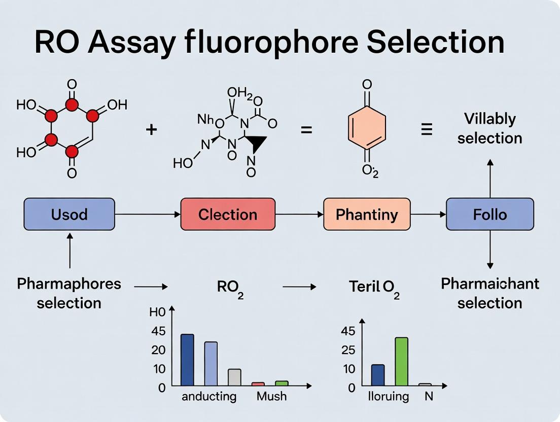
Diagram: Impact of Fluorophore Brightness on RO Assay Outcomes
Diagram Title: Fluorophore Brightness Dictates RO Assay Success for Low Expression Targets
Diagram: Key Considerations in Fluorophore Selection Workflow
Diagram Title: Workflow for Selecting a Fluorophore in RO Assay Design
This technical support center provides troubleshooting and guidance for researchers conducting Radiometric Oxygen (RO) assays, specifically within the context of selecting bright fluorophores for low-expression target research. Understanding the photophysical parameters—extinction coefficient (ε), quantum yield (Φ), and brightness (ε × Φ)—is critical for optimizing assay sensitivity and success.
Troubleshooting Guides & FAQs
Q1: My RO assay signal is weak even with confirmed transfection. What photophysical parameter should I investigate first? A: First, calculate the brightness (ε × Φ) of your chosen fluorophore. For low-expression targets, brightness is the primary figure of merit. A high extinction coefficient ensures strong light absorption, while a high quantum yield ensures efficient conversion of absorbed light to emitted fluorescence. Prioritize fluorophores with a brightness >50,000 M⁻¹cm⁻¹. Also, verify that your instrument's laser lines optimally match the fluorophore's excitation peak.
Q2: How do I differentiate between poor fluorophore performance and actual low protein expression? A: Perform a systematic control experiment:
- Protocol: Transfert cells with a well-characterized, bright fluorescent protein (e.g., EGFP) using the same protocol. Fix and image.
- Analysis: If the control signal is strong, the issue likely lies with your experimental fluorophore's photophysics or maturation in your cellular environment. If the control signal is also weak, the issue is likely with your transfection/expression efficiency or imaging settings.
- Check Quantum Yield Sensitivity: Some fluorophores have quantum yields sensitive to local environment (pH, halides). Ensure your assay buffer is compatible.
Q3: My chosen red fluorophore has a high extinction coefficient but still performs poorly in live-cell RO assays. Why? A: The quantum yield in the cellular environment may be low. The published Φ is often measured in ideal buffer conditions. In cells, factors like aggregation, binding site polarity, or quenching can reduce the effective Φ. Consult literature for reported Φ values in live cells. Consider fluorophores engineered for reduced environmental sensitivity.
Q4: How do I balance brightness with other properties for my low-expression target assay? A: Use the following decision table:
| Issue / Constraint | Prioritized Parameter | Recommended Action |
|---|---|---|
| Very low target abundance | Brightness (ε × Φ) | Select the brightest photostable fluorophore in your desired wavelength. |
| Concerns about photobleaching | Quantum Yield (Φ) & Photostability | A higher Φ means less time spent in excited state, often correlating with reduced radical generation and bleaching. |
| Using a low-power light source | Extinction Coefficient (ε) | Maximize ε at the available excitation wavelength to improve absorption efficiency. |
| Multicolor imaging with fixed lasers | Excitation Match & Brightness | Choose the brightest fluorophore whose excitation peak aligns with your available laser lines, even if it's not the absolute brightest overall. |
Table 1: Photophysical Properties of Common Fluorophores for Low-Expression Imaging
Data sourced from current fluorophore manufacturer specifications and peer-reviewed publications.
| Fluorophore | Ex Max (nm) | Em Max (nm) | Extinction Coefficient ε (M⁻¹cm⁻¹) | Quantum Yield Φ | Brightness (ε × Φ) | Notes for RO Assays |
|---|---|---|---|---|---|---|
| EGFP | 488 | 507 | 56,000 | 0.60 | 33,600 | Standard control; mature in ~30 min. |
| mNeonGreen | 506 | 517 | 116,000 | 0.80 | 92,800 | Excellent brightness for low expression. |
| mCherry | 587 | 610 | 72,000 | 0.22 | 15,840 | Moderate brightness; photostable. |
| mScarlet-I | 569 | 594 | 104,000 | 0.70 | 72,800 | Bright red alternative. |
| SIR-Tubulin | 652 | 674 | 148,000 | 0.36 | 53,280 | High ε; silicon-rhodamine, cell-permeable. |
| JF₆₄₉-HaloTag | 646 | 664 | 152,000 | 0.54 | 82,080 | Exceptional brightness for small labels. |
Experimental Protocol: Determining Optimal Fluorophore for Low-Expression RO Assay
Title: Comparative Brightness Validation for RO Assay Fluorophore Selection.
Objective: To empirically determine the most effective fluorophore for imaging a low-expression protein target.
Materials: See "The Scientist's Toolkit" below.
Methodology:
- Construct Preparation: Clone your target gene fused in-frame with 3-4 different candidate fluorescent protein (FP) tags (e.g., mNeonGreen, EGFP, mScarlet-I, mCherry) into identical expression vectors.
- Cell Seeding & Transfection: Seed identical densities of cells into a multi-well imaging plate. Transfert each FP-tagged construct using a consistent, optimized protocol. Include an untransfected control.
- Sample Preparation: 24-48 hours post-transfection, perform the RO assay protocol. Fix cells with 4% PFA for 15 minutes at room temperature.
- Image Acquisition: Using a confocal or widefield microscope with consistent settings (laser power, exposure time, gain) across all wells, capture images. Critical: Ensure no pixel saturation.
- Data Analysis:
- Measure the mean fluorescence intensity (MFI) of labeled structures in a defined ROI for >50 cells per construct.
- Measure the background MFI from untransfected cells.
- Calculate Signal-to-Background Ratio (SBR) = (MFIsample - MFIbackground) / MFI_background.
- Plot SBR for each FP construct. The highest SBR indicates the most effective tag for your specific target and assay conditions.
Visualizations
Diagram 1: Photophysics & Assay Sensitivity Relationship
Diagram 2: Fluorophore Selection Workflow for RO Assays
The Scientist's Toolkit: Research Reagent Solutions
| Item | Function in Experiment | Example/Note |
|---|---|---|
| Bright Fluorescent Protein Plasmids | Genetically encoded tags for labeling low-expression targets. | mNeonGreen, mScarlet-I, mEGFP. Ensure identical backbone vectors for comparison. |
| Cell-Permeant HaloTag/SNAP-tag Ligands | Small, bright synthetic dyes for labeling self-labeling tags. | JF₆₄₉, SiR dyes. Offer superior brightness compared to many FPs. |
| Validated Transfection Reagent | For consistent, low-toxicity delivery of FP constructs. | Lipofectamine 3000, FuGENE HD. Optimize for your cell line. |
| Fixed-Cell Mounting Medium with Antifade | Preserves fluorescence signal during imaging. | ProLong Diamond, Vectashield. Critical for quantitative comparison. |
| RO Assay-Specific Buffer Kits | Provides optimized chemical environment for the oxygen sensing reaction. | Commercial RO assay kits ensure reagent compatibility and reproducibility. |
| Calibrated Fluorescence Microscope | Instrument with stable light source and sensitive detectors. | Confocal or widefield with scientific CMOS camera. Regular power calibration is essential. |
Troubleshooting Guides & FAQs
Q1: In my RO assay, the signal from my target protein is barely above background, even with a bright fluorophore. What are the primary factors to check? A1: First, verify your assay's Signal-to-Noise Ratio (SNR) calculation. A common issue is high optical noise. Ensure you are using an appropriate control (e.g., cells without the fluorophore-conjugated reagent) to define background. Second, confirm fluorophore selection: for low expression targets, prioritize fluorophores with high extinction coefficients and quantum yields (e.g., APC, Brilliant Violet 421) over FITC or PE. Third, optimize your fixation and permeabilization protocol, as over-fixation can quench fluorescence.
Q2: How do I quantitatively determine if my SNR is sufficient for robust detection of a low-expression target? A2: A widely accepted minimum SNR for confident detection is ≥3. Calculate SNR as (Mean Signal Intensity - Mean Background Intensity) / Standard Deviation of Background Intensity. Use the table below for benchmark values from common fluorophores in model systems.
Table 1: Typical SNR Ranges for Common Fluorophores in Low Expression RO Assays (Cell-based)
| Fluorophore | Extinction Coefficient (M⁻¹cm⁻¹) | Quantum Yield | Typical SNR Range (Low Expressor) | Recommended Application |
|---|---|---|---|---|
| FITC | ~68,000 | 0.79 | 1.5 - 3 | High expression targets only |
| PE | ~1,960,000 | 0.82 | 4 - 8 | Moderate expression |
| APC | ~700,000 | 0.68 | 6 - 12 | Low expression ideal |
| Brilliant Violet 421 | >1,500,000 | 0.80 | 8 - 15 | Best for low expression |
| Alexa Fluor 647 | 270,000 | 0.33 | 5 - 10 | Good for multiplexing |
Q3: What is the step-by-step protocol to empirically determine the optimal exposure time to maximize SNR for my assay? A3: Title: Protocol for Determining Optimal Camera Exposure Time
- Prepare three samples: (a) Untreated/negative control cells, (b) Low-expression sample, (c) Known positive control (if available).
- Image the negative control sample starting at a very low exposure time (e.g., 10ms).
- Increase exposure in increments (e.g., 50ms) and capture images.
- For each image, measure the mean signal intensity and standard deviation in a Region of Interest (ROI) over cells and a background ROI.
- Calculate SNR for each exposure time.
- Plot SNR vs. Exposure Time. The optimal time is at the beginning of the plateau phase before background noise escalates dramatically.
- Validate this time on your low-expression sample.
Q4: My background noise is inconsistently high across replicates. What are the likely causes and solutions? A4: Inconsistent noise often stems from reagent or preparation issues.
- Cause: Incomplete washing of unbound primary or secondary antibodies.
- Solution: Increase wash volume and number of steps (e.g., 3x 5-minute washes with gentle agitation). Use a wash buffer with detergent (e.g., 0.1% Tween-20).
- Cause: Autofluorescence of cells or plate, or reagent precipitation.
- Solution: Include a no-antibody control. Centrifuge antibody stocks before use. Consider using a reagent like TrueBlack to quench lipofuscin autofluorescence.
- Cause: Non-specific binding of antibodies.
- Solution: Increase the concentration of blocking agent (e.g., 5% BSA or serum from the host species of your detection antibody) and extend blocking time to 1 hour at RT.
Experimental Protocol: Validating Fluorophore Performance for Low Expression Targets
Title: Quantitative Comparison of Fluorophore SNR in a Model Low-Expression RO Assay
Objective: To directly compare the Signal-to-Noise Ratio (SNR) delivered by four different fluorophores conjugated to the same detection antibody, targeting a low-abundance nuclear protein.
Materials: See "Research Reagent Solutions" table.
Methodology:
- Cell Culture & Induction: Seed HEK293 cells with a stably integrated, inducible low-expression construct of your target protein (e.g., a nuclear receptor) in a 96-well imaging plate.
- Induction: Treat wells with a low dose of inducer (e.g., 10nM ligand) for 24h to simulate physiological low expression. Include non-induced controls.
- Fixation & Permeabilization: Fix cells with 4% PFA for 15 min, permeabilize with 0.5% Triton X-100 for 10 min.
- Immunostaining:
- Block with 5% BSA for 1 hour.
- Incubate with primary antibody (mouse anti-target) overnight at 4°C.
- Wash 3x with PBS.
- Divide wells into four groups. Incubate each group with one of the four secondary antibody-fluorophore conjugates (Goat anti-Mouse IgG conjugated to AF488, PE, APC, or Brilliant Violet 421) for 1 hour at RT in the dark.
- Wash 3x with PBS. Add nuclear stain (Hoechst) and imaging medium.
- Image Acquisition: Using a high-content imager, acquire images for all channels using identical laser power/gain settings where possible. For each fluorophore channel, perform the exposure time optimization protocol (FAQ Q3) to find its optimal setting.
- Image Analysis:
- Use nuclear segmentation from the Hoechst channel.
- Measure the mean fluorescence intensity (MFI) of the target signal within each nucleus.
- Measure the MFI and standard deviation (SD) of background areas (cell-free regions).
- Calculate SNR per cell: (Cell MFI - Background MFI) / Background SD.
- Calculate the average SNR for 100+ cells per condition.
- Data Presentation: Plot average SNR for each fluorophore condition as a bar graph. Statistically compare groups using ANOVA.
Table 2: Research Reagent Solutions for Low Expression Assay Development
| Item | Function & Rationale |
|---|---|
| Brilliant Violet 421 Conjugate | Polymer dye technology offering exceptional brightness; ideal for low expression targets due to very high SNR. |
| TrueBlack Lipofuscin Autofluorescence Quencher | Reduces background from cellular autofluorescence, directly improving SNR. |
| High-Quality BSA (IgG-Free) | Effective blocking agent to minimize non-specific antibody binding and lower background noise. |
| Black/Clear Bottom 96-Well Imaging Plate | Minimizes well-to-well crosstalk and background fluorescence while allowing high-resolution imaging. |
| Triton X-100 (0.1-0.5%) | Standard permeabilization agent for intracellular (RO) targets; concentration must be optimized to balance access and morphology. |
| Anti-Fade Imaging Mountant | Presves fluorescence signal during acquisition, preventing signal decay which can affect SNR measurements. |
Signaling Pathway & Experimental Workflow Diagrams
Title: Key Steps in Generating Signal and Noise in RO Assays
Title: Experimental Workflow for Fluorophore SNR Comparison
Troubleshooting Guides & FAQs
Q1: My RO assay signal is weak despite using a bright fluorophore on a low-expression target. What could be the issue? A: Weak signal can stem from fluorophore-quencher proximity inefficiency or spectral mismatch. For low-expression targets, ensure you are using a dye with the highest possible extinction coefficient and quantum yield for your instrument's lasers and filters. Verify that the quencher effectively absorbs the donor's emission. Polymer dyes or optimized tandems are often superior for this application.
Q2: I observe high background fluorescence in my no-template controls with a tandem dye. How can I troubleshoot this? A: Tandem dyes are prone to degradation, leading to donor emission "leak" into the acceptor channel. 1) Check dye integrity by analyzing emission spectra; a peak at the donor's emission wavelength indicates breakdown. 2) Ensure reagents are fresh and stored in the dark at recommended temperatures. 3) Switch to a more photostable polymer dye or a different tandem conjugate if degradation is confirmed.
Q3: My traditional dye (e.g., FAM) shows photobleaching during repeated measurements in a live-cell RO assay. What is the solution? A: Traditional cyanine and fluorescein dyes are susceptible to photobleaching. 1) Reduce light exposure intensity or duration. 2) Use an antifade reagent if compatible with your assay. 3) Consider switching to a more photostable dye class, such as ATTO dyes or polymer-based dyes, which exhibit superior resistance to photobleaching, critical for longitudinal low-expression studies.
Q4: The fluorescence intensity of my polymer dye seems inconsistent between assay batches. What should I check? A: Polymer dye performance is highly dependent on conjugation chemistry and buffer conditions. 1) Strictly control conjugation stoichiometry and purification. 2) Ensure consistent assay buffer pH and ionic strength, as polymer dye emission can be environment-sensitive. 3) Use a standardized validation protocol with control oligonucleotides for each new batch.
Q5: How do I choose between a tandem dye and a polymer dye for a multiplexed RO assay targeting low-abundance markers? A: The choice balances brightness, stability, and spectral overlap. For low-abundance targets, brightness is paramount. See the quantitative comparison table below. Polymer dyes generally offer the highest brightness and photostability but may have broader emission spectra. Tandem dyes offer good brightness and narrower emissions but require verification of stability. Ensure your detector can resolve the chosen dye's emission.
Quantitative Data Comparison
Table 1: Key Characteristics of Fluorophore Classes for RO Assays
| Fluorophore Class | Example Dyes | Typical Extinction Coefficient (M⁻¹cm⁻¹) | Quantum Yield | Photostability | Relative Brightness (vs. FAM) | Best For Low-Expression? |
|---|---|---|---|---|---|---|
| Traditional Dyes | FAM, Cy3, Cy5 | ~70,000 - 250,000 | 0.3 - 0.7 | Moderate | 1x (reference) | No (unless target is not very low) |
| Tandem Dyes | PE-Cy7, APC-Cy5.5 | ~2,000,000* | 0.4 - 0.8 | Moderate-Low (can degrade) | 10x - 30x | Yes, but check stability |
| Polymer Dyes | Brilliant Violet 421, Spark YG 570 | ~1,500,000 - 6,000,000 | 0.7 - 0.9 | High | 50x - 100x+ | Yes, optimal choice |
*Tandem dye EC is typically that of the primary donor (e.g., PE or APC).
Experimental Protocols
Protocol 1: Validating Fluorophore Integrity for Low-Expression Target RO Assay
- Objective: Confirm tandem dye conjugation integrity and polymer dye performance.
- Materials: See "Research Reagent Solutions" below.
- Method:
- Prepare a 100 µL solution of the labeled probe (1 µM) in assay buffer.
- Using a spectrofluorometer, acquire the full emission spectrum (excite at the donor's absorbance max).
- For Tandem Dyes: Check for a secondary peak near the donor dye's original emission wavelength (e.g., ~575 nm for PE-Cy7), indicating degradation.
- For Polymer Dyes: Check for a single, sharp emission peak. Compare the spectrum and peak intensity to a manufacturer-provided reference.
- A clean, single peak confirms dye integrity. Proceed with assay optimization.
Protocol 2: Optimizing Quencher Selection for Maximum Signal-to-Background
- Objective: Maximize quenching efficiency to lower background for sensitive low-expression detection.
- Method:
- Synthesize or obtain identical target oligonucleotide probes conjugated with your chosen fluorophore but paired with different quenchers (e.g., BHQ-2, Iowa Black RQ, QSY).
- Prepare assay plates with a dilution series of synthetic target RNA, plus no-template controls (NTC).
- Run the RO assay using standard thermocycling conditions.
- Calculate the ΔRn (signal minus background) for each target concentration and the background signal for NTCs for each quencher-fluorophore pair.
- Select the pair yielding the highest ΔRn for low concentrations and the lowest NTC signal.
Visualizations
Diagram Title: RO Assay Fluorophore Decision Logic
Diagram Title: Tandem Dye Degradation Causes Signal Leak
The Scientist's Toolkit
Table 2: Essential Research Reagent Solutions for Fluorophore-Based RO Assays
| Item | Function in Low-Expression RO Assay |
|---|---|
| Polymer or Tandem Dye-Labeled Probes | Provides the high brightness essential for detecting low-abundance RNA/DNA targets. |
| Dual-Labeled Probes with Dark Quenchers (e.g., BHQ, Iowa Black) | Minimizes initial background fluorescence, maximizing signal-to-noise ratio upon cleavage. |
| Nuclease-Free Water & Buffers | Prevents degradation of oligonucleotide probes and target, crucial for assay consistency. |
| High-Efficiency DNA/RNA Polymerase (e.g., Taq Gold, RTase Mix) | Ensures robust amplification of the low-expression target to generate measurable signal. |
| Spectrofluorometer or Plate Reader | Validates dye integrity and measures emission spectra pre-experiment. |
| Real-Time PCR Instrument with Multi-Channel Detection | Precisely monitors the fluorescence increase during amplification in multiplexed assays. |
| Synthetic Target RNA/DNA Oligos | Serves as positive controls for assay optimization and generating standard curves. |
Troubleshooting Guides & FAQs
Q1: Our fluorescent signal fades rapidly during live-cell imaging of a low-expression RO assay. What is the primary culprit and how can we mitigate it? A: The primary culprit is likely poor photostability of the fluorophore. Under constant illumination, especially with high-intensity lasers in confocal microscopy, fluorophores photobleach, leading to signal loss. This is critical in low-expression research where signal is weak.
- Solution: Select fluorophores with high photostability ratings (e.g., Alexa Fluor 647, CF dyes, ATTO 655). Implement imaging protocols with lower laser power, shorter exposure times, and use of oxygen scavenging systems (e.g., Glucose Oxidase/Catalase) in live-cell experiments.
Q2: We conjugated an antibody with a bright fluorophore, but the probe shows high non-specific binding and precipitation. What went wrong? A: This often results from an excessively high Molar Ratio of Fluorophore to Protein (F/P). Over-labeling can compromise the solubility of the conjugate, cause aggregation, and increase hydrophobic interactions leading to non-specific binding.
- Solution: Aim for a moderate F/P ratio (typically 3-8 for IgG antibodies, but verify for your specific dye). Purify the conjugate rigorously using size-exclusion chromatography (SEC) or dialysis to remove free dye and aggregates. Refer to Table 1 for target F/P ratios.
Q3: Our labeled protein aggregates immediately upon buffer exchange into PBS for an RO assay. How do we resolve this? A: This is a direct solubility issue. Many organic fluorophores are poorly soluble in aqueous buffers. The conjugation process can expose hydrophobic regions of the dye.
- Solution: Ensure the dye stock is in a high-quality, anhydrous DMSO. Post-conjugation, consider switching to a buffer with added solubilizing agents (e.g., 0.1% BSA, 0.05% Tween-20, or 5-10% glycerol). For problematic dyes, test alternative buffers like Tris or HEPES.
Q4: We need maximum brightness for a low-abundance target, but are concerned about photostability and conjugate performance. How do we balance F/P ratio for optimal signal? A: You must optimize the F/P ratio to maximize brightness without inducing solubility problems or altering protein function. A higher F/P increases brightness but risks the issues in Q2.
- Solution: Perform a conjugate titration. Create a series of conjugates with different F/P ratios (see Protocol 1). Test them side-by-side in your assay, measuring specific signal, background, and photobleaching rate. The optimal ratio is where signal-to-noise is maximized before background rises sharply.
Q5: What are the key controls for validating fluorophore performance in a new RO assay system? A:
- Unlabeled Target Control: To assess autofluorescence.
- Free Dye Control: To check for non-specific binding of unreacted dye.
- Isotype Control (Conjugated): To set baselines for non-specific antibody binding.
- Photobleaching Control: A fixed sample imaged over time to quantify signal decay.
- Solubility Control: Monitor conjugate solution for turbidity before and after buffer exchange.
Data Presentation
Table 1: Characteristics of Common Bright Fluorophores for Low-Expression Research
| Fluorophore | Excitation (nm) | Emission (nm) | Relative Brightness* | Photostability | Recommended F/P Ratio (IgG) | Solubility Notes |
|---|---|---|---|---|---|---|
| Alexa Fluor 488 | 495 | 519 | High | High | 4-8 | Excellent in aqueous buffers. |
| CF 555 | 555 | 565 | Very High | Very High | 3-7 | Good; avoid freeze-thaw cycles. |
| DyLight 550 | 562 | 576 | High | Medium-High | 4-9 | May require additives (BSA). |
| Alexa Fluor 647 | 650 | 665 | High | Excellent | 3-6 | Excellent; ideal for low expression. |
| CF 680 | 680 | 698 | Very High | Excellent | 3-7 | Good in PBS. |
| ATTO 700 | 700 | 719 | High | Excellent | 2-5 | Moderate; use fresh DMSO stock. |
*Brightness = Extinction Coefficient x Quantum Yield.
Experimental Protocols
Protocol 1: Determining Optimal Fluorophore-to-Protein (F/P) Ratio
Objective: To conjugate an antibody with a fluorescent dye at varying ratios and identify the optimal balance of brightness and specificity.
Materials: See "The Scientist's Toolkit" below. Method:
- Prepare Antibody: Buffer-exchange target antibody into Conjugation Buffer (e.g., 0.1M NaHCO₃, pH 8.3) using a desalting column to a final concentration of 1-2 mg/mL.
- Prepare Dye Stock: Dissolve NHS-ester dye in anhydrous DMSO to 10 mg/mL immediately before use.
- Conjugation Reaction: Set up three separate vials with constant antibody amount (e.g., 100 µg). Add different volumes of dye stock to achieve theoretical F/P ratios of 4, 8, and 12. Mix gently and react in the dark at room temperature for 1 hour.
- Purification: Quench the reaction with 1M Tris-HCl, pH 7.5. Purify each conjugate using a size-exclusion spin column equilibrated in PBS + 0.05% sodium azide + 2% BSA (for solubility).
- Calculate Actual F/P Ratio:
- Measure absorbance at 280 nm (A280, protein) and at the dye's λmax (Adye).
- Use the formula: F/P = (Adye / εdye) / [(A280 - (CF * Adye)) / εantibody]
- Where CF (correction factor) is provided by the dye manufacturer.
- Validation: Test each conjugate via flow cytometry or immunofluorescence on cells with high and low target expression. Calculate the Signal-to-Noise Ratio (SNR).
Visualizations
Diagram 1: Fluorophore Selection Workflow for RO Assays
Diagram 2: Impact of F/P Ratio on Assay Performance
The Scientist's Toolkit: Essential Reagents for Fluorophore Conjugation & Testing
| Item | Function |
|---|---|
| NHS-Ester Dye | Reactive dye form that couples to primary amines (lysines) on proteins. |
| Anhydrous DMSO | High-quality solvent for dye stock preparation; critical for solubility and reaction efficiency. |
| Conjugation Buffer (e.g., 0.1-1.0M Carbonate-Bicarbonate, pH 8.3-9.0) | Provides optimal pH for efficient NHS-ester reaction with amine groups. |
| Size-Exclusion Spin Column / Desalting Column | Removes free, unreacted dye and allows buffer exchange into storage buffer. |
| BSA (Bovine Serum Albumin) or Carrier Protein | Added to storage buffer (0.1-1%) to improve conjugate solubility and long-term stability. |
| Spectrophotometer / Nanodrop | For precise measurement of protein and dye absorbance to calculate concentration and F/P ratio. |
| Oxygen Scavenging System (e.g., Glucose Oxidase + Catalase + Glucose) | Reduces photobleaching in live-cell imaging by removing dissolved oxygen. |
| Antifade Mounting Medium | Reduces photobleaching in fixed-cell imaging. |
Building a Robust Assay: Methodological Strategies for Low-Signal RO Detection
Troubleshooting Guides & FAQs
FAQ 1: Why do I observe a strong signal in flow cytometry but a weak or no signal in my imaging-based RO (Radiometric or Resonance Energy Transfer) platform for the same low-expression target? Answer: This is a common alignment issue due to fundamental differences in detection. Flow cytometry measures total cellular fluorescence from thousands of cells in suspension, aggregating signal. Imaging captures signal per pixel from individual, often adherent, cells with spatial context. For low-expression targets, the instantaneous signal per pixel may be below the camera's detection threshold, even if the total cellular fluorescence is adequate for the flow cytometer's photomultiplier tubes (PMTs). Solution: Switch to a brighter, more photostable fluorophore (e.g., Alexa Fluor 647 over FITC) for imaging to improve the signal-to-noise ratio (SNR) per pixel.
FAQ 2: How do I correct for higher background autofluorescence in imaging compared to flow cytometry when selecting fluorophores? Answer: Autofluorescence is often more pronounced in imaging due to excitation of cellular components across a broad spectrum. Flow cytometry can use spectral unmixing more efficiently.
- Troubleshooting Guide:
- Profile Background: Image untransfected/untreated cells under your standard assay conditions to establish autofluorescence levels per channel.
- Shift Emission: Choose fluorophores with emissions >600 nm (e.g., Cy5, Alexa Fluor 680), where cellular autofluorescence is significantly lower.
- Optimize Filters: Use narrow-band emission filters matched precisely to your fluorophore's emission peak to exclude background.
FAQ 3: My FRET/BRET efficiency calculated from imaging data doesn't match my flow cytometry data. What are the key assay format-specific factors? Answer: Misalignment often stems from environmental differences and detection mechanics.
- Troubleshooting Guide:
- Environmental Control: Imaging assays are more sensitive to temperature and CO₂ fluctuations during live-cell imaging, which can affect protein interaction. Use an environmental chamber.
- Photobleaching: Imaging causes more rapid photobleaching, skewing donor-acceptor ratios over time. Use a highly photostable dye pair (e.g., SNAP/CLIP tags with cell-permeable dyes) and minimize exposure.
- Calibration: Ensure both platforms use identical donor-acceptor pairing (e.g., GFP-RFP for both). Normalize data using positive and negative control constructs measured on the same platform.
FAQ 4: What are the critical protocol adjustments for validating low-expression target engagement using a bright fluorophore switch from flow to imaging? Answer: A direct transfer of protocol will likely fail. See the detailed experimental protocol below.
Detailed Experimental Protocol: Validating Low-Expression Target Engagement Across Platforms
Aim: To quantitatively compare receptor occupancy (RO) for a low-expression GPCR using a bright fluorescent ligand in both flow cytometry and live-cell imaging.
Key Reagents & Solutions:
- Cell Line: HEK293T stably expressing target GPCR at low levels (<50,000 copies/cell).
- Bright Fluorescent Ligand (BFL): SNAP-Lumi4-Tb (Donor, Terbium cryptate) and SNAP-Red (Acceptor, ~650 nm emission). Chosen for high brightness and photostability.
- Control: Unlabeled competing antagonist (1 µM).
- Imaging Buffer: Phenol-red free HBSS with 20 mM HEPES, 0.1% BSA.
Methodology:
- Sample Preparation: Seed cells 24h prior in a black-walled, clear-bottom 96-well plate for imaging and in parallel in a culture dish for flow cytometry.
- Labeling (Pre-treatment): Incubate cells with 100 nM SNAP-Lumi4-Tb and SNAP-Red (combined) for 60 min at 37°C in imaging buffer. Include wells with excess unlabeled competitor (1 µM) for nonspecific binding (NSB) determination.
- Wash: Rinse cells 3x with buffer to remove unbound ligand.
- Parallel Measurement:
- Flow Cytometry: Harvest imaging-plate cells (trypsin), resuspend in buffer, and analyze immediately. Use a 405nm laser, collect donor emission at 620/14 nm and acceptor at 665/8 nm. Gate on single, live cells.
- Live-Cell Imaging: Image the sister plate using a high-sensitivity widefield or confocal microscope. Use 405nm excitation, collect time-gated luminescence (for FRET) or fluorescence at ~650 nm. Acquire 5 fields per well.
- Data Analysis:
- Calculate specific binding (Total signal - NSB signal) for each cell.
- Flow: Report median fluorescence intensity (MFI) of the specific population.
- Imaging: Report the mean pixel intensity within a cell ROI, averaged across cells.
Data Presentation: Quantitative Comparison of Platform Performance
Table 1: Key Performance Metrics for Low-Expression RO Assay (Hypothetical Data)
| Metric | Flow Cytometry | Imaging (Widefield) | Implication for Low-Expression RO |
|---|---|---|---|
| Detection Limit | ~500 receptors/cell | ~5,000 receptors/cell* | Imaging requires brighter probes for same sensitivity. |
| Temporal Resolution | Single time point (Snapshot) | Real-time, seconds to minutes | Imaging is superior for kinetic association/dissociation studies. |
| Spatial Information | None | Subcellular localization | Imaging can differentiate membrane-bound vs. internalized signal. |
| Throughput | High (10,000+ cells/sec) | Low to Medium (10-100 cells/field) | Flow is better for population statistics and screening. |
| Photobleaching Concern | Low | High | Critical for imaging; mandates photostable fluorophores. |
| Typical Brightness Metric | Molecules of Equivalent Soluble Fluorophore (MESF) | Signal-to-Background Ratio (SBR) per pixel | SBR is the critical imaging metric to optimize. |
*Assumes standard sCMOS camera and a bright red fluorophore (e.g., Alexa Fluor 647).
The Scientist's Toolkit: Research Reagent Solutions
Table 2: Essential Reagents for Cross-Platform Low-Expression RO Assays
| Reagent | Function & Rationale | Example Product/Brand |
|---|---|---|
| Time-Gated Lanthanide Probes | Eliminates short-lived background autofluorescence in imaging; enables TR-FRET in flow. Critical for low-expression SNR. | Terbium (Tb) cryptate (Lumi4-Tb), Europium (Eu) chelates |
| Bright, Photostable Organic Dyes | Maximizes emitted photons per pixel in imaging; withstands prolonged illumination. | Alexa Fluor 647, CF dyes, Cy5 derivatives |
| Self-Labeling Protein Tags | Allows specific, covalent labeling of target proteins with bright synthetic fluorophores. | SNAP-tag, HaloTag, CLIP-tag |
| Phenol-Red Free Media | Reduces background fluorescence, especially in the green-red spectrum, for live-cell imaging. | Gibco FluoroBrite DMEM |
| Anti-Fading Mounting Medium | Preserves fluorescence signal intensity for fixed-cell imaging across multiple z-planes. | ProLong Diamond, VECTASHIELD |
| Cell Dissociation Reagent (Non-enzymatic) | For harvesting adherent imaging samples for flow cytometry without cleaving cell-surface epitopes. | EDTA-based solutions (e.g., Gibco Versene) |
Visualizations
Diagram 1: Fluorophore Selection Workflow for Platform Alignment
Diagram 2: Key Pathways in GPCR RO Assay Detection
Pairing Fluorophores with High-Sensitivity Detectors (e.g., Violet/UV Lasers, APDs)
Troubleshooting Guides & FAQs
Q1: In my RO assay for low-expression targets, my violet laser (405 nm) excited signal is weak even with a "bright" fluorophore like Alexa Fluor 488. What could be wrong? A: This is often a mismatch between the fluorophore's excitation peak and the laser line. Alexa Fluor 488 has a primary peak at ~495 nm, making it suboptimal for 405 nm excitation. Its extinction coefficient at 405 nm is less than 10% of its peak. For a 405 nm laser, choose fluorophores with strong violet absorption: e.g., Pacific Blue (Ex: 410 nm), Alexa Fluor 430 (Ex: 434 nm), or Dylight 405. Verify the match by consulting the fluorophore's excitation spectrum.
Q2: My APD detector is delivering high noise and inconsistent photon counts in UV excitation experiments. How can I improve signal-to-noise? A: APDs are extremely sensitive to stray light and overvoltage. First, ensure complete darkness; even minor light leaks are critical. Check that the APD is operating at its recommended bias voltage, as even a 0.5V over-bias can drastically increase dark current. Use high-quality, UV-blocking emission filters specific to your fluorophore to minimize background. Allow the instrument and APD to thermally stabilize for 30+ minutes before acquisition.
Q3: When using tandem dyes (e.g., PE-Cy7) with a UV laser, I get unexpected emission in the acceptor channel without the donor. What's happening? A: This indicates direct excitation of the acceptor dye (Cy7) by the UV/violet laser, a common phenomenon called "FRET bypass" or "cross-talk." Tandem dyes are designed for blue/green excitation of the donor (PE). UV/violet photons can directly excite acceptor cyanine dyes. Switch to a dye pair where the acceptor has minimal absorbance at the UV laser wavelength, or use a laser line closer to the donor's peak.
Q4: For quantifying very low expression levels, should I prioritize a brighter fluorophore or a more sensitive detector (like an APD over a PMT)? A: In the context of your thesis on low-expression research, the hierarchy should be: 1) Fluorophore Brightness: Choose a fluorophore with a high product of extinction coefficient and quantum yield (see Table 1) matched perfectly to your laser. 2) Optical Efficiency: Maximize light collection with high-NA objectives and clean filters. 3) Detector Sensitivity: An APD can provide lower noise and single-photon sensitivity, offering a marginal gain crucial for photon-limited applications. The brightest fluorophore on a standard PMT often outperforms a dim fluorophore on an APD.
Q5: I observe rapid photobleaching of my violet-excited fluorophore during prolonged live-cell RO assays. How can I mitigate this? A: Violet/UV photons carry higher energy, accelerating photobleaching. Implement the following: 1) Use an oxygen-scavenging system (e.g., Glucose Oxidase/Catalase) in your imaging medium. 2) Reduce laser power to the minimum required and use a faster acquisition time. 3) Consider a fluorophore from the "self-healing" Janelia Fluor (JF) or HaloTag ligand families, which are engineered for enhanced photostability. 4) Ensure your mounting medium contains an anti-fade reagent.
Table 1: Brightness & Suitability of Violet/UV-Excited Fluorophores for Low-Expression RO Assays
| Fluorophore | Peak Excitation (nm) | Extinction Coefficient (ε) at 405 nm (M⁻¹cm⁻¹)* | Quantum Yield (Φ) | Relative Brightness at 405 nm (ε * Φ) | Recommended Detector |
|---|---|---|---|---|---|
| Pacific Blue | 410 | ~46,000 (peak) | 0.99 | ~45,540 | APD / High-QE PMT |
| Alexa Fluor 430 | 434 | ~17,000 | 0.59 | ~10,030 | Standard PMT |
| Dylight 405 | 400 | ~35,000 | 0.85 | ~29,750 | APD / High-QE PMT |
| Alexa Fluor 488 | 495 | ~3,000 (at 405 nm) | 0.92 | ~2,760 | Standard PMT |
| CF405S | 404 | ~40,000 | 0.96 | ~38,400 | APD / High-QE PMT |
| Hoechst 33342 (DNA stain) | 350 | ~42,000 (at 350 nm) | 0.08 | ~3,360 | APD for low signal |
*ε at laser line is critical. Values are approximate; consult manufacturer data.
Table 2: Detector Comparison for Low-Photon-Flux Applications
| Detector Type | Typical Quantum Efficiency (QE) at 500-600 nm | Dark Current (Noise) | Gain | Suitability for Low-Expression RO Assays |
|---|---|---|---|---|
| Standard PMT (GaAsP) | 40-45% | Moderate | 10⁵ - 10⁶ | Good for moderately bright fluorophores. |
| High-Sensitivity PMT | 45-50% | Low | 10⁵ - 10⁶ | Better for dim signals, common in flow cytometers. |
| Silicon APD (Analog) | 70-80% | Very Low | 50-100 | Excellent for photon counting, ideal for ultra-low signal. |
| Hybrid Detector (HyD) | 45-50% | Very Low | 10⁶ | Excellent, with wide dynamic range and low noise. |
Experimental Protocols
Protocol: Validating Fluorophore-Brightness and Detector Sensitivity for Low-Expression Cell Surface RO Assays
Objective: To quantitatively compare the signal-to-noise ratio (SNR) of different violet-excited fluorophores conjugated to antibodies against a low-expression surface target, using both PMT and APD detection.
Materials: See "The Scientist's Toolkit" below. Method:
- Sample Preparation:
- Harvest cells expressing your target of interest at low levels (e.g., <1000 copies/cell). Include an isotype control.
- Aliquot 1x10⁶ cells per tube into 5 tubes.
- Stain each tube with a titrated optimal concentration (determined separately) of a different violet-excited antibody conjugate (e.g., Pacific Blue, CF405S, Dylight 405, Alexa Fluor 430, and an unstained control). Incubate for 30 min on ice in the dark.
- Wash cells twice with cold PBS + 2% FBS.
- Resuspend in 300 µL of PBS for analysis.
Instrument Setup & Calibration:
- Power on the violet (405 nm) laser and allow 20 minutes for stabilization.
- For the detector comparison, configure one channel to be read by a standard PMT and a parallel, identical optical path (same bandpass filter) to be read by an APD, if your system allows. Otherwise, perform sequential runs.
- Set PMT voltage to a standard linear range. Set APD bias to the manufacturer's specified optimal voltage.
- Run unstained cells and adjust detector gains/voltages so the population mean is at 10¹-10² on a log scale.
Data Acquisition:
- Acquire a minimum of 50,000 events per sample.
- Record the Median Fluorescence Intensity (MFI) of the stained positive population and the Standard Deviation (SD) of the isotype control population.
SNR Calculation & Analysis:
- For each fluorophore-detector pair, calculate: SNR = (MFIsample - MFIisotype) / (2 * SD_isotype).
- Plot SNR for each fluorophore on both detectors (see Diagram 2).
- The combination yielding the highest SNR is optimal for your specific low-expression assay.
Protocol: Direct Excitation Check for Tandem Dyes under Violet Laser
- Prepare three bead or cell samples: unstained, stained with a non-tandem violet-excited dye (e.g., Pacific Blue) as a reference, and stained with the tandem dye of interest (e.g., PE-Cy7).
- Using your violet (405 nm) laser, collect fluorescence in the donor channel (e.g., 575/25 nm for PE) and the acceptor channel (e.g., 780/60 nm for Cy7).
- Analyze: Significant signal from the tandem dye in the acceptor channel indicates direct excitation of Cy7 by 405 nm light, necessitating a dye or laser change.
Visualizations
Title: Workflow for Optimizing Fluorophore-Detector Pairing
Title: Comparative SNR of Violet Fluorophores on Detectors
The Scientist's Toolkit: Research Reagent Solutions
| Item | Function in Low-Expression RO Assays |
|---|---|
| High-Efficiency Violet (405 nm) Laser | Provides excitation photons matched to fluorophores with strong violet absorbance (e.g., Pacific Blue). Stability is critical for quantitation. |
| Anti-Fade Mounting Medium (e.g., ProLong, Fluoroshield) | Reduces photobleaching, especially under high-energy violet/UV light, preserving signal during prolonged imaging. |
| Avalanche Photodiode (APD) Detector Module | Converts photons to electrical signal with very high quantum efficiency and low noise, enabling detection of faint signals from low-copy-number targets. |
| Violet-Optimized Bandpass Filter Sets | Precisely separates fluorophore emission from laser scatter and autofluorescence, maximizing signal purity. |
| Bright, Violet-Excited Fluorophores (e.g., CF405S, Janelia Fluor 585) | Engineered for high extinction coefficient at ~400-430 nm and high quantum yield, maximizing photons emitted per excitation event. |
| High-NA Objective Lens (e.g., NA 1.4) | Collects a greater percentage of emitted photons from the sample, directly increasing signal intensity at the detector. |
| Cell Staining Buffer with Non-Specific Blockers | Reduces background from non-specific antibody binding, improving the signal-to-noise ratio for low-abundance targets. |
| Counting Beads or Reference Fluorophore Standards | Allows for instrument calibration and quantitative comparison of fluorescence intensity between experiments and days. |
Troubleshooting Guides & FAQs
FAQ: General Fluorophore Selection
Q: How do I choose a bright fluorophore for a low-expression target without causing antibody aggregation? A: Prioritize fluorophores with high extinction coefficients and quantum yields (e.g., Alexa Fluor 647, CF680) in the far-red/NIR spectrum to minimize background. Use controlled, site-specific conjugation chemistries (e.g., enzyme-mediated) to maintain antibody integrity. Avoid over-labeling; a dye-to-antibody ratio (DAR) of 2-4 is often optimal.
Q: My antibody function (binding affinity) drops significantly after conjugation. What is the most likely cause? A: This is typically due to conjugation at or near the antigen-binding site (paratope). To resolve, use site-specific conjugation methods that target engineered cysteines (THIOMAB), the Fc glycans (glycoengineering), or specific peptide tags. Avoid lysine-based random conjugation if the antibody has lysines in the complementarity-determining regions (CDRs).
Q: I need maximum brightness for my RO assay, but my signal-to-noise ratio is poor. What should I optimize? A: 1) Fluorophore Selection: Choose dyes with high photostability and brightness suitable for your instrument's lasers/filters. 2) Conjugation Control: Optimize DAR to balance brightness with quenching effects at high DAR. 3) Assay Buffer: Include quenching suppressors (e.g., Trolox, cyclooctatetraene) and ensure proper blocking to reduce non-specific binding.
FAQ: Specific Experimental Issues
Q: After conjugating a bright, high-DAR fluorophore, I observe high non-specific binding in my cell-based assay. A: High DAR can increase hydrophobicity. Troubleshoot by:
- Purifying the conjugate via size-exclusion chromatography to remove aggregates.
- Increasing the concentration of detergent (e.g., Tween-20) or protein-based blockers (e.g., BSA) in your assay buffer.
- Switching to a more hydrophilic fluorophore or reducing the DAR.
Q: My fluorophore-antibody conjugate precipitates during the reaction or storage. A: Precipitation indicates aggregation. Ensure:
- The antibody is in a conjugation-friendly buffer (PBS, pH 7.4, without amines like Tris or azide). Perform a buffer exchange beforehand.
- The dye stock (in DMSO) is anhydrous and fresh. Add it slowly to the antibody solution with gentle mixing.
- The final conjugate is formulated in a stabilizing buffer with carrier protein (e.g., 1% BSA) and stored at 4°C in the dark.
Key Experimental Protocols
Protocol 1: Site-Specific Conjugation via Engineered Cysteines (THIOMAB-style)
Objective: Attach maleimide-activated bright fluorophores to a site-engineered antibody to achieve a uniform DAR of 2, preserving function.
- Reduce Engineered Cysteines: Incubate antibody (1 mg/mL in PBS, pH 7.0, with 1 mM EDTA) with 10-fold molar excess of TCEP for 2h at 37°C.
- Purify: Remove excess TCEP using a desalting column (Zeba Spin, 7K MWCO) equilibrated with conjugation buffer (PBS, 1 mM EDTA, pH 7.0).
- Conjugate: Immediately react reduced antibody with 3.5-fold molar excess of maleimide-fluorophore (from DMSO stock) for 2h at room temperature, protected from light.
- Quench & Purify: Add 100-fold molar excess of free L-cysteine to quench for 15 min. Purify conjugate via size-exclusion chromatography (e.g., Superdex 200 Increase) in PBS, pH 7.4.
- Analyze: Determine DAR by UV-Vis spectroscopy and confirm binding by flow cytometry.
Protocol 2: Optimizing DAR for Maximum Brightness in RO Assays
Objective: Empirically determine the optimal DAR for a given bright fluorophore-antibody pair.
- Prepare Conjugates at Varying DARs: Perform standard NHS-ester lysine conjugation at different antibody:dye molar ratios (e.g., 1:2, 1:4, 1:8, 1:12). Use identical reaction time and temperature.
- Purify: Use spin desalting columns to remove free dye. Determine exact DAR for each batch by UV-Vis.
- Test in Assay: Use a standardized RO assay (e.g., cell-based staining of high and low expression lines). Measure median fluorescence intensity (MFI) and background.
- Calculate Brightness Index: (MFIsample - MFIbackground) / (Antibody Concentration). Plot vs. DAR. The peak is the optimal DAR before quenching/function loss occurs.
Data Presentation
Table 1: Bright Fluorophore Properties for Low-Expression Target Detection
| Fluorophore | Ex/Em Max (nm) | Extinction Coefficient (M⁻¹cm⁻¹) | Quantum Yield | Recommended Conjugation Chemistry | Optimal DAR Range for Low Expression |
|---|---|---|---|---|---|
| Alexa Fluor 647 | 650/668 | 270,000 | 0.33 | NHS-ester (Lysine), Maleimide (Cysteine) | 2-4 |
| CF680 | 679/702 | 180,000 | 0.54 | NHS-ester | 3-5 |
| Dylight 800 | 777/794 | 290,000 | 0.19 | Maleimide, NHS-ester | 2-4 |
| PE/Cyanine7 | 488/774 | N/A (Protein) | N/A | Streptavidin-Biotin | 1 (Complex) |
Table 2: Troubleshooting Common Conjugation Problems
| Problem | Potential Cause | Solution |
|---|---|---|
| Low Binding Affinity | Conjugation at/near paratope | Use site-specific conjugation; map conjugation site. |
| High Non-Specific Binding | High DAR, hydrophobic dye | Reduce DAR; use hydrophilic dye; optimize blocking. |
| Fluorophore Quenching | DAR too high (>6 for many dyes) | Conjugate to lower DAR; use dyes with less self-quenching. |
| Antibody Aggregation | Harsh reaction conditions, dye hydrophobicity | Use gentler chemistry (e.g., pH-controlled NHS ester); add mild detergent during reaction. |
Diagrams
The Scientist's Toolkit: Research Reagent Solutions
| Item | Function in Conjugation/Brightness Optimization |
|---|---|
| TCEP (Tris(2-carboxyethyl)phosphine) | Reduces engineered disulfide bonds for site-specific cysteine conjugation. |
| Maleimide-Activated Fluorophores | Reacts specifically with free thiols (-SH) on reduced cysteines for controlled labeling. |
| NHS-Ester-Activated Fluorophores | Reacts with primary amines (lysines) on antibodies for common, but random, conjugation. |
| Zeba Spin Desalting Columns | Rapidly removes small molecule reagents (TCEP, free dye) via buffer exchange. |
| Superdex 200 Increase Column | High-resolution size-exclusion chromatography for removing antibody aggregates post-conjugation. |
| UV-Vis Spectrophotometer | Essential for accurately determining antibody concentration and Dye-to-Antibody Ratio (DAR). |
| Cyclic Olefin Copolymer (COC) Plates | Low-autofluorescence plates for sensitive RO assays detecting low-expression targets. |
| Antibody Stabilizer/PBS Formulation Buffer | For long-term storage of conjugated antibodies to prevent aggregation and loss of function. |
FAQs & Troubleshooting Guides
Q1: My low-expression target signal is being overwhelmed by spectral spillover from a very bright fluorophore in a neighboring channel. How can I mitigate this? A: This is a common issue in multiplexed panels. Implement the following steps:
- Panel Redesign: Re-evaluate your fluorophore assignment. Pair the brightest fluorophores (e.g., PE, APC) with antigens of the lowest expression, and assign dimmer fluorophores (e.g., FITC) to high-abundance targets. This balances signal intensities.
- Optimize Staining Index: Titrate all antibodies to achieve the optimal Staining Index (SI = [Median Positive – Median Negative] / (2 * SD of Negative]), not just the brightest signal. Over-concentration of bright fluorophores exacerbates spillover.
- Apply Comprehensive Compensation: Use single-stained controls for every fluorophore in your panel, prepared with the same biological sample (e.g., cells or beads). Verify compensation matrices in your analysis software, ensuring spillover spreading is correctly subtracted.
- Consider a "Cascade" Approach: For ultra-bright fluorophores like PE, use conjugated secondary antibodies or tandem dyes (e.g., PE-Cy7) to shift emission further into the red/infrared, distancing it from dimmer targets in adjacent channels.
Q2: When designing a panel for rare event detection, what are the key considerations for fluorophore selection? A: Sensitivity is paramount. Adhere to these principles:
- Brightest Fluorophores for Rarest Targets: Assign the fluorophores with the highest photon yield (e.g., PE, Superbright polymers, APC) exclusively to your rare population markers.
- Dedicated "Background" Channels: Reserve one or two channels for dim or non-critical markers to create a cleaner background in the high-sensitivity channels.
- Minimize Spillover into Critical Channels: Avoid placing very bright fluorophores in channels that directly spill over into your rare event detection channel. Consult the instrument's spectral viewer.
- Validate with Biological Controls: Include a fully stained control sample with a known frequency of your rare population and a fluorescence-minus-one (FMO) control for the key marker to precisely set gating boundaries.
Q3: I am experiencing high background and poor resolution in my defined channels. What could be the cause? A: High background often stems from suboptimal experimental conditions.
- Antibody Titration: Re-titrate antibodies. Excess antibody leads to non-specific binding and elevated background.
- Sample Preparation: Ensure thorough cell washing to remove unbound antibody and fixatives/dead cell markers that can cause nonspecific staining. Use a viability dye to exclude dead cells.
- Buffer Optimization: Increase the concentration of Fc receptor blocking agent and use high-quality PBS/BSA or commercial staining buffers.
- Instrument Performance: Regularly perform quality control (QC) using calibration beads to ensure laser alignment and detector voltages are optimal. Poor instrument sensitivity directly impacts resolution.
Q4: How do I validate that my multiplexed panel is working correctly for my RO assay? A: A rigorous validation workflow is essential.
- Single Stain Controls: Confirm each antibody-fluorophore conjugate binds specifically and yields a detectable signal.
- Full Panel vs. FMO Controls: Run your full panel alongside FMO controls for every marker. This defines positive/negative populations and reveals any compensation errors or interactions.
- Biological Controls: Use cell lines or samples with known expression patterns (positive and negative) to confirm panel specificity.
- Reproducibility Check: Repeat the staining and acquisition on at least three independent days to assess intra-assay variability.
Experimental Protocols
Protocol 1: Antibody Titration for Optimal Staining Index
Objective: To determine the antibody concentration that delivers the highest signal-to-noise ratio for each marker in your panel. Materials: Cell sample, serial dilutions of antibody conjugates, flow cytometry staining buffer, flow cytometer. Method:
- Prepare a master cell suspension at 5-10 x 10^6 cells/mL.
- Aliquot equal cell volumes (e.g., 100 µL) into as many tubes as needed for your antibody dilution series (e.g., 6 points).
- Prepare a 2X serial dilution series of the antibody, typically spanning a range from the manufacturer's recommended concentration down to 1/16 or 1/32 of that amount.
- Add 100 µL of each antibody dilution to the cell aliquots. Include an unstained control.
- Incubate in the dark for 30 minutes at 4°C.
- Wash cells twice with 2 mL of staining buffer and resuspend in a fixed volume (e.g., 300 µL) for acquisition.
- Acquire data, ensuring sufficient events are collected. Analyze the Median Fluorescence Intensity (MFI) of the positive population and the negative population. Calculate the Staining Index for each dilution.
- Select the dilution that yields the highest Staining Index, not necessarily the highest MFI.
Protocol 2: Preparation of Single-Stain Compensation Controls
Objective: To create accurate controls for calculating spectral spillover (compensation) in software. Materials: Cells or compensation beads, each antibody-fluorophore conjugate from the panel, flow cytometry staining buffer. Method (Using Cells):
- For each fluorophore in your panel, prepare one tube.
- To each tube, add an aliquot of cells (or beads) and stain with a single antibody conjugate.
- Critical: Use the same antibody clone and fluorophore as in your full panel. The brightness must match.
- Use an antibody amount that gives a bright, clear signal (often at the titrated optimal concentration).
- For tandem dyes (e.g., PE-Cy7), include a separate control stained with the parent fluorophore only (e.g., PE) to monitor tandem dye degradation.
- Include an unstained control.
- Process all tubes identically (incubation time, wash steps) as your full panel.
- Acquire these single-stain controls using the same cytometer settings (voltages, thresholds) as your experimental samples.
Data Presentation
Table 1: Common Bright Fluorophores & Recommended Applications
| Fluorophore | Relative Brightness | Recommended Channel (e.g., Laser/Filter) | Ideal For | Caution |
|---|---|---|---|---|
| PE (Phycoerythrin) | Very High (~2400) | 488nm / 575-585nm | Lowest expression targets, rare events | High spillover into PE-Cy5, PE-Cy5.5, PE-Cy7 channels. |
| Superbright 600 | Very High | 405nm / ~610nm | Low expression targets with violet excitation. | Requires instrument with violet laser. |
| APC (Allophycocyanin) | High (~570) | 640nm / 660-680nm | Low expression targets, red laser panels. | Can have spillover into APC-Cy7, APC-Fire 810. |
| PE-Cy7 (Tandem) | High | 488nm / 780-810nm | Mid-to-high expression; shifts PE signal. | Susceptible to degradation; monitor with PE single stain. |
| BV421 (Brilliant Violet 421) | High | 405nm / 420-450nm | Low expression with violet laser. | Can spill into BV510 channel. |
| FITC | Low (~90) | 488nm / 515-545nm | High abundance targets, viability dyes. | Not suitable for low-expression markers. |
Table 2: Troubleshooting Quick Reference
| Problem | Potential Cause | Solution |
|---|---|---|
| Poor population resolution | Under-titrated antibody, high background | Titrate antibody, increase wash steps, use viability dye. |
| Unexpected signal in a channel | Spectral spillover, antibody cross-reactivity | Check compensation with fresh single stains, verify antibody specificity. |
| Dim signal on critical marker | Fluorophore too dim, antigen loss | Assign a brighter fluorophore, optimize cell fixation/permeabilization. |
| High variance between replicates | Inconsistent pipetting, cell count | Standardize cell concentration and all staining volumes. |
The Scientist's Toolkit: Research Reagent Solutions
| Item | Function |
|---|---|
| UltraComp eBeads / Compensation Beads | Uniform particles for creating consistent, cell-free single-stain compensation controls. |
| Cell Staining Buffer (with Fc Block) | Reduces nonspecific antibody binding via Fc receptors, lowering background. |
| Viability Dye (Fixable) | Distinguishes live from dead cells; dead cells cause nonspecific antibody uptake. |
| Antibody Stabilizer Solution | Preserves conjugated antibodies, especially critical for sensitive tandem dyes. |
| Prestained Reference Cells | Commercial cell standards for daily instrument QC to ensure sensitivity and reproducibility. |
Visualizations
Technical Support Center
Troubleshooting Guides & FAQs
Q1: My low-expression RO assay has high background after the final wash. What should I adjust? A: Excessive background often stems from insufficient washing or non-specific antibody binding. For low-expression targets, optimize by:
- Increasing wash volume (3x well volume) and duration (5 minutes per wash with gentle agitation).
- Adding a mild detergent (e.g., 0.05% Tween-20) to your wash buffer.
- Incorporating a blocking step with 5% BSA or a commercial protein-free blocker for 1 hour at RT before primary antibody incubation.
- Titrating your primary antibody to find the optimal signal-to-noise ratio; for bright fluorophores like CF568 or Alexa Fluor 647, a lower concentration than recommended may suffice.
Q2: How can I amplify a weak signal from a low-abundance target without increasing background? A: Implement a tyramide signal amplification (TSA) step. This enzymatic method dramatically increases fluorophore deposition at the target site.
- Protocol: After primary antibody incubation and washing, incubate with an HRP-conjugated secondary antibody (30 min, RT). Wash thoroughly. Apply fluorophore-conjugated tyramide reagent (e.g., Alexa Fluor 488 Tyramide) at the recommended dilution for 2-10 minutes. Stop the reaction with a thorough wash. Critical: You must quench endogenous peroxidases (e.g., with 3% H₂O₂ for 10 min) before the primary antibody step to prevent high background.
Q3: What is the optimal incubation time for primary antibodies in low-expression studies? A: Incubation time is a balance between sensitivity and practicality. The table below summarizes optimized strategies:
Table 1: Primary Antibody Incubation Optimization
| Incubation Condition | Typical Duration | Recommended For | Key Consideration |
|---|---|---|---|
| Room Temperature (RT) | 1-2 hours | High-affinity antibodies, abundant targets | Fast, but may yield suboptimal signal for low expression. |
| 4°C Overnight | 16-18 hours | Low-expression targets, bright fluorophores | Gold standard. Maximizes binding specificity and signal-to-noise. |
| 37°C | 30-60 minutes | Accelerated protocols | Can increase non-specific binding; requires rigorous validation. |
Q4: My bright fluorophore is photobleaching quickly during imaging. How can I mitigate this? A: Photobleaching is common with bright but less stable fluorophores. Solutions include:
- Use an antifade mounting medium (e.g., with p-phenylenediamine or commercial ProLong/Vectashield products).
- Reduce exposure time and light intensity during acquisition.
- Store slides at -20°C in the dark immediately after preparation.
- Consider more photostable fluorophores for repeated imaging: e.g., Alexa Fluor 488/555/647 or CF dyes over traditional FITC or TRITC.
Q5: How many amplification steps are too many? I am not seeing a linear increase in signal. A: Signal amplification is not linear and reaches a plateau. Excessive amplification (e.g., >2 TSA layers) exponentially increases background and noise. For most low-expression targets, a single amplification step (e.g., a well-optimized TSA or a polymer-based detection system) is sufficient. Always run a no-primary-antibody control and a target-positive control to define the dynamic range of your amplification.
Experimental Protocols
Protocol: Tyramide Signal Amplification (TSA) for Low-Expression RO Assay Objective: To detect a low-abundance receptor target (RO) using bright fluorophore amplification.
- Sample Preparation: Fix and permeabilize cells according to standard protocol.
- Endogenous Peroxidase Block: Incubate samples in 3% H₂O₂ in PBS for 10 minutes at RT. Wash 3x with PBS.
- Blocking: Incubate in protein-free blocking buffer for 1 hour at RT.
- Primary Antibody: Incubate with anti-RO primary antibody (optimized dilution in blocking buffer) overnight at 4°C. Wash 3x (5 min each) with PBST (PBS + 0.05% Tween-20).
- HRP-Secondary: Incubate with HRP-conjugated secondary antibody for 30 minutes at RT. Wash 3x (5 min each) with PBST.
- TSA Reaction: Prepare fluorophore-conjugated tyramide working solution per manufacturer instructions. Apply to sample for exactly 5 minutes. Wash thoroughly 4x (5 min each) with PBST.
- Counterstain & Mount: Apply DAPI (if needed) and mount with antifade medium.
Protocol: Optimization of Washes for High S/N in Fluorophore Imaging Objective: To determine the wash stringency that minimizes background for bright fluorophores.
- Prepare identical samples stained for a low-expression target with a bright fluorophore (e.g., Alexa Fluor 647).
- Variable: Wash stringency. Divide samples into groups washed with (a) PBS, (b) PBST (0.05% Tween), (c) PBST (0.1% Tween). Perform 3 washes of 2, 5, or 10 minutes each.
- Imaging: Image all samples under identical acquisition settings.
- Analysis: Quantify mean signal intensity from the target region and an adjacent background region. Calculate Signal-to-Noise Ratio (S/N = (SignalMean - BackgroundMean) / Background_STDev). The condition with the highest S/N indicates optimal wash stringency.
Diagrams
Title: TSA Signal Amplification Workflow
Title: Low-Expression Assay Optimization Logic
The Scientist's Toolkit
Table 2: Key Research Reagent Solutions for RO Assay with Bright Fluorophores
| Reagent / Material | Function & Rationale |
|---|---|
| Bright, Photostable Fluorophores (e.g., Alexa Fluor 647, CF568, ATTO 700) | High molar brightness and resistance to photobleaching are critical for detecting low-expression targets without signal loss during imaging. |
| Tyramide Signal Amplification (TSA) Kit | Enzyme-based method for depositing numerous fluorophore molecules per target, providing orders-of-magnitude signal enhancement for low-abundance proteins. |
| Protein-Free Blocking Buffer | Reduces non-specific background staining more effectively than protein-based blockers when using high-sensitivity amplification methods. |
| HRP-Conjugated Secondary Antibodies | Required enzyme conjugate for catalyzing the TSA reaction. Must be species-specific and highly validated for minimal cross-reactivity. |
| Antifade Mounting Medium | Preserves fluorescence intensity of bright but susceptible fluorophores during storage and repeated microscopy sessions. |
| Low-Binding Microcentrifuge Tubes | Prevents adsorption of dilute primary antibodies to tube walls, ensuring consistent antibody concentration during long 4°C incubations. |
Solving Low Signal & High Background: A Troubleshooting Framework for RO Assays
Troubleshooting Guides & FAQs
FAQ: Common Experimental Issues
Q1: My RO assay shows a very weak signal. How do I determine if my protein of interest is expressed at low levels or if the fluorophore is underperforming? A: Begin with a systematic control experiment. Transfect your construct alongside a positive control plasmid expressing a well-characterized, brightly-tagged protein (e.g., GFP-H2B) using the same protocol. If the positive control is also dim, the issue is likely with transfection efficiency, imaging settings, or fluorophore health. If the positive control is bright but your target is dim, proceed to a western blot to confirm protein expression levels independently of the fluorophore.
Q2: What are the key parameters to check for fluorophore performance in live-cell imaging? A:
- Photostability: Perform a continuous time-lapse under standard imaging conditions. A rapid decay in signal indicates poor photostability.
- Brightness: Compare the mean fluorescence intensity of your tag to other common tags (e.g., mNeonGreen vs. EGFP) under identical imaging conditions.
- Maturation Time: For time-course experiments, ensure the fluorophore has fully matured. Refer to vendor data on maturation half-times.
- Environmental Sensitivity: Some fluorophores (e.g., pH-sensitive variants) may dim in certain cellular compartments.
Q3: Which bright fluorophores are recommended for low-expression targets in receptor occupancy (RO) assays? A: For detecting low-abundance proteins, priority should be given to fluorophores with high extinction coefficients and quantum yields. Recent benchmarks favor:
- Green: mNeonGreen, SiriusGFP, Clover
- Red: mScarlet, mCherry2, mRuby3
- Far-Red/NIR: miRFP670nano, mCardinal
Experimental Protocols
Protocol 1: Validating Fluorophore Performance vs. Expression Objective: Decouple fluorophore performance from biological expression levels. Materials: See "Research Reagent Solutions" table. Steps:
- Sample Preparation: Plate cells in a 4-well imaging dish. Co-transfect wells with:
- Well A: Target protein fused to Fluorophore X.
- Well B: Control protein (e.g., H2B) fused to Fluorophore X.
- Well C: Target protein fused to Fluorophore Y (a known bright standard).
- Well D: Untransfected cells (background control).
- Fixation & Stain: 24h post-transfection, fix cells and perform immunofluorescence (IF) staining using a validated primary antibody against your target protein and a different, highly cross-absorbed secondary antibody conjugated to a spectrally distinct fluorophore.
- Imaging: Acquire images using identical settings for both fluorescence channels (fluorophore tag and IF signal).
- Analysis: Quantify the correlation between the fluorescence intensity from the live-cell tag (Fluorophore X/Y) and the immunofluorescence signal (independent validation). Use the table below for data comparison.
Quantitative Comparison of Selected Fluorophores for Low-Expression Targets
| Fluorophore | Excitation Peak (nm) | Emission Peak (nm) | Extinction Coefficient (M⁻¹cm⁻¹) | Quantum Yield | Relative Brightness (vs EGFP) | Maturation t½ (37°C) | Recommended for Low Expression? |
|---|---|---|---|---|---|---|---|
| EGFP | 488 | 507 | 56,000 | 0.60 | 1.0 | ~30 min | Baseline |
| mNeonGreen | 506 | 517 | 116,000 | 0.80 | 2.8 | ~15 min | Yes |
| SiriusGFP | 499 | 511 | 70,000 | 0.92 | 1.9 | ~10 min | Yes |
| mCherry | 587 | 610 | 72,000 | 0.22 | 0.5 | ~15 min | No |
| mScarlet | 569 | 594 | 100,000 | 0.70 | 2.1 | ~5 min | Yes |
| miRFP670nano | 646 | 670 | 98,000 | 0.14 | 0.4 | ~50 min | No* |
Note: Despite lower quantum yield, miRFP670nano's far-red excitation minimizes autofluorescence, which can improve signal-to-background for some low-expression targets.
Protocol 2: Direct Comparison Assay for Fluorophore Selection Objective: Empirically select the optimal fluorophore for your specific low-expression target. Steps:
- Clone your target gene into vectors containing the candidate bright fluorophores (e.g., mNeonGreen, mScarlet) using an identical linker sequence.
- Transfect these constructs in parallel into your cell model, ensuring identical DNA amounts and transfection conditions.
- Image live cells 24-48h post-transfection using channel-specific settings optimized for each fluorophore to avoid cross-talk.
- Quantify the signal-to-background ratio (SBR) and the signal-to-noise ratio (SNR) for each fluorophore-target pair.
- Plot the results as shown in the workflow diagram.
Diagrams
Title: Troubleshooting Workflow for Low Signal
Title: Protocol for Empirical Fluorophore Selection
The Scientist's Toolkit: Research Reagent Solutions
| Item | Function in Experiment | Example/Notes |
|---|---|---|
| Bright Fluorescent Protein Vectors | Tagging the low-expression protein of interest for direct visualization. | pcDNA3.1-mNeonGreen-N, pmScarlet-C1. Ensure consistent promoter and cloning backbone. |
| Validated Primary Antibody | Independent validation of protein expression level via immunofluorescence. | Must be specific, high-affinity, and validated for IF. Target a different epitope than the tag. |
| Cross-Adsorbed Secondary Antibody | Highly specific detection of primary antibody with minimal cross-talk. | Conjugated to a fluorophore spectrally distinct from the live-cell tag (e.g., use CF568 if live tag is green). |
| Positive Control Plasmid | Control for transfection and imaging system health. | e.g., H2B-GFP, Gap43-mCherry. Should localize distinctly. |
| Low-Autofluorescence Imaging Medium | Reduces background noise to improve SNR for dim signals. | Phenol-red free medium, with buffering system (e.g., HEPES). |
| Mounting Medium with Antifade | Preserves fluorescence signal during fixed-cell imaging. | Essential for quantitative IF comparison assays. |
| Transfection Reagent (Validated) | Ensures consistent delivery of plasmid DNA across compared conditions. | Lipofectamine 3000, PEI, or electroporation. Optimization is critical. |
Troubleshooting Guide
Q1: After titrating my antibody, I observe high background fluorescence despite good specific signal. What could be the cause and how can I resolve it?
A: High background is often due to antibody over-titration or non-specific binding. Within the context of RO assay research on low-expression targets, this obscures the critical specific signal.
- Solution: Re-perform the titration, extending the range to include lower antibody concentrations. Increase the concentration of your blocking agent (e.g., use 5% BSA or serum matching your secondary antibody host) and include a wash step with a mild detergent like 0.1% Tween-20. For intracellular targets, ensure cells are adequately fixed and permeabilized.
Q2: My stain index decreased when I switched to a brighter fluorophore for my low-expression target. Why did this happen?
A: This paradoxical result typically stems from increased background. Brighter fluorophores (e.g., PE, Brilliant Violet 421) can amplify both specific signal and any non-specific binding or cellular autofluorescence.
- Solution: Re-optimize the antibody titration specifically for the new, brighter conjugate. You will likely need to use a lower antibody concentration. Also, review your panel design; ensure the bright fluorophore is on the low-expression target and not on a highly expressed one causing spillover.
Q3: During antibody titration, my positive and negative cell populations are not distinct, making it impossible to calculate an accurate Stain Index. What should I do?
A: This is common when the target expression is very low or the negative control population is inappropriate.
- Solution: First, verify your negative control. Use a fluorescence-minus-one (FMO) control for the specific marker-fluorophore conjugate you are titrating. This is critical for low-expression analysis. If populations remain merged, consider using a brighter fluorophore-conjugate or a different antibody clone with higher affinity for the low-expression target.
Q4: How does fluorophore selection impact antibody titration requirements in RO assays?
A: Fluorophore characteristics (brightness, stability, spillover profile) directly dictate titration parameters. Bright fluorophores (high photon yield) reach optimal Stain Index at lower antibody concentrations than dim fluorophores. For low-expression targets, a bright fluorophore is essential, but requires more precise titration to balance signal-to-noise.
Frequently Asked Questions (FAQs)
Q: What is the precise formula for calculating Stain Index, and how do I apply it to titration data?
A: Stain Index (SI) quantifies the separation between positive and negative populations. The formula is:
SI = (Median_Positive − Median_Negative) / (2 × SD_Negative)
For titration, calculate the SI for each antibody concentration tested. The optimal concentration is typically at or just before the plateau of the SI curve, not necessarily the point of highest MFI.
Q: For my low-expression target research, should I prioritize antibody concentration or fluorophore brightness first?
A: Follow this hierarchy: 1) Clone Selection: Choose a validated, high-affinity antibody clone for your target. 2) Fluorophore Selection: Pair it with the brightest fluorophore compatible with your instrument's lasers/filters and panel design. 3) Titration: Perform a meticulous titration of this specific conjugate to find the optimal concentration for maximum SI.
Q: What are the critical controls for a rigorous antibody-fluorophore conjugate titration experiment?
A: Essential controls include:
- Unstained Cells: For instrument settings and autofluorescence.
- FMO Control: The most critical control for setting the negative population gate for SI calculation.
- Isotype Control (with same conjugate): Less informative than FMO but can indicate Fc receptor or non-specific binding issues.
- Single-Color Controls: For compensation, especially important for bright fluorophores with broad spillover.
Q: Can I use a previously titrated antibody concentration if I change the fluorophore it's conjugated to?
A: No. The optimal antibody concentration is conjugate-specific. A brighter fluorophore will generally require a lower antibody concentration to achieve the same level of specific staining while minimizing background. You must re-titrate for each new conjugate.
Data Presentation
Table 1: Impact of Fluorophore Brightness on Optimal Antibody Titration for a Low-Expression Target
| Fluorophore Conjugate | Relative Brightness | Typical Optimal Antibody Concentration (µg/10^6 cells) | Typical Achievable Stain Index (Low-Exp. Target) | Key Consideration for RO Assays |
|---|---|---|---|---|
| Brilliant Violet 421 | Very High | 0.125 - 0.25 | 8 - 15 | High spillover into violet filters; requires careful compensation. |
| PE | Very High | 0.25 - 0.5 | 7 - 14 | Prone to photo-bleaching; monitor laser power. |
| APC | High | 0.5 - 1.0 | 5 - 10 | Stable signal, good for time-course experiments. |
| FITC | Moderate | 1.0 - 2.0 | 3 - 7 | Higher autofluorescence in some cell types. |
| Alexa Fluor 488 | Moderate/High | 0.75 - 1.5 | 4 - 8 | More photostable than FITC. |
Table 2: Troubleshooting Common Stain Index Problems
| Problem | Possible Cause | Recommended Action |
|---|---|---|
| Low Stain Index | 1. Under- or over-titrated antibody.2. Fluorophore too dim for target.3. Excessive spillover from bright channels. | 1. Perform/check titration with FMO controls.2. Switch to a brighter fluorophore.3. Re-panel, moving bright fluorophores to less crowded regions. |
| High Background | 1. Antibody concentration too high.2. Inadequate blocking or washing.3. Secondary antibody cross-reactivity. | 1. Reduce antibody concentration.2. Optimize blocking buffer; add extra washes.3. Use highly cross-adsorbed secondary antibodies. |
| Inconsistent SI Between Repeats | 1. Inconsistent cell count or viability.2. Pipetting error in antibody dilution.3. Variable incubation time/temp. | 1. Standardize cell counting and dead cell removal.2. Create a master mix for replicates.3. Use a timer and calibrated water bath/incubator. |
Experimental Protocols
Protocol: Titration of an Antibody-Fluorophore Conjugate for Flow Cytometry
Objective: To determine the optimal concentration of a specific antibody-fluorophore conjugate that yields the maximum Stain Index for a target antigen, critical for research on low-expression targets.
Materials: See "The Scientist's Toolkit" below.
Procedure:
- Cell Preparation: Harvest and wash your cells. For surface staining, aliquot approximately 1-2 x 10^5 cells per tube/well. For intracellular targets, perform fixation and permeabilization at this stage.
- Antibody Dilution Series: Prepare a 2-fold serial dilution of your antibody-conjugate in staining buffer (e.g., PBS + 2% FBS). A typical starting range is 8x the manufacturer's recommended concentration down to a 1:64 dilution. Include a "0" concentration tube (FMO control).
- Staining:
- Add 100 µL of cell suspension to each tube.
- Add 100 µL of the appropriate antibody dilution to each tube. Mix gently.
- Incubate: Protect from light. Incubate for 30 minutes at 4°C (surface stain) or as optimized for your target.
- Washing: Add 2 mL of staining buffer to each tube. Centrifuge at 300-400 x g for 5 minutes. Aspirate supernatant carefully.
- Resuspension and Acquisition: Resuspend cell pellets in 200-300 µL of staining buffer or fixative. Acquire data on a flow cytometer, ensuring voltages are set appropriately using unstained and single-stained controls.
- Analysis:
- Gate on your live, single-cell population.
- For each concentration, use the FMO control to define the negative population.
- Record the Median Fluorescence Intensity (MFI) of both the positive and negative populations.
- Calculate the Stain Index (SI) for each concentration.
- Determination of Optimal Concentration: Plot SI (y-axis) vs. antibody concentration (x-axis, log scale). The optimal concentration is at the plateau of the curve, where further increases in antibody do not significantly improve SI.
Mandatory Visualization
Title: Antibody-Fluorophore Conjugate Titration Workflow
Title: Stain Index-Based Titration Decision Logic
The Scientist's Toolkit: Research Reagent Solutions
| Item | Function & Importance for Titration & Low-Expression Targets |
|---|---|
| High-Affinity Antibody Clones | The foundation. For low-expression targets, a clone with high affinity and specificity is non-negotiable to capture weak signal. |
| Bright Fluorophore Conjugates | Signal amplifiers. Fluorophores like Brilliant Violet and PE provide the necessary photon yield to distinguish low-expression signals from noise. |
| Staining Buffer (with Carrier) | Reduces non-specific binding. Buffers with BSA (2-5%) or serum block Fc receptors and minimize background, critical for a clean SI. |
| Fluorescence-Minus-One (FMO) Controls | The definitive negative control. Essential for accurately gating dim populations and calculating a true Stain Index during titration. |
| Viability Dye | Excludes dead cells. Dead cells exhibit high autofluorescence and non-specific antibody binding, which drastically increases background and skews SI. |
| Precision Pipettes & Master Mix Tubes | Ensures reproducibility. Accurate serial dilutions and consistent staining volumes are critical for reliable, repeatable titration results. |
| Flow Cytometer with Calibrated Beads | Ensures instrument consistency. Daily quality control with calibration beads standardizes performance, allowing SI values to be compared over time. |
Troubleshooting Guides & FAQs
Q1: My flow cytometry data shows high background fluorescence in my unstained/control sample after fixation. What could be the cause? A: This is often due to non-specific antibody binding via Fc receptors on immune cells, or autofluorescence increased by fixation. Ensure you are using an Fc blocking reagent (e.g., purified anti-CD16/32, human Fc block) before staining. For intracellular targets, the fixation/permeabilization process itself can increase autofluorescence; titrate your fixative concentration and time, and use bright fluorophores that allow you to distinguish specific signal from this elevated background.
Q2: I am staining for a low-expression intracellular phospho-protein. My positive signal is weak and overlaps with the background. How can I improve resolution? A: In the context of phospho-protein/RO assay research, fluorophore selection is critical. Choose bright fluorophores (e.g., PE, BV421, APC) for the low-expression target. Combine this with rigorous optimization of the permeabilization buffer (type, concentration, and duration) to maximize antibody access while minimizing background. Always include a matched isotype control processed through the identical fixation/permeabilization protocol.
Q3: After permeabilization, my cell surface marker signals are diminished. What went wrong? A: Harsh or prolonged permeabilization can damage cell surface epitopes. Implement a two-step protocol: stain surface markers first, then fix and permeabilize for intracellular staining. Use a mild detergent (e.g., saponin) for intracellular staining after formaldehyde fixation. Validate each step with known positive controls.
Q4: My FMO controls show higher fluorescence than expected. Is this related to Fc blocking? A: Possibly. Inadequate Fc blocking can cause antibodies in your panel to bind non-specifically to FcR+ cells, elevating signal in all channels. Extend the Fc blocking incubation time or increase the reagent concentration. Furthermore, check if your fixation has created sticky cells; including a protein-based buffer (e.g., 1% BSA in PBS) during all staining and wash steps can reduce this non-specific binding.
Key Research Reagent Solutions
| Reagent | Primary Function | Key Consideration |
|---|---|---|
| Fc Block (Anti-CD16/32) | Binds to Fcγ receptors on immune cells, preventing non-specific antibody binding. | Use before any staining step. Species-specific. |
| Formaldehyde (PFA) | Cross-linking fixative. Preserves cellular structure and protein epitopes. | Concentration (1-4%) and time (10-30 min) must be optimized to balance preservation and autofluorescence. |
| Methanol | Precipitating fixative/permeabilizer. Excellent for many intracellular targets, especially nuclear. | Can destroy some surface epitopes and increase autofluorescence. Use cold and for consistent durations. |
| Saponin | Mild detergent for permeabilization after formaldehyde fixation. Creates pores in membranes. | Requires presence in all subsequent antibody buffers to maintain permeability. Reversible. |
| Triton X-100 | Stronger non-ionic detergent for permeabilization. | Can be harsher on epitopes. Useful for difficult-to-access targets. Optimize concentration (0.1-0.5%). |
| Bright Fluorophores (PE, APC, BV421) | High photon-output fluorophores for detecting low-abundance targets. | Essential for RO assays/low-expression research. Pair with dimmer fluorophores (FITC, PerCP) on high-expression markers. |
Experimental Protocol: Intracellular Staining for Low-Abundance Phospho-Protein
- Harvest & Wash: Harvest cells, wash once in cold FACS Buffer (PBS + 1% BSA + 0.1% NaN3).
- Fc Block: Resuspend cell pellet in FACS Buffer containing Fc block reagent (1:100 dilution). Incubate for 15 minutes on ice.
- Surface Stain: Add directly conjugated surface marker antibodies (with bright fluorophores reserved for low-expression targets). Incubate 30 min on ice, protected from light.
- Wash: Wash twice with 2 mL cold FACS Buffer. Centrifuge at 300-400 x g for 5 min.
- Fixation: Resuspend cells in 100 µL of freshly prepared 4% PFA in PBS. Incubate for 20 minutes at room temperature (RT).
- Wash: Wash twice with 2 mL FACS Buffer.
- Permeabilization: Resuspend cells in 100 µL of ice-cold 90% methanol. Vortex gently while adding. Incubate for 30 minutes on ice or at -20°C.
- Wash: Wash twice with 2 mL FACS Buffer. Cells can be stored in buffer at 4°C overnight if needed.
- Intracellular Stain: Resuspend cell pellet in 100 µL FACS Buffer containing anti-phospho-protein antibody conjugated to a bright fluorophore (e.g., PE). Incubate for 60 minutes at RT, protected from light.
- Wash & Analyze: Wash twice, resuspend in FACS Buffer, and analyze on a flow cytometer.
Visualizations
Diagram 1: Workflow for Intracellular RO Assay Staining
Diagram 2: Causes of High Background in Flow
Diagram 3: Fluorophore Selection Logic for Low Expression
Within the context of research focused on robust, quantitative readouts (RO assays) for targets with low expression—requiring exceptionally bright fluorophores—managing photobleaching is not merely a technical nuisance but a fundamental determinant of data reliability. This technical support center provides targeted guidance to address specific experimental challenges in this domain.
Troubleshooting Guides & FAQs
Q1: My weak fluorescence signal disappears completely after a few seconds of imaging. What should I check first? A: First, verify the fluorophore's compatibility with your laser lines. Using a dye like Alexa Fluor 488 with a 405nm laser will cause rapid bleaching. Second, immediately check your antifade mounting medium. If it's expired, improperly stored, or omitted, signal loss is drastic. For fixed-cell RO assays, switch to a next-generation antifade like ProLong Diamond or SlowFade Glass.
Q2: I'm studying low-abundance receptors using TIRF microscopy. Which dyes offer the best combination of brightness and photostability? A: For TIRF and single-molecule imaging of low-expression targets, photostability is paramount. The following table summarizes top performers:
Table 1: High-Performance Fluorophores for Demanding Imaging Applications
| Fluorophore | Excitation/Emission (nm) | Relative Brightness | Relative Photostability | Best For |
|---|---|---|---|---|
| Alexa Fluor 647 | 650/668 | High | Very High | TIRF, STORM, low-light detection |
| CF660C | 663/682 | Very High | Excellent | RO assays, super-resolution |
| ATTO 655 | 663/684 | High | Exceptional | Single-molecule tracking |
| BrightJanus 550 | 550/570 | Exceptional | High | Low-expression targets in confocal |
Q3: My antifade reagent is causing background fluorescence or quenching my signal. How can I optimize? A: This indicates a mismatch between the antifade chemistry and your fluorophore. Consult the manufacturer's chart. For instance, traditional DABCO is poor for far-red dyes, while p-phenylenediamine can quench FITC. Perform a simple test: prepare slides with and without antifade, and with different commercial formulations, imaging a control sample with identical settings to quantify signal-to-noise retention over time.
Q4: What is a validated protocol for testing dye stability in my specific assay system? A: Follow this quantitative photobleaching assay protocol:
Protocol: Quantitative Photobleaching Half-Life Assay
- Sample Preparation: Prepare identical samples labeled with your test fluorophores. Use your standard fixation/permeabilization and staining protocol.
- Mounting: Apply a controlled volume (e.g., 50 µL) of the candidate antifade mounting medium and seal with a coverslip.
- Image Acquisition Setup: Use a confocal or widefield microscope. Define a fixed region of interest (ROI). Set laser power and exposure time to a level that gives a good initial signal but is within routine use. Crucially, keep all settings identical between samples.
- Time-Series Acquisition: Acquire images continuously at a set interval (e.g., every 500ms or 1s) for 5-10 minutes.
- Data Analysis: Measure the mean fluorescence intensity within the ROI over time. Plot intensity vs. time. Fit the curve to a single-exponential decay function. Calculate the time point at which the intensity drops to 50% of its initial value (t1/2). The dye/antifade combination with the longest t1/2 offers the best photostability for your system.
Q5: Are there antifade reagents suitable for live-cell imaging of dynamic RO assays? A: Traditional hard-set mounts are not suitable. For live-cell imaging, consider:
- Oxygen-Scavenging Systems: Such as Oxyrase or buffers containing glucose oxidase/catalase, which reduce dissolved oxygen.
- Commercial Live-Cell Solutions: ProLong Live Antifade reagent or LiveCell Shield are formulated to mitigate phototoxicity and bleaching in living samples.
Visualizing Fluorophore Selection & Photobleaching Pathways
Title: Logical Workflow for Combating Photobleaching in Imaging
Title: Research Reagent Solutions for Photostable Imaging
Addressing Non-Specific Binding and Spillover in Multiplex Panels
Troubleshooting Guides & FAQs
Q1: What are the primary causes of high background/noise in my multiplex flow cytometry data, and how do I differentiate non-specific binding from spillover? A1: High background can stem from both phenomena. Non-specific binding (NSB) typically appears as a uniform shift in fluorescence across all channels for a stained sample compared to an unstained control. Spillover (spectral overlap) manifests as signal in a detector not matched to the primary fluorophore, often creating a diagonal pattern in bi-exponential plots. To differentiate, run single-color controls: isolated spillover will show only in specific off-target channels, while NSB will show broad elevation.
Q2: My panel uses bright fluorophores (e.g., PE, APC) for low-expression targets, but my dim signals are getting buried. What optimization steps should I take? A2: This is a common challenge in "bright fluorophores low expression" experimental design. Follow this protocol:
- Titrate All Antibodies: Use a serial dilution (e.g., 1:50, 1:100, 1:200, 1:400) to find the optimal signal-to-noise ratio for each reagent.
- Implement a Staining Buffer with Additives: Use PBS containing 1% BSA, 0.1% Sodium Azide, and add:
- Fc Receptor Block: (e.g., human Fc block) for 10 minutes prior to staining.
- Non-Specific Blocking Reagents: 5-10% normal serum (from the host species of your secondary antibodies) or 0.1-1 mg/ml of purified, non-reactive IgG.
- Wash Extensively: Perform two post-staining washes with 2-3 ml of buffer.
- Utilize "Spillover Spreading" Matrix: After compensation, analyze if spreading error from bright markers is obscuring dim populations. Consider reassigning fluorophores.
Q3: How do I systematically design a panel to minimize spillover when I need to pair bright fluorophores with low-expression targets? A3: Adopt a spillover-aware panel design protocol:
- Rank Targets: List targets by expression level (low, medium, high).
- Rank Fluorophores: List available fluorophores by brightness (from manufacturer's data or published indices).
- Apply the "High-Low" Pairing Rule: Pair your brightest fluorophores (e.g., PE, Super Bright) with your lowest-expression targets.
- Consult Spillover Matrix: Use your cytometer's spillover matrix (or a universal one like from Fluorofinder or BD's spectrum viewer) to check for high overlap values (>10%). Avoid pairing a very bright fluorophore with a high-expression target if its spillover spreads into the channel of your dim, low-expression target.
- Leverage Tandem Dyes Cautiously: While bright, they can have lot-to-lot variability and increased spillover. Use them with well-expressed targets where slight variability won't be catastrophic.
Data Presentation
Table 1: Common Fluorophore Brightness Index and Spillover Potential
| Fluorophore | Relative Brightness (Approx. Index) | Common Excitation Laser(s) | High Spillover Risk Channels (Example) |
|---|---|---|---|
| FITC | 1 (Reference) | 488 nm | PE, PerCP-Cy5.5 |
| PE | 8-12 | 488 nm, 561 nm | FITC, PE-Cy7 |
| PE-Cy7 | 5-7 (Tandem) | 488 nm, 561 nm | APC-Cy7, PerCP-Cy5.5 |
| APC | 6-8 | 640 nm | Alexa Fluor 700, PerCP-Cy5.5 |
| APC-Cy7 | 4-6 (Tandem) | 640 nm | PE-Cy7, BV711 |
| BV421 | 7-9 | 405 nm | BV510, FITC |
| Super Bright 600 | 12+ | 640 nm | BV650, APC |
Table 2: Optimization Reagents and Their Functions for Reducing NSB
| Reagent | Recommended Concentration | Primary Function | Incubation Step |
|---|---|---|---|
| Fc Block (Human/Mouse) | 5 µl/test, 10 min | Blocks antibody binding to Fc receptors | Before surface staining |
| Normal Mouse/ Rat Serum | 2-5% (v/v) | Provides non-specific protein block | In staining buffer |
| Bovine Serum Albumin (BSA) | 0.5-1% (w/v) | Carrier protein to reduce adsorption | In all staining/wash buffers |
| Sodium Azide | 0.1% (w/v) | Inhibits internalization | In storage/staining buffers |
Experimental Protocols
Protocol: Comprehensive Single-Color Control Setup for Spillover Compensation and NSB Assessment Objective: To generate accurate compensation controls and assess non-specific binding for each fluorophore in your panel. Materials: Cell sample (viable, >90% viability), antibody conjugates, staining buffer (PBS + 1% BSA + 0.1% azide), Fc block, compensation beads (positive and negative). Method:
- Prepare Cells: Aliquot 1x10^5 cells per single-color control tube + one unstained cell tube.
- Fc Block (Cells only): Resuspend cell pellets in 50 µl staining buffer containing Fc block. Incubate 10 min on ice. (Omit for beads).
- Stain: Add the predetermined optimal amount of a single antibody conjugate to the appropriate tube. Include a "Beads Only" tube for each fluorophore.
- Incubate: 30 minutes on ice, protected from light.
- Wash: Add 2 ml cold buffer, centrifuge (300-400 x g, 5 min), decant supernatant.
- Resuspend: Resuspend all cell controls in 200-300 µl buffer. Resuspend bead controls in the volume recommended by the manufacturer.
- Acquire: Run samples on the cytometer, collecting sufficient events. Use the unstained cells and negative beads to set negative populations.
- Analyze: Calculate compensation using the bead or cell controls. Overlay histograms of unstained vs. stained controls to check for NSB (broad shift).
Protocol: Titration for Optimal Signal-to-Noise Ratio Objective: To determine the antibody concentration that yields the highest specific signal with the lowest background. Method:
- Prepare a cell sample with at least 5 aliquots of 1x10^5 cells each.
- Perform a 4-fold serial dilution of your antibody in staining buffer (e.g., 1:25, 1:100, 1:400, 1:1600). Include a negative control (no antibody).
- Stain cells with each dilution following your standard staining protocol.
- Acquire data and plot the Median Fluorescence Intensity (MFI) of the positive population against the antibody dilution.
- The optimal dilution is typically at the "knee" of the curve, just before the MFI plateaus, maximizing the Stain Index (SI).
SI = (MFI_positive - MFI_negative) / (2 * SD_negative).
The Scientist's Toolkit: Key Research Reagent Solutions
| Item | Function in Addressing NSB/Spillover |
|---|---|
| UltraComp eBeads / Compensation Beads | Provide consistent, bright positive signals for calculating spillover compensation matrices without cellular autofluorescence. |
| Fc Receptor Blocking Solution | Monoclonal antibody to CD16/32 (mouse) or human Fc block, prevents antibody binding via non-antigen-specific Fc interactions. |
| Cell Staining Buffer (with BSA & Azide) | Standardized buffer formulation reduces hydrophobic and ionic NSB. Azide prevents receptor internalization during staining. |
| Normal Serum (from secondary host species) | Blocks non-specific interactions through serum proteins, critical when using indirect staining or prone tissues. |
| Fluorophore-Conjugated Antibody Titration Kit | Pre-optimized dilution series to empirically determine the best concentration for specific antibody lots. |
| Viability Dye (Fixable Viability Stain) | Distinguishes live from dead cells; dead cells cause extensive NSB and must be excluded from analysis. |
| Antibody Stabilizer/Preservative | For custom conjugations, maintains fluorophore integrity, preventing breakdown and release of proteins that increase background. |
Diagrams
Benchmarking Performance: Validation and Comparative Analysis of Next-Gen Fluorophores
Technical Support Center: Troubleshooting & FAQs for RO Assay Fluorophore Selection in Low Expression Research
This support center addresses common challenges in selecting and validating bright fluorophores for Receptor Occupancy (RO) assays, particularly in low expression level research critical for drug development.
Frequently Asked Questions (FAQs)
Q1: Our assay sensitivity is insufficient for detecting low receptor expression levels. What fluorophore properties should we prioritize? A: For low expression targets, prioritize fluorophores with high molar brightness (product of extinction coefficient and quantum yield). Consider photostability to allow longer signal integration times. Table 1 compares key properties. Implement the "Signal-to-Noise Maximization Protocol" below.
Q2: How can we improve assay specificity and reduce background in flow cytometry-based RO assays? A: High background often stems from non-specific antibody binding or fluorophore aggregation. Use a validated secondary detection system with minimal cross-reactivity. Employ a rigorous titration of both primary and secondary reagents. Include full minus one (FMO) and isotype controls in every experiment. Refer to the "Specificity Validation Workflow" diagram.
Q3: Inter-assay reproducibility is poor. What are the critical steps to control? A: Reproducibility hinges on consistent sample processing, reagent handling, and instrument calibration.
- Reagent Stability: Aliquot antibodies and fluorophore conjugates to avoid freeze-thaw cycles.
- Protocol: Standardize cell fixation/permeabilization times and temperatures.
- Instrumentation: Perform daily calibration using standard beads. Track laser powers and PMT voltages.
- Data Analysis: Use standardized gating templates. Document all adjustments.
Q4: Our bright fluorophore conjugate appears to quench over time. How do we test and ensure conjugate stability? A: Perform a longitudinal stability test. Aliquot the conjugate and store under recommended conditions. Test each aliquot monthly using a control cell line or bead set with known target expression. Plot the Mean Fluorescence Intensity (MFI) over time. A drop >20% indicates significant degradation. Consider switching to more stable fluorophore chemistries (e.g., some polymer dyes).
Table 1: Comparison of Fluorophore Properties for Low Expression RO Assays
| Fluorophore | Excitation (nm) | Emission (nm) | Molar Brightness* | Relative Photostability | Recommended for Low Expression? |
|---|---|---|---|---|---|
| PE (R-PE) | 565 | 575 | Very High (~2x10^6) | Moderate | Yes,首选 |
| APC | 650 | 660 | High (~7x10^5) | Moderate | Yes |
| Alexa Fluor 488 | 495 | 519 | High (~7x10^4) | High | Marginal (brightness limited) |
| BV421 | 405 | 421 | High (~1x10^6) | High | Yes |
| PE-Cy7 | 565 | 785 | N/A (Tandem) | Low | Caution (low photostability) |
| Super Bright 600 | 488 | 600 | Extreme (~3x10^6) | High | Yes, emerging top choice |
*Molar Brightness (ε × QY); approximate values for comparison.
Table 2: Impact of Validation Parameters on RO Assay Outcomes
| Parameter | Typical Target Threshold | Consequence of Not Meeting Threshold |
|---|---|---|
| Sensitivity | Detect 100 copies/cell | Inability to quantify RO on therapeutically relevant subsets. |
| Specificity | Signal:Background > 5:1 | False positive occupancy readings; misleading pharmacology. |
| Reproducibility | Inter-assay CV < 15% | Inability to track RO dynamics over a clinical trial. |
| Stability | <20% signal loss in 6mo | Increased cost and variability; failed longitudinal studies. |
Experimental Protocols
Protocol: Signal-to-Noise Maximization for Low Expression Targets
- Cell Preparation: Use a positive control cell line with known, low expression of the target receptor. Include a negative control (KO or isotype).
- Titration: Titrate both the primary detection antibody (or therapeutic analog) and the fluorophore-conjugated secondary reagent. Use the lowest concentration that yields maximal separation from negative control.
- Staining: Perform staining in a buffer containing a proven blocking agent (e.g., Fc block, 5% BSA) to reduce non-specific binding. Incubate at 4°C in the dark.
- Fixation: If required, use a gentle, standardized fixation protocol (e.g., 1-2% PFA for 15 min at RT).
- Acquisition: On flow cytometer, use a low flow rate. Adjust PMT voltage so the negative population is in the first decade of the log scale. Collect a high event count (e.g., >50,000 target cell events).
- Analysis: Calculate the Stain Index (SI) = (MFIpositive - MFInegative) / (2 × SD_negative). Aim for SI > 5.
Protocol: Conjugate Stability Longitudinal Test
- Aliquot: Upon receipt, aliquot the fluorophore-conjugated antibody into single-use volumes.
- Storage: Store aliquots under manufacturer's recommended conditions (often 4°C in the dark for ready-to-use formulations, or -80°C for lyophilized products).
- Monthly Testing: On a set day each month, thaw/use one aliquot. Stain control beads or cells with a standardized protocol.
- Data Recording: Acquire data on a calibrated instrument using identical settings. Record the MFI of the positive population and the Stain Index.
- Analysis: Plot MFI and SI vs. time. Establish a lab-specific stability cutoff (e.g., 80% of initial MFI).
Diagrams
Title: Specificity Validation Workflow for RO Assays
Title: Pillars of Assay Reproducibility
The Scientist's Toolkit: Research Reagent Solutions
Table 3: Essential Materials for Low Expression RO Assay Development
| Item | Function & Rationale |
|---|---|
| Bright Fluorophore Conjugates (e.g., PE, APC, Super Bright polymers) | Maximizes signal from few antigenic sites; crucial for detecting low copy numbers. |
| Validated, Low Cross-Reactivity Secondary Antibodies | Ensures specific amplification of primary signal without background from sample components. |
| Ultra-compensation Beads (Positive & Negative) | Enables accurate spectral overlap compensation on cytometers, preserving dim population detection. |
| Cell Lines: Stable Low-Expresser & KO Isogenic Control | Provides consistent biological positive and negative controls for optimization and validation. |
| Fc Receptor Blocking Solution | Reduces non-specific antibody binding via Fc receptors, a major source of background. |
| Fluorophore Stabilizing Mounting Medium (for imaging) | Slows photobleaching, allowing longer exposure times to capture weak signals. |
| Calibration Beads (e.g., Rainbow, PMT) | For daily instrument performance tracking and ensuring longitudinal reproducibility. |
| Standardized Fixation/Permeabilization Kit | Provides consistent cell treatment for intracellular or nuclear target RO assays. |
Troubleshooting & FAQ Guide
Q1: My Brilliant Violet 421 (BV421) signal is very dim. What could be wrong? A: BV421 is prone to quenching. Ensure you are using a violet laser (405nm) with proper voltage settings. Avoid exposure to light during staining and fixative steps. Check if your target is of very low expression; consider switching to a brighter fluorophore like Brilliant Violet 605 (BV605) or a PE tandem for such targets.
Q2: I'm seeing excessive spillover spreading into other channels when using Super Bright polymers. How can I mitigate this? A: Super Bright dyes are extremely bright but have broader emission spectra. This requires extensive optimization of your compensation matrix using single-stained controls. Use a fluorescence-minus-one (FMO) control to set gates accurately. Consider reducing the antibody titration or using a panel design tool that accounts for high spillover.
Q3: My PE/Cyanine7 (PE-Cy7) tandem is showing poor staining or unexpected positivity. A: PE-Cy7 tandems are sensitive to fixation and light exposure. Avoid using paraformaldehyde-based fixatives for extended periods (>30 minutes) before acquisition. Always store aliquots in the dark at 4°C. The tandem can also degrade over time; use a fresh vial. For intracellular targets, use a permeabilization buffer compatible with tandem dyes.
Q4: For low-expression targets in a RO assay, which fluorophore class is most reliable? A: For the highest sensitivity on low-expression targets, the ranking is generally: Super Bright polymers ≈ PE > Brilliant Violet > APC > FITC. However, instrument configuration is key. Use the brightest fluorophore (e.g., PE, Super Bright 600) on your instrument's most sensitive detector (often the 488-blue laser/585nm filter). Always validate with an FMO control.
Q5: How do I choose between a Brilliant Violet dye and a PE tandem for my panel? A: Refer to the comparison table below. Prioritize Brilliant Violet dyes for panels with many colors from the violet laser to avoid excessive spillover. Use PE tandems (e.g., PE-Cy7, PE-CF594) to expand your panel on the blue/yellow-green laser, but be mindful of their sensitivity to fixation.
Quantitative Fluorophore Comparison Data
Table 1: Key Properties of High-Brightness Fluorophore Classes
| Fluorophore Class | Example Dyes | Excitation Laser (nm) | Relative Brightness (vs FITC) | Photostability | Sensitivity to Fixation | Primary Use Case |
|---|---|---|---|---|---|---|
| Brilliant Violet | BV421, BV605, BV711 | 405 (Violet) | 3-6 (BV421) to 10+ (BV650) | High | Low | High-parameter panels, low expression targets |
| Super Bright | SB436, SB600, SB702 | 405, 488, 561, 640 | 20-40+ (Extremely High) | Very High | Low | Ultra-low expression targets, demanding assays |
| PE Tandem | PE, PE-Cy7, PE-CF594 | 488 (Blue) | 20-25 (PE) | Moderate | High (for tandems) | Maximizing sensitivity on main detector |
| APC Tandem | APC, APC-Cy7, APC-Fire750 | 640 (Red) | 7-10 (APC) | Moderate | High (for tandems) | Expanding panels on red laser |
Table 2: Selection Guide for Low-Expression Target Detection (RO Assay Context)
| Priority | Fluorophore Recommendation | Rationale | Critical Validation Control |
|---|---|---|---|
| 1st Choice | Super Bright 600 (ex: 488nm) | Extreme brightness maximizes signal-to-noise for dim populations. | FMO |
| 2nd Choice | PE or Brilliant Violet 605 | Very bright, stable, good for most instruments. | FMO & Compensation Beads |
| 3rd Choice | APC | Good brightness on red laser, lower spillover than PE tandems. | FMO |
| Avoid | FITC, PerCP-Cy5.5 | Lower inherent brightness can obscure dim positive signals. | --- |
Experimental Protocols
Protocol 1: Optimizing Staining for Low-Expression Targets with Bright Fluorophores
- Harvest & Wash Cells: Suspend 1x10^6 cells in cold FACS buffer (PBS + 2% FBS + 1mM EDTA).
- Fc Block: Incubate with human/mouse Fc receptor blocking reagent for 10 minutes on ice.
- Surface Staining: Titrate your bright fluorophore-conjugated antibody (e.g., Super Bright 600 anti-CDxxx). Use a range from 0.06 µg to 1 µg per test. Incubate for 30 minutes in the dark at 4°C.
- Wash: Wash cells twice with 2 mL FACS buffer.
- Fixation (if required): For time-delayed acquisition, fix in 1% PFA for 20 minutes in the dark at 4°C. Note: Avoid fixatives for PE-Cy7 if possible.
- Acquisition: Acquire on flow cytometer within 24 hours. Use the lowest acceptable flow rate for higher sensitivity.
Protocol 2: Spillover Spread and Compensation Matrix Validation for High-Parameter Panels
- Prepare Single-Stain Controls: For each bright fluorophore (BV, Super Bright, PE tandem), stain individual aliquots of cells or compensation beads with that antibody only.
- Prepare FMO Controls: For each critical low-expression marker, prepare a sample stained with all antibodies except the one targeting that marker.
- Acquire Controls First: Collect data for all single-stain and FMO controls using the same voltages/conditions as your experimental panel.
- Calculate Compensation: Use software (e.g., FACSDiva, FlowJo) to generate a compensation matrix from single-stains.
- Apply & Validate: Apply the matrix to your FMO controls. The FMO for a bright dye should show no median fluorescence shift in its channel compared to the unstained.
Diagrams
Title: Experimental Workflow for Low-Expression Target Detection
Title: Laser and Detector Setup with Common Spillover
The Scientist's Toolkit: Research Reagent Solutions
Table 3: Essential Reagents for Fluorophore Comparison & Low-Expression Assays
| Reagent / Material | Function & Importance |
|---|---|
| UltraComp eBeads / Compensation Beads | Prevents cellular autofluorescence interference, ensuring accurate single-stain controls for compensation. |
| Fc Receptor Blocking Solution | Reduces nonspecific antibody binding, critical for clean background with high-sensitivity detection. |
| Cell Staining Buffer (with EDTA) | Preserves cell viability and prevents clumping during washes and acquisition. |
| FMO Control Antibody Cocktails | The gold standard for setting positive/negative gates, especially for dim populations. |
| Viability Dye (e.g., Fixable Viability Stain) | Allows exclusion of dead cells which cause nonspecific binding and high autofluorescence. |
| Mirror Panel Antibodies | Antibodies conjugated to different, spectrally distinct fluorophores targeting the same antigen. Validates staining specificity. |
Troubleshooting Guide & FAQs
Q1: Our quantitative Receptor Occupancy (RO) assay for a low-density checkpoint receptor (e.g., LAG-3) shows unacceptably high background signal, obscuring the specific binding signal. What are the primary causes and solutions?
A: High background in low-target-density RO assays is often due to non-specific antibody binding or fluorophore aggregation.
- Solution 1: Titrate the Detection Antibody. Perform a checkerboard titration of your fluorophore-conjugated detection antibody against a cell line with known negative and low-positive expression. Use the table below to identify the optimal concentration.
- Solution 2: Implement a Blocking Step. Use an excess of unlabeled, Fc-blocking reagent or isotype control antibody (2.5 µg/mL for 15 mins) prior to staining to reduce non-specific Fc receptor-mediated binding.
- Solution 3: Evaluate Fluorophore. Switch to a brighter, more photostable fluorophore (e.g., PE, APC) that allows for lower antibody concentrations, reducing background. See Table 1.
Q2: When validating the assay, the signal-to-noise ratio (S/N) is low, making it difficult to distinguish between occupied and unoccupied receptors. How can we improve it?
A: Low S/N is central to the thesis on fluorophore selection for low expression targets. Improvement requires optimizing both signal generation and noise reduction.
- Primary Action: Select a High-Stokes Shift, Bright Fluorophore. Bright fluorophores (high extinction coefficient and quantum yield) generate more photons per antibody bound. This is critical for detecting sparse targets. Data summarized in Table 1.
- Protocol - S/N Validation Experiment:
- Prepare three cell aliquots: (1) Untreated cells (max signal control), (2) Cells saturated with therapeutic antibody (min signal/occupied control), (3) Isotype control.
- Stain all aliquots with an identical, titrated concentration of your chosen bright detection antibody.
- Acquire data on a flow cytometer, ensuring PMT voltages are set using unstained cells.
- Calculate S/N: (Median Fluorescence Intensity (MFI) of Untreated Cells) / (MFI of Saturated Cells). Target an S/N > 3 for a robust assay.
Q3: The assay shows poor reproducibility between technical replicates, especially in the mid-range occupancy values. What steps should we take?
A: This often stems from inconsistent staining or data acquisition.
- Solution: Standardize the Staining Protocol Rigorously.
- Use counting-based cell aliquots, not volume-based.
- Maintain consistent incubation times and temperatures (e.g., 60 mins on ice).
- Use a validated, cell surface-specific wash buffer (e.g., PBS + 2% FBS + 2mM EDTA).
- Critical Step: Fix cells with a low concentration of paraformaldehyde (1% for 10 mins on ice) after the final wash to stabilize the signal, provided it doesn't affect the antibody-epitope interaction.
- Acquire data on the cytometer within a fixed, short timeframe (e.g., 24 hours) using the same instrument settings.
Data Presentation
Table 1: Fluorophore Properties for Low-Density Target Detection
| Fluorophore | Excitation (nm) | Emission (nm) | Extinction Coefficient (M⁻¹cm⁻¹) | Quantum Yield | Relative Brightness (vs FITC) | Suitability for Low-Density RO |
|---|---|---|---|---|---|---|
| FITC | 495 | 519 | 68,000 | 0.79 | 1.0 | Low - Prone to photobleaching. |
| PE | 565 | 578 | ~1,960,000 | 0.82 | ~230 | High - Exceptionally bright. |
| APC | 650 | 660 | 700,000 | 0.68 | ~70 | High - Bright, good for spectral overlap management. |
| Brilliant Violet 421 | 405 | 421 | > 100,000 | High | ~40-60 | Moderate - Good for violet laser. |
| PE/Cy7 | 565 | 785 | NA (Tandem) | NA | ~190* | High - Bright for near-IR detection. |
*Tandem dye brightness is dependent on conjugation efficiency and stability.
Experimental Protocols
Protocol 1: Quantitative RO Assay for a Low-Density Checkpoint Receptor
Principle: This assay quantifies the percentage of cell surface receptors bound (occupied) by a therapeutic antibody by comparing total available epitopes in untreated cells to remaining unoccupied epitopes in drug-treated cells.
Reagents:
- Cells expressing the target receptor at low density.
- Therapeutic antibody (Therapeutic Ab).
- Fluorophore-conjugated detection antibody (Detect Ab), specific to a different epitope on the receptor than the Therapeutic Ab.
- Flow cytometry staining buffer.
- Flow cytometer.
Procedure:
- Cell Preparation: Aliquot 5e5 cells/tube into two main tubes: (A) No treatment, (B) Therapeutic Ab-treated.
- Saturation (Occupancy Induction): Incubate Tube B with a saturating concentration (e.g., 10 µg/mL) of Therapeutic Ab for 60 minutes on ice. Tube A receives buffer only.
- Wash: Wash all cells twice with 2 mL of cold buffer.
- Staining with Detection Antibody: Resuspend cell pellets in 100 µL of buffer containing a pre-titrated, optimal concentration of the bright fluorophore-conjugated Detect Ab. Incubate for 60 minutes on ice in the dark.
- Wash & Fix: Wash cells twice. Resuspend in 200 µL of buffer with 1% PFA for fixation (optional, validate first).
- Acquisition: Acquire data on a flow cytometer. Collect at least 10,000 viable cell events per sample.
- Analysis: Gate on single, live cells. Record the Median Fluorescence Intensity (MFI) of the detection channel.
- Calculation:
- % Receptor Occupancy = [1 - (MFI of Therapeutic Ab-Treated Sample / MFI of Untreated Sample)] x 100
Mandatory Visualization
Quantitative RO Assay Experimental Workflow
Strategic Optimization for Low-Density RO Assay S/N
The Scientist's Toolkit
Table 2: Key Research Reagent Solutions for Low-Density RO Assays
| Reagent / Material | Function & Rationale |
|---|---|
| Bright, Tandem Fluorophore-conjugated Antibodies (e.g., PE, APC, PE/Cy7) | Maximizes photon output per bound antibody, which is critical for detecting low-density targets. Central to thesis on fluorophore selection. |
| Recombinant Human Fc Block (e.g., anti-CD16/32) | Blocks Fc receptors on immune cells to minimize non-specific, background binding of detection antibodies, improving S/N. |
| Validated, Clone-Specific Detection Antibody | An antibody that binds a non-competing epitope on the target receptor, ensuring it only detects unoccupied receptors. Specificity is key. |
| Cell Surface Protein Stabilization Buffer | A specialized buffer containing protease inhibitors and cross-linkers to help preserve low-density surface antigens during processing. |
| Counting Beads for Flow Cytometry | Allows for absolute cell counting per volume, enabling highly reproducible cell numbers per staining reaction, improving replicate consistency. |
| High-Sensitivity Flow Cytometer | Instruments equipped with high-quality PMTs and low-noise electronics are essential for accurately resolving dim signals from background. |
Correlation with Orthogonal Methods (e.g., Radioligand Binding, SPR).
Troubleshooting Guide & FAQs
Q1: My fluorescent signal in the Receptor Occupancy (RO) assay is weak, despite using a "bright" fluorophore. Orthogonal SPR data shows strong binding. What could be wrong? A: This is common in low-expression systems. Brightness is function of both fluorophore properties and labeling efficiency. Check:
- Labeling Stoichiometry: A bright fluorophore on a poorly labeled antibody yields low signal. Use a labeling kit with rigorous purification and measure Degree of Labeling (DoL). Aim for DoL of 2-4.
- Quenching: Fluorophore quenching can occur if the epitope is near the membrane. Try a different dye with a longer linker or a different antibody conjugation site.
- Expression Level: Confirm membrane target density. For very low expression (<1,000 copies/cell), even bright fluorophores may struggle. Correlate with a highly sensitive orthogonal method like radioligand binding.
Q2: My RO assay EC50 does not correlate with my SPR KD. The RO values are consistently less potent (higher EC50). Why? A: Discrepancies often arise from assay format differences, not fluorophore failure.
- Cellular Context vs. Purified Protein: RO assays include cellular factors (membrane integrity, receptor dimerization, internalization) absent in SPR. This can shift functional EC50.
- Signal Saturation: Ensure your flow cytometer or imager is not saturating at high ligand concentrations, which can distort curve fitting.
- Valency: Your fluorescent probe may be bivalent (IgG), while your SPR analyte may be monovalent. This avidity effect will shift EC50. Use a monovalent Fab fragment for your fluorescent probe.
Q3: High non-specific binding is obscuring my RO signal. How can I resolve this? A: Non-specific binding competes with specific signal, critical for low-expression targets.
- Titrate Your Probe: Perform a probe titration to find the optimal signal-to-noise ratio. A table from a recent optimization is below.
- Use Blocking Agents: Include excess inert protein (e.g., BSA) or Fc block.
- Validate with Orthogonal Competition: Run a parallel radioligand binding assay with the same cell line and cold competitor to define non-specific binding level.
Table 1: Probe Titration to Minimize Non-Specific Binding
| Probe Concentration (nM) | Specific Signal (MFI) | Non-Specific Signal (MFI + Isotype) | S/N Ratio | Recommended for Low Expression? |
|---|---|---|---|---|
| 10 | 550 | 80 | 6.9 | No (Signal too low) |
| 50 | 2500 | 350 | 7.1 | Yes |
| 100 | 4800 | 1200 | 4.0 | No (High background) |
| 200 | 5000 | 3000 | 1.7 | No |
Q4: How do I formally correlate RO data with radioligand binding data? A: Follow this integrated protocol to generate comparable parameters.
Protocol: Correlative Analysis of RO by Flow Cytometry and Radioligand Binding
- Cell Preparation: Use the same cell batch, aliquot for parallel assays.
- RO Assay (Flow Cytometry):
- Harvest cells, wash.
- Incubate cells with serial dilutions of unlabeled competitor drug (3h, 4°C).
- Add optimal concentration of fluorescent-tagged detection antibody (from Table 1) (1h, 4°C, dark).
- Wash, resuspend in buffer with viability dye.
- Acquire on flow cytometer. Gate on live, single cells. Report Median Fluorescence Intensity (MFI).
- Radioligand Binding Assay:
- Harvest, homogenize cells to make membrane preparation.
- Incubate membranes with fixed concentration of radioligand and same serial dilutions of unlabeled competitor (1h, RT).
- Filter harvest to separate bound/free radioligand.
- Quantify bound radioactivity via scintillation counting.
- Data Analysis:
- Fit both data sets to a 4-parameter logistic model or use competitive binding models.
- Calculate IC50 values. Convert to Ki using Cheng-Prusoff equation for binding assays.
- Plot Ki(RO) vs. Ki(Radio). Use linear regression to assess correlation (see Diagram 1).
The Scientist's Toolkit: Research Reagent Solutions
| Item | Function in RO/Orthogonal Correlation |
|---|---|
| Bright, Photostable Fluorophores (e.g., Alexa Fluor 647, PE/DyLight 650) | High photon output per probe improves detection of low-expression targets. Resistance to photobleaching is critical for imaging and long acquisition. |
| Antibody Labeling Kits with Size-Exclusion Purification | Ensures removal of unconjugated dye, which causes high background, and allows for precise measurement of Degree of Labeling (DoL). |
| Monovalent Fab Fragments (Fluorescent or Radioactive) | Eliminates avidity effects, allowing for more direct correlation between RO EC50 and solution-phase KD from SPR or radioligand binding. |
| Scintillation Proximity Assay (SPA) Beads | Enables homogeneous, no-wash radioligand binding assays, facilitating higher throughput correlation data. |
| Biotinylated Ligands & Streptavidin Sensors (for SPR) | Provides a versatile capture system for immobilizing receptors of interest on the SPR chip surface in a defined orientation. |
| Cell Dissociation Enzymes (Non-Trypsin) | For harvesting adherent cells without cleaving the target epitope, preserving receptor integrity for both RO and membrane preparation. |
Pathway & Workflow Visualizations
Title: Workflow for RO Assay Validation with Orthogonal Methods
Title: Competitive Binding in RO vs. Orthogonal Assays
Technical Support Center
Troubleshooting Guides & FAQs
Q1: Why is my MFI shift for a low-expression target statistically insignificant despite using a bright fluorophore? A: This is often due to high background or poor signal-to-noise ratio. Ensure you:
- Use an Fc receptor blocking reagent to minimize non-specific binding.
- Titrate your antibody to achieve an optimal signal-to-noise ratio; over-concentration increases background.
- Include a fluorescence-minus-one (FMO) control to set the correct gate boundary. For low expression, the median fluorescence intensity (MFI) of the FMO is critical for accurate subtraction.
Q2: What is the best gating strategy to identify a dim positive population? A: A hierarchical, sequential gating strategy is essential.
- Step 1: Gate on single cells using FSC-A vs. FSC-H.
- Step 2: Gate on live cells using a viability dye.
- Step 3: Use an FMO control for the target channel to set the positive population gate. Do not rely on an isotype control. Position the gate to capture the dimmest 0.1-0.5% of the FMO population.
- Step 4: Report both the percentage of positive cells and the ΔMFI (MFI of positive population minus MFI of the FMO control).
Q3: How should I report MFI data for low expression targets in publications? A: Transparency is key. Present the data in a table format that includes:
- The fluorophore used and its relative brightness.
- The MFI of the stained sample.
- The MFI of the critical control (FMO).
- The calculated ΔMFI.
- The stain index (SI) or signal-to-background ratio (ΔMFI / (2 * SD of FMO)).
Q4: My bright fluorophore is causing spillover into my target channel, obscuring the dim signal. How can I fix this? A: This requires optimal panel design and compensation.
- Panel Design: Assign the brightest fluorophores to the lowest expression antigens, but avoid pairing them with highly expressed markers on the same cell if their emission spectra overlap significantly.
- Compensation: Use single-stain controls from the same cellular sample (not beads) for all fluorophores. For the bright fluorophore causing spillover, use a control that is positive for that bright fluorophore but negative for your dim target.
- Spread: Calculate and review the compensation matrix for large spread values, which indicate problematic spillover that may mask dim populations.
Data Presentation Tables
Table 1: Comparison of Reporting Methods for Low Expression Targets
| Metric | Calculation | Advantage | Limitation |
|---|---|---|---|
| % Positive | Cells within marker-positive gate | Intuitive, standard | Gate-sensitive, ignores intensity |
| ΔMFI | MFIsample - MFIFMO | Quantifies intensity shift | Does not account for variance |
| Stain Index (SI) | (MFIsample - MFIFMO) / (2 * SD_FMO) | Incorporates signal spread | Requires high-quality FMO data |
| Fold Change | MFIsample / MFIFMO | Simple ratio | Can exaggerate small shifts |
Table 2: Bright Fluorophore Suitability for Low Expression Targets (RO Assay Context)
| Fluorophore | Excitation Laser | Relative Brightness | Best Paired With | Considerations for Low Expression |
|---|---|---|---|---|
| PE | 488nm, 561nm | Very High | Low-abundance targets | High background if not titrated; significant spillover. |
| APC | 640nm | Very High | Low-abundance targets | Good for panels avoiding PE. |
| Brilliant Violet 421 | 405nm | High | Low/Moderate targets | Can have high autofluorescence in some cell types. |
| FITC | 488nm | Low | High-abundance targets only | Not recommended for low expression. |
Experimental Protocols
Protocol: Optimizing Detection of a Low Expression Target using an RO Assay Objective: To accurately measure the MFI shift of a low-expression cell surface receptor (Target X) after drug treatment.
Materials: See "The Scientist's Toolkit" below. Method:
- Cell Preparation: Harvest and wash cells. Split into treatment (drug) and untreated control groups. Incubate per study protocol.
- Blocking: Resuspend cell pellets in 100µL of FACS buffer containing Fc block. Incubate for 10 minutes on ice.
- Staining: Without washing, add titrated antibody cocktails. Include:
- Full Stain: All markers including Target X-(Bright Fluorophore, e.g., PE).
- FMO Control: All markers except the antibody for Target X.
- Viability Dye Control.
- Single Stain Controls for compensation. Incubate for 30 minutes in the dark on ice.
- Washing & Acquisition: Wash cells twice, resuspend in FACS buffer, and acquire data on a flow cytometer with consistent voltage settings.
- Gating & Analysis: Apply the sequential gating strategy (Diagram 1). Use the FMO control to set the gate for Target X positivity. Report data as outlined in Table 1.
Diagrams
Title: Sequential Gating for Dim Populations
Title: Low Expression MFI Analysis Workflow
The Scientist's Toolkit: Research Reagent Solutions
| Item | Function | Example/Note |
|---|---|---|
| High-Sensitivity Flow Cytometer | Instrument capable of detecting low signal levels with low noise. | Instruments with high-quality PMT detectors and digital signal processing. |
| Bright Fluorophore-Conjugated Antibody | Maximizes signal from low-abundance targets. | PE, APC, or Brilliant Violet dyes conjugated to target-specific antibodies. |
| Fc Receptor Blocking Reagent | Reduces non-specific antibody binding, lowering background. | Human Fc Block (CD16/32), species-specific serum, or purified IgG. |
| Viability Dye | Distinguishes live from dead cells to exclude autofluorescent debris. | Fixable viability dyes (e.g., Zombie NIR, PI for no fixation). |
| Ultra-clean FACS Buffer | Prevents cell clumping and non-specific staining. | PBS with 0.5-2% BSA or FBS and optional 1mM EDTA. |
| Compensation Beads or Cells | For creating an accurate spillover matrix. | Antibody capture beads; use cells positive for single fluorophores if possible. |
| Fluorescence-Minus-One (FMO) Control | The critical control for setting gates for dim/negative populations. | Contains all antibodies in the panel except the one being gated on. |
Conclusion
Selecting the optimal bright fluorophore is a pivotal, multi-factorial decision that can determine the success or failure of an RO assay for low-expression targets. A successful strategy integrates foundational knowledge of fluorophore properties with methodological optimization tailored to sensitive detection platforms, rigorous troubleshooting to enhance signal-to-noise, and thorough validation against comparative benchmarks. As therapeutic targets become increasingly subtle and expression levels lower, the demand for ultra-bright, photostable dyes and sophisticated conjugation methods will grow. Future directions will likely involve novel nanocrystal or infrared fluorophores, advanced computational panel design tools, and standardized validation frameworks to ensure RO data is robust, reproducible, and clinically actionable, ultimately accelerating the development of precision biologics and cell therapies.