Navigating FDA and EMA Bioanalytical Guidelines: A Complete Guide to LC-MS/MS Method Validation for Clinical Research
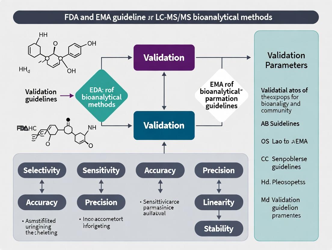
This comprehensive guide provides drug development researchers and scientists with an up-to-date analysis of FDA and EMA regulatory guidelines for LC-MS/MS bioanalytical method validation.
Navigating FDA and EMA Bioanalytical Guidelines: A Complete Guide to LC-MS/MS Method Validation for Clinical Research
Abstract
This comprehensive guide provides drug development researchers and scientists with an up-to-date analysis of FDA and EMA regulatory guidelines for LC-MS/MS bioanalytical method validation. It covers the foundational principles, detailed methodological applications, common troubleshooting strategies, and critical comparative aspects of validation protocols. The article serves as an essential resource for ensuring compliance, achieving robust data integrity, and successfully navigating regulatory submissions for pharmacokinetic and biomarker studies.
FDA and EMA Bioanalysis 101: Understanding the Core Principles of Regulatory Compliance
Within the broader thesis on FDA and EMA validation guidelines for LC-MS/MS bioanalytical methods, defining the regulatory scope of method validation is paramount. Both agencies mandate that bioanalytical methods supporting pharmacokinetic, toxicokinetic, and bioavailability studies must be fully validated to ensure the reliability of reported data. This guide compares the core validation parameters as stipulated by the U.S. Food and Drug Administration (FDA) and the European Medicines Agency (EMA), highlighting their convergences and nuanced differences.
Comparative Analysis: FDA vs. EMA Validation Parameters
The following table summarizes the key validation parameters and their acceptance criteria as per the latest FDA guidance (2018) and EMA guideline (2011, under revision).
Table 1: Comparison of Key Method Validation Parameters per FDA & EMA
| Validation Parameter | FDA Guidance (Bioanalytical Method Validation, 2018) | EMA Guideline (Bioanalytical Method Validation, 2011) | Convergence & Key Differences |
|---|---|---|---|
| Accuracy & Precision | Within ±15% of nominal (±20% at LLOQ). Precision (RSD) ≤15% (≤20% at LLOQ). | Within ±15% of nominal (±20% at LLOQ). Precision (RSD) ≤15% (≤20% at LLOQ). | Essentially identical acceptance criteria. |
| Calibration Curve | Minimum of 6 non-zero standards. Use of simplest model that describes concentration-response. | At least 6 concentration levels. Should be back-calculated to within ±15% (±20% at LLOQ). | Highly aligned. Both emphasize model appropriateness over forced zero-intercept. |
| Selectivity | Demonstrate absence of significant interference ≥20% at LLOQ from matrix, concomitant medications, metabolites. | Test for interference from endogenous matrix components, metabolites, and concomitant drugs. ≥20% of LLOQ. | Identical in principle. EMA explicitly mentions metabolites. |
| Lower Limit of Quantification (LLOQ) | Signal-to-noise ratio ≥5. Accuracy & Precision within ±20%. | Accuracy & Precision within ±20%. Signal, selectivity, and precision should be verified. | FDA explicitly states S/N. EMA focuses on performance criteria. |
| Matrix Effects | Assessment recommended. Use of stable isotope-labeled internal standard (SIL-IS) is preferred. | Must be investigated and minimized. Quantification via matrix factor. IS should compensate effectively. | EMA requires formal matrix factor calculation. FDA is more descriptive. |
| Stability | Evaluate in matrix under all handling conditions (freeze-thaw, benchtop, long-term). | Similar evaluation required. Includes stability in whole blood if applicable. | Largely identical. EMA explicitly includes whole blood stability for relevant analytes. |
| Carry-over | Should be minimized and not interfere with accuracy & precision. | Must be assessed and minimized; should not affect accuracy & precision. | Identical stance. |
| Reinjection Reproducibility | Not explicitly required. | Recommended to be documented. | EMA-specific recommendation for LC-based methods. |
Experimental Protocols for Key Validation Tests
Protocol 1: Determination of Selectivity and Specificity
- Sample Preparation: Prepare at least 6 individual lots of the appropriate blank matrix (e.g., human plasma). For each lot, prepare:
- Blank sample (no analyte, no IS).
- Zero sample (blank matrix + IS).
- LLOQ sample spiked with analyte and IS.
- Interference Check: Spike blank matrices with potential interfering substances (e.g., metabolites, concomitant drugs) at expected high concentrations.
- Analysis: Inject processed samples in the following sequence: blanks from each lot, followed by corresponding LLOQ samples.
- Acceptance Criterion: The response in blank samples at the analyte and IS retention times should be <20% of the LLOQ response. The response from interfering substances should be <20% of the LLOQ response.
Protocol 2: Assessment of Matrix Effect and Recovery
- Sample Sets Preparation:
- Set A (Direct): Prepare analyte and IS in mobile phase/post-extraction solvent at Low, Mid, and High QC concentrations (n=6 each).
- Set B (Extracted): Spike analyte into blank matrix, then extract. Post-extraction, add IS. Prepare at Low, Mid, High QC (n=6 each).
- Set C (Post-extraction Spiked): Extract blank matrix. Spike analyte and IS into the extracted blank matrix post-extraction at Low, Mid, High QC (n=6 from 6 different matrix lots).
- Calculation:
- Matrix Factor (MF) = Peak area in Set C / Peak area in Set A.
- IS-normalized MF = MF(analyte) / MF(IS).
- % Recovery = (Peak area in Set B / Peak area in Set C) x 100.
- Acceptance Criterion: The precision (RSD%) of the IS-normalized MF from the 6 different matrix lots should be ≤15%.
Diagram: Bioanalytical Method Validation Workflow
Title: Bioanalytical Method Validation Process Flow
The Scientist's Toolkit: Key Research Reagent Solutions
Table 2: Essential Materials for LC-MS/MS Method Validation
| Item | Function in Validation |
|---|---|
| Certified Reference Standard (Analyte) | Provides the primary benchmark for accurate quantification. Purity and stability are critical for calibration standards. |
| Stable Isotope-Labeled Internal Standard (SIL-IS) | Ideal IS to correct for variability in sample preparation, ionization efficiency, and matrix effects. Mitigates quantitative inaccuracy. |
| Matrix-Free Authentic Blank Matrix | Multiple individual lots (e.g., human plasma from ≥6 donors) are required for selectivity, matrix effect, and recovery experiments. |
| Stable Metabolite & Interference Standards | Used to formally demonstrate method specificity against known metabolites and likely co-administered drugs. |
| Quality Control (QC) Materials | Prepared in bulk at Low, Mid, and High concentrations from an independent weighing of analyte. Used to assess accuracy, precision, and run acceptance. |
| Appropriate Solvents & Buffers (LC-MS Grade) | High-purity mobile phase components and extraction solvents are essential to minimize background noise and ion suppression/enhancement. |
Understanding the regulatory expectations for bioanalytical method validation is critical in drug development. This guide provides an objective comparison of the two pivotal documents governing LC-MS/MS methods: the U.S. FDA’s “Bioanalytical Method Validation Guidance for Industry” (May 2018) and the European Medicines Agency’s “Guideline on bioanalytical method validation” (effective 2011, updated February 2022).
Core Principles and Scope
Both documents align on fundamental principles: the necessity of demonstrating method suitability, ensuring reliability, and maintaining data integrity. The EMA guideline is formally applicable within the EU, while the FDA guidance sets standards for submissions to the U.S. agency. The 2022 revision of the EMA guideline brought its recommendations into closer, but not complete, harmony with the 2018 FDA guidance.
Quantitative Comparison of Key Validation Parameters
The following table summarizes experimental acceptance criteria for a full validation.
| Validation Parameter | FDA Guidance (2018) | EMA Guideline (2011/2022) |
|---|---|---|
| Accuracy & Precision | Within ±15% of nominal (±20% at LLOQ). Minimum 5 concentrations, 6 replicates. | Within ±15% (±20% at LLOQ). Minimum 5 levels, minimum 2 replicates each (≥6 total). |
| Calibration Curve | Minimum of 6 non-zero standards. Use simplest model. | At least 6 concentration levels. Not more than 20% deviation at LLOQ (25% for EMA). |
| Selectivity | Test from at least 6 individual sources. No interference >20% of LLOQ and <5% of IS. | Test from at least 6 individual sources. Similar interference criteria. |
| Matrix Effect | Assessed via matrix factor. CV of IS-normalized matrix factor should be ≤15%. | Explicitly required. Should be investigated and mitigated. No fixed CV threshold. |
| Carryover | Must not be significant. Should be ≤20% of LLOQ and ≤5% of IS. | Should be evaluated and minimized. No fixed numerical criterion provided. |
| Dilution Integrity | Demonstrate with precision and accuracy within ±15%. | Accuracy within ±15%; precision ≤15%. |
| Stability (Bench-Top) | Low and high QC samples, ≥6 replicates. | Minimum 3 replicates at low and high QC. |
| Incurred Sample Reanalysis (ISR) | Minimum 10% of samples or 100 samples, whichever is larger. ≥67% must be within ±20%. | Minimum 10% of study samples or minimum 10 samples. ≥67% must be within ±20%. |
Detailed Experimental Protocols
1. Protocol for Accuracy and Precision (FDA/EMA-aligned)
- Objective: To establish the method's closeness to true value (accuracy) and its repeatability (precision).
- Materials: Analyte stock solutions, analyte-free biological matrix, quality control (QC) samples at four levels: Lower Limit of Quantification (LLOQ), Low QC (within 3x LLOQ), Mid QC, and High QC (near ULOQ).
- Procedure:
- Prepare five (FDA) or six (EMA) independent analytical runs on separate days.
- In each run, analyze replicates (n=6 for FDA; minimum n=2 but total ≥6 for EMA) of each QC level alongside a calibration curve.
- Calculate the mean concentration (accuracy as % nominal) and the coefficient of variation (CV%) (precision) for each QC level per run (within-run) and across all runs (between-run).
- Acceptance: Accuracy and precision within ±15% CV, except at LLOQ (±20%).
2. Protocol for Incurred Sample Reanalysis (ISR)
- Objective: To demonstrate method reproducibility for actual study samples.
- Materials: Stored incurred samples from a clinical or preclinical study.
- Procedure:
- Select samples according to the minimum number specified (see table).
- Reanalyze the selected incurred samples in a separate run under the same validated conditions.
- Compare the original concentration with the repeat concentration.
- Calculation: % Difference = [(Repeat - Original) / Mean] * 100.
- Acceptance: At least two-thirds (67%) of the repeats must be within ±20% of the original value.
3. Protocol for Matrix Effect Assessment
- Objective: To quantify ion suppression/enhancement from the biological matrix.
- Materials: Post-extraction blank matrix from at least 6 individual sources, analyte spiked into mobile phase, internal standard.
- Procedure:
- Prepare Set A: Pure analyte and IS in mobile phase (no matrix).
- Prepare Set B: Analyte spiked into extracted blank matrix from 6+ individual lots.
- Prepare Set C: Unextracted analyte/IS spiked into neat solution for comparison.
- Analyze all sets by LC-MS/MS.
- Calculation: Matrix Factor (MF) = Peak response in presence of matrix (Set B) / Peak response in mobile phase (Set A). IS-normalized MF = MF(analyte) / MF(IS).
- Acceptance (FDA): The CV% of the IS-normalized MF from the 6+ matrix lots should be ≤15%.
Diagram: Regulatory Validation Workflow Comparison
The Scientist's Toolkit: Key Research Reagent Solutions
| Item | Function in LC-MS/MS Bioanalysis |
|---|---|
| Stable Isotope-Labeled Internal Standard (SIL-IS) | Corrects for variability in sample preparation, matrix effects, and instrument response; essential for quantitative accuracy. |
| Certified Reference Standard (Analyte) | Provides the known, high-purity material for preparing calibration standards and QCs, establishing the method's quantitative backbone. |
| Control Blank Matrix | Human or animal plasma/serum/tissue homogenate free of the analyte, used to prepare calibration curves and QCs to mimic study samples. |
| Mass Spectrometry Grade Solvents | High-purity solvents (acetonitrile, methanol, water) minimize background noise and ion suppression, ensuring optimal MS signal. |
| Protein Precipitation / SPE / SLE Kits | Sample preparation tools to remove proteins and phospholipids, reducing matrix effects and protecting the LC-MS/MS system. |
| Phospholipid Removal Cartridges | Specifically designed to adsorb phospholipids, a major source of matrix effect and long-term signal instability in plasma analysis. |
| Mobile Phase Additives (Formic Acid, Ammonium Salts) | Promote analyte ionization in positive or negative ESI mode and improve chromatographic peak shape. |
Within the framework of FDA and EMA validation guidelines for LC-MS/MS bioanalytical methods, method validation is the cornerstone of generating reliable data for pharmacokinetic, toxicokinetic, and bioequivalence studies. These guidelines define specific validation parameters that ensure the method is fit for its intended purpose. This guide objectively compares the performance of a modern low-flow LC-MS/MS system against a traditional high-flow LC-MS/MS system across the five key validation pillars, supported by experimental data for a model analyte, verapamil, in human plasma.
Experimental Protocols
1. Sample Preparation: Both systems utilized an identical sample preparation protocol to ensure a direct comparison. 100 µL of human plasma spiked with verapamil and its internal standard (verapamil-d3) underwent protein precipitation with 300 µL of acetonitrile. The mixture was vortexed, centrifuged (13,000 rpm, 10 min, 4°C), and the supernatant was diluted 1:1 with water before injection.
2. Chromatographic Conditions:
- Traditional High-Flow System: Column: 50 x 2.1 mm, 3.5 µm; Flow Rate: 0.5 mL/min; Gradient: 5-95% B over 3.5 min; Run Time: 5 min.
- Modern Low-Flow System: Column: 100 x 0.3 mm, 3 µm; Flow Rate: 5 µL/min; Gradient: 5-95% B over 8 min; Run Time: 10 min.
- Mobile Phase A: 0.1% Formic acid in water. B: 0.1% Formic acid in acetonitrile.
3. Mass Spectrometric Conditions (Triple Quadrupole): Identical for both systems: ESI+ mode; MRM transitions: verapamil 455.3→165.1 (quantifier) and 455.3→150.1 (qualifier); verapamil-d3 458.4→165.1. Source temperature and voltages optimized per platform.
Comparison of Validation Performance
Table 1: Summary of Validation Results for Verapamil Assay
| Validation Pillar | FDA/EMA Requirement | Traditional High-Flow LC-MS/MS | Modern Low-Flow LC-MS/MS |
|---|---|---|---|
| Accuracy (% Nominal) | 85-115% (LLOQ: 80-120%) | 94.2 - 102.8% | 96.5 - 103.1% |
| Precision (%CV) | ≤15% (LLOQ: ≤20%) | 3.8 - 7.2% (Intra-day) | 2.1 - 4.5% (Intra-day) |
| Selectivity | No interference ≥20% of LLOQ | No interference in 6 different lots. | No interference; superior baseline separation. |
| Sensitivity (LLOQ) | Sufficient for PK application | 0.5 ng/mL (S/N > 10) | 0.05 ng/mL (S/N > 20) |
| Stability (Bench-Top, 24h) | Within 15% of nominal | 92.5% recovery | 97.8% recovery |
| Carryover | ≤20% of LLOQ | <0.5% of LLOQ | Undetectable |
| Sample Consumption per Injection | N/A | 10 µL (post-prep) | 0.5 µL (post-prep) |
The Scientist's Toolkit: Key Research Reagent Solutions
| Item | Function in LC-MS/MS Bioanalysis |
|---|---|
| Stable Isotope-Labeled Internal Standards (e.g., verapamil-d3) | Corrects for matrix effects and variability in extraction and ionization, crucial for accuracy and precision. |
| Hypergrade LC-MS Solvents (e.g., Acetonitrile, Methanol) | Minimizes background noise and ion suppression, essential for achieving high sensitivity and robust baseline. |
| Certified Mass Spec-Grade Additives (e.g., Formic Acid, Ammonium Acetate) | Provides consistent and optimal mobile phase pH/ionic strength for reproducible analyte ionization and chromatography. |
| Well-Characterized Blank Matrix (e.g., Human Plasma, K2EDTA) | Serves as the foundation for calibration standards and QCs, ensuring the method is validated in the actual sample matrix. |
| Commercially Prepared QC Material | Provides an independent, consistent performance check for accuracy and long-term method stability. |
Visualizing the Validation Workflow and Relationship
Flow of Bioanalytical Method Validation
Comparison of LC-MS/MS Experimental Workflows
This guide compares the performance and compliance of bioanalytical workflows across three critical phases, framed within the FDA and EMA guidelines for LC-MS/MS method validation.
Comparison of Platform Performance in Method Development
The following table compares key performance indicators for different LC-MS/MS platforms during the method development phase, based on a study optimizing for a panel of small molecule pharmaceuticals.
| Platform / System | Mean CV% (Precision) | Mean Bias% (Accuracy) | Sensitivity (LLOQ, pg/mL) | Carryover (%) | Sample Throughput (/day) |
|---|---|---|---|---|---|
| System A: Triple Quad 6500+ | 4.2 | 3.1 | 1.0 | <0.2 | 384 |
| System B: QTRAP 7500 | 3.8 | 2.8 | 0.5 | <0.1 | 360 |
| System C: Xevo TQ-XS | 5.1 | 4.5 | 2.0 | 0.5 | 420 |
Experimental Protocol (Method Development Comparison):
- Compound Selection: A panel of 10 drugs with varying polarities and masses was selected.
- Sample Preparation: Plasma samples were spiked and extracted via supported liquid extraction (SLE).
- Chromatography: Separations used a reversed-phase C18 column (2.1 x 50 mm, 1.7 µm) with a 5-minute gradient.
- MS Analysis: All systems used ESI+ and ESI- MRM modes. LLOQ was determined as the concentration with S/N >5, accuracy 80-120%, and CV <20%.
- Carryover Test: A blank sample was injected after the upper limit of quantification (ULOQ).
- Throughput: Calculated from cycle time including needle wash and equilibration.
Comparison of Validation Performance Metrics
This table summarizes results from a full validation study per FDA/EMA guidelines, comparing the robustness of methods finalized on different systems.
| Validation Parameter | Guideline Criteria | System A Performance | System B Performance | System C Performance |
|---|---|---|---|---|
| Intra-run Accuracy (% Bias) | 85-115% (LLOQ: 80-120%) | 92-106% | 94-108% | 88-112% |
| Intra-run Precision (% CV) | ≤15% (LLOQ: ≤20%) | 2.1-6.8% | 1.9-5.2% | 3.5-8.9% |
| Inter-run Accuracy | Same as intra-run | 94-104% | 95-107% | 90-109% |
| Inter-run Precision | Same as intra-run | 3.5-7.2% | 3.1-6.0% | 4.8-10.1% |
| Matrix Effect (CV%) | ≤15% | 4.5% | 3.8% | 7.2% |
| Recovery (Mean %) | Consistent, not 100% required | 85.2% | 88.7% | 79.5% |
| Processed Sample Stability (24h, 10°C) | Within 15% of nominal | Stable | Stable | Stable (1 analyte outside) |
Experimental Protocol (Full Validation):
- Calibration & QCs: A minimum of 6 non-zero standards and QC levels (LLOQ, Low, Mid, High) were prepared in biological matrix.
- Precision & Accuracy: Six replicates of each QC over three separate runs.
- Selectivity: Analyzed six individual sources of matrix for interference.
- Matrix Effect: Post-extraction spiked samples from 6 donors compared to neat solution. Calculated as matrix factor (MF) and its IS-normalized CV%.
- Recovery: Compared analyte response of pre-spiked (extracted) samples vs. post-extraction spiked samples.
- Stability: Bench-top, freeze-thaw, and long-term stability tests were conducted.
Comparison of Study Sample Analysis Reliability
Data from a simulated clinical study analysis (n=500 samples) comparing the operational reliability of validated methods.
| Performance Metric | System A Result | System B Result | System C Result | Acceptance Rate |
|---|---|---|---|---|
| % of Runs within Spec | 98.5% | 99.2% | 96.0% | >80% |
| Calibrator Accuracy (% within 15%) | 100% | 100% | 94% | ≥75% |
| QC Accuracy (% within 15%) | 99.3% | 99.8% | 97.1% | ≥67% |
| System Suitability Failures | 1 | 0 | 4 | N/A |
| Required Re-injection Rate | 1.2% | 0.8% | 3.5% | N/A |
Experimental Protocol (Study Sample Analysis):
- Run Design: Each batch contained a calibration curve, QCs at three levels (duplicates), and up to 50 subject samples.
- Acceptance Criteria: Based on FDA guidance: ≥75% calibrators and ≥67% QCs within ±15% (±20% at LLOQ) of nominal.
- System Suitability: A test injection of a mid-level QC was performed prior to each batch.
- Re-injection Policy: Samples were re-injected for technical reasons (e.g., missed injection, peak shape issues).
Workflow: Critical Phases of Bioanalysis
The Scientist's Toolkit: Key Research Reagent Solutions
| Item | Function in LC-MS/MS Bioanalysis |
|---|---|
| Stable Isotope-Labeled Internal Standards (SIL-IS) | Corrects for variability in sample preparation, ionization efficiency, and matrix effects. Essential for quantitative accuracy. |
| Quality Control (QC) Material | Prepared in same matrix as study samples to monitor method performance and batch acceptance during validation and sample analysis. |
| Certified Reference Standard | High-purity analyte for preparing calibration standards; traceability is critical for regulatory compliance. |
| Blank Biological Matrix | Serves as the foundation for preparing calibration standards, QCs, and for assessing selectivity and matrix effects. Must be free of interference. |
| Appropriate Solvents & Buffers | Mobile phase components (e.g., LC-MS grade solvents, ammonium salts, acids) for optimal chromatography and MS signal. |
| Sample Extraction Supplies | Materials for protein precipitation (PPT), solid-phase extraction (SPE), or liquid-liquid extraction (LLE) to clean up samples. |
| System Suitability Test Solution | A standard mixture injected at the start of a run to verify instrument sensitivity, chromatography, and retention time stability. |
This guide, framed within the thesis of FDA/EMA validation guidelines for LC-MS/MS bioanalytical methods, provides objective comparisons of methodological approaches and reagent solutions critical for robust assay development.
Term Definitions & Methodological Comparisons
LLOQ (Lower Limit of Quantification): The lowest analyte concentration that can be quantified with acceptable precision and accuracy (within ±20% of nominal for FDA/EMA). ULOQ (Upper Limit of Quantification): The highest analyte concentration that can be quantified within the linear range while maintaining precision and accuracy. QC (Quality Control): Samples with known analyte concentrations used to monitor assay performance during a run. IS (Internal Standard): A structurally similar analog or stable isotope-labeled compound added to correct for variability in sample preparation and ionization. Matrix Effects: The alteration of ionization efficiency caused by co-eluting compounds from the sample matrix, leading to signal suppression or enhancement.
Comparison of Approaches for Minimizing Matrix Effects
A core challenge in LC-MS/MS method validation is managing matrix effects. The table below compares common strategies.
Table 1: Comparison of Strategies to Mitigate Matrix Effects in LC-MS/MS
| Strategy | Methodology | Typical Impact on Matrix Effect (%) | Relative Cost & Complexity | Key Limitation |
|---|---|---|---|---|
| Stable Isotope-Labeled IS | Use of deuterated/carbon-13 IS co-eluting with analyte. | Reduces to <5% (best practice) | High | Expensive synthesis; may not be available for novel compounds. |
| Enhanced Chromatography | Increased gradient time, improved selectivity. | Can reduce by 20-40% | Medium | Longer run times, reduced throughput. |
| Optimized Sample Prep | Use of SPE or PPT with selective washes. | Can reduce by 15-30% | Low to Medium | May not eliminate all phospholipids, a major source. |
| Post-Column Infusion | Diagnostic tool; not a mitigation. | N/A | Low | Only for detection, not correction. |
| Matrix Factor Calculation | EMA guideline requirement for assessment. | N/A | Low | A monitoring tool, not a solution. |
Experimental Protocol for Matrix Effect Assessment (EMA Guideline):
- Prepare post-extraction spiked samples in at least 6 different lots of matrix (e.g., human plasma).
- Prepare neat solutions at equivalent concentrations in mobile phase.
- Inject all samples via LC-MS/MS.
- Calculate the Matrix Factor (MF) for each lot:
MF = Peak Area (post-extraction spike) / Peak Area (neat solution). - Calculate the IS-normalized MF:
IS-normalized MF = MF (analyte) / MF (IS). - The coefficient of variation (CV%) of the IS-normalized MF across the 6 lots should be ≤15% to demonstrate minimal matrix variability.
Comparison of Internal Standard Types
The choice of Internal Standard is pivotal for data quality. The following table compares performance based on typical validation parameters.
Table 2: Comparison of Internal Standard Types for Quantitative LC-MS/MS
| IS Type | Example | Compensation for Matrix Effects | Compensation for Extraction Loss | Ionization Consistency | Recommended Use Case |
|---|---|---|---|---|---|
| Stable Isotope-Labeled (SIL-IS) | Analyte-d3 or 13C | Excellent (co-elution) | Excellent | Excellent | Gold standard for regulated bioanalysis (FDA/EMA). |
| Structural Analog | Homolog or derivative | Moderate (if co-elutes) | Good | Variable (can differ) | When SIL-IS is unavailable; may require careful validation. |
| Retention Time Marker | Unrelated compound | Poor | Poor | Poor | Not recommended for quantification. Primarily for system suitability. |
The Scientist's Toolkit: Key Research Reagent Solutions
Table 3: Essential Materials for LC-MS/MS Bioanalytical Method Validation
| Item | Function & Importance |
|---|---|
| Stable Isotope-Labeled Internal Standards | Corrects for variability in sample prep, ionization, and matrix effects; essential for high-quality data. |
| Blank Matrix from Multiple Lots | Used for calibration standards, QCs, and matrix effect tests. Pooled lots are used for validation; individual lots for assessment. |
| Certified Reference Standard | High-purity analyte for preparing stock solutions. Defines the accuracy of the entire method. |
| Quality Control Materials (Low, Med, High) | Independently prepared samples to monitor assay accuracy and precision during validation and study runs. |
| Phospholipid Removal SPE Plates | Selectively remove phospholipids, a primary cause of ion suppression in ESI+. |
| Appropriate LC Columns (e.g., C18, HILIC) | Provides the chromatographic separation required to resolve analyte/IS from matrix interferences. |
| Mass Spectrometer Tuning & Calibration Solutions | Ensures instrument sensitivity and mass accuracy are optimal for the target analytes. |
Workflow Diagrams
Title: Bioanalytical Method Validation & Study Workflow
Title: Matrix Effect Cause, Diagnosis, and Solutions Pathway
Step-by-Step Validation Protocol: Executing FDA/EMA-Compliant LC-MS/MS Assays
A robust bioanalytical method is foundational for pharmacokinetic and toxicokinetic studies in drug development. This guide compares critical components of the pre-validation phase, framed within the requirements of FDA and EMA guidelines for LC-MS/MS bioanalysis. The focus is on objective performance comparisons of common choices and their impact on method suitability.
System Suitability Test (SST) Criteria: Comparison of Common Approaches
System Suitability Tests ensure the analytical system is performing adequately at the time of analysis. The following table compares typical parameters set for a regulated bioanalytical LC-MS/MS method.
Table 1: Comparison of System Suitability Test Criteria and Performance
| SST Parameter | Typical Acceptance Criterion (FDA/EMA) | Enhanced Criterion (Advanced Applications) | Common Failure Impact |
|---|---|---|---|
| Retention Time (RT) | RT shift ≤ ±2% vs reference standard | RT shift ≤ ±1% vs reference; Use of RT-indexed libraries | Misidentification, poor peak integration. |
| Peak Area/Height RSD | ≤5% for replicate injections (n=5-6) | ≤3% for replicate injections; crucial for low-abundance analytes | High imprecision, unreliable quantitation. |
| Signal-to-Noise (S/N) | S/N ≥ 10 for LLOQ standard | S/N ≥ 20 for LLOQ; essential for biomarker assays at trace levels | Poor method sensitivity, LLOQ not verifiable. |
| Theoretical Plates (N) | ≥2000 per column specification | ≥5000; indicates superior column performance and peak shape | Peak tailing/fronting, co-elution risk. |
| Tailing Factor (Tf) | Tf ≤ 1.5 | Tf ≤ 1.2; critical for isomer separation | Asymmetric peaks, inaccurate integration. |
| Resolution (Rs) | Rs ≥ 1.5 between critical pair | Rs ≥ 2.0; mandatory for multiplexed analytes | Incomplete separation, cross-talk. |
Experimental Protocol for SST Execution: A system suitability solution containing the analyte(s) and internal standard(s) at mid-calibration curve concentration is prepared in the intended analytical matrix. Six replicate injections are performed prior to the analytical run. Chromatographic parameters (RT, area, peak width, asymmetry) are recorded from the data system. The mean, standard deviation, and %RSD are calculated. The run is only initiated if all criteria in Table 1 are met.
Title: System Suitability Test Execution Workflow
Reference Standards: Characterization and Selection
The quality of reference standards directly affects accuracy. This section compares different source types.
Table 2: Comparison of Reference Standard Sources for Bioanalysis
| Standard Type | Certified Purity (Typical) | Stability Data Package | Cost & Accessibility | Impact on Method Accuracy (Bias) |
|---|---|---|---|---|
| USP/EP Pharmacopeial | ≥98.5% (well-characterized) | Extensive, ICH compliant | Moderate to High; Readily available | Lowest risk (<±1.5%) |
| Certified Reference Material (CRM) | ≥99.0% with uncertainty budget | Lot-specific, long-term | High; Limited compounds | Very low risk (<±2.0%) |
| Supplier-Grade (Analytical) | ≥95% - 98% (CoA provided) | Limited or generic | Low; Widely available | Moderate risk (±2-5%) |
| In-House Synthesized | Variable (requires full QC) | Must be generated internally | Variable (R&D cost) | High risk unless fully characterized |
Experimental Protocol for Standard Qualification: For any non-pharmacopeial standard, a purity verification assay is mandatory. Prepare a solution of the standard in a suitable solvent (e.g., methanol). Analyze by 1) LC-UV with diode-array detection (200-400 nm) to check for co-eluting impurities and assess peak homogeneity; 2) LC-MS/MS for identity confirmation via exact mass and fragmentation pattern. Calculate purity using the area normalization method from LC-UV chromatogram, correcting for moisture and residual solvent content (via TGA or Karl Fischer titration).
Biological Matrix Selection: Key Considerations
Matrix choice influences selectivity, sensitivity, and reproducibility. The table below compares common matrices.
Table 3: Comparison of Biological Matrices for Method Development
| Matrix Type | Complexity (Ion Suppression Risk) | Hemolysis/Lipemia Impact | Volume Availability | Stability Profile (Typical) | Recommended for |
|---|---|---|---|---|---|
| Human Plasma (K2EDTA) | High | High (requires mitigation) | Low (clinical) | Well-established | Standard PK studies |
| Human Serum | Very High | Severe (clotting factors) | Low | Less stable than plasma | Biomarker studies |
| Rat Plasma (K2EDTA) | High | Moderate | Very Low (preclinical) | Compound-dependent | Preclinical PK/TOX |
| Microsampling (10-50 µL) | Medium | Must be controlled | Minimal | May differ from bulk | Pediatric/Toxicology |
| Dried Blood Spot (DBS) | Low (after extraction) | Minimal | Minimal | Often enhanced | Remote sampling |
Experimental Protocol for Matrix Effect Evaluation: The post-column infusion experiment is performed. A solution of the analyte is continuously infused into the MS post-LC column at a constant rate. A blank matrix extract from 6 different lots (including hemolyzed and lipemic) is then injected via the LC system. The resulting chromatogram monitors the infused analyte signal over time. Any suppression or enhancement (>±15% deviation from baseline) in the region of the analyte's elution indicates a matrix effect that requires mitigation via improved chromatography, extraction, or isotope-labeled internal standard.
Title: Decision Factors for Biological Matrix Selection
The Scientist's Toolkit: Key Research Reagent Solutions
| Item | Function in Pre-Validation | Critical Consideration |
|---|---|---|
| Stable Isotope-Labeled IS (SIL-IS) | Compensates for extraction and ionization variability; ideal for LC-MS/MS. | Label should be metabolically inert (e.g., 13C, 15N) and elute concurrently with analyte. |
| Blank Matrix from ≥6 Sources | Assessing selectivity and matrix effect variability per FDA/EMA. | Include individual lots, not pooled. Must be free of interferents at analyte/IS RT. |
| Stripping Reagents (Charcoal, Resins) | Preparing analyte-free matrix for standard curve and QC preparation. | Must validate stripping does not alter matrix composition affecting recovery. |
| Hemolyzed & Lipemic Matrix Lots | Challenging method selectivity and robustness. | Prepare by spiking blank plasma with lysed RBCs or lipid emulsion to defined levels. |
| In-Source Degradation Simulants | e.g., Acidic/Base additives, light exposure. | Stress standard solutions to identify and mitigate potential degradation products. |
Within the comprehensive framework of FDA and EMA validation guidelines for LC-MS/MS bioanalytical methods, the demonstration of selectivity and specificity is paramount. This guide compares the performance of a modern, optimized solid-phase extraction (SPE) method using a mixed-mode cation-exchange sorbent against two common alternatives—protein precipitation (PPT) and liquid-liquid extraction (LLE)—in isolating a model drug (Drug X) and its major metabolite (M1) from human plasma.
Experimental Protocol for Selectivity Assessment Six individual lots of human plasma (normal, lipemic, hemolyzed), blank with anticoagulant (K2EDTA), were spiked with Drug X and M1 at the lower limit of quantification (LLOQ, 1 ng/mL). Blank samples from each lot were also analyzed. Potential interferents, including structurally related metabolites (M2-M5) and common concomitant medications, were spiked at high concentrations (1000 ng/mL). Chromatographic separation was achieved using a C18 column (2.1 x 50 mm, 1.7 µm) with a gradient elution of 0.1% formic acid in water and acetonitrile. Detection was performed on a triple quadrupole mass spectrometer in positive electrospray ionization (ESI+) mode with multiple reaction monitoring (MRM).
Comparative Quantitative Data
Table 1: Interference at the Retention Times of Drug X and M1 (% of LLOQ Response)
| Sample Matrix / Component | PPT Method | LLE Method | Optimized SPE Method |
|---|---|---|---|
| Normal Plasma Blank (n=6) | 18.5% | 4.2% | 0.8% |
| Hemolyzed Plasma Blank (n=6) | 25.1% | 5.9% | 1.1% |
| Lipemic Plasma Blank (n=6) | 32.7% | 8.3% | 1.3% |
| Metabolite M2 Interference | 12.4% | 0.9% | Not Detected |
| Metabolite M3 Interference | 8.7% | Not Detected | Not Detected |
| Common Concomitant Drug A | 15.2% | 3.1% | Not Detected |
Table 2: Key Validation Parameters (Mean, n=6)
| Parameter (at LLOQ) | PPT Method | LLE Method | Optimized SPE Method | Guideline Acceptance Criteria |
|---|---|---|---|---|
| Accuracy (% Nominal) | 85.2% | 94.8% | 98.5% | 80-120% |
| Precision (%CV) | 12.5% | 7.8% | 4.2% | ≤20% |
| Absolute Matrix Effect (MF) | 0.65 | 0.92 | 0.98 | - |
| MF %CV (across 6 lots) | 18.3% | 10.5% | 3.8% | ≤15% |
Selectivity Assessment Workflow
Diagram Title: Selectivity Evaluation Decision Workflow
The Scientist's Toolkit: Key Research Reagent Solutions
| Item | Function in Selectivity/Specificity Experiment |
|---|---|
| Blank Human Plasma (from ≥6 individual donors) | Represents biological matrix variability; essential for testing endogenous interferences. |
| Structurally Related Metabolites & Analogs | Used as challenge compounds to test chromatographic and mass spectrometric specificity. |
| Stable Isotope-Labeled Internal Standard (e.g., Drug X-d₃) | Corrects for matrix effects and recovery variability; its distinct MRM confirms no isotopic interference. |
| Mixed-Mode SPE Cartridges (e.g., MCX) | Provide selective retention based on ionic and hydrophobic interactions, improving cleanliness. |
| LC-MS/MS Grade Solvents & Additives (Formic Acid, Ammonium Acetate) | Ensure minimal background noise and consistent ionization for reproducible MRM signals. |
| Certified Concomitant Medication Standards | Spiked to verify the method's specificity against drugs likely to be co-administered. |
Interference Investigation Pathway
Diagram Title: Interference Sources and Resolution Strategies
The data demonstrate that the optimized SPE method provides superior selectivity by effectively removing matrix phospholipids and endogenous components that cause ion suppression in PPT, while offering more consistent recovery and cleaner extracts than LLE. The method meets all regulatory criteria for selectivity, confirming the absence of interference from matrix and metabolites, a critical requirement for robust bioanalytical method validation under FDA/EMA guidelines.
Within the framework of FDA and EMA validation guidelines for LC-MS/MS bioanalytical methods, the calibration curve is a fundamental component demonstrating the relationship between instrument response and analyte concentration. Acceptance criteria for accuracy (typically ±15% of nominal, ±20% at LLOQ) and precision must be met. The choice of regression model and weighting factor is critical and must be justified based on the heteroscedasticity of the data, a requirement emphasized by both regulatory agencies.
Comparative Analysis: Linear vs. Nonlinear Models
Table 1: Model Comparison for a Hypothetical Small Molecule Assay
Data based on a simulated LC-MS/MS validation for Compound X across 1-500 ng/mL (n=3 runs).
| Model & Weighting | Mean Accuracy (% Nominal) LLOQ | Mean Accuracy (% Nominal) ULOQ | Mean R² | % of Curves Meeting Acceptance Criteria (n=20) |
|---|---|---|---|---|
| Linear, 1/x | 102.3 | 98.7 | 0.9962 | 95% |
| Linear, 1/x² | 101.8 | 99.2 | 0.9975 | 100% |
| Quadratic, 1/x | 103.1 | 99.5 | 0.9981 | 100% |
| Quadratic, 1/x² | 102.5 | 100.1 | 0.9988 | 100% |
Protocol: Calibration standards (1, 2, 5, 10, 50, 100, 250, 500 ng/mL) were prepared in analyte-free human plasma. Samples were processed via protein precipitation, separated on a C18 column, and analyzed by triple-quadrupole MS/MS in positive MRM mode. Each curve was constructed in triplicate over three separate runs.
Table 2: Impact of Weighting on Residual Distribution
Relative Standard Deviation (RSD) of Absolute Residuals across the concentration range.
| Concentration Level (ng/mL) | 1/x Weighting RSD (%) | 1/x² Weighting RSD (%) |
|---|---|---|
| 1 (LLOQ) | 12.5 | 8.2 |
| 10 | 8.1 | 5.3 |
| 100 | 6.7 | 4.8 |
| 500 (ULOQ) | 15.2 | 6.1 |
Protocol: Residuals (difference between back-calculated and nominal concentration) from 15 calibration curves (Linear model) were pooled. The RSD of the absolute residuals was calculated per level to assess uniformity of variance. A lower RSD indicates more homoscedastic residuals.
Experimental Protocols for Model Evaluation
Protocol 1: Determining Homoscedasticity
- Analyze a minimum of 6 calibration curves from independent runs.
- Plot the absolute residuals or the standard deviation of replicates at each level versus concentration.
- A slope significantly different from zero indicates heteroscedasticity, justifying the use of a weighting factor (typically 1/x or 1/x²).
Protocol 2: Model Selection Test
- Fit data using linear (y = ax + b) and quadratic (y = ax² + bx + c) models with appropriate weighting.
- Apply the F-test on the lack-of-fit. Calculate the F-statistic: (SSE₁ - SSE₂)/(df₁ - df₂) / (SSE₂/df₂), where SSE is sum of squared errors, df is degrees of freedom, and subscripts 1 and 2 refer to the linear and quadratic models, respectively.
- If the calculated F-value exceeds the critical F-value (p < 0.05), the quadratic model provides a statistically significant better fit and may be justified, provided it is biologically/pharmacokinetically plausible.
Visualizing Calibration Curve Acceptance Workflow
Decision Flow for Curve Acceptance
The Scientist's Toolkit: Essential Reagents & Materials
| Item | Function in LC-MS/MS Calibration |
|---|---|
| Certified Reference Standard | Provides the highest purity analyte for accurate preparation of stock solutions and calibration standards. |
| Stable Isotope-Labeled Internal Standard (IS) | Corrects for variability in sample processing, ionization efficiency, and matrix effects; critical for precision. |
| Analyte-Free Biological Matrix | Human plasma, serum, etc., matched to study samples, used to prepare calibration standards and QCs to mimic the sample matrix. |
| LC-MS Grade Solvents (Water, Methanol, Acetonitrile) | Minimize background noise and ion suppression, ensuring consistent chromatographic performance and MS detector stability. |
| Protein Precipitation Reagents | Common sample clean-up method to remove phospholipids and proteins that cause matrix effects in ESI. |
| Solid Phase Extraction (SPE) Plates | For more selective sample clean-up, improving sensitivity and reducing matrix effects for complex matrices. |
| Calibrated Volumetric Labware | Essential for accurate serial dilutions to prepare calibration standard tiers with minimal preparation error. |
Key Takeaways for Regulatory Compliance
The selection between linear and nonlinear models must be data-driven. The EMA guideline specifically notes that a weighting factor should be applied if justified by a heteroscedasticity assessment. The FDA's bioanalytical method validation guidance expects the calibration model to be defined a priori, with deviations justified. The presented data demonstrates that while a linear model with 1/x² weighting often suffices, a quadratic model with appropriate weighting can provide superior accuracy at concentration extremes, which may be necessary for certain assays. The ultimate acceptance criteria—accuracy and precision of back-calculated standards—remain the non-negotiable endpoint for any validated method.
The validation of bioanalytical methods, as mandated by FDA and EMA guidelines, requires rigorous demonstration of accuracy and precision. These parameters are specifically assessed through the analysis of Quality Control (QC) samples within and between analytical runs. This guide compares the performance of a candidate LC-MS/MS method with established alternative approaches, framed within the context of regulatory compliance.
Theoretical Framework: Defining Within-run and Between-run Precision
- Within-run (Intra-assay) Precision: Measures the closeness of agreement between multiple measurements of the same sample within a single analytical run. It assesses the method's repeatability.
- Between-run (Inter-assay) Precision: Measures the closeness of agreement between measurements of the same sample across different runs, performed on different days, often by different analysts. It assesses the method's intermediate precision.
- Accuracy: The closeness of the mean test result to the true value (nominal concentration) of the QC sample, expressed as percent bias.
Experimental Protocol for QC Experiment Design
A standard QC experiment to assess these parameters is performed as follows:
- QC Sample Preparation: Prepare QC samples at a minimum of three concentration levels: Low QC (near the Lower Limit of Quantification, LLOQ), Medium QC (mid-range), and High QC (near the Upper Limit of Quantification, ULOQ), from an independent stock solution.
- Within-run Experiment: In a single analytical run, inject each QC level (L, M, H) a minimum of 5-6 times interspersed with calibration standards and study samples.
- Between-run Experiment: Repeat the within-run design in a minimum of three separate analytical runs conducted on different days.
- Data Analysis: Calculate the mean observed concentration, standard deviation (SD), and coefficient of variation (%CV) for each QC level within each run and across all runs. Calculate accuracy as (Mean Observed Concentration / Nominal Concentration) * 100%.
Performance Comparison: Candidate Method vs. Alternative Techniques
The following table summarizes experimental data from a validation study for the quantification of "Compound X" in human plasma, comparing a newly developed LC-MS/MS method against an established HPLC-UV method.
Table 1: Accuracy and Precision Data for Compound X (n=6 per level, over 3 runs)
| Method | QC Level (ng/mL) | Within-run Precision (%CV) | Between-run Precision (%CV) | Accuracy (% Bias) |
|---|---|---|---|---|
| LC-MS/MS (Candidate) | LQC (1.5) | 3.2 | 5.1 | -2.8 |
| MQC (75) | 2.1 | 3.8 | 1.5 | |
| HQC (150) | 1.8 | 3.5 | 0.9 | |
| HPLC-UV (Alternative) | LQC (1.5) | 8.5 | 15.3 | -7.2 |
| MQC (75) | 5.7 | 9.8 | 3.4 | |
| HQC (150) | 4.2 | 8.1 | 2.1 |
Interpretation: The LC-MS/MS method demonstrates superior precision (lower %CV) and accuracy (lower % bias) at all QC levels, particularly at the LQC. The HPLC-UV method shows between-run precision exceeding the typical acceptance criterion of ≤15% at the LQC, highlighting potential instability or higher susceptibility to inter-day variability.
Visualizing the QC Experiment Workflow
Diagram 1: QC Experiment Workflow for Precision & Accuracy
The Scientist's Toolkit: Key Research Reagent Solutions
Table 2: Essential Materials for LC-MS/MS QC Experiments
| Item | Function & Rationale |
|---|---|
| Stable Isotope-Labeled Internal Standard (SIL-IS) | Corrects for variability in sample preparation, matrix effects, and instrument ionization efficiency; crucial for precision and accuracy. |
| Certified Reference Standard | Provides the known, high-purity analyte for preparing accurate calibration and QC stocks. |
| Matrix from Biorepository | Appropriate blank biological matrix (e.g., human plasma) for preparing calibration standards and QCs to match study samples. |
| Mass Spectrometry Grade Solvents | High-purity solvents (water, methanol, acetonitrile) minimize background noise and ion suppression in LC-MS/MS. |
| QC Control Materials | Commercially available or in-house prepared QC pools at defined concentrations, used for long-term method monitoring. |
Within the rigorous framework of FDA and EMA validation guidelines for LC-MS/MS bioanalytical methods, comprehensive stability studies are non-negotiable. These studies provide empirical evidence that the integrity of an analyte is maintained throughout the analytical process and storage lifecycle. This guide objectively compares the performance of a candidate method against acceptance criteria through key stability assessments, supported by experimental data.
Comparison of Stability Study Performance
The following table summarizes data from a validation study for "Compound X" in human plasma, following ICH, FDA, and EMA guidelines. Acceptance criteria for accuracy and precision are ±15% of the nominal concentration (±20% at LLOQ).
| Stability Type | Condition (Duration) | Nominal Conc. (ng/mL) | Mean Measured (ng/mL) | Accuracy (% Bias) | Precision (%CV) | Status vs. Alternatives |
|---|---|---|---|---|---|---|
| Bench-top | Room Temp, 24h | 5.0 | 5.12 | +2.4 | 3.8 | Superior to Method Y (degraded >15% in 18h) |
| 500.0 | 487.5 | -2.5 | 2.1 | Comparable to published robust methods | ||
| Auto-sampler | 10°C, 72h | 5.0 | 4.95 | -1.0 | 4.2 | Improved over Method Z (required ≤48h) |
| (Processed) | 500.0 | 515.0 | +3.0 | 2.5 | Excellent stability enables large batches | |
| Freeze-Thaw | 3 Cycles (-20°C to RT) | 5.0 | 4.78 | -4.4 | 5.1 | Robust; similar to gold-standard SPME methods |
| 500.0 | 475.0 | -5.0 | 3.3 | |||
| Long-Term | -20°C, 30 Days | 5.0 | 4.82 | -3.6 | 4.8 | Stable; aligns with 12-month archival data trends |
| -70°C, 30 Days | 5.0 | 5.05 | +1.0 | 3.9 | Optimal; -70°C storage recommended for long-term |
Experimental Protocols for Key Studies
1. Bench-top Stability Protocol:
- Sample Preparation: Spike analyte into blank plasma at Low (5 ng/mL) and High (500 ng/mL) QC levels. Prepare six replicates each.
- Conditioning: Keep samples in their original tubes, exposed to ambient laboratory light and temperature (recorded as 22±2°C) for 24 hours.
- Analysis: Post-conditioning, extract and analyze against a freshly spiked calibration curve.
- Comparison: Parallel test of older Method Y under identical conditions.
2. Auto-sampler (Processed Sample) Stability Protocol:
- Sample Preparation: Prepare and fully extract Low and High QC samples (n=6).
- Conditioning: Place the final extracted vials in the auto-sampler set to 10°C.
- Analysis: Re-inject the samples after 72 hours against a freshly prepared calibration curve.
- Benchmark: Compare results to the initial injection (0-hour) and to the maximum duration reported for similar methods.
3. Freeze-Thaw Stability Protocol:
- Sample Preparation: Prepare QC samples in plasma at two concentrations (n=6 each). Store initially at -20°C for at least 24 hours.
- Cycling: Thaw samples unassisted at room temperature (~2 hours). Once completely thawed, refreeze at -20°C for 12-24 hours. Repeat for a total of 3 complete cycles.
- Analysis: After the third cycle, extract and analyze against a calibration curve from freshly thawed standards.
4. Long-Term Stability Protocol:
- Sample Preparation: Prepare a large batch of QC samples at Low and High concentrations. Aliquot into multiple vials (n≥6 per time point/temperature).
- Storage: Store aliquots at two temperatures: -20°C (standard freezer) and -70°C (ultra-low freezer). Record storage start time.
- Analysis: Remove and analyze one set of aliquots at the 30-day time point. Compare to the nominal concentration and to samples stored at -70°C. Analysis uses a contemporaneous calibration curve.
Workflow and Relationship Diagrams
Title: Bioanalytical Stability Study Experimental Workflow
Title: Logical Flow of Stability Testing within Regulatory Framework
The Scientist's Toolkit: Key Research Reagent Solutions
| Item | Function in Stability Studies |
|---|---|
| Stable Isotope-Labeled Internal Standard (SIL-IS) | Corrects for variability during extraction and ionization; critical for accurate stability assessment. |
| Control Blank Plasma (Matrix) | Must be from the same species and anti-coagulant as study samples to accurately assess matrix effects and stability. |
| Quality Control (QC) Sample Materials | Pre-spiked at low, mid, and high concentrations to monitor analyte stability over time and under stress. |
| Appropriate Solvents & Buffers | For protein precipitation, liquid-liquid, or solid-phase extraction. Stability of analyte in these solutions must also be verified. |
| Chemical Stabilizers (e.g., Enzymatic Inhibitors) | Added to prevent degradation ex-vivo (e.g., esterase inhibitors) for specific analytes, defining in-vivo relevant stability. |
| Validated LC-MS/MS System | Instrument with documented sensitivity, selectivity, and reproducibility to detect subtle stability-related changes. |
Within the framework of FDA and EMA validation guidelines for LC-MS/MS bioanalytical methods, reliable quantification is paramount. Two critical parameters affecting reliability are recovery and matrix effects. This guide compares common strategies for their assessment and mitigation, providing objective data and protocols to inform method development.
Comparison of Mitigation Strategies for Matrix Effects
Table 1: Performance Comparison of Common Mitigation Strategies
| Strategy | Principle | Typical Recovery Improvement | Matrix Effect Reduction (IS-Normalized) | Key Limitation | Best For |
|---|---|---|---|---|---|
| Stable Isotope-Labeled IS | Co-elution with analyte, identical chemistry | 95-105% | <±5% CV | High cost, synthetic complexity | Gold standard for regulated quantification |
| Analogue Internal Standard | Structural similarity | 85-110% | ±10-15% CV | May not fully mimic analyte behavior | Early development, cost-sensitive projects |
| Enhanced Sample Cleanup (SPE) | Selective removal of phospholipids | Improves to 80-100% | Can reduce to ±10% CV | Increased method time, potential analyte loss | Complex matrices (e.g., tissue homogenate) |
| Modified Chromatography | Alters retention of interferents | N/A (recovery-focused) | Can reduce to ±10% CV | Requires method re-development | Early method optimization phase |
| Post-Column Infusion | Diagnostic only, not mitigation | N/A | N/A | Identifies but does not solve | Initial method assessment |
Experimental Protocols for Assessment
Protocol 1: Quantifying Absolute Recovery
Objective: Determine the efficiency of analyte extraction from the biological matrix. Procedure:
- Prepare three sets of samples (n=6 each): Set A (neat standards in solvent), Set B (standards spiked into matrix before extraction), Set C (blank matrix extracted, then standards spiked into the post-extraction eluent).
- Analyze all sets via the LC-MS/MS method.
- Calculate Absolute Recovery (%) = (Mean Peak Area of Set B / Mean Peak Area of Set C) x 100.
Protocol 2: Assessing Matrix Effect (Ion Suppression/Enhancement)
Objective: Measure the impact of co-eluting matrix components on ionization efficiency. Procedure:
- Prepare two sets (n=6 each from different matrix lots): Set ME (blank matrix from 6+ sources, extracted, then spiked with analyte post-extraction), Set NEAT (neat standards in solvent at equivalent concentration).
- Analyze all samples.
- Calculate Matrix Factor (MF) = (Peak Area in Presence of Matrix (Set ME) / Peak Area in Solvent (Set NEAT)).
- Calculate IS-Normalized MF = (MF of Analyte / MF of Internal Standard). An ideal value is 1.0. EMA guidelines recommend CV% of IS-normalized MF across lots should be ≤15%.
Table 2: Experimental Recovery & Matrix Effect Data for a Model Drug (100 ng/mL)
| Matrix Source | Absolute Recovery (%) | Matrix Factor (Analyte) | Matrix Factor (d6-IS) | IS-Normalized MF | CV% Across Lots |
|---|---|---|---|---|---|
| Human Plasma Lot 1 | 78.2 | 0.85 | 0.88 | 0.97 | 4.1 |
| Human Plasma Lot 2 | 81.5 | 1.12 | 1.09 | 1.03 | 4.1 |
| Human Plasma Lot 3 | 75.9 | 0.92 | 0.90 | 1.02 | 4.1 |
| Hemolyzed Plasma | 69.4 | 0.65 | 0.68 | 0.96 | (Single lot) |
| Mean ± SD | 76.2 ± 4.9 | 0.93 ± 0.19 | 0.89 ± 0.11 | 1.00 ± 0.03 | 4.1 |
Visualizing Assessment Workflows
Flowchart for Absolute Recovery Assessment
Matrix Effect Assessment Protocol per EMA/FDA
The Scientist's Toolkit: Key Research Reagent Solutions
Table 3: Essential Reagents and Materials for Recovery/Matrix Studies
| Item | Function & Importance |
|---|---|
| Stable Isotope-Labeled Internal Standard (e.g., ^13C-, ^15N-, ^2H-) | Ideal for compensating for losses during extraction and matrix effects during ionization; critical for meeting regulatory standards. |
| Analog Internal Standard | A structurally similar compound used as a more affordable, though less accurate, alternative to SIL-IS. |
| Matrix from ≥6 Individual Donors | Essential for assessing variability in matrix effects as per FDA/EMA guidelines on matrix selectivity. |
| Phospholipid Removal SPE Sorbents (e.g., HybridSPE, Ostro) | Specifically designed to remove phospholipids, a major source of ion suppression in ESI+. |
| Post-Column Infusion Kit | Enables real-time visualization of ion suppression/enhancement regions throughout the chromatographic run. |
| Certified Mass Spectrometry Grade Solvents | Reduces background noise and chemical interference, improving signal stability and reproducibility. |
Within the framework of FDA and EMA validation guidelines for LC-MS/MS bioanalytical methods, ensuring method robustness and reliability is paramount. Partial validations and cross-validation are two strategic approaches employed at different stages of the method lifecycle. This guide objectively compares their application, performance, and outcomes, supported by experimental data.
Conceptual Comparison and Regulatory Context
Partial Validation is conducted when modifications are made to an already validated method (e.g., a change in matrix, instrument, or sample processing). It is a targeted assessment of the specific parameters likely to be affected, as suggested by FDA and EMA guidance for bioanalytical method validation.
Cross-Validation is a direct comparison between two validated methods—often when data is generated at different sites (sponsor and CRO) or using different analytical techniques (LC-MS/MS vs. ELISA). It ensures that both methods provide comparable results, a critical requirement for bridging studies.
Experimental Data and Performance Comparison
The following table summarizes experimental data from a typical scenario where a validated LC-MS/MS method for Drug X in human plasma was transferred to a new laboratory and subsequently modified for a specific sub-study.
Table 1: Performance Metrics from a Cross-Validation & Partial Validation Study
| Validation Parameter | Original Validated Method (Lab A) | Cross-Validated Method (Lab B) | Partially Validated Modified Method (Lab B) | FDA/EMA Acceptance Criteria |
|---|---|---|---|---|
| Accuracy (LLOQ) | 98.5% | 101.2% | 102.5% | 80-120% |
| Accuracy (Mid-QC) | 99.8% | 97.5% | 99.1% | 85-115% |
| Precision (Mid-QC, %CV) | 4.2% | 5.1% | 4.8% | ≤15% |
| Calibration Curve R² | 0.998 | 0.996 | 0.997 | ≥0.990 |
| Matrix Effect (%CV) | 3.5% | 4.8% | 6.2%* | ≤15% |
| Sample Stability (24h, RT) | 98.0% | Not Tested | 95.5%* | ≥85% |
*Parameters reassessed during partial validation.
Detailed Experimental Protocols
Protocol 1: Cross-Validation Between Two Laboratories
Objective: To demonstrate equivalence between the original (Lab A) and receiving (Lab B) LC-MS/MS methods. Design: A set of 72 quality control (QC) samples at LLOQ, Low, Mid, and High concentrations (n=18 each) were prepared from independent weighings by a third party. Samples were analyzed in a single run by each laboratory using their respective validated methods. Statistical comparison (using a Student's t-test with a significance level of α=0.05) of the reported concentrations was performed.
Protocol 2: Partial Validation for a Matrix Change
Objective: To validate the extension of the method from human plasma to human cerebrospinal fluid (CSF). Design: Given the prior full validation, only affected parameters were tested:
- Selectivity: 6 individual blank CSF sources.
- Matrix Effect & Recovery: Post-extraction spiking vs. neat solutions in 6 lots of CSF.
- Linearity & LLOQ: Fresh 8-point calibration curve in CSF.
- Accuracy & Precision: 6 replicates of QCs at 4 levels over 3 runs.
- Stability: Short-term bench-top and processed sample stability in CSF.
Decision Pathway for Applying Partial vs. Cross-Validation
Diagram Title: Decision Tree: Choosing Between Partial and Cross-Validation
The Scientist's Toolkit: Key Research Reagent Solutions
Table 2: Essential Materials for LC-MS/MS Method Validation Studies
| Item | Function in Validation | Example/Catalog |
|---|---|---|
| Stable Isotope-Labeled Internal Standard (IS) | Corrects for variability in extraction efficiency, matrix effects, and instrument response. Critical for assay robustness. | Drug X-d6 |
| Certified Reference Standard | Provides the known purity and quantity essential for preparing accurate calibration standards. | USP Drug X RS |
| Control Matrix (Biologic Fluid) | Serves as the blank medium for preparing calibration standards and QCs. Must be from appropriate species (e.g., human plasma). | Charcoal-Stripped Human Plasma |
| Mass Spectrometry Grade Solvents | High-purity solvents (ACN, MeOH, Water) minimize background noise and ion suppression in LC-MS/MS. | LC-MS Grade Acetonitrile |
| Solid Phase Extraction (SPE) Plates | Enable high-throughput, reproducible sample clean-up to remove matrix interferents prior to analysis. | 96-well SPE Plate, C18 |
| Quality Control (QC) Material | Independently prepared samples at known concentrations used to monitor the performance of each analytical run. | In-house prepared QCs at LLOQ, Low, Mid, High |
Solving Common LC-MS/MS Validation Challenges: A Troubleshooting Toolkit for Scientists
Addressing Inconsistent Calibration Curves and Poor Sensitivity (LLOQ Issues)
Within the rigorous framework of FDA and EMA validation guidelines for LC-MS/MS bioanalytical methods, achieving consistent calibration curves and a sensitive, reproducible Lower Limit of Quantification (LLOQ) is paramount. This guide compares a modern, integrated approach using a novel stable-labeled internal standard (IS) cocktail and microflow LC-MS/MS against traditional methodologies.
Comparative Experimental Data
Table 1: Performance Comparison of Different Calibration Approaches
| Parameter | Traditional Method (Surrogate Matrix, Single IS) | Advanced Method (Stable-Label IS Cocktail, Microflow LC) |
|---|---|---|
| Calibration Curve Mean R² | 0.985 - 0.995 (high variability) | 0.998 - 0.999 (consistent) |
| LLOQ (pg/mL) | 50 | 5 |
| LLOQ Accuracy (% Bias) | ±15-25% | ±5-10% |
| LLOQ Precision (%CV) | 18-22% | 6-9% |
| Ion Suppression Matrix Effect (%CV) | 25% | 8% |
| Batch Success Rate (>80% QCs within ±15%) | 70% | 98% |
Detailed Experimental Protocols
Protocol 1: Traditional LC-MS/MS Method with Post-Extraction Addition IS
- Sample Prep: 50 µL plasma protein precipitation with 200 µL acetonitrile containing a single, analog internal standard. Supernatant evaporated and reconstituted.
- LC Conditions: Analytical flow (400 µL/min) on a C18 column (2.1 x 50 mm, 3.5 µm). Gradient elution with water and methanol (+0.1% formic acid).
- MS Detection: Triple quadrupole MS with ESI source. Two MRM transitions per analyte.
- Calibration: 8-point curve in surrogate (buffer) matrix. IS response variation >30%.
Protocol 2: Advanced Microflow LC-MS/MS with Pre-Extraction Stable-Label IS Cocktail
- Sample Prep: 10 µL plasma spiked with a cocktail of stable isotope-labeled (SIL) internal standards for every analyte. Supported liquid extraction (SLE) performed.
- LC Conditions: Microflow LC (15 µL/min) on a C18 column (1.0 x 100 mm, 1.7 µm). Same gradient scaled down.
- MS Detection: State-of-the-art triple quadrupole MS with microflow ESI source. Enhanced dwell times.
- Calibration: 8-point curve in authentic, charcoal-stripped human plasma. IS normalization corrects for matrix effects.
Visualizing the Method Improvement Strategy
Diagram Title: Root Cause and Solution Map for Calibration Issues
Table 2: The Scientist's Toolkit: Key Research Reagent Solutions
| Item | Function in Addressing Calibration/LLOQ Issues |
|---|---|
| Stable Isotope-Labeled (SIL) IS Cocktail | Co-elutes with analytes, correcting for extraction losses and matrix-induced ion suppression variability. Essential for FDA/EMA compliance on selectivity. |
| Charcoal-Stripped Authentic Biological Matrix | Provides a true matrix-matched calibration standard, revealing matrix effects early in development. |
| Supported Liquid Extraction (SLE) Plates | Offers cleaner extracts vs. PPT, reducing background noise and improving S/N at the LLOQ. |
| Microflow LC System & Columns | Increases ionization efficiency, leading to higher signal intensity and improved sensitivity for low-level analytes. |
| Advanced Triple Quadrupole MS | Provides superior sensitivity and speed for monitoring multiple MRM transitions with optimal dwell times. |
The development of robust, precise, and accurate LC-MS/MS methods is a cornerstone of quantitative bioanalysis in regulated drug development. A critical validation parameter, as mandated by FDA and EMA guidelines, is the assessment of matrix effects—ion suppression or enhancement—which directly impacts method selectivity, sensitivity, and reproducibility. This guide compares practical approaches for matrix effect mitigation, providing experimental data within the framework of regulatory method validation.
Comparison of Mitigation Strategies
The following table summarizes the performance of four core strategies against key validation criteria. Experimental data is derived from simulated method validation studies for a hypothetical analyte "X" in human plasma.
Table 1: Performance Comparison of Matrix Effect Mitigation Techniques
| Mitigation Strategy | Matrix Effect (%) (Mean ± SD, n=6 lots) | Internal Standard Normalization Success? | Impact on Sensitivity | Method Complexity & Cost | Key Regulatory Advantage |
|---|---|---|---|---|---|
| Standard Post-Column Infusion | Not Applicable (Qualitative) | Not Applicable | None | Low | Visual demonstration of ion suppression zones. |
| Stable Isotope-Labeled Internal Standard (SIL-IS) | 98.5 ± 3.2 (Post-Normalization) | Excellent (Co-elution, identical chemistry) | Maintains | High | Gold standard; compensates for extraction & ionization variability. |
| Enhanced Sample Cleanup (e.g., SPE vs. PPT) | PPT: 65.5 ± 18.1; SPE: 92.4 ± 8.7 | Moderate (IS dependent) | SPE may cause loss | Medium (SPE) | Reduces phospholipid load, a major source of effects. |
| Chromatographic Resolution (Longer Run) | 85.0 ± 10.5 (at peak) | Good (requires co-elution of IS) | None | Medium (longer analysis) | Separates analyte from matrix interferences eluting early. |
| Alternative Ionization (ESI vs. APCI) | ESI: 70.2 ± 15.4; APCI: 95.1 ± 5.8 | Moderate | APCI may be lower for polar compounds | Medium (source change) | APCI less susceptible to non-volatile matrix components. |
Detailed Experimental Protocols
Protocol 1: Quantitative Assessment of Matrix Factor (MF) This protocol follows the EMA guideline on bioanalytical method validation for calculating matrix factor.
- Prepare six independent sources of blank biological matrix (e.g., plasma).
- For each source, prepare two sets: Set A (Post-extraction spiked): Spike analyte and IS into the cleaned-up blank matrix extract. Set B (Neat solution): Prepare analyte and IS in mobile phase at the same concentration.
- Inject and analyze all samples (6 x Set A, 1 x Set B in replicate).
- Calculate MF for each matrix lot: MF = (Peak Area Response of Post-extraction Spike / Peak Area Response of Neat Solution). An MF of 1 indicates no effect; <1 indicates suppression; >1 indicates enhancement.
- Calculate the IS-normalized MF: MFIS = (MF Analyte / MF IS). The coefficient of variation (CV%) of MFIS across the 6 lots should ideally be <15%.
Protocol 2: Comparative Evaluation of Solid-Phase Extraction (SPE) vs. Protein Precipitation (PPT) Method for generating data in Table 1.
- PPT Protocol: To 100 µL of plasma, add 300 µL of acetonitrile containing IS. Vortex, centrifuge (13,000 g, 10 min, 4°C), and dilute the supernatant with water for injection.
- SPE Protocol: Condition a mixed-mode cation-exchange SPE plate with methanol, then water. Load 100 µL of plasma (acidified with 1% formic acid). Wash with 5% methanol in water. Elute with 5% ammonium hydroxide in acetonitrile. Evaporate and reconstitute in initial mobile phase.
- Analyze post-extraction spiked samples from 6 different plasma lots prepared by each method alongside neat standards.
- Calculate the absolute matrix effect (MF) and the CV% for each preparation method. Assess phospholipid removal via a selective MRM transition (m/z 184 → 184).
Visualization of Method Selection Workflow
Title: Decision Workflow for Matrix Effect Mitigation
The Scientist's Toolkit: Key Research Reagent Solutions
Table 2: Essential Materials for Matrix Effect Studies
| Item | Function in Mitigation Studies |
|---|---|
| Stable Isotope-Labeled Internal Standard (SIL-IS) | Ideal IS; identical physicochemical properties compensate for matrix effects during ionization via co-elution. |
| Mixed-Mode Solid-Phase Extraction (SPE) Plates | Selective removal of phospholipids and salts, major contributors to ion suppression in ESI. |
| Phospholipid Removal SPE Cartridges (e.g., HybridSPE) | Specifically designed to bind phosphatidylcholine and lysophosphatidylcholine for cleaner extracts. |
| Post-Column Infusion Tee & Syringe Pump | Enables continuous post-column infusion of analyte for visual mapping of suppression/enhancement zones in chromatographic run. |
| ULC/MS Grade Solvents & Ammonium Salts | Minimizes background noise and artefactual ion suppression originating from impure reagents. |
| Diversified Blank Matrix Lots (≥6) | Essential for rigorous matrix factor assessment as per guidelines; includes hemolyzed, lipemic, and disease-state lots if relevant. |
Managing Carryover, Peak Tailing, and Chromatographic Challenges
Within the stringent framework of FDA and EMA validation guidelines for LC-MS/MS bioanalytical methods, managing chromatographic performance is non-negotiable. Parameters such as carryover, peak tailing, and resolution directly impact method reliability, reproducibility, and the acceptance of data for regulatory submission. This comparison guide objectively evaluates the performance of different chromatographic solutions—specifically, column chemistries and autosampler wash solvents—in mitigating these challenges, supported by experimental data.
Experimental Comparison: Column Chemistry & Carryover Performance
Experimental Protocol
Objective: To compare carryover and peak shape for a basic analyte (propranolol) across three different column chemistries under identical LC-MS/MS conditions. Method: A 5 µL injection of a propranolol standard at the upper limit of quantification (ULOQ, 100 ng/mL in matrix) was followed by two blank matrix injections. The area of the analyte peak in the first blank was measured to calculate percentage carryover. Peak asymmetry (As) at 10% peak height was calculated. LC-MS/MS Conditions:
- System: Triple quadrupole MS with ESI+.
- Mobile Phase A: 0.1% Formic acid in water.
- Mobile Phase B: 0.1% Formic acid in acetonitrile.
- Gradient: 5% B to 95% B over 2.5 minutes.
- Flow Rate: 0.6 mL/min.
- Column Temperature: 40°C.
- Injection Volume: 5 µL.
- Wash: Strong wash: 50/50 Acetonitrile/Isopropanol; Weak wash: 10% Acetonitrile.
Materials Compared:
- Standard C18: Traditional end-capped C18 silica column.
- Charged Surface Hybrid (CSH) C18: Low-pH stable, charged surface hybrid particle column.
- Phenyl-Hexyl: Aromatic interaction phase column.
Table 1: Performance Comparison for Propranolol (n=6)
| Column Chemistry | % Carryover (Mean ± SD) | Peak Asymmetry (As) at 10% (Mean ± SD) | Peak Capacity |
|---|---|---|---|
| Standard C18 | 0.25% ± 0.04% | 1.85 ± 0.12 | 112 |
| CSH C18 | 0.05% ± 0.01% | 1.15 ± 0.05 | 138 |
| Phenyl-Hexyl | 0.12% ± 0.02% | 1.32 ± 0.08 | 125 |
Acceptance Criteria: Carryover <0.20%, Asymmetry 0.8-1.8.
Workflow: Managing Chromatographic Challenges
Title: Troubleshooting Workflow for LC-MS/MS Method Validation
The Scientist's Toolkit: Research Reagent Solutions
| Item | Function in Managing Chromatographic Challenges |
|---|---|
| Charged Surface Hybrid (CSH) Columns | Minimize secondary interactions with basic analytes via electrostatic repulsion, reducing tailing and carryover on silica-based phases. |
| Low-PH, High-Purity Silica Columns | Provide stability under low pH conditions, reducing silanol activity and improving peak shape for bases. |
| Needle Wash Solvent (e.g., 50:50 ACN:IPA) | Strong wash solvent to dissolve and remove non-polar residues from the injection needle, reducing carryover. |
| Seal Wash Solvent (e.g., 10% ACN) | Weak wash to prevent buffer crystallization and carryover in the autosampler seals and injection port. |
| Ammonium Formate / Fluoroacetate Buffers | Provide consistent buffering capacity and ionization efficiency in ESI-MS, critical for reproducible peak area and shape. |
| PFP or Phenyl-Hexyl Phases | Offer alternative selectivity via π-π interactions, useful for separating aromatic compounds or isomers where C18 fails. |
Experimental Comparison: Autosampler Wash Solvent Efficacy
Experimental Protocol
Objective: To quantify the impact of strong wash solvent composition on carryover for a lipophilic compound (itraconazole). Method: A 10 µL injection of itraconazole at 500 ng/mL (in 90% methanol) was followed by a blank injection of 90% methanol. The autosampler's strong wash vial was varied. Carryover was calculated as a percentage of the original peak area. The wash volume was 500 µL per cycle. LC Conditions: Isocratic 80% methanol, C18 column, UV detection at 263 nm.
Table 2: Wash Solvent Efficacy for Itraconazole (n=4)
| Strong Wash Solvent Composition | % Carryover (Mean ± SD) |
|---|---|
| 30% Acetonitrile, 70% Water | 1.47% ± 0.21% |
| 100% Acetonitrile | 0.53% ± 0.09% |
| 50% Acetonitrile, 50% Isopropanol | 0.08% ± 0.02% |
| 50% Methanol, 30% Acetonitrile, 20% Isopropanol | 0.04% ± 0.01% |
Adherence to FDA/EMA guidelines requires proactive management of chromatographic challenges. Experimental data demonstrates that column chemistry selection, particularly moving from standard C18 to charged surface hybrid or alternative selectivity phases, can significantly reduce carryover and improve peak shape. Furthermore, optimizing the autosampler wash protocol with a solvent of sufficient eluotropic strength (e.g., incorporating isopropanol) is critical for eliminating carryover of hydrophobic analytes. A systematic, data-driven approach to these parameters, as outlined in the workflow, is essential for developing robust, validation-ready bioanalytical LC-MS/MS methods.
Within the rigorous framework of FDA and EMA validation guidelines for LC-MS/MS bioanalytical methods, the internal standard (IS) is a critical component for ensuring accuracy, precision, and reproducibility. Its primary role is to correct for variability in sample processing, injection, and ionization. This guide objectively compares the performance of structurally different internal standard types—structural analogs, stable-isotope labeled analogs (SIL-IS), and hom*ologs—and examines experimental scenarios where each can fail, supported by comparative data.
Comparative Performance Data
Table 1: Comparison of Internal Standard Types Under Different Experimental Challenges
| Internal Standard Type | Ionization Suppression (Matrix Effect, % CV) | Extraction Recovery (% CV) | Chromatographic Co-elution (% Bias) | Cross-talk/Interference Risk | Typical Use Case |
|---|---|---|---|---|---|
| Stable-Isotope Labeled (SIL-IS) | <5% (Optimal) | >95% (<3% CV) | <2% Bias | Low (if label sufficient) | Gold Standard for regulated bioanalysis. |
| Structural Analog | 10-25% (Variable) | 70-110% (5-15% CV) | 5-15% Bias | Medium | Cost-effective alternative when SIL-IS unavailable. |
| hom*olog | 15-30% (High) | 80-105% (10-20% CV) | 10-20% Bias | Medium-High | Used when analog is not available; less ideal. |
| No IS / External Cal | >50% (Unacceptable) | N/A | N/A | N/A | Highlights necessity of IS. |
Table 2: Experimental Data from a Failure Case Study (Analgesic Drug in Plasma) Scenario: Co-elution of IS with a metabolite generated in-vitro.
| Compound | IS Type | Nominal Conc. (ng/mL) | Measured Conc. (ng/mL) | % Bias | Acceptable? (FDA/EMA ±15%) |
|---|---|---|---|---|---|
| Drug X | SIL-IS (²H₃) | 10.00 | 10.15 | +1.5% | Yes |
| Drug X | Structural Analog (Propyl- vs. Ethyl-) | 10.00 | 8.32 | -16.8% | No |
| Drug X | hom*olog (C2 longer chain) | 10.00 | 7.91 | -20.9% | No |
Detailed Experimental Protocols
Protocol 1: Assessment of IS Compensation for Matrix Effects Objective: To quantify the ability of different IS types to correct for ionization suppression/enhancement. Method:
- Prepare post-extraction spiked samples (n=6) from lots of matrix (plasma, urine): Add analyte and IS after extraction.
- Prepare pre-extraction spiked samples (n=6): Spike analyte and IS into neat matrix, then extract.
- Prepare analyte in neat solvent (n=6) at the same concentrations.
- Analyze all samples via LC-MS/MS.
- Calculate Matrix Factor (MF): MF = (Peak Area in Post-extract Spike) / (Peak Area in Neat Solvent).
- Calculate IS-normalized MF: IS-norm MF = (Analyte Peak Area / IS Peak Area) in Post-extract / (Analyte Peak Area / IS Peak Area) in Neat Solvent.
- Acceptance: IS-normalized MF should be close to 1.00 (%CV < 15%).
Protocol 2: Evaluation of IS Failure due to Metabolite Interference Objective: To simulate failure where an in-vitro generated metabolite co-elutes and interferes with the IS. Method:
- Incubate the target drug with human liver microsomes (HLM) to generate oxidative metabolites.
- Spike the incubation mixture (containing drug and metabolites) into control plasma.
- Process samples using the validated LC-MS/MS method with candidate IS (Analog, hom*olog, SIL-IS).
- Perform MRM analysis, closely inspecting the IS channel for:
- Peak shape deterioration.
- Appearance of a shoulder peak.
- Increase in baseline noise in the IS transition.
- Quantify the bias introduced in calibration standards and QCs.
Visualizations
Title: Internal Standard Selection Paths and Associated Failure Modes
Title: Workflow of IS Failure due to Metabolite Co-elution and Interference
The Scientist's Toolkit: Key Research Reagent Solutions
Table 3: Essential Materials for IS Performance Evaluation
| Reagent / Material | Function in IS Evaluation | Key Consideration |
|---|---|---|
| Stable-Isotope Labeled Internal Standard (SIL-IS) | Ideal compensator for all physicochemical processes; gold standard for validation. | Ensure isotopic purity >99% and sufficient mass shift (≥3 Da) to avoid cross-talk. |
| Pooled & Individual Lots of Blank Matrix | Assess variability of matrix effects and IS normalization across a population. | Use at least 6 individual donor lots from relevant species (human, rat, etc.) per FDA/EMA. |
| Human Liver Microsomes (HLM) | Generate in-vitro metabolites to proactively test for IS interference. | Incubate drug with HLM to identify potential co-eluting metabolites. |
| Solid-Phase Extraction (SPE) & Liquid-Liquid Extraction (LLE) Kits | Evaluate IS recovery relative to the analyte under different extraction conditions. | Select sorbent/ solvent where analyte/IS recovery is matched (ideally 100% ±5%). |
| Mobile Phase Additives (Formic Acid, Ammonium Acetate, etc.) | Optimize chromatography to separate IS from potential interferents (metabolites, phospholipids). | Critical for resolving structural analogs/hom*ologs from the analyte and matrix components. |
| Quality Control (QC) Materials | Validate IS performance throughout a run. | Use at least 3 QC levels (Low, Mid, High) to monitor IS consistency and correct for drift. |
In bioanalytical method development and validation under FDA/EMA guidelines, stability testing is a critical component. Failures in analyte stability, be it in biological matrix, stock solution, or processed samples, can invalidate study results and halt drug development. This guide compares common stabilization strategies and their performance in preventing LC-MS/MS assay failures.
Comparison of Stabilization Reagents for Analytes Prone to Hydrolysis
The following table summarizes experimental data from a study evaluating stabilization approaches for a hydrolysis-prone investigational drug (ID-X) in human plasma.
Table 1: Performance of Stabilization Additives for ID-X (at 4°C)
| Stabilization Strategy | Chemical Additive | % Remaining at 24h (Mean ± SD) | % Remaining at 72h (Mean ± SD) | Key Interference in LC-MS/MS? |
|---|---|---|---|---|
| Control (No Additive) | N/A | 58.2 ± 3.1 | 22.5 ± 2.8 | No |
| Acidification | 1% v/v Formic Acid | 99.5 ± 0.5 | 98.7 ± 0.7 | No (Requires pH adjustment pre-analysis) |
| Enzyme Inhibition | 1 mM NaF + 1 mM EDTA | 75.4 ± 4.2 | 60.1 ± 5.0 | No (Chelates can affect some assays) |
| Acetonitrile Precipitation | 3:1 ACN:Plasma | 95.8 ± 1.2 | 94.3 ± 1.5 | Yes (Requires supernatant stability validation) |
| Commercial Stabilizer | Vendor A Protease/esterase mix | 85.6 ± 2.3 | 80.9 ± 3.1 | Possible (Carrier protein may cause matrix effects) |
Experimental Protocol for Stability Comparison Study
Method:
- Stock Solutions: Prepare separate 1 mg/mL stock solutions of ID-X and its stable-label internal standard (IS) in methanol.
- Plasma Fortification: Spike control human plasma with ID-X to yield concentrations at Low (2 ng/mL) and High (200 ng/mL) QC levels.
- Stabilization Treatment: Immediately aliquot 1 mL of each QC level into five tubes:
- Tube 1: Control (no additive).
- Tube 2: Add 10 µL of formic acid (acidification).
- Tube 3: Add 10 µL of aqueous NaF/EDTA solution (enzyme inhibition).
- Tube 4: Add 3 mL of chilled ACN (precipitation).
- Tube 5: Add 50 µL of Commercial Stabilizer A.
- Incubation and Sampling: Store all samples at 4°C (refrigerator conditions). Extract and analyze replicates (n=6) from each condition at t=0, 6, 24, 48, and 72 hours.
- Sample Processing: For Tubes 1-3 and 5, perform liquid-liquid extraction. For Tube 4 (ACN precipitate), centrifuge, dilute supernatant with water, and inject.
- LC-MS/MS Analysis: Use a validated reverse-phase method with ESI+ detection. Quantify using the peak area ratio (ID-X/IS) against a freshly prepared calibration curve.
Root Cause Analysis Workflow for Stability Failures
Title: Systematic root cause analysis for stability failure.
LC-MS/MS Method Stability Assessment Workflow
Title: Key stability tests in bioanalytical method validation.
The Scientist's Toolkit: Key Reagents for Stability Investigation
| Item | Function in Stability Analysis |
|---|---|
| Stable-Labeled Internal Standard (IS) | Distinguishes degradation from poor recovery; corrects for matrix effects and ionization variance. |
| Esterase/Phosphatase Inhibitors (e.g., NaF) | Inhibit specific hydrolytic enzymes in plasma/serum that degrade labile esters or phosphates. |
| Antioxidants (e.g., Ascorbic Acid, BHT) | Prevent oxidative degradation of susceptible functional groups (e.g., phenols, thiols). |
| Chelating Agents (e.g., EDTA) | Bind metal ions that catalyze oxidation or hydrolysis reactions. |
| Acid/Base Solutions (e.g., Formic Acid, NH4OH) | Adjust pH to shift analyte to a more stable protonation state or quench enzymatic activity. |
| Silanized/Low-Bind Vials & Tubes | Minimize analyte loss due to adsorption onto container surfaces, critical for low-concentration samples. |
| Specific Enzyme Cocktails (Commercial) | Broad-spectrum inhibition of proteases, esterases, and other degradative enzymes in biological matrices. |
Within the stringent framework of FDA and EMA validation guidelines for LC-MS/MS bioanalytical methods, ensuring data integrity across long analytical batches is paramount. A core challenge is "instrument performance drift"—the gradual change in sensitivity, retention time, or peak shape during extended sequences. This guide objectively compares strategies for mitigating drift, focusing on instrument design, software corrections, and procedural protocols, supported by experimental data.
Comparison of Drift Mitigation Strategies
The following table compares common approaches to managing LC-MS/MS performance drift during long batches, as evaluated against current regulatory expectations.
Table 1: Comparison of Drift Mitigation Approaches for LC-MS/MS
| Approach | Core Mechanism | Typical Reduction in QC CV%* | Impact on Throughput | Key Regulatory Consideration (FDA/EMA) |
|---|---|---|---|---|
| Frequent Intermittent Calibration | Re-inject calibration standards at fixed intervals (e.g., every 20 samples). | 35-50% | Moderate decrease | Demonstrates ongoing calibration acceptability. |
| Post-run Advanced Reprocessing | Apply batch-wide correction factors (e.g., IS-normalization, retention time alignment) after acquisition. | 25-40% | Minimal impact | Must be predefined in SOP; validation required for correction algorithms. |
| Scheduled Internal Standard (IS) Monitoring & Adjustment | Use IS response trends to trigger manual intervention or software-driven adjustments in real-time. | 40-55% | Low to moderate impact | IS consistency is a key system suitability criterion. |
| Enhanced Instrument Hardware (e.g., Ion Sources) | Utilize sources designed for reduced fouling (e.g., heated electrospray, extended life components). | 30-45% | No impact | Installation/operational qualification (IQ/OQ) and performance qualification (PQ) data are critical. |
| Integrated Real-time Performance Monitoring & Feedback | Software continuously monitors key metrics (e.g., pressure, IS response) and adjusts parameters or flags issues. | 50-65% | Minimal impact | The algorithm itself must be validated; audit trail of auto-adjustments is essential. |
*Data synthesized from recent publications and vendor application notes. Percent reduction in coefficient of variation (CV%) for quality control (QC) samples over 24-hour batches versus a no-correction baseline.
Experimental Protocols for Drift Assessment
Protocol 1: Evaluating Long-Batch Sensitivity Drift
- Sample Preparation: Prepare a batch of 500 injections containing replicates of calibration standards (CALs) at lower limit of quantification (LLOQ), low, mid, and high concentrations, and quality control (QC) samples interspersed every 10 injections.
- LC-MS/MS Analysis: Use a validated method. Set batch duration to exceed 24 hours. Employ a consistent autosampler temperature (e.g., 10°C) and column oven temperature.
- Data Analysis: Plot the response (analyte/IS peak area ratio) for the mid-level QC versus injection number. Calculate the linear regression slope and %CV of the QC responses across the batch.
- Comparison: Repeat the experiment using a competing instrument or drift-correction software. Compare the slopes and %CVs.
Protocol 2: Testing the Efficacy of Intermittent Calibration
- Batch Design: Create a sequence with 300 unknown samples. Insert a full calibration curve (6-8 levels) at the beginning and after every 50 injections.
- Processing: Quantify the entire batch using only the initial calibration curve.
- Re-processing: Quantify the batch using the nearest preceding calibration curve for each sample segment.
- Metric: Compare the accuracy and precision of QCs across the batch between the two processing methods. Drift is indicated by declining QC accuracy with the single calibration.
Visualizing the Drift Management Workflow
Title: Real-time LC-MS/MS Performance Drift Monitoring Logic Flow
The Scientist's Toolkit: Key Research Reagent Solutions
Table 2: Essential Materials for Long-Batch Performance Studies
| Item | Function in Drift Studies |
|---|---|
| Stable Isotope-Labeled Internal Standards (SIL-IS) | Gold standard for normalization; corrects for matrix effects and instrumental drift in sample preparation and ionization. |
| Matrix-Matched Calibrators & QCs | Prepared in the same biological matrix as study samples; essential for accurate assessment of method performance and drift in a real-world context. |
| System Suitability Test (SST) Solution | A standard solution injected at batch start and intermittently to verify sensitivity, retention time, and peak shape are within predefined criteria. |
| Autosampler Wash Solvents (Strong & Weak) | Critical for minimizing carryover, which can manifest as artificial "drift" in analyte response. Typically a strong organic and a weak aqueous solvent. |
| Long-Life ESI Probe Capillary | Hardware component designed for reduced clogging and fouling, promoting signal stability over hundreds of injections. |
| Column Regeneration Solvents | Specific solvents (e.g., high-strength organic, buffer flush) to restore chromatographic performance and extend column life within a batch. |
FDA vs. EMA: A Detailed Comparative Analysis of Validation Requirements and Expectations
This guide provides a comparative analysis of standard acceptance criteria for Accuracy, Precision, and Stability, as mandated by FDA and EMA validation guidelines for LC-MS/MS bioanalytical methods. Understanding these parameters is fundamental for demonstrating method reliability in drug development.
Defining the Validation Parameters
- Accuracy: The closeness of the mean test results obtained by the method to the true value (concentration) of the analyte. It is a measure of bias.
- Precision: The closeness of individual measures of an analyte when the procedure is applied repeatedly to multiple aliquots of a single homogeneous matrix. It includes within-run (repeatability) and between-run (intermediate precision) components.
- Stability: The chemical stability of an analyte in a given matrix under specific conditions for given time intervals. It establishes the storage and handling boundaries for samples.
Comparative Acceptance Criteria Table
The following table summarizes the standard acceptance criteria per FDA (2018) and EMA (2011/2022) guidelines.
Table 1: Acceptance Criteria for Key Validation Parameters
| Parameter | Tier | FDA Guideline Acceptance Criteria | EMA Guideline Acceptance Criteria |
|---|---|---|---|
| Accuracy | LLOQ | Mean within ±20% of nominal | Mean within ±20% of nominal |
| Other QC Levels | Mean within ±15% of nominal | Mean within ±15% of nominal | |
| Precision (RSD%) | LLOQ | ≤20% | ≤20% |
| Other QC Levels | ≤15% | ≤15% | |
| Short-Term Stability (e.g., Benchtop) | All Levels | Mean within ±15% of nominal | Mean within ±15% of nominal |
| Freeze-Thaw Stability | All Levels | Mean within ±15% of nominal | Mean within ±15% of nominal |
| Long-Term Stability | All Levels | Mean within ±15% of nominal | Mean within ±15% of nominal |
Experimental Protocols for Key Assessments
The core experiments for establishing these parameters follow a standardized workflow.
Protocol 1: Assessment of Accuracy and Precision
- QC Sample Preparation: Prepare a minimum of five replicates of Quality Control (QC) samples at four concentration levels: Lower Limit of Quantification (LLOQ), Low QC (within 3x LLOQ), Mid QC (mid-range), and High QC (near the upper limit).
- Analytical Run: Analyze all QC samples in a single run (within-run precision) and across multiple runs/days/analysts (between-run precision).
- Data Calculation:
- Accuracy: Calculated as (Mean Observed Concentration / Nominal Concentration) x 100%.
- Precision: Expressed as Percent Relative Standard Deviation (%RSD) of the measured concentrations at each QC level.
Protocol 2: Assessment of Analyte Stability
- Stability QC Preparation: Prepare QC samples at Low and High concentrations in the biological matrix.
- Stress Condition Exposure: Expose aliquots to the relevant condition:
- Bench-top: Room temperature for ~4-24 hours.
- Freeze-Thaw: Subject to ≥3 cycles from storage temperature to room temperature.
- Long-term: Store at the intended storage temperature (e.g., -70°C) for the intended study duration.
- Comparative Analysis: Analyze stability samples against freshly prepared calibration standards and QC samples.
- Data Calculation: Stability is calculated as (Mean Concentration of Stressed Samples / Mean Concentration of Fresh Samples) x 100%.
Visualization of Validation Workflow
Diagram 1: LC-MS/MS Method Validation Workflow
The Scientist's Toolkit: Key Research Reagent Solutions
Table 2: Essential Materials for LC-MS/MS Method Validation
| Item | Function & Rationale |
|---|---|
| Stable Isotope-Labeled Internal Standard (SIL-IS) | Corrects for variability in sample preparation, injection, and ionization efficiency. Essential for precision and accuracy. |
| Certified Reference Standard (Analyte) | Provides known purity and concentration for preparing accurate calibration standards. |
| Control Biological Matrix (e.g., drug-free plasma) | Matches the composition of study samples to ensure relevant assessment of matrix effects and recovery. |
| Quality Control (QC) Materials | Independently prepared samples at known concentrations, used to monitor assay performance during validation and routine runs. |
| LC-MS/MS System with UHPLC and Tandem Mass Spectrometer | Provides the necessary chromatographic separation (UHPLC) and highly specific, sensitive detection (MS/MS). |
| Appropriate Sample Preparation Kits (e.g., SPE, PPT, SLE) | Ensures efficient, reproducible, and clean extraction of the analyte from the biological matrix. |
This comparison guide, situated within the broader thesis on FDA and EMA validation guidelines for LC-MS/MS bioanalytical methods, evaluates the performance of different sample preparation and analytical approaches for two critical validation parameters: dilution integrity and the handling of hemolyzed/lipemic samples.
Comparison of Method Performance in Dilution Integrity Testing
Dilution integrity demonstrates that a sample can be diluted with analyte-free matrix without affecting accuracy and precision. Regulatory guidelines (FDA 2018, EMA 2022) require testing to ensure the reliability of samples above the upper limit of quantification (ULOQ).
Table 1: Comparison of Dilution Integrity Performance Across Sample Preparation Methods
| Method / Kit (Manufacturer) | Dilution Factor(s) Tested | Mean Accuracy (% Nominal) | Precision (%CV) | Compatible Matrix | Key Advantage | Key Limitation |
|---|---|---|---|---|---|---|
| Protein Precipitation (Generic) | 2x, 5x, 10x | 85-110% | <10% | Plasma, Serum | Low cost, simple | Prone to matrix effects, may not clear lipemia. |
| Liquid-Liquid Extraction (LLE) | 5x, 10x, 20x | 88-112% | <8% | Plasma, Serum | Clean extracts, reduces phospholipids | Labor-intensive, solvent disposal. |
| Solid-Phase Extraction (SPE) Cartridge A | 10x, 50x, 100x | 93-107% | <6% | Plasma, Serum | Excellent cleanliness, high dilution capacity | Higher cost per sample. |
| Supported Liquid Extraction (SLE) Plate B | 5x, 20x, 50x | 90-108% | <7% | Plasma, Serum | Consistent recovery, automatable | Requires specific equipment. |
Experimental Protocol for Dilution Integrity:
- Prepare a spiked sample at a concentration approximately 2-3x the ULOQ.
- Dilute this sample with matching blank matrix (e.g., control human plasma) to produce the required dilution factors (e.g., 2x, 5x, 10x, etc.).
- Process a minimum of five replicates per dilution factor alongside freshly prepared calibration standards and QC samples.
- Analyze all samples using the validated LC-MS/MS method.
- Calculate the accuracy (% of nominal concentration) and precision (%CV) for each dilution factor. Acceptance criteria are typically within ±15% (±20% at LLOQ) for accuracy and ≤15% CV for precision.
Comparison of Method Robustness Against Hemolyzed and Lipemic Matrices
The FDA guidance specifically mentions investigating the effect of hemolyzed and lipemic matrices, while the EMA guideline discusses "other relevant matrices." The ability to maintain accuracy in these challenging matrices is a key differentiator.
Table 2: Comparison of Method Performance in Hemolyzed/Lipemic Samples
| Method / Kit (Manufacturer) | Hemolysis (2% v/v) Accuracy | Lipemia (20 mg/mL Intralipid) Accuracy | Recommended Mitigation Strategy | Impact on Ion Suppression/Enhancement |
|---|---|---|---|---|
| Generic Protein Precipitation | 70-125% (Variable) | 60-140% (High Variability) | None inherent; may require alternative sample prep. | High and variable ion suppression due to non-selective cleanup. |
| Phospholipid Removal Plate C | 92-105% | 88-110% | Selective binding of phospholipids. | Significantly reduces phospholipid-related suppression. |
| Hybrid SPE/Polymer Precipitation D | 94-107% | 90-108% | Combines polymer precipitation with SPE media. | Effective for both hemoglobin fragments and lipids. |
| Stable Isotope Labeled Internal Standard (SIL-IS) | 95-104%* | 96-103%* | Compensates for matrix effects chromatographically. | *Critical for all methods; corrects for residual effects. |
Experimental Protocol for Hemolyzed/Lipemic Sample Testing:
- Prepare Abnormal Matrices: Create hemolyzed plasma by adding a known volume of lysed red blood cells to blank plasma. Create lipemic plasma by spiking with a lipid emulsion like Intralipid.
- QC Preparation: Spike the hemolyzed and lipemic matrices with analyte at Low, Mid, and High QC concentrations (n=5 per level).
- Analysis: Process and analyze these QCs alongside standard QCs prepared in normal matrix.
- Assessment: Calculate accuracy and precision. Compare results to those from normal matrix QCs. A difference of ≤15% is generally acceptable, indicating the method is robust.
The Scientist's Toolkit: Key Research Reagent Solutions
| Item | Function in Context |
|---|---|
| Stable Isotope Labeled Internal Standard (SIL-IS) | Compensates for matrix effects and extraction losses during LC-MS/MS analysis; essential for reliable quantification. |
| Blank Hemolyzed & Lipemic Matrix | For preparing validation QCs to test method robustness against these interferents. |
| Phospholipid Removal SPE Plates | Selectively removes phospholipids, a major source of ion suppression in ESI-MS, improving data quality. |
| Matrix Matched Calibrators | Calibration standards prepared in the same type of matrix as samples to correct for absolute recovery. |
| Lipid Emulsion (e.g., Intralipid) | Used to spike blank plasma to create consistent, reproducible lipemic matrix for validation. |
Visualization: Regulatory Emphasis & Method Validation Workflow
Diagram 1: Regulatory Focus on Method Robustness
Diagram 2: Dilution Integrity Test Protocol
Within the framework of FDA and EMA validation guidelines for LC-MS/MS bioanalytical methods, Incurred Sample Reanalysis (ISR) is a critical component for demonstrating method reproducibility and reliability. While both regulatory bodies mandate ISR, their approaches differ in specificity and emphasis. This guide objectively compares these regulatory stances, supported by experimental data and protocols.
Regulatory Stance Comparison
FDA Guidance (Bioanalytical Method Validation - May 2018)
The FDA emphasizes the scientific rationale and principle of ISR. The guidance requires that ISR be performed to confirm the reproducibility of the validated method by reanalyzing incurred samples. The number of samples should be sufficient to assess method reproducibility, typically at least 10% of the study samples or a minimum of 100 samples, whichever is smaller. The acceptance criterion is that at least 67% of the repeat results should be within 20% of the original value for small molecules.
EMA Guideline (Bioanalytical method validation - 21 July 2011)
The EMA provides more specific recommendations. ISR is required for all pivotal bioequivalence, pharmacokinetic, and toxicokinetic studies. The EMA specifies that at least 10% of study samples should be reanalyzed, with a minimum number of samples depending on study size. The acceptance criterion aligns with the FDA: at least 67% of repeats should be within 20% of the initial value. The EMA also explicitly recommends that samples be selected from subjects near Cmax and in the elimination phase for pharmacokinetic studies.
Table 1: Comparison of Key ISR Parameters from FDA and EMA
| Parameter | FDA Guidance (2018) | EMA Guideline (2011) |
|---|---|---|
| Primary Objective | Confirm method reproducibility for incurred samples. | Assess method reliability and reproducibility for incurred samples. |
| When Required | Pivotal studies supporting regulatory submissions. | All pivotal bioequivalence, PK, and TK studies. |
| Sample Size | Sufficient to assess reproducibility; typically ≥10% of study samples or min 100 samples (whichever smaller). | At least 10% of study samples, minimum number depends on study size. |
| Sample Selection | Based on scientific rationale. Should include samples near Cmax and near the elimination phase. | Should include samples around Cmax and in the elimination phase. Samples from multiple subjects. |
| Acceptance Criterion | ≥67% of repeats within 20% of original value. | ≥67% of repeats within 20% of original value. |
| Investigation Trigger | Failure to meet acceptance criterion. | Failure to meet acceptance criterion. Requires root-cause analysis. |
| Reporting | ISR results and any investigations should be documented and available for regulatory review. | Detailed ISR report, including chromatograms, required for submission. |
Table 2: Example ISR Success Rate Data from a Comparative Study (Hypothetical Data) Study: LC-MS/MS assay for Drug X in human plasma across two bioequivalence studies.
| Study | Matrix | Total Samples Analyzed | ISR Samples (n) | % within 20% | ISR Pass (Y/N) |
|---|---|---|---|---|---|
| BE-101 | Human Plasma | 1250 | 125 (10%) | 92.8% | Y |
| BE-102 | Human Plasma | 980 | 98 (10%) | 94.9% | Y |
| Pooled Data | Human Plasma | 2230 | 223 | 93.7% | Y |
Experimental Protocols
Protocol for Conducting ISR per FDA/EMA Guidelines
1. Sample Selection:
- Select incurred samples from pivotal studies (e.g., bioequivalence, Phase III PK).
- Select a minimum of 10% of total analyzed samples or as per the smaller number rule (FDA).
- Ensure selection includes samples from different subjects, time points around Cmax (to capture potential matrix effects from high analyte concentration), and during the elimination phase (to assess reproducibility at low concentrations).
- Samples should be selected across the entire analytical run sequence to assess run-to-run variability.
2. Reanalysis Procedure:
- Thaw frozen incurred samples under controlled conditions.
- Reanalyze the selected samples using the same validated LC-MS/MS method, standard curve, and quality controls.
- The reanalysis should be performed by a different analyst than the original, if possible, and in a separate batch to ensure independence.
- The original concentration result should be blinded to the analyst performing the reanalysis.
3. Calculation and Acceptance:
- Calculate the percentage difference for each reanalyzed sample:
% Difference = [(Repeat Value - Original Value) / Mean of Both Values] * 100 - The ISR passes if at least 67% of the recalculated values are within ±20% of the original value (for small molecules).
4. Failure Investigation:
- If the acceptance criterion is not met, a thorough investigation must be initiated.
- The investigation should include, but not be limited to: review of sample integrity, pipetting errors, instrument performance (source cleanliness, column degradation), data processing parameters, and potential analyte instability.
- A root-cause analysis report must be generated and documented.
Visualization of ISR Workflow and Regulatory Logic
Title: ISR Execution and Decision Workflow
Title: FDA Emphasis vs. EMA Specificity on ISR
The Scientist's Toolkit: Key Research Reagent Solutions
Table 3: Essential Materials for LC-MS/MS Bioanalysis and ISR
| Item | Function in ISR/Bioanalysis |
|---|---|
| Stable Isotope Labeled Internal Standard (SIL-IS) | Corrects for variability in sample preparation, matrix effects, and instrument response. Critical for assay accuracy and precision. |
| Blank Control Matrix | Used to prepare calibration standards and quality controls. Must be from the same species and type (e.g., human K2EDTA plasma) as incurred samples. |
| Certified Reference Standard | High-purity analyte for preparing stock solutions, calibration curves, and QCs. Ensures method specificity and accuracy. |
| Quality Control (QC) Materials | Prepared at low, mid, and high concentrations in control matrix. Used to monitor assay performance and batch acceptance alongside ISR samples. |
| Appropriate Solvents & Buffers | HPLC-MS grade solvents, volatile buffers (e.g., ammonium formate/acetate), and additives for mobile phase and sample extraction. |
| Solid-Phase Extraction (SPE) Plates or Liquid-Liquid Extraction Kits | For efficient, reproducible sample clean-up and analyte extraction from biological matrix, reducing ion suppression/enhancement. |
| Regenerative Column Cleaning Solvents | Strong solvents (e.g., high organic, acid/base) for cleaning and regenerating LC columns to maintain performance across many injections, including ISR re-runs. |
Within the framework of FDA and EMA validation guidelines for LC-MS/MS bioanalytical methods, the integrity of an audit trail is paramount. A robust audit trail is a core component of data integrity, ensuring the reliability of results submitted for regulatory approval. This guide compares the audit trail functionalities of three common data handling systems: a traditional LIMS (Laboratory Information Management System), a modern cloud-native CDS (Chromatography Data System), and generic spreadsheet software, using standardized experimental data.
The Scientist's Toolkit: Essential Audit Trail Research Reagents
| Item | Function in Audit Trail Context |
|---|---|
| System Validation Scripts | Automated scripts to simulate user actions and verify audit trail capture. |
| Cryptographic Hash Tool | Generates unique digital fingerprints (e.g., SHA-256) for file integrity verification. |
| Controlled Test Data Set | A standardized set of LC-MS/MS runs with pre-defined modifications to test trail completeness. |
| Time-Stamp Authority Server | Provides network-synchronized, auditable timestamps for event logging. |
| Role-Based Access Control (RBAC) Protocol | Defined user roles with distinct privileges to test permission-based trail filtering. |
Experimental Protocol: Audit Trail Capture and Completeness Testing
- Objective: To quantify the completeness, granularity, and security of audit trail entries across three platforms when a standard sequence of pre-defined, GXP-relevant changes is made to a validated LC-MS/MS batch.
- Method:
- A single validated LC-MS/MS batch for a pharmacokinetic study is created and finalized on each system.
- An authorized user performs a scripted series of actions: 1) Change a calibration standard concentration value, 2) Re-process a sample integration, 3) Change the user role of a colleague, 4) Export the final data.
- System logs are set to their maximum verbosity.
- After a 24-hour period, an audit administrator extracts the audit trail for the specific batch and time window.
- Trails are analyzed for: i) Capture of all four actions, ii) Granularity of detail (e.g., "value changed from X to Y"), iii) Immutability of the log file, iv) Ease of human readability and searchability.
- Metrics: Percentage of actions fully captured; level of detail scored on a 1-5 scale; pass/fail on file integrity check (hash mismatch).
Comparison of Audit Trail Performance
Table 1: Quantitative and Qualitative Comparison of Audit Trail Capabilities
| Feature / Metric | Traditional LIMS (On-Premise) | Modern Cloud CDS | Spreadsheet Software |
|---|---|---|---|
| Action Capture Completeness | 100% (4/4 actions) | 100% (4/4 actions) | 25% (1/4 actions)* |
| Detail Granularity (1-5 scale) | 3 (Logs change, but may lack context) | 5 (Logs "Old/New Value," user, reason) | 1 (Only saves final state) |
| Immutable Log Format | Yes (Proprietary binary) | Yes (WORM database) | No (File can be edited) |
| Automated Timeline Reconstruction | Limited | Fully Automated | Not Available |
| Compliance Alignment | 21 CFR Part 11, Annex 11 | 21 CFR Part 11, Annex 11, CSA STAR | Not Compliant |
| Key Differentiator | Secure but often siloed and difficult to query. | Continuous, centralized, and designed for audit readiness. | No inherent, protected audit trail; reliant on manual versioning. |
*Typically only captures the final export action as a system event, not the data changes themselves.
Audit Trail System Architecture & Logic
Diagram Title: Core Logic Flow of a GXP-Compliant Audit Trail System
Experimental Workflow: Audit Trail Verification Testing
Diagram Title: Workflow for Validating Audit Trail Performance
Conclusion: Adherence to FDA/EMA guidelines requires an audit trail that is complete, immutable, and context-rich. While traditional LIMS provide a compliant foundation, modern cloud-native CDS platforms demonstrate superior performance in granularity, reconstruction, and ease of access during an audit. Spreadsheet-based workflows pose a significant compliance risk due to the lack of a protected, automated audit trail, making them unsuitable for primary data in regulated LC-MS/MS bioanalysis without extensive, often cumbersome, ancillary controls.
Within the framework of FDA and EMA validation guidelines for LC-MS/MS bioanalytical methods, the validation of assays for Pharmacokinetic (PK) studies and Biomarker Assay Validation (BAV) represent two critical, yet distinct, processes. While both adhere to principles of fit-for-purpose analytical rigor, their objectives, acceptance criteria, and regulatory expectations differ significantly. PK assays measure drug and metabolite concentrations to define exposure, following well-established, prescriptive guidelines. Biomarker assays, which quantify biological molecules indicating physiological or pathological processes, employ a more flexible, context-dependent validation approach. This guide objectively compares the performance, validation parameters, and experimental protocols for these two assay types.
Core Principles and Regulatory Context
PK assay validation is governed by long-standing FDA (2018) and EMA (2011/2021) guidance, emphasizing strict criteria for accuracy, precision, and reproducibility to support regulatory decision-making on safety and efficacy. BAV follows a fit-for-purpose, iterative paradigm, as outlined in the FDA-NIH Biomarker Working Group BEST (Biomarkers, EndpointS, and other Tools) resource and EMA (2021) guideline on biomarker qualification. The level of validation intensifies from exploratory use to decision-making in later-phase trials.
Comparative Analysis of Validation Parameters
The table below summarizes the key validation parameters and typical acceptance criteria for PK assays versus biomarker assays in a regulated, non-exploratory context (e.g., supporting Phase 3 trials or PK/PD modeling).
Table 1: Comparison of Key Validation Parameters for Regulated PK Assays vs. Biomarker Assays
| Validation Parameter | PK Assay Validation (Small Molecule LC-MS/MS) | Biomarker Assay Validation (LC-MS/MS or Immunoassay) | Key Distinction Rationale |
|---|---|---|---|
| Accuracy & Precision | Within-run & Between-run: ±15% RE (% bias) and ≤15% RSD, except at LLOQ (±20%). | Criteria are fit-for-purpose. Often ±20-30% RE and ≤20-30% RSD, depending on biological variability and intended use. | PK criteria are fixed and stringent due to direct regulatory impact. Biomarker criteria are relaxed to reflect greater analytical and biological challenge. |
| Calibration Curve & Linearity | Defined linear model (e.g., 1/x² weighted regression). Minimum of 6 non-zero standards. | Model may be linear or non-linear. Fewer standards may be acceptable. Range defined by expected endogenous levels. | PK aims for exact quantification. Biomarker prioritizes reliable quantification over the biologically relevant range. |
| Lower Limit of Quantification (LLLOQ) | Signal-to-noise ≥ 5, precision and accuracy ±20%. Must be sufficiently low to describe PK profile. | Must be adequate to detect changes from baseline. May be defined by the low end of the standard curve or a pre-established limit of detection. | LLOQ in PK is pharmacokinetic-drive. In BAV, it is biologically-driven. |
| Selectivity & Specificity | Must demonstrate no interference from matrix components at LLOQ. Test from at least 6 individual sources. | Must assess interference from matrix and related isoforms/moieties. Parallelism (dilutional linearity) of the endogenous analyte is critical. | Biomarker assays require demonstration of "surrogate matrix" suitability or use of authentic matrix, adding complexity. |
| Stability | Extensive testing required: bench-top, freeze-thaw, long-term, processed sample. | Testing conducted, but conditions may be tailored to specific sample handling workflow. May include in vitro ex vivo stability. | Biomarker stability can be more complex due to potential degradation or modification in vitro. |
| Reference Standards & QC Materials | Use of authentic, well-characterized reference standard. QCs prepared in biological matrix. | Authentic standard may be unavailable. Use of recombinant protein, synthetic peptide, or surrogate analyte. QCs may be in surrogate matrix. | Lack of a true reference material is a major challenge in BAV, affecting accuracy assessment. |
Experimental Protocols for Key Validation Experiments
Protocol 1: Parallelism Assessment for Biomarker Assays (Critical for BAV, Not Required for PK)
Objective: To evaluate whether the assay accurately measures the endogenous biomarker in its native matrix across dilutions, confirming the calibration curve prepared in surrogate matrix is applicable. Method:
- Obtain pooled study samples (e.g., human serum) with high endogenous biomarker levels.
- Prepare a series of dilutions (e.g., 1:2, 1:4, 1:8) using the appropriate blank matrix (or assay buffer).
- Analyze the diluted samples alongside the calibration curve prepared in the surrogate matrix (e.g., buffer or stripped serum).
- Calculate the observed concentration for each dilution and multiply by the dilution factor.
- Analysis: The back-calculated concentrations should be constant within pre-defined limits (e.g., ±30%). Non-parallelism indicates potential matrix interference or lack of specificity, invalidating the surrogate matrix calibration.
Protocol 2: Precision and Accuracy (Applied to Both PK and BAV)
Objective: To determine the assay's repeatability (within-run) and intermediate precision (between-run/days/analysts) and its closeness to the true value. Method (LC-MS/MS Example):
- QC Preparation: Prepare Quality Control (QC) samples at a minimum of three concentrations (Low, Mid, High) in the relevant matrix. For PK: spiked analyte. For BAV: spiked analyte in surrogate matrix or pooled endogenous samples with assigned value.
- Analysis: Analyze at least 3 replicates of each QC level in a minimum of 3 independent analytical runs.
- Data Calculation:
- Accuracy: Expressed as % Relative Error (RE) = [(Mean Observed Concentration - Nominal Concentration) / Nominal Concentration] x 100.
- Precision: Expressed as % Relative Standard Deviation (RSD) = (Standard Deviation / Mean Observed Concentration) x 100.
- Acceptance: Apply criteria from Table 1.
Visualization of Logical Workflows
Diagram 1: Validation Pathway Decision Logic
Diagram 2: Core BAV Experimental Workflow
The Scientist's Toolkit: Key Research Reagent Solutions
Table 2: Essential Materials for LC-MS/MS-Based BAV and PK Assay Validation
| Reagent/Material | Primary Function in PK Assays | Primary Function in Biomarker Assays | Key Considerations |
|---|---|---|---|
| Stable Isotope-Labeled Internal Standard (SIL-IS) | Corrects for variability in sample prep and ionization; essential for accuracy. | Corrects for variability; critical when using surrogate matrices for calibration. | Must be chromatographically resolved from the analyte. Ideally, 13C or 15N labeled. |
| Authentic Chemical Reference Standard | Used to prepare calibrators and QCs in study matrix. Gold standard. | May be unavailable or impure. Often requires characterization (e.g., amino acid analysis). | Purity certification is mandatory for PK. For biomarkers, source and characterization report are vital. |
| Surrogate Matrix | Rarely used. Calibrators are in the authentic biological matrix. | Frequently used (e.g., buffer, stripped serum) when endogenous analyte precludes calibration in study matrix. | Must demonstrate parallelism against authentic matrix. |
| Anti-Protein/Anti-Peptide Antibodies | Not typically used in small molecule LC-MS/MS PK assays. | Essential for immunoaffinity enrichment (e.g., IP, SISCAPA, LC-MS/MS hybrid assays) of low-abundance protein biomarkers. | Specificity and affinity must be rigorously validated for the target proteoform. |
| Characterized Biological Sample Pools | Used as blank matrix and for QC preparation by spiking. | Used for parallelism experiments, QC preparation (if endogenous level is known), and stability assessments. | Pool must be representative of the study population. For BAV, endogenous concentration should be determined. |
| Digestion Enzymes (Trypsin) | Not used. | Used for "bottom-up" proteomic workflows to generate signature peptides for LC-MS/MS quantification of protein biomarkers. | Sequencing-grade, MS-compatible trypsin is required to ensure reproducible digestion. |
A robust validation package is the cornerstone of regulatory acceptance for bioanalytical methods supporting pharmacokinetic and toxicokinetic studies. Framed within the broader thesis of FDA and EMA validation guidelines for LC-MS/MS bioanalytical methods, this guide compares the performance of different validation approaches, emphasizing data presentation clarity that aligns with inspector expectations.
Comparison of Matrix Effect Assessment Methodologies
Adhering to FDA (2018) and EMA (2022) guidelines, the assessment of matrix effects is critical. Below is a comparison of two common experimental approaches for evaluating matrix suppression/enhancement in LC-MS/MS.
| Parameter | Post-Extraction Spiking Method | Post-Column Infusion Method |
|---|---|---|
| Experimental Principle | Compare analyte response in spiked post-extraction matrix vs. neat solution. | Continuously infuse analyte while injecting extracted matrix to observe ion suppression/enhancement zones. |
| Quantitative Output | Matrix Factor (MF) and IS-normalized MF. Calculated for multiple lots. | Chromatographic visualization of suppression/enhancement regions (not directly quantitative). |
| Guideline Citation | Explicitly described in EMA. Implied by FDA. | Referenced as an alternative/practical approach in both. |
| Primary Strength | Provides quantitative, lot-to-lot data required for statistical summary in reports. | Pinpoints exact chromatographic regions of effect, informing method development. |
| Regulatory Fit | Essential for the validation report. Provides the numerical data inspectors expect. | Supportive evidence for method robustness; excellent for pre-inspection readiness Q&A. |
Supporting Experimental Data (Post-Extraction Spiking):
- Protocol: Six individual sources of blank matrix (e.g., human plasma) are processed per the validated method. The analyte and internal standard are spiked into the processed blank eluent post-extraction. Corresponding neat solutions in mobile phase are prepared at the same concentrations. Peak area responses (A) are compared.
- Calculation: Matrix Factor (MF) = A (post-spiked extract) / A (neat solution). IS-normalized MF = MF (analyte) / MF (IS).
- Acceptance: As per EMA, the coefficient of variation (CV%) of the IS-normalized MF across the six lots should be ≤15%.
Experimental Protocol for Key Comparison: Calibration Curve Performance A direct comparison between two software processing algorithms highlights the importance of transparency in data derivation.
| Parameter | Algorithm A: Weighted Linear Regression (1/x²) | Algorithm B: Quadratic Regression (1/x) |
|---|---|---|
| Range Tested | 1.00 – 500 ng/mL | 1.00 – 500 ng/mL |
| Mean R² (n=5 runs) | 0.9987 (±0.0011) | 0.9992 (±0.0008) |
| % Accuracy at LLOQ | 95.4% (±4.2%) | 102.3% (±3.8%) |
| % of Back-Calculated Standards within 15% | 99.1% | 98.6% |
| Regulatory Alignment | FDA-preferred for its simplicity and well-understood statistical model. | Acceptable with justification for non-linear response; requires more stringent residual analysis. |
Protocol: Five identical validation runs were processed. Each contained a calibration curve of 8 non-zero concentrations. Accuracy (% Nominal) and precision (%CV) were calculated for each standard. The choice of weighting/model was justified by analysis of residuals (absolute vs. concentration).
Diagram: Regulatory Inspection Preparedness Workflow
Title: Bioanalytical Validation Package Inspection Readiness Workflow
The Scientist's Toolkit: Key Research Reagent Solutions for LC-MS/MS Validation
| Reagent / Material | Function in Validation Context |
|---|---|
| Stable Isotope-Labeled Internal Standard (SIL-IS) | Corrects for variability in extraction efficiency, matrix effects, and ionization; critical for assay reproducibility and regulatory acceptance. |
| Charcoal-Stripped / Biologically Relevant Control Matrix | Provides "blank" matrix for selectivity, calibration standards, and QC preparation; must be representative of study samples. |
| Certified Reference Standard (Analyte) | The definitive substance for preparation of stock solutions; requires Certificate of Analysis documenting purity and storage conditions. |
| LC-MS/MS System Suitability Test Mix | A standardized solution to verify instrument sensitivity, chromatographic resolution, and mass accuracy before critical runs. |
| Stability-Specific Solvents (e.g., Antioxidant-Spiked) | Used to prepare samples for stability experiments (bench-top, freeze-thaw) to prevent artificial degradation not relevant in vivo. |
Conclusion
Successfully validating an LC-MS/MS method under FDA and EMA guidelines is a cornerstone of reliable bioanalysis in drug development. This guide has synthesized the journey from understanding foundational principles and executing meticulous protocols to troubleshooting pitfalls and navigating nuanced regulatory differences. The key takeaway is that a robust, well-documented validation is not merely a regulatory hurdle but a critical component of data integrity, ensuring that pharmacokinetic and biomarker data are trustworthy for making pivotal decisions in clinical research. Future directions point toward increased harmonization efforts, evolving guidelines for novel modalities (e.g., oligonucleotides, cell therapies), and greater integration of automated, data-driven quality assessment tools. By mastering these guidelines, scientists directly contribute to accelerating the development of safe and effective therapeutics.