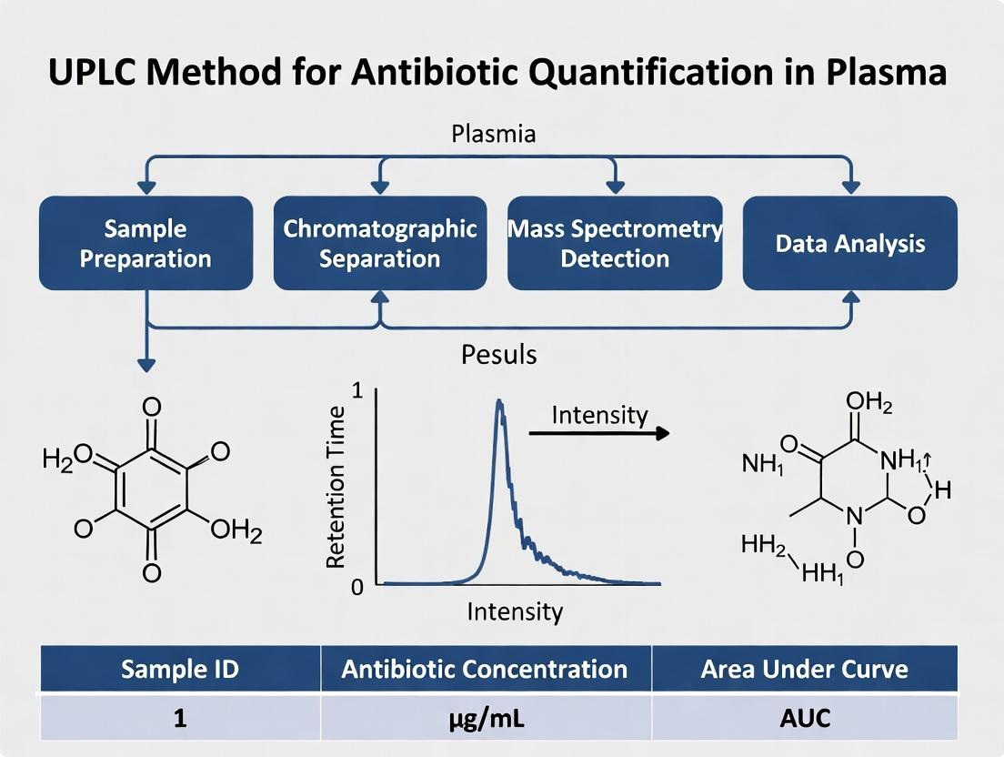
Optimized UPLC Method for Antibiotic Quantification in Plasma: Development, Validation, and Clinical Application
This article provides a comprehensive guide to developing and applying Ultra-Performance Liquid Chromatography (UPLC) methods for the precise quantification of antibiotics in plasma.
Optimized UPLC Method for Antibiotic Quantification in Plasma: Development, Validation, and Clinical Application
Abstract
This article provides a comprehensive guide to developing and applying Ultra-Performance Liquid Chromatography (UPLC) methods for the precise quantification of antibiotics in plasma. Tailored for researchers and drug development professionals, the content explores the critical need for therapeutic drug monitoring (TDM) of antibiotics, outlines the development of a robust UPLC method from column selection to detection, addresses common troubleshooting and optimization challenges, and details the rigorous validation process per ICH/FDA guidelines. The guide concludes by synthesizing best practices and highlighting the method's impact on pharmacokinetic studies, personalized dosing, and combating antimicrobial resistance.
Why Quantify Antibiotics in Plasma? The Critical Role of UPLC in TDM and PK/PD Studies
The Imperative for Therapeutic Drug Monitoring (TDM) of Antibiotics
Therapeutic Drug Monitoring (TDM) of antibiotics is a critical component of precision medicine in infectious diseases. It involves measuring drug concentrations in a patient's plasma to individualize dosing regimens, thereby maximizing efficacy and minimizing toxicity. The need for TDM is driven by significant inter-patient variability in pharmacokinetics (PK) due to factors such as age, organ dysfunction, critical illness, and drug-drug interactions. For antibiotics with a narrow therapeutic index—where the toxic dose is close to the effective dose—TDM is essential to avoid under-dosing (leading to treatment failure and antimicrobial resistance) and over-dosing (leading to organ toxicity).
Key Antibiotics Requiring TDM and Their PK/PD Targets
The following table summarizes the primary antibiotics for which TDM is strongly recommended, along with their associated pharmacokinetic/pharmacodynamic (PK/PD) targets.
Table 1: Key Antibiotics for TDM and Their PK/PD Targets
| Antibiotic Class | Example Drugs | Primary Toxicity Concern | Key PK/PD Index & Target |
|---|---|---|---|
| Glycopeptides | Vancomycin | Nephrotoxicity, Ototoxicity | AUC₂₄/MIC ≥ 400-600 (for S. aureus) |
| Aminoglycosides | Gentamicin, Tobramycin, Amikacin | Nephrotoxicity, Ototoxicity | Cmax/MIC > 8-10 (for efficacy); Trough < 1-2 mg/L (to limit toxicity) |
| Beta-lactams | Piperacillin, Meropenem, Ceftazidime | Neurotoxicity (high levels) | %fT >MIC (40-100% of dosing interval, depending on bug/drug) |
| Lipopeptides | Daptomycin | Myopathy, CPK elevation | Cmin (trough) > 24.3 mg/L linked to toxicity |
| Oxazolidinones | Linezolid | Myelosuppression, Neuropathy | AUC₂₄ > 200 mg·h/L linked to toxicity; fT > MIC |
| Triazoles | Voriconazole, Posaconazole | Hepatotoxicity, Neurotoxicity | Trough ranges: Voriconazole 1-5.5 mg/L; Posaconazole > 1 mg/L (prophylaxis) > 1.8 mg/L (treatment) |
UPLC-Based TDM Protocol: Quantification of Vancomycin in Human Plasma
This detailed protocol is framed within a research thesis developing a robust Ultra-Performance Liquid Chromatography (UPLC) method for the simultaneous quantification of multiple antibiotics in plasma.
Research Reagent Solutions & Materials
Table 2: Scientist's Toolkit - Key Reagents and Materials
| Item | Function / Explanation |
|---|---|
| UPLC System (e.g., Waters ACQUITY, Agilent 1290) | High-pressure chromatographic system for fast, high-resolution separation. |
| PDA or DAD Detector | Photodiode Array or Diode Array Detector for identifying and quantifying analytes based on UV-Vis spectra. |
| C18 UPLC Column (1.7µm, 2.1 x 50-100mm) | Stationary phase for analytical separation. Small particle size provides high efficiency. |
| Vancomycin Certified Reference Standard | Primary standard for preparing calibration curves and quality controls. |
| Internal Standard (IS) (e.g., Teicoplanin or API-analog) | Compound added in constant amount to all samples to correct for variability in extraction and injection. |
| Mass Spectrometry-grade Acetonitrile & Methanol | Low UV-absorbance, high-purity solvents for mobile phase and protein precipitation. |
| Ammonium Formate/Formic Acid | Buffers and ion-pairing agents to optimize mobile phase pH and improve peak shape. |
| Drug-Free Human Plasma | Matrix for preparing calibration standards (CS) and quality control (QC) samples. |
| Micro-centrifuge & Vortex Mixer | For sample preparation and protein precipitation. |
| 0.22 µm PVDF Syringe Filters | For filtering the supernatant post-precipitation to protect the UPLC column. |
Detailed Experimental Protocol
A. Solution Preparation
- Stock Solutions (1 mg/mL): Accurately weigh vancomycin and internal standard. Dissolve in appropriate solvent (e.g., water/methanol mix). Store at -80°C.
- Working Solutions: Serially dilute stock solutions in water to prepare working standards for spiking.
- Mobile Phase:
- A: 20 mM Ammonium formate in water, pH adjusted to 3.5 with formic acid.
- B: Acetonitrile.
- Calibrators & QCs: Spike drug-free human plasma with working solutions to prepare a 7-point calibration curve (e.g., 2.5 – 100 mg/L) and QC samples at Low, Mid, and High concentrations.
B. Sample Preparation (Protein Precipitation)
- Aliquot 100 µL of plasma sample (calibrator, QC, or patient sample) into a 1.5 mL microcentrifuge tube.
- Add 20 µL of internal standard working solution.
- Add 300 µL of cold acetonitrile.
- Vortex vigorously for 2 minutes.
- Centrifuge at 14,000 rpm (≈18,000 x g) for 10 minutes at 4°C.
- Carefully transfer 200 µL of the clear supernatant to a clean vial, optionally filtering through a 0.22 µm PVDF filter.
- Inject 2-5 µL into the UPLC system.
C. UPLC-PDA Analytical Conditions
- Column: C18 (1.7 µm, 2.1 x 50 mm)
- Column Temperature: 40 °C
- Flow Rate: 0.4 mL/min
- Injection Volume: 3 µL
- Gradient Program:
Time (min) %A %B 0.0 95 5 2.0 70 30 2.5 5 95 3.5 5 95 3.6 95 5 5.0 95 5 - Detection: PDA, 210 nm (vancomycin) and appropriate wavelength for IS.
D. Data Analysis
- Plot peak area ratio (Vancomycin/IS) against nominal vancomycin concentration for calibrators.
- Fit data using linear regression (weighted 1/x²).
- Calculate concentrations of QCs and unknown samples from the regression equation.
- Validate method per ICH/FDA guidelines: specificity, linearity, accuracy, precision, recovery, matrix effect, and stability.
Visualized Workflows and Relationships
UPLC TDM Workflow for Antibiotic Optimization
Consequences of Sub-Optimal Antibiotic Exposure
This document serves as a detailed application note for the development and validation of an Ultra-Performance Liquid Chromatography (UPLC) method for the quantification of antibiotic compounds in plasma. The broader thesis aims to establish a robust, sensitive, and high-throughput analytical platform to support pharmacokinetic studies and therapeutic drug monitoring. This note specifically addresses the critical challenges encountered in plasma bioanalysis and provides standardized protocols to mitigate them.
Table 1: Key Challenges in Plasma Analysis for Antibiotics and Corresponding Mitigation Strategies
| Challenge | Primary Impact on UPLC-MS/MS Analysis | Recommended Mitigation Strategy | Typical Performance Target |
|---|---|---|---|
| Matrix Effects | Ion suppression/enhancement, leading to inaccurate quantification. | Use of Stable Isotope-Labeled Internal Standards (SIL-IS); Efficient sample clean-up (e.g., SPE). | Matrix Factor: 85-115%; CV < 15%. |
| Low Concentrations | Signal below Limit of Quantification (LOQ), poor precision at Cmin. | Micro-volume sample processing (< 100 µL); Selective detection (MS/MS); On-line sample preconcentration. | LOQ ≤ 10% of Cmin; LOD ≈ 1/3 of LOQ. |
| Diverse Chemistries | Inconsistent extraction recovery, variable chromatographic retention. | Generic protein precipitation followed by selective SPE; Use of charged-surface hybrid (CSH) columns. | Recovery: 70-120% for all analytes. |
Detailed Experimental Protocol: UPLC-MS/MS Method for Multi-class Antibiotics
Protocol Title: Simultaneous Quantification of Fluoroquinolones, Beta-Lactams, and Glycopeptides in Human Plasma.
2.1. Materials & Reagents (The Scientist's Toolkit) Table 2: Research Reagent Solutions & Essential Materials
| Item | Function / Rationale |
|---|---|
| Stable Isotope-Labeled IS (e.g., Ciprofloxacin-d8, Piperacillin-d5) | Corrects for losses during sample prep and matrix effects during MS ionization. |
| Oasis HLB Solid-Phase Extraction (SPE) Cartridge (30 mg) | Provides mixed-mode reversed-phase and weak anion exchange retention for diverse polar antibiotics. |
| Acetonitrile (LC-MS Grade) | Protein precipitation agent and mobile phase component; high purity reduces background noise. |
| Ammonium Formate Buffer (10mM, pH 3.5) | Volatile mobile phase additive for consistent ionization and peak shaping in ESI+. |
| Acquity UPLC HSS T3 Column (2.1 x 100 mm, 1.8 µm) | High-Strength Silica column with trifunctional C18 for retention of polar compounds at low pH. |
| Mass Spectrometric Tuning Solution (e.g., NaI/KI) | Calibrates and optimizes mass accuracy and sensitivity of the triple quadrupole detector. |
2.2. Sample Preparation Workflow (Protein Precipitation followed by SPE)
- Aliquot & Spike: Pipette 50 µL of plasma sample into a 1.5 mL microcentrifuge tube.
- Add Internal Standard: Add 10 µL of a working solution containing all SIL-IS in methanol:water (50:50, v/v).
- Protein Precipitation: Add 150 µL of cold acetonitrile, vortex for 30 seconds, and centrifuge at 14,000 x g for 10 minutes at 4°C.
- SPE Conditioning & Loading: Condition an Oasis HLB cartridge with 1 mL methanol, then 1 mL water. Load the supernatant from step 3.
- Wash & Elute: Wash with 1 mL of 5% methanol in water. Elute analytes with 1 mL of methanol containing 2% formic acid.
- Evaporation & Reconstitution: Evaporate the eluate to dryness under a gentle nitrogen stream at 40°C. Reconstitute in 100 µL of initial mobile phase (98% 10mM ammonium formate, pH 3.5 / 2% acetonitrile), vortex, and transfer to a UPLC vial.
2.3. UPLC-MS/MS Conditions
- Chromatography: Acquity UPLC I-Class System.
- Column: HSS T3 (2.1 x 100 mm, 1.8 µm). Temperature: 40°C.
- Mobile Phase: A: 10mM Ammonium Formate (pH 3.5), B: Acetonitrile.
- Gradient: 2% B (0-0.5 min), 2% → 95% B (0.5-8.0 min), 95% B (8.0-9.0 min), re-equilibrate at 2% B (9.1-12.0 min).
- Flow Rate: 0.4 mL/min. Injection Volume: 5 µL (partial loop with needle overfill).
- Detection: Xevo TQ-S Micro Triple Quadrupole Mass Spectrometer with ESI+.
- Data Acquisition: Multiple Reaction Monitoring (MRM). Dwell time ≥ 20 ms per transition.
Visualization of Workflows and Relationships
Diagram 1: Plasma Analysis Challenge Mitigation Pathway
Diagram 2: Sample Preparation & Analysis Workflow
Why UPLC? Advantages Over HPLC for Speed, Sensitivity, and Resolution in Bioanalysis
Within the context of developing an Ultra-Performance Liquid Chromatography (UPLC) method for the quantification of novel beta-lactam antibiotics in human plasma, the selection of the chromatographic platform is foundational. High-Performance Liquid Chromatography (HPLC) has long been the standard for bioanalytical quantification. However, the evolution towards UPLC represents a paradigm shift, driven by the need for higher throughput, superior sensitivity, and enhanced resolution in modern drug development. This application note delineates the core advantages of UPLC over HPLC, providing experimental protocols and data from our ongoing thesis research on antibiotic pharmacokinetics.
Core Advantages: UPLC vs. HPLC
UPLC technology utilizes columns packed with smaller particles (<2.2 µm) and operates at significantly higher pressures (up to 15,000 psi or 1000 bar) compared to traditional HPLC (3-5 µm particles, ~4000 psi). This fundamental difference yields marked improvements in key performance metrics.
Table 1: Comparative System Parameters and Performance Outcomes
| Parameter | Traditional HPLC | UPLC | Advantage in Bioanalysis |
|---|---|---|---|
| Typical Particle Size | 3-5 µm | 1.7-1.8 µm | Reduced band broadening, higher peak capacity. |
| Operating Pressure | Up to 4000 psi (600 bar) | Up to 15,000 psi (1000 bar) | Enables use of smaller particles for efficiency. |
| Linear Velocity | ~1-2 mm/sec | ~2-5 mm/sec | Faster separations. |
| Typical Run Time | 10-30 minutes | 3-10 minutes | 3-5x increase in throughput; ideal for high-sample-volume studies. |
| Peak Width | 10-30 seconds | 2-5 seconds | Sharper peaks, lower detection limits. |
| Theoretical Plates | ~10,000-15,000/column | ~20,000-40,000/column | Superior resolving power for complex matrices. |
| Solvent Consumption | Higher (mL/min flow) | Lower (~50% reduction) | Cost-effective and environmentally friendly. |
Table 2: Experimental Comparison from Antibiotic in Plasma Study
| Analytic (Cephalosporin) | System | Runtime (min) | Resolution (Rs) from Key Interferent | Signal-to-Noise (S/N) at LLOQ | Injection Volume (µL) |
|---|---|---|---|---|---|
| Ceftriaxone | HPLC (C18, 5µm, 150 x 4.6 mm) | 12.0 | 2.5 | 12 | 50 |
| Ceftriaxone | UPLC (HSS C18, 1.8µm, 100 x 2.1 mm) | 3.5 | 4.1 | 45 | 10 |
| Cefepime | HPLC (C18, 5µm, 150 x 4.6 mm) | 15.0 | 1.8 (co-elution risk) | 9 | 50 |
| Cefepime | UPLC (HSS C18, 1.8µm, 100 x 2.1 mm) | 4.2 | 3.5 (baseline resolved) | 52 | 10 |
Detailed Experimental Protocols
Protocol 1: UPLC-MS/MS Method for Plasma Antibiotic Quantification
Objective: To quantify a panel of beta-lactam antibiotics in 50 µL human plasma with a 5-minute runtime. Materials: See "The Scientist's Toolkit" below. Procedure:
- Sample Preparation (Protein Precipitation):
- Aliquot 50 µL of plasma calibration standard, QC, or study sample into a 1.5 mL microcentrifuge tube.
- Add 150 µL of internal standard (IS) working solution in acetonitrile (e.g., stable isotope-labeled antibiotic).
- Vortex mix vigorously for 1 minute.
- Centrifuge at 16,000 × g for 10 minutes at 4°C.
- Transfer 100 µL of the clear supernatant to a certified UPLC vial with insert.
- UPLC Conditions:
- System: Acquity UPLC H-Class (or equivalent).
- Column: Acquity UPLC HSS T3 (1.8 µm, 2.1 x 100 mm), maintained at 45°C.
- Mobile Phase A: 0.1% Formic acid in water.
- Mobile Phase B: 0.1% Formic acid in acetonitrile.
- Flow Rate: 0.5 mL/min.
- Gradient:
- 0-0.5 min: 2% B
- 0.5-3.5 min: 2% → 95% B (linear gradient)
- 3.5-4.0 min: 95% B (wash)
- 4.0-4.1 min: 95% → 2% B
- 4.1-5.0 min: 2% B (re-equilibration)
- Injection Volume: 2-10 µL (partial loop with needle overfill).
- MS/MS Detection (Triple Quadrupole):
- Ionization Mode: Electrospray Ionization (ESI), positive.
- Capillary Voltage: 3.0 kV.
- Source Temp.: 150°C.
- Desolvation Temp.: 500°C.
- Desolvation Gas Flow: 1000 L/hr.
- Data Acquisition: Multiple Reaction Monitoring (MRM). Example transition for Ceftriaxone: 555.1 → 396.0 (collision energy: 18 eV).
Protocol 2: Direct Method Transfer from HPLC to UPLC
Objective: To transfer and optimize an existing HPLC method for meropenem to UPLC, improving speed and sensitivity. Procedure:
- Column Selection: Choose a UPLC column with similar chemistry (e.g., C18) but smaller particles (1.7-1.8 µm). Scale column dimensions: (HPLC Column Length / Particle Size HPLC) ≈ (UPLC Column Length / Particle Size UPLC).
- Flow Rate Scaling: Adjust linearly based on column cross-sectional area: F_UPLC = F_HPLC × (r_UPLC² / r_HPLC²), where r is column radius.
- Gradient Scaling: Maintain the same number of column volumes. Calculate: t_UPLC = t_HPLC × (F_HPLC / F_UPLC) × (V_UPLC / V_HPLC), where V is column void volume.
- Injection Volume Scaling: Scale relative to column void volume: Inj_UPLC ≈ Inj_HPLC × (V_UPLC / V_HPLC).
- Re-optimization: Fine-tune gradient slope, initial/final %B, and column temperature to achieve optimal resolution.
Visualizations
UPLC-MS/MS Plasma Bioanalysis Workflow
Mechanistic Basis for UPLC Performance Gains
The Scientist's Toolkit: Key Research Reagent Solutions
Table 3: Essential Materials for UPLC Bioanalysis of Antibiotics in Plasma
| Item / Reagent | Function & Rationale | Example Product/Chemical |
|---|---|---|
| Sub-2µm UPLC Column | Core of the separation; provides high efficiency and resolution. | Waters Acquity UPLC HSS T3 (1.8 µm, 2.1 x 100 mm) |
| LC-MS Grade Solvents | Minimize background noise, prevent system contamination, and ensure reproducibility. | Acetonitrile and Water (0.1% Formic acid additive) |
| Stable Isotope-Labeled Internal Standard (IS) | Corrects for matrix effects and variability in extraction/ionization; essential for accurate quantification. | Ceftriaxone-d3, Meropenem-d6 |
| Protein Precipitation Reagent | Rapid and simple removal of plasma proteins to prevent column fouling and ion suppression. | Cold Acetonitrile (often containing IS) |
| Certified Low-Volume Vials & Inserts | Ensure precise, accurate, and contamination-free injections for UPLC's narrow peaks. | 12 x 32 mm Screw Neck Vials, 250 µL Polypropylene Inserts |
| Mass Spectrometer Tuning & Calibration Solution | Optimizes and calibrates MS/MS instrument response for specific analytes. | Sodium Formate or proprietary mixes for mass axis calibration; analyte-specific tuning. |
The development and validation of a universal UPLC (Ultra-Performance Liquid Chromatography) method for the simultaneous quantification of multiple antibiotic classes in human plasma is a cornerstone of modern pharmacokinetic (PK) research and therapeutic drug monitoring (TDM). Precise plasma concentration data is critical for optimizing efficacy, minimizing toxicity, and combating antimicrobial resistance. This application note details protocols and considerations for monitoring key antibiotic classes within this analytical framework.
Key Antibiotic Classes and TDM Rationale
Beta-Lactams (Penicillins, Cephalosporins, Carbapenems): Time-dependent killers requiring plasma concentrations to exceed the minimum inhibitory concentration (MIC) for a significant portion of the dosing interval (fT > MIC). Subtherapeutic levels drive resistance, while excessive levels (particularly of penicillins) can cause neurotoxicity.
Glycopeptides (Vancomycin, Teicoplanin): Concentration-dependent activity with a post-antibiotic effect. Monitoring is essential to achieve a target area under the curve (AUC) to MIC ratio while avoiding nephrotoxicity and ototoxicity.
Aminoglycosides (Gentamicin, Tobramycin, Amikacin): Concentration-dependent killers with a significant post-antibiotic effect. Peak concentrations correlate with efficacy, while trough concentrations are monitored to prevent dose-dependent nephro- and ototoxicity.
Other Key Classes:
- Fluoroquinolones (e.g., Ciprofloxacin): AUC/MIC is the primary PK/PD index. TDM prevents treatment failure and limits toxicity risks (CNS, tendonitis).
- Oxazolidinones (Linezolid): TDM is used to maximize efficacy (fT > MIC) and mitigate myelosuppression and neuropathy associated with prolonged exposure.
- Polymyxins (Colistin): Complex pharmacokinetics of the prodrug (colistimethate) and active moiety necessitate TDM to optimize antibacterial effect and prevent acute kidney injury.
Quantitative PK/PD Targets for TDM
Table 1: Key Pharmacokinetic/Pharmacodynamic Targets for Major Antibiotic Classes
| Antibiotic Class | Primary PK/PD Index | Typical TDM Target | Toxic Threshold |
|---|---|---|---|
| Beta-Lactams | fT > MIC | 100% fT > 1-4x MIC | Variable; often >5-10x MIC (CNS toxicity) |
| Vancomycin | AUC24/MIC | AUC24 400-600 mg·h/L (for MIC ≤1 mg/L) | Trough >15-20 mg/L (nephrotoxicity) |
| Aminoglycosides | Cmax/MIC | Cmax/MIC >8-10 | Trough: Gent >1 mg/L; Amik >5 mg/L |
| Fluoroquinolones | AUC24/MIC | AUC24/MIC >125 (e.g., Pseudomonas) | CNS, QT prolongation risk |
| Linezolid | fT > MIC & AUC24 | Trough: 2-8 mg/L | Trough >10 mg/L (myelosuppression) |
UPLC-MS/MS Protocol for Multi-Class Antibiotic Quantification in Plasma
Title: UPLC-MS/MS Workflow for Plasma Antibiotics
Detailed Protocol
1. Sample Preparation (Protein Precipitation)
- Aliquot 100 µL of human plasma (calibrator, QC, or patient sample) into a microcentrifuge tube.
- Add 10 µL of internal standard (IS) working solution (stable isotope-labeled analogs of each target antibiotic).
- Add 300 µL of ice-cold acetonitrile for protein precipitation.
- Vortex vigorously for 1 minute, then centrifuge at 13,000 x g for 10 minutes at 4°C.
- Transfer 150 µL of the clear supernatant to a new vial and dilute with 150 µL of LC-MS grade water. Mix gently.
- Transfer to a total recovery vial for UPLC-MS/MS analysis.
2. UPLC Chromatographic Conditions
- System: Acquity UPLC H-Class.
- Column: Acquity UPLC BEH C18 (1.7 µm, 2.1 x 50 mm).
- Column Temperature: 40°C.
- Flow Rate: 0.4 mL/min.
- Injection Volume: 2 µL.
- Mobile Phase:
- A: Water with 0.1% Formic Acid.
- B: Acetonitrile with 0.1% Formic Acid.
- Gradient Program:
- 0-1.0 min: 2% B
- 1.0-4.0 min: 2% → 40% B
- 4.0-5.0 min: 40% → 95% B
- 5.0-6.0 min: Hold at 95% B
- 6.0-6.1 min: 95% → 2% B
- 6.1-8.0 min: Re-equilibrate at 2% B
3. MS/MS Detection Conditions
- System: Xevo TQ-S micro Triple Quadrupole Mass Spectrometer.
- Ionization: Electrospray Ionization (ESI), positive and negative switching.
- Capillary Voltage: 2.8 kV (ESI+), 2.5 kV (ESI-).
- Source Temperature: 150°C.
- Desolvation Temperature: 500°C.
- Desolvation Gas Flow: 1000 L/hr.
- Data Acquisition: Multiple Reaction Monitoring (MRM). Key transitions must be optimized for each compound (see Table 2).
Table 2: Example MRM Transitions for Select Antibiotics
| Antibiotic (Class) | Precursor Ion (m/z) | Product Ion (m/z) | Cone Voltage (V) | Collision Energy (eV) | Polarity |
|---|---|---|---|---|---|
| Meropenem (Beta-Lactam) | 384.1 | 141.1 | 18 | 10 | + |
| Piperacillin (Beta-Lactam) | 518.1 | 143.1 | 26 | 20 | + |
| Vancomycin (Glycopeptide) | 725.4 | 144.1 | 40 | 22 | + |
| Gentamicin C1a (Aminoglycoside) | 450.3 | 322.2 | 30 | 12 | + |
| Ciprofloxacin (Fluoroquinolone) | 332.1 | 288.1 | 40 | 25 | + |
| Linezolid (Oxazolidinone) | 338.1 | 296.1 | 25 | 12 | + |
4. Method Validation The developed method must be validated per FDA/EMA bioanalytical guidelines for:
- Selectivity/Specificity: No interference from blank plasma.
- Linearity: Calibration curve (e.g., 0.1 - 50 µg/mL) with R² > 0.99.
- Accuracy & Precision: Within ±15% for QCs (LLOQ: ±20%).
- Recovery & Matrix Effect: Consistent and minimal ion suppression/enhancement.
- Stability: Bench-top, processed, and long-term freezer stability assessments.
The Scientist's Toolkit: Research Reagent Solutions
Table 3: Essential Materials for UPLC-MS/MS Antibiotic Quantification
| Item | Function & Importance |
|---|---|
| Stable Isotope-Labeled Internal Standards (e.g., ¹³C, ²H) | Corrects for variability in sample prep and ionization efficiency; essential for accuracy. |
| Mass Spectrometry-Grade Acetonitrile & Methanol | High-purity solvents prevent background noise and instrument contamination. |
| LC-MS Grade Water & Formic Acid | Ensures clean baseline and consistent ionization for gradient elution. |
| Pooled Human Plasma (Drug-Free) | Matrix for preparing calibration standards and quality control samples. |
| UPLC BEH C18 or HSS T3 Column (1.7-1.8 µm) | Provides high-resolution, rapid separation of polar to moderately polar antibiotics. |
| Phosphate-Buffered Saline (PBS) | Used for making stock solutions and dilution blanks to match plasma pH/ionic strength. |
| Supported Liquid Extraction (SLE) or Solid-Phase Extraction (SPE) Plates | For more complex, sensitive methods requiring advanced sample clean-up. |
| Reference Standards of Target Antibiotics | Certified pure material for accurate preparation of stock solutions and calibration curves. |
Key Pharmacodynamic Pathways in Antibiotic Action
Title: Core Antibiotic PD Pathways & Resistance
Linking Plasma Concentrations to Pharmacokinetic/Pharmacodynamic (PK/PD) Targets
Within the scope of a thesis developing and validating an Ultra-Performance Liquid Chromatography (UPLC) method for quantifying antibiotics in plasma, a critical subsequent step is linking the measured plasma concentrations to pharmacokinetic/pharmacodynamic (PK/PD) targets. This application note details protocols for deriving PK parameters, establishing PK/PD indices, and applying these to optimize dosing regimens for antibacterial efficacy and suppress resistance.
PK/PD Indices and Therapeutic Targets for Antibiotics
Successful antibiotic therapy requires that drug exposure, defined by the concentration-time profile in plasma and at the infection site, exceeds a threshold relative to the pathogen's susceptibility. The primary PK/PD indices are summarized in Table 1.
Table 1: Primary PK/PD Indices for Major Antibiotic Classes
| Antibiotic Class | Key PK/PD Index | Typical Therapeutic Target | Rationale |
|---|---|---|---|
| β-lactams (Penicillins, Cephalosporins, Carbapenems) | %T>MIC (Time above MIC) | 40-70% of dosing interval | Time-dependent killing; efficacy depends on duration of exposure. |
| Fluoroquinolones | AUC₂₄/MIC (Area Under Curve to MIC) | ≥125 for Gram-negatives; ≥30-50 for Gram-positives | Concentration-dependent killing & persistent effects. |
| Aminoglycosides | Cₘₐₓ/MIC (Peak to MIC) | 8-10 | Concentration-dependent killing; high peak maximizes efficacy. |
| Glycopeptides (Vancomycin) | AUC₂₄/MIC | ≥400 (for S. aureus) | Best predictor for clinical outcomes, balancing efficacy and toxicity. |
| Oxazolidinones (Linezolid) | AUC₂₄/MIC & %T>MIC | AUC/MIC: 80-120; %T>MIC: ≥85% | Mixed pattern; both time and total exposure are important. |
Core Protocols
Protocol 2.1: From UPLC Data to Pharmacokinetic Parameters
Objective: To calculate non-compartmental analysis (NCA) PK parameters from plasma concentration-time data generated via a validated UPLC assay.
Materials & Reagents:
- Plasma concentration-time data from UPLC analysis.
- PK analysis software (e.g., Phoenix WinNonlin, PKanalix, R package
PK). - Known dosing regimen (dose, route, infusion time).
Procedure:
- Data Preparation: Compile measured plasma concentrations with corresponding exact sampling times (post-dose) in a spreadsheet.
- Non-Compartmental Analysis (NCA):
- Input concentration-time data and dosing information into the software.
- Select appropriate model (e.g., plasma, IV bolus or infusion, extravascular).
- The software will calculate:
- Cₘₐₓ: Maximum observed concentration.
- Tₘₐₓ: Time of Cₘₐₓ.
- AUC₀–t: Area under the concentration-time curve from zero to last measurable time (t), calculated via the linear/log trapezoidal rule.
- AUC₀–∞: AUC from zero to infinity (AUC₀–t + Cₗᴀꜱₜ/λ₂).
- t₁/₂: Terminal elimination half-life (0.693/λ₂).
- CL: Systemic Clearance (Dose / AUC₀–∞).
- Vd: Volume of Distribution (Dose / (AUC₀–∞ * λ₂)).
- Output: Report all calculated parameters with units. Visualize with a linear and log-linear concentration-time plot.
Protocol 2.2: Determining PK/PD Index Attainment for a Specific Pathogen
Objective: To assess whether a simulated or observed dosing regimen achieves the PK/PD target for a pathogen with a known Minimum Inhibitory Concentration (MIC).
Materials & Reagents:
- PK parameters from Protocol 2.1 or a population PK model.
- Pathogen MIC value (from microdilution assay).
- Simulation software (e.g., Berkeley Madonna, R, NONMEM).
Procedure:
- Define Target: Based on antibiotic class (Table 1), select the relevant PK/PD index (e.g., AUC₂₄/MIC) and its target value (e.g., ≥125).
- Simulate Profile:
- For a given dose and regimen, simulate the plasma concentration-time profile over 24h using the PK parameters (e.g., one-compartment model:
Cₜ = (Dose/(Vd*kel)) * exp(-kel*time)). - For %T>MIC, simulate a profile at steady-state.
- For a given dose and regimen, simulate the plasma concentration-time profile over 24h using the PK parameters (e.g., one-compartment model:
- Calculate Index:
- AUC₂₄/MIC: Calculate simulated AUC over 24h (AUC₂₄) and divide by the MIC.
- %T>MIC: From the simulated steady-state profile, determine the cumulative time during a dosing interval that concentrations exceed the MIC, expressed as a percentage of the interval.
- Cₘₐₓ/MIC: Divide the simulated peak (Cₘₐₓ) by the MIC.
- Attainment Assessment: Compare the calculated index to the therapeutic target. Perform Monte Carlo simulations (Protocol 2.3) for robust probability estimates.
Protocol 2.3: Monte Carlo Simulation (MCS) for Probability of Target Attainment (PTA)
Objective: To estimate the likelihood (%) that a given dosing regimen will achieve a predefined PK/PD target in a population, accounting for variability in PK and MIC.
Procedure:
- Define Input Distributions:
- PK Variability: Obtain mean and variance (e.g., CV%) for key parameters (Clearance-CL, Volume-Vd) from literature or population PK studies. Assume log-normal distribution.
- MIC Distribution: Obtain the MIC₅₀, MIC₉₀, and range for the target pathogen from surveillance databases (e.g., EUCAST, CLSI).
- Set Simulation Conditions: Specify the dosing regimen (dose, interval, infusion time), number of virtual subjects (e.g., 10,000), and the PK/PD target (e.g., AUC₂₄/MIC ≥125).
- Run Simulation: For each virtual subject:
- Randomly sample a CL and Vd from their distributions.
- Randomly sample an MIC from the pathogen MIC distribution.
- Calculate the PK/PD index based on the sampled PK and MIC.
- Record if the target is attained (1) or not (0).
- Analyze Output: The Probability of Target Attainment (PTA) for a specific MIC is the percentage of subjects attaining the target. The Cumulative Fraction of Response (CFR) is the expected PTA across the entire MIC distribution, weighted by the frequency of each MIC.
Table 2: Example PTA Output for a Fluoroquinolone Regimen (Target: AUC₂₄/MIC ≥125)
| MIC (mg/L) | Probability of Target Attainment (PTA, %) |
|---|---|
| 0.06 | 99.8 |
| 0.125 | 98.5 |
| 0.25 | 92.1 |
| 0.5 | 75.4 |
| 1.0 | 45.2 |
| 2.0 | 12.7 |
| 4.0 | 1.1 |
| CFR (for given MIC distribution): | 88.5% |
The Scientist's Toolkit: Research Reagent Solutions
Table 3: Essential Materials for PK/PD Studies of Antibiotics in Plasma
| Item | Function & Application |
|---|---|
| Validated UPLC-MS/MS System | High-sensitivity, specific quantification of antibiotic concentrations in biological matrices (plasma). The core analytical tool. |
| Stable Isotope-Labeled Internal Standards (e.g., ¹³C, ²H) | Corrects for variability in sample preparation, matrix effects, and instrument response, ensuring assay accuracy and precision. |
| Protein Precipitation Plates (e.g., 96-well) | For high-throughput sample preparation to remove plasma proteins prior to UPLC injection. |
| Pharmacokinetic Modeling Software (WinNonlin, NONMEM) | For calculating PK parameters, simulating concentration-time profiles, and performing population PK/PD analysis. |
| Statistical Software (R, Python with SciPy) | For data analysis, custom PK calculations, and running Monte Carlo simulations. |
| Lyophilized Human Plasma (Control) | Used as a blank matrix for preparing calibration standards and quality control samples during UPLC method validation and sample analysis. |
| Cation-Adjusted Mueller-Hinton Broth (CAMHB) | Standardized medium for performing broth microdilution to determine the Minimum Inhibitory Concentration (MIC) of the target pathogen. |
| EUCAST or CLSI MIC Panels | Pre-configured panels for standardized, reproducible determination of antibiotic MICs against clinical bacterial isolates. |
Visualizing the PK/PD Workflow and Relationships
Diagram Title: PK/PD Target Attainment Workflow
Diagram Title: PK/PD Index Links to Antibacterial Activity
Step-by-Step Development of a Robust UPLC Method for Plasma Antibiotic Analysis
Core UPLC System Components and Configuration for Bioanalysis
Within the context of developing a robust Ultra-Performance Liquid Chromatography (UPLC) method for the quantification of antibiotics in human plasma, the selection and configuration of core system components are paramount. This protocol details the essential hardware, software, and operational parameters necessary to achieve the high sensitivity, resolution, and throughput required for bioanalytical applications in drug development.
Core System Components
A UPLC system for bioanalysis consists of several integrated modules. Their specifications directly impact method performance for antibiotic analysis in complex matrices like plasma.
Table 1: Core UPLC System Components and Specifications for Bioanalysis
| Component | Key Specification/Model Example | Function in Bioanalysis |
|---|---|---|
| Binary Pump | Max Pressure: 18,000-19,000 psi; Flow Precision: <0.075% RSD | Delivers a precise, high-pressure gradient of mobile phase for rapid and efficient chromatographic separation. |
| Autosampler | Temperature-controlled (4-10°C); Carryover: <0.005% | Introduces plasma extract samples with high precision while maintaining analyte stability. Cooling is critical for labile compounds. |
| Column Heater | Temperature Range: 10-90°C; Stability: ±0.5°C | Maintains a constant column temperature for reproducible retention times and optimal peak shape. |
| Column | BEH C18, 1.7 µm, 2.1 x 50-100 mm; Temperature Limit: 90°C | The stationary phase where separation occurs. Sub-2 µm particles provide high efficiency. C18 chemistry is common for antibiotics. |
| Detector (PDA) | Sampling Rate: 20-80 Hz; Bandwidth: 1.2 nm | Provides UV/Vis spectral data for peak purity assessment and selective quantification of antibiotics with chromophores. |
| Detector (MS/MS) | Triple Quadrupole; ESI Source; Dwell Time: ~10 msec/transition | Enables highly selective and sensitive detection and quantification via multiple reaction monitoring (MRM), essential for trace levels in plasma. |
| Sample Organizer | Capacity for >100 vials; Temperature-controlled | Holds calibration standards, quality controls (QCs), and study samples in a regulated sequence. |
| Data System | Empower or MassLynx Software | Controls the instrument, acquires data, and processes chromatograms for quantitative analysis. |
Critical System Configuration Protocol
Protocol 2.1: Initial System Setup and Performance Qualification for Antibiotic Analysis
Objective: To configure and qualify the UPLC-MS/MS system for the sensitive and reproducible quantification of antibiotics in plasma.
Materials:
- UPLC system with binary pump, cooled autosampler, column heater, and tandem quadrupole mass spectrometer.
- Analytical column: Acquity UPLC BEH C18, 1.7 µm, 2.1 x 50 mm.
- Mobile Phase A: 0.1% Formic acid in water (LC-MS grade).
- Mobile Phase B: 0.1% Formic acid in acetonitrile (LC-MS grade).
- System Suitability Solution: A standard containing the target antibiotic(s) at a mid-range concentration in a solvent that mimics the initial mobile phase composition.
- Needle Wash Solution: 50:50 Water:Methanol with 0.1% formic acid.
Procedure:
- Mobile Phase Preparation: Degas all mobile phases by sonication under vacuum for 10 minutes or using an inline degasser. Filter through a 0.22 µm nylon membrane.
- Column Installation: Install the BEH C18 column in the column heater. Set the heater to 40°C (optimal for many beta-lactams and fluoroquinolones).
- Pump Priming & Purging: Prime all pump lines with their respective degassed mobile phases. Perform a high-flow purge (2 mL/min for 5 minutes) to remove air bubbles.
- Autosampler Configuration:
- Set the sample compartment temperature to 6°C.
- Program the needle wash to occur with the designated wash solution for 5 seconds before and after each injection.
- Set the injection volume to 2-5 µL (partial loop with needle overfill mode).
- MS Source Configuration:
- Install the appropriate ion source (e.g., ESI). Set source temperature to 150°C and desolvation gas temperature to 500°C.
- Set desolvation gas flow to 1000 L/hr and cone gas flow to 150 L/hr (Nitrogen).
- Optimize capillary voltage, cone voltage, and MRM transitions for each target antibiotic using direct infusion.
- Gradient Programming: Establish a chromatographic gradient.
- Initial: 95% A, 5% B.
- Ramp to 5% A, 95% B over 2.5 minutes.
- Hold for 0.5 minutes.
- Return to initial conditions in 0.1 minutes and re-equilibrate for 1.0 minutes.
- Total run time: 4.0 minutes. Flow rate: 0.4 mL/min.
- System Suitability Test: Inject the system suitability solution six times consecutively.
- Acceptance Criteria: Retention time RSD ≤ 1%; Peak area RSD ≤ 2%; Peak asymmetry factor between 0.8 and 1.5.
Bioanalytical Workflow for Antibiotic Quantification
Title: UPLC-MS/MS Bioanalysis Workflow
The Scientist's Toolkit: Key Research Reagent Solutions
Table 2: Essential Materials for UPLC Bioanalysis of Antibiotics in Plasma
| Item | Function in Protocol | Critical Consideration for Antibiotics |
|---|---|---|
| Ammonium Acetate Buffer (pH 4.5) | Mobile phase additive for LC-MS; improves ionization and peak shape. | Optimal pH for stability and detection of many beta-lactam and macrolide antibiotics. |
| Oasis HLB or MCX SPE Cartridges | Solid-phase extraction for selective sample clean-up and analyte enrichment. | HLB is versatile for a broad range; MCX is ideal for basic compounds. Essential for low-concentration analytes. |
| Acetonitrile (LC-MS Grade) | Organic mobile phase component and protein precipitation solvent. | Low UV absorbance and high MS compatibility. More effective than methanol for precipitating plasma proteins. |
| Formic Acid (LC-MS Grade) | Mobile phase additive to promote positive ionization (proton donation) in ESI+. | Concentration typically 0.1%. Enhances sensitivity for most antibiotics analyzed in positive ion mode. |
| Stable Isotope-Labeled Internal Standards (SIL-IS) | Added to each sample before processing to correct for variability in extraction and ionization. | e.g., ^13C- or ^2H-labeled version of the target analyte. Crucial for achieving high accuracy and precision in quantitative bioanalysis. |
| Blank Matrix (Plasma) | Used to prepare calibration standards and quality controls. | Must be analyte-free. Species-matched (e.g., human) and from an appropriate anticoagulant (e.g., K2EDTA). |
Method Development and Optimization Logic
Title: UPLC Method Development Decision Tree
Detailed Sample Preparation Protocol (SPE)
Protocol 6.1: Solid-Phase Extraction (SPE) of Beta-Lactam Antibiotics from Plasma
Objective: To isolate and concentrate target antibiotics from human plasma using mixed-mode cation exchange (MCX) SPE.
Procedure:
- Conditioning: Load an Oasis MCX 30 mg cartridge on a vacuum manifold. Condition with 1 mL of methanol followed by 1 mL of water. Do not let the sorbent dry.
- Loading: Piper 100 µL of plasma sample (calibrator, QC, or unknown) into a tube. Add 20 µL of working SIL-IS solution and 300 µL of 4% phosphoric acid. Vortex mix for 10 seconds. Load the entire mixture onto the conditioned cartridge at a flow of ~1 mL/min.
- Washing: Wash sequentially with 1 mL of 2% formic acid in water, followed by 1 mL of methanol. Dry the cartridge under full vacuum for 3 minutes to remove residual methanol.
- Elution: Elute analytes into a clean collection tube with 1 mL of 5% ammonium hydroxide in acetonitrile. Let the eluent gravity-flow for 30 seconds before applying vacuum.
- Evaporation & Reconstitution: Evaporate the eluent to dryness under a gentle stream of nitrogen at 40°C. Reconstitute the dry residue with 100 µL of initial mobile phase (e.g., 95:5 Water:ACN with 0.1% FA). Vortex for 60 seconds and centrifuge at 13,000 x g for 5 minutes. Transfer supernatant to a low-volume autosampler vial for UPLC-MS/MS analysis.
Within the development of an Ultra-Performance Liquid Chromatography (UPLC) method for the quantification of antibiotics in plasma, sample preparation is a critical first step. The complexity of the plasma matrix, containing proteins, lipids, salts, and endogenous compounds, necessitates robust, reproducible, and efficient cleanup strategies to ensure method specificity, sensitivity, and instrument longevity. This application note details three fundamental techniques—Protein Precipitation (PPT), Liquid-Liquid Extraction (LLE), and Solid-Phase Extraction (SPE)—framed within the context of antibiotic bioanalysis. The selection of an appropriate strategy is contingent on the physicochemical properties of the target antibiotic(s), required sensitivity, throughput, and available resources.
The following table summarizes the key characteristics, advantages, and limitations of each technique.
Table 1: Comparison of Sample Preparation Techniques for Antibiotic Quantification in Plasma
| Parameter | Protein Precipitation (PPT) | Liquid-Liquid Extraction (LLE) | Solid-Phase Extraction (SPE) |
|---|---|---|---|
| Principle | Denaturation of proteins using organic solvent/acids. | Partitioning of analyte between two immiscible liquids. | Selective adsorption/desorption from a solid sorbent. |
| Complexity | Low | Medium | Medium to High |
| Cost per Sample | Very Low | Low | Medium to High |
| Throughput | High (amenable to 96-well plate) | Medium | Medium to High (automation possible) |
| Recovery (%) | Variable (60-85%) | High (70-95%) | High and Consistent (80-105%) |
| Cleanup Efficiency | Low (co-precipitation of analytes, high matrix effect) | Medium (depends on solvent choice) | High (selective wash steps) |
| Ideal For | Rapid screening, high-throughput, highly polar compounds. | Non-polar to medium-polar analytes, cost-effective scale-up. | Complex matrices, trace-level analysis, demanding LC-MS/MS methods. |
| Key Limitation | High ion suppression/enhancement in MS, dirty extracts. | Emulsion formation, use of large solvent volumes. | Method development time, sorbent cost, potential for channeling. |
Recovery can be compromised due to analyte binding to precipitated protein pellets.
Detailed Protocols & Application Notes
Protocol 1: Protein Precipitation (PPT) for Fluoroquinolones in Plasma
- Objective: Rapid deproteinization for the analysis of ciprofloxacin and levofloxacin.
- Research Reagent Solutions:
- Acetonitrile (ACN) with 1% Formic Acid: Precipitating agent. Acidification aids in breaking protein-analyte bonds and improves recovery for basic analytes.
- Internal Standard (IS) Solution: Deuterated analog of the target antibiotic (e.g., Ciprofloxacin-d8) in methanol.
- Ammonium Acetate Buffer (10 mM, pH 3.0): Reconstitution solution to match initial mobile phase conditions.
Procedure:
- Pipette 100 µL of plasma sample (calibrator, QC, or unknown) into a 1.5 mL microcentrifuge tube.
- Add 25 µL of Internal Standard working solution.
- Vortex-mix for 10 seconds.
- Add 300 µL of ice-cold ACN with 1% Formic Acid.
- Vortex vigorously for 2 minutes to ensure complete protein denaturation and mixing.
- Centrifuge at 14,000 x g for 10 minutes at 4°C.
- Carefully transfer 200 µL of the clear supernatant to a fresh vial.
- Evaporate to dryness under a gentle stream of nitrogen at 40°C.
- Reconstitute the dry residue with 150 µL of 10 mM Ammonium Acetate buffer (pH 3.0).
- Vortex for 1 minute and centrifuge at 14,000 x g for 5 minutes.
- Transfer the supernatant to a UPLC vial with insert for analysis.
Protocol 2: Liquid-Liquid Extraction (LLE) for Macrolides in Plasma
- Objective: Selective extraction of azithromycin and clarithromycin from plasma.
- Research Reagent Solutions:
- Saturation Solution: Potassium Carbonate (K₂CO₃) in water (2 M). Adjusts pH to basic conditions, keeping analytes in neutral form for efficient organic solvent partitioning.
- Extraction Solvent: Ethyl Acetate:Hexane (70:30, v/v). Optimized for macrolide antibiotic recovery.
- Reconstitution Solution: Methanol:Water (50:50, v/v) with 0.1% Formic Acid.
Procedure:
- Aliquot 200 µL of plasma into a glass extraction tube.
- Add 50 µL of IS working solution and 100 µL of 2 M K₂CO₃ saturation solution.
- Vortex-mix for 30 seconds.
- Add 1.5 mL of Ethyl Acetate:Hexane (70:30) extraction solvent.
- Seal the tube and mix by horizontal shaking for 15 minutes.
- Centrifuge at 3000 x g for 5 minutes for clear phase separation.
- Transfer the entire upper organic layer to a clean evaporation tube.
- Repeat the extraction (steps 4-7) with a fresh 1.0 mL of solvent and combine the organic layers.
- Evaporate the combined organic extracts to complete dryness under nitrogen at 40°C.
- Reconstitute the dry extract in 100 µL of Methanol:Water (50:50) with 0.1% Formic Acid.
- Vortex for 2 minutes, centrifuge, and transfer to a UPLC vial.
Protocol 3: Solid-Phase Extraction (SPE) for Beta-Lactams in Plasma
- Objective: High-cleanup extraction for sensitive LC-MS/MS quantification of amoxicillin and piperacillin.
- Research Reagent Solutions:
- Conditioning Solvents: Methanol, followed by 20 mM Phosphate Buffer (pH 7.0).
- Wash Solution 1: 5% Methanol in Water.
- Wash Solution 2: n-Hexane (for lipid removal).
- Elution Solvent: Acetonitrile:Methanol (80:20, v/v) with 2% Formic Acid.
- Strong Cation Exchange (SCX) Cartridges: 30 mg, 1 mL sorbent beds.
Procedure:
- Condition the SPE cartridge sequentially with 1 mL of Methanol and 1 mL of 20 mM Phosphate Buffer (pH 7.0). Do not let the sorbent bed dry.
- Dilute 200 µL of plasma with 200 µL of phosphate buffer (pH 7.0) and add 50 µL of IS. Vortex.
- Load the diluted plasma sample onto the conditioned cartridge at a slow, dropwise rate (~1 drop/sec).
- Wash the cartridge with 1 mL of 5% Methanol in Water, followed by 1 mL of n-Hexane.
- Dry the cartridge under full vacuum for 5 minutes to remove residual water and hexane.
- Elute the analytes into a clean collection tube with 2 x 0.5 mL aliquots of Acetonitrile:Methanol (80:20) with 2% Formic Acid.
- Evaporate the eluate to dryness under nitrogen at 35°C (higher temperatures degrade beta-lactams).
- Reconstitute in 150 µL of mobile phase initial conditions (e.g., 95% Water, 5% ACN, 0.1% Formic Acid).
- Vortex, centrifuge, and transfer to a UPLC vial.
Visualized Workflows
Protein Precipitation Workflow for Plasma
Liquid-Liquid Extraction Workflow for Plasma
Solid Phase Extraction Workflow for Plasma
The Scientist's Toolkit: Essential Research Reagent Solutions
Table 2: Key Reagents and Materials for Sample Preparation in Antibiotic Analysis
| Item | Function & Rationale |
|---|---|
| Deuterated Internal Standards (e.g., Ciprofloxacin-d8) | Corrects for variability in extraction efficiency, evaporation, and matrix effects; essential for accurate quantification. |
| LC-MS Grade Organic Solvents (ACN, MeOH, Acetone) | Minimizes background interference, ensures chromatographic purity, and prevents system contamination. |
| Solid-Phase Extraction Cartridges (e.g., Mixed-Mode, HLB, SCX) | Provide selective retention and cleanup based on analyte polarity, charge, and hydrophobicity. |
| Weak & Strong Buffers (Ammonium Acetate, Formate, Phosphate) | Control sample pH during extraction to optimize analyte charge state for efficient recovery in LLE/SPE. |
| Protein Precipitating Agents (Trichloroacetic Acid, ZnSO4) | Alternative to ACN/MeOH; can offer better recovery for specific analyte classes. |
| Evaporation System (Nitrogen Turbovap) | Gentle, rapid removal of extraction solvents for sample concentration and reconstitution in LC-compatible buffer. |
| Phosphate-Buffered Saline (PBS) | For dilution of viscous plasma samples prior to loading on SPE cartridges to improve flow and binding kinetics. |
| Lipid Removal Sorbents (e.g., HybridSPE-PPT+) | Specialized cartridges for selective phospholipid depletion, significantly reducing a major source of matrix effect. |
The choice between PPT, LLE, and SPE for UPLC-based antibiotic quantification in plasma is a balance of speed, cleanliness, and recovery. For rapid, high-throughput analysis where sensitivity is not paramount, PPT is suitable. LLE offers a good compromise with better cleanup for less polar analytes. For the development of robust, sensitive, and GLP-compliant methods—particularly for challenging analytes like beta-lactams—SPE is the gold standard, providing superior matrix removal and analyte enrichment. The protocols and insights provided herein serve as a foundational guide for researchers developing UPLC methods in pharmacokinetic and therapeutic drug monitoring studies.
1. Introduction Within the development of a UPLC (Ultra-Performance Liquid Chromatography) method for the quantification of novel beta-lactam antibiotics in human plasma, optimization of the chromatographic core parameters is paramount. This application note details systematic strategies for optimizing column chemistry, mobile phase composition, gradient profile, and column temperature to achieve baseline resolution of target analytes from endogenous plasma interferences, maximize sensitivity, and minimize run time. All protocols are contextualized for bioanalytical method development in support of pharmacokinetic studies.
2. Core Parameter Optimization Strategies & Data
2.1 Column Chemistry Selection The stationary phase is the primary determinant of selectivity. For polar, ionizable antibiotics, charged surface hybrid (CSH) and ethylene-bridged hybrid (BEH) C18 columns with low bleed characteristics are recommended.
Table 1: Evaluation of Stationary Phases for Beta-Lactam Separation
| Column Chemistry | Particle Size | Dimensions (mm) | Key Property | Result for Analyte X |
|---|---|---|---|---|
| BEH C18 | 1.7 µm | 2.1 x 50 | High pH stability (1-12) | Good peak shape, moderate retention |
| CSH C18 | 1.7 µm | 2.1 x 50 | Surface charge at low pH | Enhanced retention of bases, sharper peaks |
| HSS T3 | 1.8 µm | 2.1 x 100 | Aqueous stability (100%) | Excellent for polar metabolites |
| BEH Shield RP18 | 1.7 µm | 2.1 x 50 | Embedded polar group | Reduced hydrophobic collapse, unique selectivity |
Protocol 2.1: Column Screening
- Conditioning: Flush each candidate column with 10 column volumes of starting mobile phase at 0.2 mL/min.
- Test Injection: Inject a standard solution (1 µg/mL of each analyte in 5% organic solvent) using a generic gradient (5-95% organic in 5 min).
- Mobile Phase: Use 0.1% formic acid in water (A) and 0.1% formic acid in acetonitrile (B).
- Detection: Monitor at 260 nm (typical β-lactam absorbance) and by MS/MS in positive ESI mode.
- Evaluation Criteria: Record retention factor (k > 2), peak asymmetry (As 0.8-1.2), and resolution (Rs > 1.5 from nearest interference).
2.2 Mobile Phase & pH Optimization Mobile phase pH critically influences the ionization state of ionizable antibiotics and silanol interactions.
Table 2: Effect of Mobile Phase pH on Chromatographic Metrics
| pH (Ammonium Formate Buffer) | Organic Modifier | Analyte pKa | Retention Time (min) | Peak Capacity |
|---|---|---|---|---|
| 3.0 | Acetonitrile | 2.1, 8.7 | 4.2 | 85 |
| 4.5 | Acetonitrile | 2.1, 8.7 | 5.1 | 92 |
| 6.0 | Acetonitrile | 2.1, 8.7 | 3.8 | 78 |
| 4.5 | Methanol | 2.1, 8.7 | 7.3 | 65 |
Protocol 2.2: pH & Modifier Scouting
- Buffer Preparation: Prepare 10 mM ammonium formate buffers at pH 3.0, 4.5, and 6.0 using formic acid/ammonium hydroxide. Filter through 0.22 µm PVDF.
- Modifier: Test acetonitrile and methanol separately.
- Isocratic Scouting: Perform short isocratic runs at 20% organic for each pH/modifier combination.
- Gradient Refinement: Based on isocratic data, design a 5-minute gradient from 5% to 50% organic.
- Analysis: Plot retention time vs. pH to identify the region of greatest sensitivity to change, indicating an optimal, robust working point.
2.3 Gradient Profile Optimization A well-designed gradient balances resolution and speed.
Protocol 2.3: Gradient Steepness Optimization
- Initial Conditions: From the optimal pH/modifier, set initial %B to 5%.
- Design Experiments: Create three linear gradients to 95% B over 3, 5, and 7 minutes.
- Hold & Re-equilibration: Hold at 95% B for 0.5 min, then re-equilibrate at 5% B for 1.5x the gradient time.
- Calculate Metrics: Determine the resolution (Rs) between the critical pair and the peak capacity for each run.
- Optimize: Use modeling software or a manual compromise to select the gradient time yielding Rs > 2.0 with the shortest run time.
2.4 Column Temperature Effects Temperature affects viscosity, retention, and selectivity.
Table 3: Impact of Column Temperature on Separation
| Temperature (°C) | Backpressure (psi) | Retention Time (min) | Theoretical Plates (N) |
|---|---|---|---|
| 30 | 8500 | 5.21 | 12500 |
| 40 | 7200 | 4.95 | 13500 |
| 50 | 6100 | 4.72 | 14200 |
| 60 | 5300 | 4.51 | 13800 |
Protocol 2.4: Temperature Study
- Set Oven: Allow the column compartment to equilibrate at each test temperature (30, 40, 50, 60°C) for 15 min.
- Isocratic Run: Perform an isocratic run (e.g., 20% B) at each temperature.
- Analyze: Plot ln(retention factor) vs. 1/T (Van 't Hoff plot) to assess thermodynamic behavior. Select a temperature that offers a good compromise between efficiency, pressure, and run time, typically 40-50°C for UPLC.
3. The Scientist's Toolkit: Essential Research Reagents & Materials
Table 4: Key Reagent Solutions for UPLC Method Development
| Item | Function & Specification |
|---|---|
| Ammonium Formate (MS Grade) | Volatile buffer salt for mobile phase, compatible with MS detection. |
| Formic Acid (LC-MS Grade) | Ion-pairing agent and pH modifier for positive ion mode ESI. |
| Acetonitrile (LC-MS Grade) | Low-viscosity organic modifier for UPLC; provides sharp peaks. |
| Drug Substance/Metabolite Standards | High-purity (>95%) reference materials for identification and calibration. |
| Control Human Plasma (K2EDTA) | Matrix for preparing calibration standards and quality controls. |
| Protein Precipitation Reagent | e.g., 3:1 v/v Acetonitrile with 0.1% Formic Acid; for sample cleanup. |
| Water (LC-MS Grade, 18.2 MΩ·cm) | Mobile phase base, minimizes background ions and column contamination. |
4. Visualized Workflows & Relationships
UPLC Method Optimization Decision Pathway
Core Parameters Impact on Chromatographic Resolution
Within the context of developing a robust Ultra-Performance Liquid Chromatography (UPLC) method for antibiotic quantification in plasma, the choice of detection system is paramount. This application note provides a comparative analysis of two prevalent detection techniques: Ultraviolet/Photodiode Array (UV/PDA) detection and tandem mass spectrometry (MS/MS). The focus is on their respective specificity and sensitivity in the complex biological matrix of plasma, critical for accurate pharmacokinetic studies and therapeutic drug monitoring in drug development.
Comparative Performance Data
The following tables summarize key performance metrics for UV/PDA and MS/MS detection in the analysis of antibiotics in plasma.
Table 1: General Comparison of Detection Techniques
| Parameter | UV/PDA Detection | Tandem MS (MS/MS) Detection |
|---|---|---|
| Principle | Absorption of UV-Vis light by chromophores | Mass-to-charge ratio (m/z) of ions, with fragmentation |
| Typical Sensitivity (LLOQ) | 0.1 - 1 µg/mL (100 - 1000 ng/mL) | 0.1 - 10 ng/mL (often 100-1000x more sensitive) |
| Specificity | Moderate (co-eluting compounds with similar λ can interfere) | Very High (specific precursor → product ion transitions) |
| Dynamic Range | 10² - 10³ | 10³ - 10⁵ |
| Matrix Effect (Plasma) | Significant; requires extensive sample cleanup | Can be significant but mitigated by MRM and stable isotope IS |
| Sample Throughput | High | High (with modern systems) |
| Method Development Complexity | Low to Moderate | High |
| Instrument Cost | Relatively Low | High |
Table 2: Example Antibiotic Analysis in Plasma (Theoretical Data Based on Current Literature)
| Antibiotic (Class) | UV/PDA (LLOQ) | MS/MS (LLOQ) | Key Advantage of MS/MS |
|---|---|---|---|
| Ciprofloxacin (Fluoroquinolone) | 50 ng/mL | 0.5 ng/mL | Enables PK studies at sub-therapeutic levels |
| Vancomycin (Glycopeptide) | 500 ng/mL | 50 ng/mL | Essential for precise TDM in critical care |
| Meropenem (Carbapenem) | 200 ng/mL | 5 ng/mL | Captures rapid pharmacokinetics in critically ill patients |
| Linezolid (Oxazolidinone) | 250 ng/mL | 10 ng/mL | Improved specificity in complex matrices |
Detailed Experimental Protocols
Protocol A: UPLC-UV/PDA Method for β-Lactam Antibiotics in Plasma
Objective: Quantify amoxicillin and clavulanate in human plasma. Reagent Solutions:
- Mobile Phase A: 0.1% Formic acid in water.
- Mobile Phase B: 0.1% Formic acid in acetonitrile.
- Precipitation Solution: Acetonitrile (containing internal standard, e.g., phenacetin).
- Standard Solutions: Serial dilutions of analytes in blank plasma (range 0.1-50 µg/mL).
Procedure:
- Sample Preparation: Piper 100 µL of plasma sample into a microcentrifuge tube.
- Protein Precipitation: Add 300 µL of ice-cold precipitation solution. Vortex vigorously for 1 minute.
- Centrifugation: Centrifuge at 14,000 x g for 10 minutes at 4°C.
- Supernatant Transfer: Transfer 200 µL of clear supernatant to a fresh vial.
- Evaporation & Reconstitution: Evaporate to dryness under a gentle nitrogen stream at 40°C. Reconstitute the residue in 100 µL of initial mobile phase conditions (95% A / 5% B). Vortex for 30 seconds.
- UPLC-UV/PDA Analysis:
- Column: C18, 1.7 µm, 2.1 x 50 mm.
- Flow Rate: 0.4 mL/min.
- Gradient: 5% B to 95% B over 5 minutes.
- Detection: PDA, 220 nm (amoxicillin) and 230 nm (clavulanate).
- Injection Volume: 5 µL.
- Data Analysis: Plot peak area ratio (analyte/IS) vs. concentration to generate a calibration curve.
Protocol B: UPLC-MS/MS Method for Fluoroquinolones in Plasma
Objective: Quantify ciprofloxacin and levofloxacin in rat plasma with high sensitivity. Reagent Solutions:
- Mobile Phase A: 5 mM Ammonium formate in water.
- Mobile Phase B: 5 mM Ammonium formate in methanol.
- Extraction Solution: 70:30 (v/v) Ethyl Acetate: Dichloromethane.
- Internal Standard (IS) Solution: D8-Ciprofloxacin in methanol.
- Standard Solutions: Serial dilutions of analytes in blank plasma (range 0.1-500 ng/mL).
Procedure:
- Sample Preparation: Piper 50 µL of plasma sample into a tube.
- IS Addition: Add 25 µL of IS solution.
- Liquid-Liquid Extraction: Add 1 mL of extraction solution. Vortex for 3 minutes.
- Centrifugation: Centrifuge at 10,000 x g for 5 minutes.
- Organic Layer Transfer: Transfer the upper organic layer to a clean tube.
- Evaporation & Reconstitution: Evaporate to dryness under nitrogen at 40°C. Reconstitute in 100 µL of initial mobile phase (90% A / 10% B).
- UPLC-MS/MS Analysis:
- Column: HSS T3, 1.8 µm, 2.1 x 100 mm.
- Flow Rate: 0.3 mL/min.
- Gradient: 10% B to 95% B over 6 minutes.
- Ionization: ESI positive mode.
- MS/MS Transitions (MRM):
- Ciprofloxacin: 332.1 → 288.1 (CE 22 eV), 332.1 → 231.1 (CE 35 eV)
- Levofloxacin: 362.1 → 318.1 (CE 20 eV)
- D8-Ciprofloxacin (IS): 340.2 → 296.2 (CE 22 eV)
- Injection Volume: 2 µL.
- Data Analysis: Use peak area ratios (analyte/IS) from the primary MRM transition for quantification, using the secondary for confirmation.
Visualization of Workflows and Relationships
Workflow: Antibiotic Analysis in Plasma
Detection Choice: Key Parameter Trade-offs
The Scientist's Toolkit: Essential Research Reagent Solutions
Table 3: Key Reagents & Materials for UPLC Antibiotic Quantification
| Item | Function/Explanation |
|---|---|
| Stable Isotope-Labeled Internal Standards (e.g., ¹³C, ²H) | Corrects for matrix effects and losses during sample preparation; crucial for MS/MS accuracy. |
| Mass Spectrometry-Grade Solvents (ACN, MeOH, Water) | Minimize chemical noise and ion suppression in the MS source, ensuring reproducible ionization. |
| Volatile Buffers (Ammonium Formate/Acetate) | Compatible with MS detection; facilitate analyte ionization and maintain stable pH in mobile phase. |
| Solid Phase Extraction (SPE) Cartridges (e.g., Mixed-Mode) | Selective extraction of antibiotics from plasma, removing phospholipids (major source of matrix effect). |
| Protein Precipitation Plates (96-well format) | Enable high-throughput sample preparation with minimal manual handling and transfer steps. |
| UPLC Columns (Sub-2µm Particle, e.g., BEH C18, HSS T3) | Provide high resolution and peak capacity for separating analytes from matrix interferences. |
| Phospholipid Removal SPE Plates | Specifically designed to remove phospholipids, significantly reducing ion suppression in ESI-MS. |
| PDA Detector with High-Resolution Scanning | Allows spectral confirmation of peak purity and selection of optimal wavelength for each analyte. |
Application Notes
In the development of a UPLC-MS/MS method for quantifying antibiotics in plasma, selecting an appropriate internal standard (IS) is critical for ensuring method accuracy, precision, and robustness. The choice between stable isotope-labeled analogs (SIL-IS) and structural (or non-labeled) analogs is fundamental. This decision impacts the ability of the IS to correct for analyte losses during sample preparation, matrix effects during ionization, and instrument variability.
Key Considerations:
- Stable-Labeled Analogs (SIL-IS): These are chemically identical to the target analyte but incorporate non-radioactive heavy isotopes (e.g., ^2H, ^13C, ^15N). They represent the ideal IS as they co-elute with the analyte and exhibit nearly identical physicochemical and chromatographic behavior, ensuring precise compensation for matrix effects and recovery losses. Their primary limitation is cost and availability.
- Structural Analogs: These are compounds with a molecular structure similar to the target analyte but not identical. They are more readily available and less expensive. However, differences in retention time, extraction recovery, and ionization efficiency can lead to suboptimal correction for matrix effects, potentially compromising accuracy, especially in complex matrices like plasma.
Within our thesis on UPLC-MS/MS method development for novel beta-lactam antibiotics in human plasma, a direct comparison was performed to evaluate the impact of IS choice on method performance.
Table 1: Method Validation Parameters for Antibiotic X with Different Internal Standards
| Parameter | Stable-Labeled Analog (Antibiotic X-^13C3) | Structural Analog (Antibiotic Y) |
|---|---|---|
| Linear Range (µg/mL) | 0.1 - 50 | 0.1 - 50 |
| Coefficient of Determination (R²) | 0.9992 | 0.9985 |
| Accuracy (% Bias) | -2.1 to +3.8 | -8.5 to +6.2 |
| Precision (% CV) | Intra-day: <5.1 | Intra-day: <7.8 |
| Inter-day: <6.4 | Inter-day: <9.5 | |
| Matrix Effect (%) | 95-102 (CV < 3%) | 88-115 (CV < 8%) |
| Extraction Recovery (%) | 98.5 ± 2.1 | 85.3 ± 5.7 |
| Process Efficiency (%) | 96.5 ± 3.0 | 78.4 ± 7.2 |
| Cost per mg | $450 | $50 |
Table 2: Stability Assessment in Plasma (Room Temperature, 24h)
| Analytic | Internal Standard Type | % Change from T0 |
|---|---|---|
| Antibiotic X | Stable-Labeled Analog | +1.2 |
| Antibiotic X | Structural Analog | -4.7 |
Experimental Protocols
Protocol 1: Comparison of Matrix Effect for SIL-IS vs. Structural Analog IS
Objective: To quantitatively assess the ability of each IS type to correct for ion suppression/enhancement in human plasma.
Materials: See "The Scientist's Toolkit" below.
Procedure:
- Prepare neat standard solutions of the analyte (Antibiotic X) and the two candidate IS (SIL-IS and Structural Analog) in methanol at appropriate concentrations.
- Prepare Post-Extraction Spiked Samples (Set A): a. Extract 100 µL of blank plasma from 6 different individual donors using your developed protein precipitation method (e.g., with 300 µL of acetonitrile containing 0.1% formic acid). b. Centrifuge at 14,000 x g for 10 minutes at 4°C. c. Transfer the supernatant and spike with a known, low concentration of the analyte and the IS.
- Prepare Neat Solution Samples (Set B): Spike the same amounts of analyte and IS into pure mobile phase (no plasma matrix).
- Analyze all samples (Set A and Set B) using the candidate UPLC-MS/MS method.
- Calculation: For each IS type, calculate the matrix factor (MF) for the analyte and the IS.
- MFAnalyte = Peak Area (Set A, Analyte) / Peak Area (Set B, Analyte)
- MFIS = Peak Area (Set A, IS) / Peak Area (Set B, IS)
- Normalized MF = MFAnalyte / MFIS
- The variability (CV%) of the normalized MF across the 6 different plasma lots is the key metric. A CV < 5% indicates excellent IS compensation.
Protocol 2: Sample Preparation and UPLC-MS/MS Analysis for Plasma Antibiotics
Objective: To detail the sample preparation and chromatographic conditions used for the quantitative analysis.
Procedure:
- Sample Preparation: a. Thaw frozen plasma samples on ice and vortex for 10 seconds. b. Aliquot 50 µL of plasma into a 1.5 mL microcentrifuge tube. c. Add 10 µL of the working internal standard solution (prepared in methanol). d. Add 150 µL of precipitation solvent (Acetonitrile:MeOH, 80:20 v/v, with 0.1% Formic Acid). e. Vortex vigorously for 2 minutes. f. Centrifuge at 18,000 x g for 15 minutes at 4°C. g. Transfer 150 µL of the clear supernatant to a fresh vial containing 50 µL of water. Vortex to mix. h. Transfer to a total recovery UPLC vial for injection.
- UPLC Conditions:
- Column: CORTECS T3 (2.1 x 100 mm, 1.6 µm)
- Temperature: 40°C
- Flow Rate: 0.4 mL/min
- Mobile Phase A: Water with 0.1% Formic Acid
- Mobile Phase B: Acetonitrile with 0.1% Formic Acid
- Gradient: 2% B (0-0.5 min), 2% to 95% B (0.5-5.0 min), 95% B (5.0-6.0 min), re-equilibrate at 2% B for 2.5 min.
- Injection Volume: 2 µL
- MS/MS Conditions (ESI+):
- Source Temp: 150°C
- Desolvation Temp: 500°C
- Cone Gas Flow: 150 L/Hr
- Desolvation Gas Flow: 1000 L/Hr
- Capillary Voltage: 1.0 kV
- Monitor 2-3 MRM transitions per compound for quantification and confirmation.
Diagrams
Title: Decision Flowchart for Internal Standard Type
Title: Plasma Sample Preparation and Analysis Workflow
The Scientist's Toolkit
Table 3: Key Research Reagent Solutions for UPLC-MS/MS Antibiotic Assay
| Item | Function & Specification |
|---|---|
| Stable Isotope-Labeled Internal Standard | Chemically identical IS for optimal correction of matrix effects and recovery. Purity >98%, isotopic enrichment >99%. |
| Structural Analog Internal Standard | Chemically similar IS, a cost-effective alternative. Must have similar retention and extraction to analyte. |
| Mass Spectrometry-Grade Solvents | Acetonitrile, Methanol, Water. Minimize background noise and ion suppression in MS detection. |
| Acid Additives (MS Grade) | Formic Acid, Ammonium Formate. Enhance analyte ionization in positive or negative ESI mode. |
| UPLC Column (C18/T3/HSS) | Sub-2µm particle columns for high resolution & speed. Select based on analyte polarity (e.g., T3 for polar antibiotics). |
| Control Human Plasma (K2EDTA) | Drug-free matrix for preparing calibration standards and quality controls. Use from multiple donors for MF tests. |
| Protein Precipitation Plates/Tubes | 96-well plates or microcentrifuge tubes compatible with organic solvents for high-throughput processing. |
| UPLC Vials & Caps | Low volume insert vials with pre-slit caps to ensure sample integrity and prevent evaporation. |
Troubleshooting Common UPLC Issues: Peak Shape, Matrix Effects, and Sensitivity Challenges
This application note is presented within the context of a broader thesis research project aimed at developing and validating a robust, sensitive, and fast Ultra-Performance Liquid Chromatography (UPLC) method for the quantification of novel beta-lactam antibiotics in human plasma. Achieving optimal chromatographic peak shape is a non-negotiable prerequisite for method validation, as it directly impacts critical parameters including resolution, sensitivity, reproducibility, and accurate integration. Poor peak morphology—manifesting as tailing, fronting, or excessive broadening—can compromise the entire analytical workflow, leading to inaccurate quantification and unreliable pharmacokinetic data. This document provides a diagnostic framework and practical, evidence-based protocols for identifying and rectifying common peak shape anomalies.
Fundamentals of Peak Shape Anomalies
Peak shape is typically evaluated using the asymmetry factor (As) or tailing factor (Tf), where a value of 1.0 represents perfect symmetry. Values >1.2 generally indicate tailing, while values <0.8 suggest fronting. Broad peaks are characterized by increased peak width (W) or reduced plate count (N). The root causes are often interrelated and can be chemical, instrumental, or column-related.
Table 1: Diagnostic Guide to Poor Peak Shape in UPLC Antibiotic Analysis
| Peak Anomaly | Primary Indicators | Common Causes in Plasma Analysis | Immediate Diagnostic Checks |
|---|---|---|---|
| Tailing | As > 1.2, Tf > 1.2 | 1. Secondary interactions with active silanols.2. Column overload (mass or volume).3. Inadequate mobile phase pH control.4. Extra-column volume post-column. | 1. Inject a neat standard to rule out matrix.2. Reduce injection volume by 50%.3. Check mobile phase pH and buffer capacity. |
| Fronting | As < 0.8, Tf < 0.8 | 1. Column channeling or void formation.2. Sample solvent stronger than mobile phase.3. Overloading of the stationary phase. | 1. Visual check for column bed damage.2. Ensure sample is in starting mobile phase.3. Reduce analyte concentration. |
| Broad Peaks | High W, Low N | 1. Excessive extra-column volume (tubing, detector cell).2. Inadequate column temperature control.3. Slow detector response time.4. Poorly optimized gradient delay volume. | 1. Verify all tubing is 0.005" ID or smaller.2. Increase column temperature (e.g., 40-50°C).3. Set detector sampling rate appropriately. |
The Scientist's Toolkit: Research Reagent Solutions
Table 2: Essential Materials for UPLC Method Troubleshooting in Bioanalysis
| Item | Function & Rationale |
|---|---|
| Hybrid C18 Column (e.g., BEH C18) | Robust, high-pressure stable column with minimized residual silanol activity, essential for basic antibiotic compounds. |
| Mass Spectrometry-Grade Acids & Buffers | High-purity formic acid, ammonium formate/acetate to ensure reproducible ionization and pH control, minimizing background noise. |
| Solid-Phase Extraction (SPE) Plates (C18) | For efficient plasma sample cleanup, removing phospholipids and proteins that cause matrix effects and column fouling. |
| UPLC Guard Column (VanGuard FIT) | Protects the expensive analytical column from particulate matter and irreversibly adsorbed plasma matrix components. |
| Needle Wash Solvent (e.g., 50:50 Water:MeOH) | Critical for preventing carryover of high-concentration plasma samples, a common source of ghost peaks and integration errors. |
| Column Regeneration Solvents | Sequential flushes of water, acetonitrile, and a strong solvent like 80:20 ACN:IPA with 0.1% formic acid to restore column performance. |
Experimental Protocols for Diagnosis and Correction
Protocol 4.1: Systematic Diagnosis of Peak Shape Issues
Objective: To isolate the root cause of a peak shape problem (column, instrument, or method conditions).
- Prepare Solutions: Prepare a mid-level calibration standard of the target antibiotic in neat mobile phase A.
- Baseline Instrument Check: Install a known, well-performing column (e.g., a certified reference column). Inject the neat standard using a generic, well-established gradient. Evaluate peak shape.
- Test Method Conditions: Switch to the new method's mobile phases and gradient. Inject the same neat standard. Note any change in peak symmetry.
- Test with Matrix: Inject a processed plasma sample (blank matrix spiked post-extraction). Compare peak shape to the neat standard from Step 3. Significant degradation indicates a matrix effect or insufficient sample cleanup.
- Test with Original Column: Reinstall the column from the problematic method. Repeat injection of the neat standard from Step 1. Degraded peak shape indicates column degradation or mismatch.
Protocol 4.2: Protocol for Mitigating Tailing of Basic Antibiotics
Objective: To reduce silanol interactions causing tailing for amine-containing beta-lactams.
- Increase Buffer Concentration: Prepare mobile phase A with 10-20 mM ammonium formate (pH 3.5 with formic acid) instead of 5 mM. The higher ionic strength better masks silanols.
- Add a Competing Amine: Add 0.1% (v/v) triethylamine (TEA) or 0.2% (v/v) diethylamine (DEA) to mobile phase A. Caution: MS compatibility must be tested; may cause ion suppression.
- Adjust pH: If the analyte's pKa allows, lower the mobile phase pH to 2.5-3.0 (using formic acid). This protonates basic silanols, reducing ionic interaction.
- Switch Stationary Phase: Use a charged surface hybrid (CSH) column or a column end-capped with sterically bulky groups. These are specifically designed for superior peak shape with basic compounds.
- Re-evaluate: Inject the problematic plasma sample and measure the asymmetry factor. Optimize one parameter at a time.
Protocol 4.3: Protocol for Correcting Broad and Fronting Peaks
Objective: To improve efficiency (plate count) and peak symmetry.
- Minimize Extra-Column Volume:
- Use the shortest possible length of 0.005" ID tubing between the column outlet and the detector flow cell.
- Ensure the detector flow cell volume is appropriate for the column's internal diameter (e.g., ≤2 µL for 2.1 mm ID columns).
- Optimize Sample Solvent: Reconstitute the dried SPE extract in the starting mobile phase composition (e.g., 5% acetonitrile in aqueous buffer), not a strong organic solvent.
- Optimize Thermal Conditions: Place the column in a forced-air oven set to 40-50°C. This reduces viscosity, improving mass transfer and efficiency.
- Adjust Detector Settings: Set the detector data acquisition rate to ≥20 points per peak (e.g., 10 Hz for UPLC). Reduce the response time constant to the manufacturer's recommended UPLC setting.
- Verify Column Health: If fronting persists, the column inlet frit may be blocked or the bed may be voided. Replace the guard column or the analytical column.
Visual Workflow for Troubleshooting
Title: Diagnostic Workflow for Peak Shape Issues
Title: Mechanism of Amine Modifier Action
Ion suppression and enhancement represent critical challenges in Electrospray Ionization Mass Spectrometry (ESI-MS) when developing robust UPLC-MS/MS methods for antibiotic quantification in complex matrices like plasma. Within the broader thesis research on a novel UPLC method for multi-class antibiotic quantification, this document details application notes and protocols to address these matrix effects (MEs). Effective mitigation is essential for achieving the required sensitivity, accuracy, and regulatory compliance in bioanalytical drug development.
Core Mechanisms and Impact Assessment
Matrix effects originate from co-eluting, non-volatile, or semi-volatile compounds that alter the ionization efficiency of the target analyte. Their impact is quantitatively assessed using the post-extraction spike method: ME (%) = (Peak Area of post-extraction spike / Peak Area of neat standard solution) × 100 A value of 100% indicates no effect, <100% indicates suppression, and >100% indicates enhancement. In initial method development for plasma antibiotics (e.g., fluoroquinolones, beta-lactams, macrolides), MEs ranged from severe suppression (45%) to significant enhancement (180%), compromising data integrity.
Source Optimization Protocols
ESI Source Parameter Optimization
Objective: To fine-tune the ESI interface for maximum desolvation and ion transmission while minimizing non-specific interactions. Protocol:
- Prepare Solutions: Use a standard mixture of target antibiotics at 100 ng/mL in a 50:50 water:methanol + 0.1% formic acid.
- Infusion Setup: Connect a syringe pump for continuous infusion of the standard at 10 µL/min. Simultaneously, inject a blank plasma extract via the UPLC system.
- Parameter Screening: Systematically vary the following parameters, monitoring the signal intensity and stability of the infused standard:
- Drying Gas Temperature: 200°C to 400°C.
- Drying Gas Flow: 5 to 12 L/min.
- Nebulizer Gas Pressure: 15 to 50 psi.
- Capillary Voltage: 2.0 to 4.0 kV (positive mode).
- Nozzle Voltage/Skimmer Offset: 0 to 300 V.
- Optimal Conditions: The combination yielding the highest, most stable signal for the infused standard during co-elution of matrix components is selected. For the thesis method, optimal conditions were: Drying Gas: 325°C, 10 L/min; Nebulizer: 35 psi; Capillary Voltage: 3.0 kV; Nozzle Voltage: 150 V.
Mobile Phase and Gradient Optimization
Objective: To temporally separate analytes from major ion-suppressing components (e.g., phospholipids, salts). Protocol:
- Phospholipid Mapping: Inject a blank plasma extract and use a precursor ion scan of m/z 184 (positive mode) or m/z 153 (negative mode) to identify phospholipid elution regions.
- Gradient Adjustment: Design a chromatographic gradient that shifts analyte elution windows away from the high-intensity phospholipid regions (typically 2-6 minutes in reversed-phase). This may involve altering the initial organic percentage, gradient slope, and total run time.
- Mobile Phase Modifiers: Test different additives:
- Formic Acid (0.1%)
- Ammonium Formate (2-10 mM) + Formic Acid
- Acetic Acid (0.1-1%) Evaluate for improved analyte ionization and reduced adduct formation.
Sample Clean-up Improvement Protocols
Comparative Evaluation of Clean-up Techniques
Objective: To quantitatively compare the efficiency of different sample preparation methods in removing phospholipids and reducing ME.
Protocol for Parallel Sample Preparation:
- Sample: Pooled human plasma (K2EDTA).
- Spiking: Fortify plasma with a mixture of target antibiotics at 50 ng/mL and IS at 100 ng/mL.
- Precipitation (PPT):
- Mix 100 µL plasma with 300 µL of cold acetonitrile containing 1% formic acid.
- Vortex for 1 min, centrifuge at 14,000 g for 10 min (4°C).
- Transfer supernatant, evaporate under nitrogen at 40°C, reconstitute in 100 µL initial mobile phase.
- Solid-Phase Extraction (SPE):
- Use a mixed-mode cationic exchange (MCX) or phospholipid removal (PLR) 96-well plate.
- Condition with 1 mL methanol, then 1 mL water.
- Load 100 µL of acidified plasma (with 1% formic acid).
- Wash with 1 mL 2% formic acid in water, then 1 mL methanol.
- Elute with 1 mL of 5% ammonium hydroxide in methanol.
- Evaporate and reconstitute as above.
- Liquid-Liquid Extraction (LLE):
- Mix 100 µL plasma with 50 µL of internal standard and 1 mL of ethyl acetate:hexane (80:20, v/v).
- Shake for 15 min, centrifuge at 5,000 g for 5 min.
- Freeze the aqueous layer at -80°C for 15 min, decant organic layer.
- Evaporate and reconstitute.
Table 1: Comparison of Clean-up Efficiency on Matrix Effects and Phospholipid Removal
| Clean-up Method | Avg. ME % (Range) | Phospholipid Reduction vs PPT (%) | Avg. Absolute Recovery % (RSD) |
|---|---|---|---|
| Protein Precipitation (PPT) | 62% (45-180%) | 0% (Baseline) | 85% (15%) |
| Liquid-Liquid Extraction (LLE) | 88% (75-110%) | ~75% | 70% (12%) |
| Mixed-Mode SPE (MCX) | 95% (90-102%) | ~95% | 92% (7%) |
| Phospholipid Removal SPE (PLR) | 98% (96-105%) | >99% | 88% (5%) |
Protocol for On-line Clean-up Using 2D-LC Configuration
Objective: To implement a robust, automated two-dimensional liquid chromatography (2D-LC) setup for on-line matrix removal.
Detailed Workflow:
- System Configuration: Two binary pumps, a 2-position/10-port dual-loop valve, and a column manager.
- First Dimension (Clean-up):
- Column: Cyano or HILIC guard column (e.g., 10 x 2.1 mm, 5 µm).
- Mobile Phase: A: 10 mM Ammonium Acetate in water (pH 4.5), B: Acetonitrile.
- Flow: 0.5 mL/min. Isocratic at 95% B for 1 min.
- Injection: 10 µL of precipitated plasma sample.
- Early eluting matrix components (salts, polar acids) are directed to waste.
- Heart-Cut Transfer:
- At 1.0 min, valve switches, transferring the analyte-containing effluent (~0.8-1.2 min window) to the second-dimension loop.
- Second Dimension (Analytical Separation):
- Column: C18 reversed-phase (e.g., 50 x 2.1 mm, 1.7 µm).
- Flow: 0.4 mL/min. Gradient from 5% to 95% organic over 5 min.
- Analytes are focused, separated, and introduced to the ESI source.
- Re-equilibration: Valve switches back at 1.5 min, re-equilibrating both systems.
The Scientist's Toolkit
Table 2: Key Research Reagent Solutions for ME Mitigation
| Item | Function & Rationale |
|---|---|
| Phospholipid Removal (PLR) SPE Cartridges | Selectively binds phospholipids via zirconia-coated silica, dramatically reducing a major source of ion suppression. |
| Mixed-Mode Cation Exchange (MCX) Sorbent | Combines reversed-phase and strong cation exchange for clean-up of basic antibiotics (e.g., macrolides) from plasma. |
| Deuterated Internal Standards (ISTD) | Corrects for analyte-specific ion suppression/enhancement by co-eluting with the target, compensating for signal fluctuations. |
| HybridSPE-Precipitation Plates | Integrates protein precipitation with selective phospholipid removal in a single filtration step for high-throughput. |
| UPLC Columns with Charged Surface Hybrid (CSH) Technology | Provides improved peak shape for basic compounds at low pH, enhancing separation from interferences and sensitivity. |
| LC-MS Grade Ammonium Formate & Formic Acid | High-purity mobile phase additives ensure consistent ionization and minimize background noise. |
Diagrams
Title: Sample Preparation & ME Mitigation Workflow
Title: ESI Source Parameter Optimization Logic
Managing High Background Noise and Improving Signal-to-Noise Ratio
Application Notes
In the development and application of a UPLC (Ultra-Performance Liquid Chromatography) method for the precise quantification of antibiotics in complex biological matrices like plasma, managing background noise and optimizing the signal-to-noise ratio (S/N) is critical. High background noise can obscure target analyte peaks, leading to poor detection limits, inaccurate quantification, and reduced method robustness. This is particularly challenging due to the inherent complexity of plasma, which contains endogenous compounds (proteins, lipids, metabolites) that co-elute and interfere with the analytes of interest.
A multi-pronged strategy is essential, focusing on sample preparation, chromatographic separation, and detector optimization. The overarching goal is to maximize the signal from the target antibiotic while minimizing noise from both the instrument and the sample matrix. Successful implementation directly impacts the sensitivity, specificity, and reliability of the pharmacokinetic and pharmacodynamic data generated in drug development.
Key Experimental Protocols
Protocol 1: Optimized Solid-Phase Extraction (SPE) for Plasma Cleanup
Objective: To selectively isolate target antibiotics from plasma, removing proteinaceous and phospholipid interference that contribute to background noise in the chromatographic baseline. Procedure:
- Plasma Pre-treatment: Thaw study samples on ice. Aliquot 200 µL of plasma into a microcentrifuge tube. Precipitate proteins by adding 400 µL of ice-cold acetonitrile (containing 0.1% formic acid) with internal standard. Vortex for 1 minute, then centrifuge at 14,000 x g for 10 minutes at 4°C.
- SPE Cartridge Conditioning: Load a hybridSPE-Phospholipid (or equivalent mixed-mode cation-exchange) cartridge (30 mg/1 mL). Condition sequentially with 1 mL methanol and 1 mL water.
- Sample Loading: Transfer the supernatant from step 1 to the conditioned cartridge. Allow it to pass through by gravity flow (~1-2 minutes).
- Washing: Wash the cartridge with 1 mL of 5% methanol in water (v/v) to remove polar impurities. Dry the cartridge under full vacuum for 5 minutes.
- Elution: Elute the target analytes with 1 mL of a freshly prepared elution solution (e.g., 80:20 acetonitrile:methanol with 2% ammonium hydroxide). Collect the eluate in a clean tube.
- Reconstitution: Evaporate the eluate to dryness under a gentle stream of nitrogen at 40°C. Reconstitute the dry residue in 100 µL of initial mobile phase (e.g., 95% aqueous / 5% organic). Vortex for 30 seconds and centrifuge at 14,000 x g for 5 minutes before transferring to a UPLC vial.
Protocol 2: UPLC Gradient Optimization for Peak Separation
Objective: To achieve baseline resolution of target antibiotic peaks from nearby endogenous peaks, reducing co-elution noise. Procedure:
- Initial Conditions: Set column temperature to 45°C. Use a dedicated C18 column (e.g., 2.1 x 100 mm, 1.7 µm particle size). Flow rate: 0.4 mL/min. Mobile phase A: 0.1% formic acid in water. Mobile phase B: 0.1% formic acid in acetonitrile.
- Scouting Run: Perform a generic 10-minute linear gradient from 5% B to 95% B. Identify the retention window of the target antibiotics and regions of high endogenous noise.
- Gradient Steepness Adjustment: In the critical retention window, flatten the gradient slope (e.g., change from 2% B/min to 0.5% B/min) to improve resolution between closely eluting peaks.
- Mobile Phase pH Adjustment: If analytes are ionizable, test buffers (e.g., ammonium formate, ammonium acetate) at pH 3.0, 4.5, and 6.0 to shift selectivity and move analyte peaks away from noisy regions.
- Final Method: Based on scouting results, design a multi-segment gradient that rapidly elutes early noise, provides a shallow gradient during analyte elution, and then applies a strong wash step.
Protocol 3: Tandem Mass Spectrometry (MS/MS) Parameter Optimization
Objective: To maximize selective ion transmission and detection efficiency, thereby boosting S/N. Procedure:
- Source Optimization: Directly infuse a standard solution (100 ng/mL in mobile phase) via a syringe pump. Optimize for the target analyte:
- Capillary Voltage: Test between 2.5 - 3.5 kV in 0.1 kV steps.
- Source Temperature: Test between 120°C - 150°C and 300°C - 500°C for desolvation.
- Cone Gas and Desolvation Gas Flows: Adjust for maximum signal intensity.
- MRM Development: For each antibiotic, select the precursor ion [M+H]+ or [M-H]-. Use collision-induced dissociation (CID) to generate product ions. Optimize collision energy (CE) for 2-3 transitions per compound (one quantifier, one or two qualifiers). Select the transition with the highest, most stable signal.
- Dwell Time and Scheduling: Set dwell time to achieve ≥ 15 data points across the peak. Use scheduled MRM to focus acquisition only around the expected retention time window of each analyte, increasing dwell time and thus S/N.
Data Presentation
Table 1: Impact of Sample Preparation on Background Noise and S/N for Vancomycin in Plasma
| Preparation Method | Avg. Baseline Noise (µV) | Vancomycin Peak Height (µV) | Signal-to-Noise Ratio (S/N) | % Matrix Effect |
|---|---|---|---|---|
| Protein Precipitation Only | 125.4 ± 15.2 | 1,850 ± 210 | 14.8 ± 1.5 | -25.6 ± 3.1 |
| HybridSPE Phospholipid | 41.7 ± 5.8 | 1,920 ± 195 | 46.1 ± 4.8 | -5.2 ± 1.8 |
| Liquid-Liquid Extraction | 58.3 ± 7.1 | 1,780 ± 205 | 30.5 ± 3.2 | -12.4 ± 2.5 |
Table 2: Effect of Chromatographic Peak Width and Dwell Time on S/N in MS Detection
| Peak Width at Base (seconds) | MRM Dwell Time (ms) | Effective Data Points per Peak | Measured S/N (100 pg on-column) |
|---|---|---|---|
| 3.0 | 10 | ~15 | 125 |
| 6.0 | 50 | ~20 | 310 |
| 8.0 | 100 | ~16 | 285 |
| 6.0 | 100 | ~12 | 290 |
Diagrams
Title: Workflow for S/N Improvement in UPLC-MS Bioanalysis
Title: Key Noise Sources and Mitigation Strategies
The Scientist's Toolkit: Research Reagent Solutions
| Item | Function & Rationale |
|---|---|
| HybridSPE-Phospholipid Cartridges | Specialized SPE sorbent that selectively binds phospholipids via zirconia-coated silica, dramatically reducing a major source of matrix effect and background noise in MS. |
| Stable Isotope-Labeled Internal Standards (SIL-IS) | Deuterated or 13C-labeled analogs of target antibiotics. Corrects for losses during sample prep and matrix-induced signal suppression/variation, improving accuracy and precision. |
| LC-MS Grade Solvents & Additives | Ultra-pure methanol, acetonitrile, water, and formic acid/ammonium salts minimize chemical background noise introduced by the mobile phase. |
| UPLC C18 Columns (1.7-1.8 µm) | Columns with small, stable particles provide high efficiency and sharp peaks, increasing peak height (signal) relative to baseline width (noise). |
| Ammonium Formate/Acetate Buffers | Provide consistent mobile phase pH control for reproducible retention of ionizable antibiotics, moving them away from regions of endogenous interference. |
| Regenerated Cellulose or PVDF Syringe Filters | For final filtrate clarification post-reconstitution, preventing particulate matter from entering and damaging the UPLC system. |
Addressing Carryover and Ensuring System Cleanliness
Within the context of developing and validating a robust Ultra-Performance Liquid Chromatography (UPLC) method for antibiotic quantification in biological matrices like plasma, addressing carryover and ensuring system cleanliness are paramount. Carryover, the unintended transfer of analyte from a previous injection to a subsequent one, can lead to inaccurate quantification, compromised data integrity, and failed method validation. This is especially critical in therapeutic drug monitoring (TDM) of antibiotics where clinical decisions hinge on precise plasma concentrations. This application note details protocols and experimental strategies to identify, mitigate, and monitor carryover, ensuring the reliability of UPLC-MS/MS methods in antibiotic research and development.
Quantifying and Defining Acceptable Carryover
Carryover is typically expressed as a percentage, calculated by comparing the peak area of the analyte in a blank injection following a high-concentration standard to the peak area of the high-concentration standard itself. Regulatory guidelines from agencies like the FDA and EMA, as well as scientific literature, provide benchmarks for acceptance.
Table 1: Carryover Acceptance Criteria and Experimental Data
| Analyte Class | Typical Acceptance Criterion | Example: Cefepime in Plasma | Example: Vancomycin in Plasma |
|---|---|---|---|
| General Bioanalysis | ≤20% of LLOQ area or ≤0.1% of high QC | ≤0.15% of ULOQ (100 µg/mL) | ≤0.20% of ULOQ (80 µg/mL) |
| Regulatory Guidance (FDA/EMA) | Should be minimized and demonstrated to be negligible | Documented as 0.12% in validated method | Documented as 0.18% in validated method |
| Impact on LLOQ | Must not contribute >20% of LLOQ signal | Blank after ULOQ showed 0.05 µg/mL signal (<20% of LLOQ at 0.25 µg/mL) | Blank after ULOQ showed 0.08 µg/mL signal (<20% of LLOQ at 0.5 µg/mL) |
Experimental Protocols for Carryover Assessment
Protocol 3.1: Standard Carryover Test
Objective: To quantify the percentage carryover in a UPLC-MS/MS system.
Materials: Blank plasma matrix, stock solutions of target antibiotic (e.g., meropenem), UPLC-MS/MS system, appropriate analytical column.
Procedure:
- Prepare a blank matrix sample (processed plasma with no analyte).
- Prepare a high-concentration standard at the upper limit of quantification (ULOQ), e.g., 80 µg/mL for vancomycin.
- Inject the ULOQ standard in triplicate.
- Immediately following the third ULOQ injection, inject the processed blank matrix sample.
- Chromatographically analyze all injections.
- Calculation: % Carryover = (Peak Area in Blank Post-ULOQ) / (Mean Peak Area of ULOQ) × 100.
Protocol 3.2: System Cleanliness and Carryover Mitigation Protocol
Objective: To establish a robust wash procedure for the autosampler needle and injection valve to minimize carryover.
Materials: Strong wash solvents (see Reagent Toolkit), weak wash solvents.
Procedure:
- Diagnostic Phase: Perform Protocol 3.1. If carryover exceeds 0.2%, proceed.
- Wash Solvent Optimization: Test combinations of wash solvents in the autosampler's wash ports. A typical sequence is:
- Weak Wash: 5-10% organic solvent in water (e.g., 10% methanol). Used for the initial rinse to prevent protein precipitation in the needle.
- Strong Wash: High organic content solvent (e.g., 50:50 Acetonitrile:Isopropanol) or a solvent matching the initial mobile phase composition. Used for deep cleaning.
- Needle Surface Wash: An external wash port that rinses the exterior of the needle.
- Wash Cycle Programming: Program the autosampler to perform an extended wash cycle (e.g., 3-5 wash draws and dispenses) with the strong wash solvent after analyzing a high-concentration sample and before the next injection.
- Validation: Re-run Protocol 3.1 using the optimized wash program. Acceptability is achieved when results meet criteria in Table 1.
The Scientist's Toolkit: Research Reagent Solutions
Table 2: Essential Materials for Carryover Mitigation in UPLC-MS/MS Bioanalysis
| Item | Function & Rationale |
|---|---|
| Needle Wash Solvents (Acetonitrile:IPA, 50:50 v/v) | A strong, non-polar solvent mixture effective at dissolving and flushing hydrophobic compounds and lipids from the autosampler syringe and injection valve. |
| Needle Wash Solvents (Water:Methanol, 90:10 v/v) | A weak wash solvent used initially to remove polar buffer salts and prevent precipitation of plasma proteins inside the needle. |
| Mobile Phase Additive (Formic Acid, 0.1%) | Improves peak shape and ionization for many antibiotics. Its consistent use in washes and mobile phase prevents adsorption to active silanol sites. |
| Column Wash Solvent (High-Organic Flush, e.g., 95% Acetonitrile) | Used in a periodic column cleaning protocol (back-flush recommended) to elute strongly retained, non-polar matrix components. |
| In-Line Filter or Guard Column | Protects the analytical column from particulate matter and irreversibly retained compounds, a source of system back-pressure and carryover. |
| Plasma Protein Precipitation (PPT) Reagents (e.g., Acetonitrile with 1% Formic Acid) | Efficiently removes proteins which can foul the system. Cleaner samples reduce overall contamination burden. |
Visualization of Workflows and Relationships
Title: Carryover Investigation and Mitigation Decision Tree
Title: Autosampler Wash Cycle Workflow for Cleanliness
1. Introduction Within the context of developing a robust Ultra-Performance Liquid Chromatography (UPLC) method for the quantification of novel beta-lactam antibiotics in human plasma, method transfer and validation are critical. This protocol details the application notes for ensuring the method's reproducibility across different days (inter-day) and different chromatographic systems (inter-system), a prerequisite for multi-center clinical trials and routine therapeutic drug monitoring.
2. Research Reagent Solutions & Essential Materials
| Item | Function in UPLC Antibiotic Quantification |
|---|---|
| Analytical Reference Standard | High-purity antibiotic compound for preparing calibration standards and determining system suitability. |
| Stable Isotope-Labeled Internal Standard (IS) | Corrects for variability in sample preparation, injection, and ionization (e.g., antibiotic-d3 or d5). |
| Drug-Free Human Plasma | Matrix for preparing calibration curves and quality control (QC) samples to match patient samples. |
| Protein Precipitation Solvent | Typically cold acetonitrile or methanol with 0.1% formic acid; deproteinates plasma for clean extract. |
| Mobile Phase A | Aqueous component (e.g., 0.1% formic acid in water) for UPLC separation. |
| Mobile Phase B | Organic component (e.g., 0.1% formic acid in acetonitrile) for UPLC gradient elution. |
| QC Samples (LQC, MQC, HQC) | Plasma samples spiked with antibiotic at low, mid, and high concentrations to monitor assay performance. |
3. Experimental Protocols
3.1. Protocol for Inter-day Reproducibility Testing
- Objective: To assess the method's precision and accuracy over multiple, non-consecutive days.
- Procedure:
- Prepare fresh calibration standards (e.g., 0.1, 0.5, 1, 5, 10, 25 µg/mL) and QC samples (LQC: 0.3 µg/mL, MQC: 5 µg/mL, HQC: 20 µg/mL) in drug-free plasma on each day of analysis.
- Process samples (n=6 per QC level per day) via protein precipitation: Mix 100 µL plasma with 20 µL IS and 300 µL cold acetonitrile. Vortex, centrifuge (13,000 x g, 10 min, 4°C), and inject supernatant.
- Analyze all samples on the same UPLC-MS/MS system following the validated method.
- Repeat the entire process on three separate days (Day 1, Day 2, Day 3).
- Calculate the mean concentration, accuracy (% nominal), and precision (%CV) for each QC level within each day and between all days.
3.2. Protocol for Inter-system Reproducibility Testing
- Objective: To validate method transfer between a primary (Lab A) and a secondary (Lab B) UPLC-MS/MS system.
- Procedure:
- Prepare a single, large batch of calibration and QC samples in plasma. Aliquot and freeze at -80°C. Ship identical aliquots on dry ice to the receiving laboratory.
- Primary System (Lab A): Perform triplicate runs of the full batch on the original, validated UPLC-MS/MS system.
- Secondary System (Lab B): A trained analyst performs triplicate runs using the identical analytical method, column lot, and reagent sources.
- Both labs follow the same sample processing protocol (Section 3.1, Step 2).
- Compare key system suitability parameters (retention time, peak area, peak width, signal-to-noise) and QC accuracy/precision between the two systems.
4. Data Presentation
Table 1: Summary of Inter-day Reproducibility Data for Antibiotic X in Plasma
| QC Level (µg/mL) | Day | Mean Found (µg/mL) | Accuracy (%) | Precision (%CV) |
|---|---|---|---|---|
| LQC (0.3) | 1 | 0.29 | 96.7 | 4.1 |
| 2 | 0.31 | 103.3 | 3.8 | |
| 3 | 0.30 | 100.0 | 4.5 | |
| Inter-day | 0.300 | 100.0 | 4.5 | |
| MQC (5) | 1 | 4.95 | 99.0 | 2.2 |
| 2 | 5.10 | 102.0 | 1.9 | |
| 3 | 4.88 | 97.6 | 2.5 | |
| Inter-day | 4.977 | 99.5 | 2.8 | |
| HQC (20) | 1 | 20.4 | 102.0 | 1.8 |
| 2 | 19.7 | 98.5 | 1.5 | |
| 3 | 20.1 | 100.5 | 2.0 | |
| Inter-day | 20.067 | 100.3 | 1.9 |
Table 2: Inter-system Comparison of Key Method Parameters
| Parameter | Acceptance Criteria | Primary System (Lab A) | Secondary System (Lab B) |
|---|---|---|---|
| Retention Time (min) | RSD < 2% | 2.15 ± 0.02 | 2.18 ± 0.03 |
| Peak Area Ratio (IS-Norm.) - MQC | RSD < 5% | 1.02 ± 0.03 | 0.99 ± 0.04 |
| Theoretical Plates | > 5000 | 12450 | 11800 |
| Tailing Factor | < 2.0 | 1.1 | 1.2 |
| Accuracy (MQC, % nominal) | 85-115% | 99.0% | 101.5% |
5. Visualization
Diagram Title: Inter-system Method Transfer Workflow
Diagram Title: Key Parameters for UPLC Method Robustness
Validating Your UPLC Method: Meeting ICH M10 and FDA Guidelines for Bioanalytical Assays
Application Notes
In the development and validation of an Ultra-Performance Liquid Chromatography (UPLC) method for antibiotic quantification in complex biological matrices like plasma, establishing robust validation parameters is critical for regulatory acceptance and scientific reliability. These parameters ensure the method is suitable for its intended purpose in pharmacokinetic studies, therapeutic drug monitoring, and bioequivalence assessments.
Selectivity is the ability of the method to unequivocally identify and quantify the target antibiotic(s) in the presence of endogenous plasma components (proteins, lipids, electrolytes), co-administered drugs, and potential metabolites. For UPLC, this is primarily assessed by comparing chromatograms of blank plasma, plasma spiked with the antibiotic, and real study samples.
Linearity defines the method's ability to produce detector responses that are directly proportional to the concentration of the antibiotic across a specified range (e.g., the expected therapeutic window). It is foundational for accurate quantification.
Accuracy expresses the closeness of agreement between the measured value (from the UPLC method) and the true value (the known spiked concentration) of the antibiotic in plasma. It is typically reported as percent recovery.
Precision describes the closeness of agreement between a series of measurements from multiple sampling of the same homogeneous plasma sample under prescribed conditions. It includes repeatability (intra-day) and intermediate precision (inter-day, inter-analyst, inter-instrument).
Table 1: Typical Acceptance Criteria for UPLC Method Validation in Plasma Analysis
| Parameter | Evaluation Method | Typical Acceptance Criteria (for Antibiotics) | Example Result for a Hypothetical Fluoroquinolone UPLC-UV Method |
|---|---|---|---|
| Selectivity | Chromatographic resolution (Rs) from nearest peak. | Rs ≥ 2.0. No interference ≥ 20% of LLOQ or ≥ 5% of analyte. | Rs = 4.2 from nearest endogenous peak. No interference observed. |
| Linearity | Correlation coefficient (r) and residual plot. | r ≥ 0.998. Y-intercept not significantly different from zero. | r = 0.9998 over 0.1–20 µg/mL. |
| Accuracy | % Recovery at QC levels (LLOQ, Low, Mid, High). | Mean recovery within 85–115% (80–120% at LLOQ). | LLOQ: 98.5%, Low: 102.1%, Mid: 99.8%, High: 101.3%. |
| Precision (Repeatability) | Relative Standard Deviation (%RSD) of replicates. | %RSD ≤ 15% (≤20% at LLOQ). | Intra-day %RSD (n=6): 1.8–3.2%. |
| Precision (Intermediate) | %RSD across multiple runs/days/analysts. | %RSD ≤ 15% (≤20% at LLOQ). | Inter-day %RSD (n=18 over 3 days): 4.1%. |
Table 2: Illustrative Accuracy and Precision Data from a Validation Run
| Nominal Conc. (µg/mL) | Mean Measured Conc. (µg/mL) | Accuracy (% Recovery) | Intra-day Precision (%RSD, n=6) | Inter-day Precision (%RSD, n=18) |
|---|---|---|---|---|
| 0.1 (LLOQ) | 0.098 | 98.0% | 5.2% | 7.8% |
| 0.3 (Low QC) | 0.291 | 97.0% | 3.5% | 4.9% |
| 5.0 (Mid QC) | 5.10 | 102.0% | 2.1% | 3.2% |
| 16.0 (High QC) | 15.8 | 98.8% | 1.9% | 2.8% |
Experimental Protocols
Protocol 3.1: Selectivity and Specificity Assessment
- Sample Preparation: Prepare six independent lots of blank human plasma from different donors (including hemolyzed and lipemic if possible). For each lot, prepare: a. Unprocessed (blank) plasma. b. Plasma spiked with the target antibiotic at the Lower Limit of Quantification (LLOQ). c. Plasma spiked with the antibiotic at a high concentration. d. Plasma spiked with potentially co-administered drugs and major metabolites.
- Extraction & Analysis: Process all samples per the developed UPLC sample preparation protocol (e.g., protein precipitation, solid-phase extraction). Inject onto the UPLC system.
- Data Analysis: Overlay chromatograms. Confirm the absence of interfering peaks at the retention time of the antibiotic in all blank lots. Measure resolution from the nearest eluting peak. Verify no interference from metabolites or co-medications.
Protocol 3.2: Linearity and Calibration Curve Establishment
- Calibration Standards: Prepare a minimum of six non-zero calibration standards in replicate (n=3) spanning the entire range (e.g., 0.1, 0.5, 1.0, 5.0, 10.0, 20.0 µg/mL) by spiking the antibiotic into blank plasma. Include a blank (no analyte) and a zero sample (processed plasma with internal standard).
- Analysis: Process and analyze calibration standards in random order across multiple runs.
- Curve Fitting: Plot peak area ratio (analyte/internal standard) vs. nominal concentration. Apply linear regression (weighting of 1/x or 1/x² is often required for bioanalysis). Calculate the correlation coefficient (r), slope, intercept, and % deviation of each back-calculated standard.
Protocol 3.3: Accuracy and Precision (Intra-day & Inter-day)
- QC Sample Preparation: Prepare Quality Control (QC) samples at four concentrations: LLOQ, Low (3x LLOQ), Mid (~50% of range), and High (~75-85% of range) in bulk, aliquot, and store at the intended study conditions.
- Intra-day (Repeatability): In a single run, process and analyze six replicates of each QC level. Calculate the mean, standard deviation (SD), and %RSD for each level.
- Inter-day (Intermediate Precision): Repeat the analysis of six replicates at each QC level on three separate days, by two different analysts if possible, using different columns or instrument calibrations. Pool all data (n=18 per level) to calculate overall mean, SD, and %RSD.
Visualizations
Title: Selectivity Assessment Protocol Workflow
Title: Core Validation Parameters Relationship
The Scientist's Toolkit: Research Reagent Solutions
Table 3: Essential Materials for UPLC Antibiotic Quantification in Plasma
| Item | Function & Rationale |
|---|---|
| UPLC System (e.g., Acquity, Nexera) | Provides high-pressure separation with sub-2µm particle columns for superior resolution, speed, and sensitivity vs. HPLC. Essential for resolving antibiotics from complex plasma matrices. |
| C18 UPLC Column (e.g., 1.7µm, 2.1x50mm) | The core stationary phase for reversed-phase separation. Small particles enhance efficiency and peak capacity, critical for selectivity. |
| Mass Spectrometer (e.g., TQD, QTOF) | Gold-standard detector for bioanalysis. Provides high selectivity (via MRM) and sensitivity, often required for low-concentration antibiotics and metabolite differentiation. |
| Stable Isotope-Labeled Internal Standard (e.g., [13C6]-antibiotic) | Corrects for variability in sample preparation, injection, and ionization. Improves accuracy and precision significantly. |
| Protein Precipitation Reagents (e.g., Acetonitrile with 0.1% Formic Acid) | Efficiently removes plasma proteins, precipitating >95% of proteins to minimize matrix effects and protect the UPLC column. |
| Solid-Phase Extraction (SPE) Plates (e.g., Oasis HLB 96-well) | For more complex methods, provides cleaner extracts than protein precipitation, reducing ion suppression and improving sensitivity. |
| Mobile Phase Additives (e.g., Ammonium Formate, Formic Acid) | Modifies pH and ionic strength to optimize analyte ionization (for MS) and chromatographic peak shape (sharp, symmetric peaks). |
| Authentic Antibiotic Reference Standard (USP/EP grade) | Used to prepare calibration standards. High purity is mandatory for accurate quantification and establishing linearity. |
| Control Human Plasma (K2EDTA anticoagulant) | Matrix-matched blanks for preparing calibration standards and QCs. Should be screened to be free of interfering substances. |
Determining Lower Limit of Quantification (LLOQ) and Limit of Detection (LOD) for Trace Analysis
Within the broader thesis research focused on developing and validating an Ultra-Performance Liquid Chromatography (UPLC) method for the quantification of novel beta-lactam antibiotics in human plasma, establishing the method's sensitivity is paramount. Accurate determination of the Lower Limit of Quantification (LLOQ) and the Limit of Detection (LOD) is critical for ensuring reliable measurement of trace drug concentrations in pharmacokinetic studies. These parameters define the method's capability to detect and precisely quantify the analyte at the lowest levels, directly impacting the study of drug absorption, distribution, metabolism, and excretion (ADME). This application note details the protocols and statistical approaches for determining LLOQ and LOD in compliance with ICH M10 and FDA Bioanalytical Method Validation guidelines.
Key Definitions and Regulatory Considerations
Limit of Detection (LOD): The lowest analyte concentration that can be reliably distinguished from the blank (noise), but not necessarily quantified with acceptable precision and accuracy. It is a signal-to-noise based parameter. Lower Limit of Quantification (LLOQ): The lowest concentration of an analyte in a sample that can be quantitatively determined with acceptable precision (coefficient of variation, CV ≤ 20%) and accuracy (80-120%). It is the first point on the calibration curve.
Experimental Protocols for LOD and LLOQ Determination
Protocol A: Signal-to-Noise Ratio Method (Common for Chromatographic Techniques)
This method is applicable to analytical procedures that exhibit baseline noise.
Materials:
- UPLC system with UV or MS/MS detector.
- Drug-free (blank) human plasma.
- Antibiotic stock solution and serial dilutions.
- Appropriate internal standard (e.g., stable isotope-labeled antibiotic).
Procedure:
- Prepare Samples: Inject a blank plasma sample (processed through the full extraction protocol) and a series of low-concentration samples (e.g., 0.1, 0.5, 1.0 ng/mL).
- Chromatographic Analysis: Perform UPLC-MS/MS analysis using the developed method.
- Measure Signal and Noise: In the chromatogram of the low-level sample, measure the height of the analyte peak (H) and the peak-to-peak noise (N) in a region close to the analyte retention time.
- Calculate S/N: Compute the Signal-to-Noise ratio (S/N = H/N).
- Determine LOD: The LOD is the analyte concentration that yields an S/N ≥ 3.
- Determine LLOQ: The LLOQ is the analyte concentration that yields an S/N ≥ 10 and meets precision (CV ≤ 20%) and accuracy (80-120%) criteria upon replicate analysis (n=5).
Protocol B: Standard Deviation of the Response and the Slope Method (Statistical Approach)
This robust ICH-recommended method uses the calibration curve data.
Procedure:
- Prepare Calibration Curve: Prepare a minimum of six calibration standards in drug-free plasma, spanning the expected range down to the anticipated LLOQ (e.g., 0.5, 1, 2, 5, 10, 25, 50, 100 ng/mL). Process and analyze each standard in replicate (n=3-5).
- Construct Calibration Curve: Plot analyte response (peak area ratio to internal standard) vs. concentration. Perform linear regression analysis to obtain the slope (S).
- Calculate Standard Deviation: Calculate the standard deviation (σ) of the response. This can be derived from:
- The standard deviation of the y-intercepts of multiple regression lines, or
- The residual standard deviation of the regression line, or
- The standard deviation of responses from multiple analyses of blank matrices.
- Calculate LOD and LLOQ:
- LOD = 3.3 * (σ / S)
- LLOQ = 10 * (σ / S)
- Experimental Verification: Prepare and analyze at least five independent samples at the calculated LLOQ concentration. The mean accuracy must be within 80-120% of the nominal value with a CV ≤ 20%.
Protocol C: Visual Evaluation Method
Used as a supportive, non-exclusive method.
Procedure: Analyze samples with known concentrations of the analyte. The LOD is determined by the lowest concentration level at which the analyte can be reliably detected visually in the chromatogram. The LLOQ is the lowest concentration at which the analyte peak is identifiable, discrete, and reproducible with the required precision and accuracy.
Summarized Data and Comparison of Methods
Table 1: LOD and LLOQ Values for a Hypothetical Beta-Lactam Antibiotic (Cefepime Analog) via UPLC-MS/MS
| Determination Method | LOD (ng/mL) | LLOQ (ng/mL) | Precision at LLOQ (%CV) | Accuracy at LLOQ (%) |
|---|---|---|---|---|
| Signal-to-Noise (S/N) | 0.15 | 0.50 | 6.2 | 98.5 |
| Standard Deviation & Slope | 0.18 | 0.55 | 8.5 | 101.2 |
| Visual Evaluation (supported) | 0.20 | 0.50 | 7.1 | 96.8 |
Table 2: Key Decision Criteria for LLOQ Acceptance (ICH M10)
| Parameter | Acceptance Criterion | Verification Experiment |
|---|---|---|
| Precision | Coefficient of Variation (CV) ≤ 20% | Analyze ≥5 LLOQ samples independently |
| Accuracy | Mean value within 80-120% of nominal concentration | Same as above |
| Signal-to-Noise | S/N ≥ 10 (recommended) | Measured from LLOQ sample chromatogram |
| Chromatographic | Analyte response identifiable, discrete, reproducible | Visual inspection |
The Scientist's Toolkit: Research Reagent Solutions
Table 3: Essential Materials for LLOQ/LOD Determination in Plasma Antibiotic Analysis
| Item / Reagent Solution | Function / Explanation |
|---|---|
| Charcoal-Stripped Human Plasma | Drug-free matrix for preparing calibration standards and QCs, minimizing matrix interference. |
| Stable Isotope-Labeled Internal Standard (IS) | Corrects for variability in sample preparation and ionization efficiency in MS. Crucial for precision at low levels. |
| Mass Spectrometry-Grade Solvents | Acetonitrile, methanol, and water with minimal impurities to reduce background noise. |
| Solid Phase Extraction (SPE) Cartridges | For selective extraction and pre-concentration of the antibiotic from plasma, improving S/N. |
| Low-Binding Microcentrifuge Tubes & Pipette Tips | Minimizes analyte adsorption to surfaces, critical for recovery of trace amounts. |
| Certified Reference Standard | High-purity antibiotic compound for accurate preparation of stock and working solutions. |
Visualized Workflows and Relationships
Diagram Title: LLOQ/LOD Determination Workflow for UPLC-MS/MS
Diagram Title: LOD & LLOQ Definitions and Relationship
Assessing Recovery, Matrix Effects, and Stability Under Various Conditions.
Application Notes and Protocols
Thesis Context: Development and Validation of a Robust UPLC-MS/MS Method for the Quantification of Novel Beta-Lactam Antibiotics in Human Plasma for Pharmacokinetic Studies.
1. Introduction Accurate quantification of antibiotics in biological matrices is critical for pharmacokinetic/pharmacodynamic (PK/PD) modeling and therapeutic drug monitoring (TDM). This protocol details the experiments essential for validating the bioanalytical method's reliability, focusing on recovery, matrix effects (ME), and stability under conditions mimicking sample handling, storage, and analysis. These parameters are fundamental to the broader thesis establishing a UPLC-MS/MS method for simultaneous quantification of ceftazidime, avibactam, and a novel beta-lactamase inhibitor in plasma.
2. Experimental Protocols
2.1. Protocol for Assessment of Absolute Matrix Effect, Recovery, and Process Efficiency Objective: To differentiate and quantify ion suppression/enhancement from the ESI source and efficiency of the extraction procedure. Materials: Drug-free plasma from at least 6 individual donors (including hemolyzed and lipemic), analyte stock solutions, stable isotope-labeled internal standards (SIL-IS), methanol, acetonitrile, formic acid. Procedure:
- Prepare three sets of samples in quintuplicate (n=5): Set A (Post-extraction spiked): Extract 100 µL of blank plasma from each donor via protein precipitation (PP) with 300 µL of acetonitrile containing 0.1% formic acid. Spike known analyte concentrations into the clean supernatant. Set B (Neat solution): Prepare analyte in the reconstitution solvent (e.g., water:methanol, 95:5, v/v) at identical concentrations to Set A. Set C (Pre-extraction spiked): Spike analytes into 100 µL of blank plasma, then perform PP extraction as above.
- Add a fixed amount of SIL-IS to all samples post-reconstitution.
- Analyze all samples by UPLC-MS/MS.
- Calculations: Absolute Matrix Effect (ME%) = (Mean Peak Area of Set A / Mean Peak Area of Set B) × 100. Recovery (RE%) = (Mean Peak Area of Set C / Mean Peak Area of Set A) × 100. Process Efficiency (PE%) = (Mean Peak Area of Set C / Mean Peak Area of Set B) × 100 = (ME% × RE%)/100.
2.2. Protocol for Stability Assessment Under Various Conditions Objective: To evaluate analyte integrity in plasma during typical pre-analysis and storage scenarios. Materials: Quality Control (QC) samples at Low, Mid, and High concentrations in pooled plasma. Procedure:
- Bench-Top Stability: Keep QC samples at room temperature (20-25°C) for 24 hours before processing and analysis. Compare to freshly prepared QCs.
- Processed Sample Stability (Autosampler Stability): Keep extracted QC samples in the autosampler (4-10°C) for 24-48 hours. Re-inject and compare to initial injection.
- Freeze-Thaw Stability: Subject QC samples to three complete freeze (-80°C) and thaw (room temperature) cycles. Analyze after the third cycle vs. fresh QCs.
- Long-Term Stability: Store QC samples at -80°C for 30, 60, and 90 days. Analyze alongside freshly prepared calibration standards.
- Calculation: Stability is expressed as % nominal concentration. Acceptance criterion: 85-115%.
3. Data Presentation
Table 1: Summary of Recovery, Matrix Effects, and Process Efficiency for Target Analytes (n=6 lots).
| Analytic | Concentration (ng/mL) | Absolute Matrix Effect (%) [CV%] | Recovery (%) [CV%] | Process Efficiency (%) [CV%] |
|---|---|---|---|---|
| Ceftazidime | 50 (Low QC) | 98.5 [3.2] | 88.2 [4.1] | 86.9 [4.5] |
| Ceftazidime | 5000 (High QC) | 101.2 [2.8] | 87.5 [3.7] | 88.5 [4.0] |
| Avibactam | 30 (Low QC) | 105.3 [5.1] | 85.4 [5.3] | 89.9 [5.8] |
| Avibactam | 3000 (High QC) | 103.8 [4.5] | 86.1 [4.9] | 89.4 [5.1] |
| Novel Inhibitor | 20 (Low QC) | 92.7 [6.0] | 90.1 [4.8] | 83.5 [5.5] |
| Novel Inhibitor | 2000 (High QC) | 94.2 [5.5] | 89.8 [4.5] | 84.6 [5.0] |
Table 2: Stability Assessment of Antibiotics in Human Plasma.
| Stability Condition | Ceftazidime (% Nominal) | Avibactam (% Nominal) | Novel Inhibitor (% Nominal) |
|---|---|---|---|
| Bench-Top (24h) | 98.7 | 96.5 | 102.3 |
| Autosampler (48h, 4°C) | 99.1 | 97.8 | 101.5 |
| 3 Freeze-Thaw Cycles | 96.4 | 94.2 | 98.9 |
| Long-Term (-80°C, 90 days) | 95.8 | 93.5 | 97.2 |
4. Diagrams
Title: Recovery & Matrix Effect Workflow
Title: Stability Testing Protocol Diagram
5. The Scientist's Toolkit: Key Research Reagent Solutions
| Item | Function in Experiment |
|---|---|
| Charcoal-Stripped Human Plasma | Provides a standardized, analyte-free matrix for preparing calibration standards, minimizing lot-to-lot variability. |
| Stable Isotope-Labeled Internal Standards (SIL-IS) | Corrects for variability in sample preparation and ionization efficiency; essential for accurate matrix effect compensation. |
| Acetonitrile with 0.1% Formic Acid | Common protein precipitation solvent; denatures and precipitates plasma proteins while providing acidity for positive-mode ESI. |
| Ammonium Formate / Formic Acid Buffers | Common mobile phase additives for UPLC-MS/MS; volatile and compatible with MS detection, aiding in peak shape and ionization. |
| Quality Control (QC) Pools (L, M, H) | Prepared in bulk from a different source than calibration standards; used to monitor method accuracy and precision during validation and routine runs. |
| Whole Blood with Anticoagulant (K2EDTA) | Source for preparing study-specific plasma samples; anticoagulant choice is critical to avoid analyte degradation or chelation. |
This application note is framed within a broader thesis developing a robust Ultra-Performance Liquid Chromatography (UPLC) method for the quantification of broad-spectrum antibiotics (e.g., fluoroquinolones, beta-lactams) in human plasma. Accurate quantification is critical for pharmacokinetic/pharmacodynamic (PK/PD) studies, therapeutic drug monitoring (TDM), and drug development. This document benchmarks the novel UPLC method against established High-Performance Liquid Chromatography (HPLC) and immunoassay techniques, providing detailed protocols and comparative data.
Quantitative Benchmarking Data
Table 1: Method Comparison for Ciprofloxacin Quantification in Plasma
| Parameter | Immunoassay (ELISA) | Conventional HPLC | Novel UPLC Method |
|---|---|---|---|
| Total Run Time | ~3 hours (batch) | 22 min | 5.5 min |
| Linear Range (µg/mL) | 0.5 - 10.0 | 0.1 - 20.0 | 0.05 - 25.0 |
| LOD / LOQ (µg/mL) | 0.2 / 0.5 | 0.03 / 0.1 | 0.01 / 0.05 |
| Intra-day Precision (%RSD) | 8.5% | 4.2% | 1.8% |
| Inter-day Precision (%RSD) | 12.1% | 6.7% | 3.1% |
| Accuracy (% Bias) | -15 to +20% | -5 to +8% | -3 to +4% |
| Sample Volume Required | 50 µL | 100 µL | 50 µL |
| Solvent Consumption per Run | N/A | ~10 mL | ~2 mL |
Table 2: Cross-Reactivity Comparison of Immunoassays for Beta-Lactams
| Antibiotic | Immunoassay A (% Cross-Reactivity) | Immunoassay B (% Cross-Reactivity) |
|---|---|---|
| Amoxicillin | 100% (Target) | 100% (Target) |
| Ampicillin | 92% | 88% |
| Piperacillin | <5% | 65% |
| Cefotaxime | <1% | <1% |
| Meropenem | <0.1% | <0.1% |
Experimental Protocols
Protocol 1: UPLC-MS/MS for Antibiotic Quantification in Plasma
A. Sample Preparation (Protein Precipitation)
- Thaw frozen plasma samples on ice.
- Aliquot 50 µL of plasma into a 1.5 mL microcentrifuge tube.
- Add 150 µL of internal standard (IS) solution in acetonitrile (e.g., deuterated antibiotic at 1 µg/mL).
- Vortex vigorously for 1 minute.
- Centrifuge at 16,000 × g for 10 minutes at 4°C.
- Transfer 150 µL of the clear supernatant to a UPLC vial with insert.
- Inject 2-5 µL into the UPLC-MS/MS system.
B. UPLC Conditions (Acquity H-Class System)
- Column: Acquity UPLC BEH C18 (1.7 µm, 2.1 x 50 mm)
- Mobile Phase A: 0.1% Formic acid in water
- Mobile Phase B: 0.1% Formic acid in acetonitrile
- Flow Rate: 0.4 mL/min
- Gradient: 5% B to 95% B over 4.0 min, hold 0.5 min, re-equilibrate for 1.0 min.
- Column Temp: 40°C
- Sample Temp: 10°C
C. MS/MS Detection (Xevo TQ-S Micro)
- Ionization Mode: Electrospray Ionization (ESI+)
- Capillary Voltage: 3.0 kV
- Source Temp: 150°C
- Desolvation Temp: 500°C
- Data Acquisition: Multiple Reaction Monitoring (MRM)
Protocol 2: Comparative HPLC-UV Analysis
A. Sample Preparation Follow steps 1-6 from Protocol 1A, but use 100 µL plasma and 300 µL precipitant.
B. HPLC Conditions (Agilent 1260 Infinity II)
- Column: Zorbax Eclipse Plus C18 (5 µm, 4.6 x 150 mm)
- Mobile Phase: 25:75 (v/v) Acetonitrile: 20 mM Phosphate buffer (pH 3.0)
- Flow Rate: 1.0 mL/min
- Isocratic Run: 22 minutes
- Detection: UV at 270 nm (for fluoroquinolones)
- Injection Volume: 20 µL
Protocol 3: Competitive Enzyme-Linked Immunosorbent Assay (ELISA)
- Coating: Coat 96-well plate with antibiotic-BSA conjugate (100 µL/well, 2 µg/mL in carbonate buffer). Incubate overnight at 4°C.
- Blocking: Wash 3x with PBS-T (0.05% Tween-20). Block with 200 µL/well 1% BSA in PBS for 2 hours at 37°C. Wash 3x.
- Competition: Add 50 µL/well of plasma standard or sample (appropriately diluted) followed by 50 µL/well of primary anti-antibiotic antibody. Incubate 1 hour at 37°C. Wash 5x.
- Detection: Add 100 µL/well of HRP-conjugated secondary antibody (1:5000 dilution). Incubate 1 hour at 37°C. Wash 5x.
- Development: Add 100 µL/well TMB substrate. Incubate 15 min in dark.
- Stop & Read: Add 50 µL/well 1M H2SO4. Measure absorbance at 450 nm immediately.
Visualizations
Diagram 1: Method Selection Logic for Antibiotic Assay
Diagram 2: UPLC-MS/MS Plasma Analysis Workflow
Diagram 3: Core Technique Comparison Summary
The Scientist's Toolkit: Research Reagent Solutions
Table 3: Essential Materials for UPLC Antibiotic Quantification
| Item | Function & Critical Note |
|---|---|
| Acquity UPLC BEH C18 Column (1.7 µm) | Provides high-resolution, fast separations under high pressure (>10,000 psi). Particle size is key for UPLC performance. |
| Mass Spectrometry Grade Solvents (ACN, MeOH, Water) | Minimizes background noise and ion suppression in MS detection. Essential for reproducibility. |
| Stable Isotope-Labeled Internal Standards (e.g., D5-Ciprofloxacin) | Corrects for matrix effects and variability in extraction/ionization. Critical for accuracy in complex matrices like plasma. |
| Phosphate-Buffered Saline (PBS) & PBS-Tween | Used for sample dilution and as a washing buffer in immunoassay protocols. |
| Drug-Free Human Plasma | Serves as the blank matrix for preparing calibration standards and quality control samples. Must be screened for analytes. |
| Protein Precipitation Plates (96-well) | Enable high-throughput sample preparation when processing large PK/PD sample sets. |
| TMB (3,3',5,5'-Tetramethylbenzidine) Substrate | Chromogenic HRP substrate for colorimetric detection in ELISA. Sensitivity depends on formulation purity. |
| Anti-Antibody Polyclonal/Monoclonal Antibodies | Key reagent for immunoassays. Specificity and affinity directly define assay performance and cross-reactivity profile. |
Introduction This document presents a comprehensive validation report for a multiplexed Ultra-Performance Liquid Chromatography-Tandem Mass Spectrometry (UPLC-MS/MS) assay for the quantification of vancomycin and meropenem in human plasma. This work forms a critical methodological chapter within a broader thesis investigating optimized UPLC-MS/MS protocols for therapeutic drug monitoring (TDM) of broad-spectrum antibiotics in critically ill patients. Robust, rapid, and simultaneous quantification of these antibiotics is essential for personalized dosing to maximize efficacy and minimize toxicity or resistance development.
1. Research Reagent Solutions & Essential Materials
| Item | Function |
|---|---|
| Vancomycin & Meropenem Reference Standards | Certified pure compounds for preparing calibration and quality control (QC) samples, ensuring accurate quantification. |
| Deuterated Internal Standards (e.g., Vancomycin-d₈, Meropenem-d₆) | Correct for variability in sample preparation, injection, and ionization; critical for assay precision. |
| Mass Spectrometry-Grade Methanol & Acetonitrile | Used for protein precipitation, mobile phase preparation, and system cleaning; high purity minimizes background noise. |
| Ammonium Formate or Formic Acid (MS-grade) | Mobile phase additives to control pH and enhance ionization efficiency in the MS source (ESI positive mode). |
| Blank Human Plasma (Lithium Heparin) | Matrix for preparing calibration standards and QCs, ensuring the validation reflects the analysis of real patient samples. |
| HybridSPE-Phospholipid Depletion Plates or equivalent | Remove phospholipids from plasma extracts, reducing matrix effects and ion suppression in the MS/MS detection. |
| C18 UPLC Column (e.g., 2.1 x 50 mm, 1.7-1.8 µm) | Provides high-resolution, rapid separation of analytes from matrix interferences with minimal carryover. |
2. Experimental Protocol: UPLC-MS/MS Method
2.1. Sample Preparation (Protein Precipitation with Phospholipid Removal)
- Thaw plasma samples, calibrators, and QCs on ice.
- Aliquot 50 µL of plasma into a well of a 96-well HybridSPE plate.
- Add 150 µL of a working solution containing the deuterated internal standards (e.g., 1 µg/mL in acetonitrile with 1% formic acid).
- Seal the plate, vortex mix for 5 minutes, and then incubate for 10 minutes at room temperature.
- Apply vacuum (~5-10 in. Hg) to pass the extract into a clean collection plate.
- Dilute the eluent with 100 µL of water, seal, and vortex briefly.
- Transfer to autosampler vials for UPLC-MS/MS analysis.
2.2. Chromatographic Conditions
- System: Acquity UPLC H-Class.
- Column: Acquity UPLC BEH C18 (2.1 x 50 mm, 1.7 µm).
- Column Temperature: 40 °C.
- Mobile Phase A: 0.1% Formic Acid in Water.
- Mobile Phase B: 0.1% Formic Acid in Acetonitrile.
- Flow Rate: 0.45 mL/min.
- Gradient: 0-0.5 min: 2% B; 0.5-2.0 min: 2% → 50% B; 2.0-2.5 min: 50% → 95% B; 2.5-3.0 min: 95% B; 3.0-3.1 min: 95% → 2% B; 3.1-4.0 min: 2% B (re-equilibration).
- Injection Volume: 2 µL.
2.3. Mass Spectrometric Conditions
- System: Xevo TQ-S micro Triple Quadrupole Mass Spectrometer.
- Ionization Mode: Electrospray Ionization (ESI), Positive.
- Capillary Voltage: 3.0 kV.
- Source Temperature: 150 °C.
- Desolvation Temperature: 500 °C.
- Desolvation Gas Flow: 1000 L/hr.
- Data Acquisition: Multiple Reaction Monitoring (MRM). Transitions and parameters are summarized in Table 1.
3. Validation Results & Data Presentation
Table 1: Optimized MRM Transitions and Cone/Collision Voltages
| Analytic | Precursor Ion (m/z) | Product Ion (m/z) | Cone Voltage (V) | Collision Energy (eV) | Function |
|---|---|---|---|---|---|
| Vancomycin | 725.8 | 144.2* | 30 | 22 | Quantifier |
| Vancomycin | 725.8 | 100.2 | 30 | 40 | Qualifier |
| Vancomycin-d₈ | 734.1 | 147.2 | 30 | 22 | Internal Standard |
| Meropenem | 384.1 | 141.1* | 18 | 12 | Quantifier |
| Meropenem | 384.1 | 68.1 | 18 | 28 | Qualifier |
| Meropenem-d₆ | 390.2 | 147.2 | 18 | 12 | Internal Standard |
*Primary quantifier ion.
Table 2: Summary of Method Validation Parameters
| Validation Parameter | Vancomycin | Meropenem | Acceptance Criteria |
|---|---|---|---|
| Calibration Range | 1.0 – 100.0 µg/mL | 0.5 – 50.0 µg/mL | R² ≥ 0.995 |
| LLOQ | 1.0 µg/mL | 0.5 µg/mL | Accuracy 80-120%, CV <20% |
| Intra-day Accuracy (% Bias) | -3.2% to +4.1% | -4.5% to +5.2% | ±15% (±20% at LLOQ) |
| Intra-day Precision (%CV) | 2.1% – 5.8% | 2.8% – 7.1% | ≤15% (≤20% at LLOQ) |
| Inter-day Accuracy (% Bias) | -4.8% to +5.5% | -5.9% to +6.3% | ±15% (±20% at LLOQ) |
| Inter-day Precision (%CV) | 4.5% – 8.2% | 5.1% – 9.5% | ≤15% (≤20% at LLOQ) |
| Matrix Effect (Mean %) | 97.5% (CV 6.2%) | 102.3% (CV 7.8%) | 85-115%, CV <15% |
| Extraction Recovery (Mean %) | 88.4% | 85.7% | Consistent and high |
| Processed Sample Stability (24h, 10°C) | 98.2% | 96.7% | 85-115% of nominal |
4. Visualized Workflows
UPLC-MS/MS Sample Analysis Workflow
Validation Parameter Logical Sequence
Conclusion
A well-developed, optimized, and fully validated UPLC method is indispensable for the accurate quantification of antibiotics in plasma, forming the bedrock of effective Therapeutic Drug Monitoring and robust pharmacokinetic research. By mastering the foundational principles, methodological steps, troubleshooting techniques, and rigorous validation standards outlined, researchers can generate reliable data critical for optimizing antibiotic dosing regimens. This directly contributes to improving clinical outcomes, minimizing toxicity, and combating antimicrobial resistance. Future directions include the development of multiplexed assays for simultaneous quantification of antibiotic cocktails, integration with automated sample preparation for high-throughput clinical labs, and the application of these methods in special populations like critically ill patients and pediatrics to further advance personalized medicine.