Patch Clamp Electrophysiology in Ion Channel Drug Discovery: From Gold Standard to High-Throughput Screening
This article provides a comprehensive overview of patch clamp electrophysiology and its pivotal role in modern ion channel drug screening.
Patch Clamp Electrophysiology in Ion Channel Drug Discovery: From Gold Standard to High-Throughput Screening
Abstract
This article provides a comprehensive overview of patch clamp electrophysiology and its pivotal role in modern ion channel drug screening. Tailored for researchers, scientists, and drug development professionals, it covers foundational principles, explores advanced methodological applications like Automated and Population Patch Clamp, and addresses key troubleshooting and optimization strategies. It further validates the technology through comparative analysis with other screening methods and synthesizes future directions, including the impact of cryo-EM, AI, and organellar channel screening on reinvigorating ion channel drug discovery.
Ion Channels as Drug Targets: Why Patch Clamp is the Unrivaled Gold Standard
Ion channels are integral membrane proteins that regulate the flow of ions across cellular membranes, serving as critical regulators of electrical signaling, calcium homeostasis, and overall cellular excitability [1] [2]. With over 200 genes encoding ion channels in the human genome, they constitute the second-largest category of pharmacologically targetable proteins after G protein-coupled receptors [3] [2]. Their dysfunction underlies a wide spectrum of disorders known as channelopathies, affecting neurological, cardiovascular, and muscular systems [1] [3]. The pivotal role of ion channels in human physiology and disease has rendered them crucial targets for therapeutic intervention, with ion channel-modulating drugs representing a global market valued at approximately $12 billion in 2022 and projected to reach $16 billion by 2030 [4]. This Application Note examines the critical role of ion channels in disease and therapeutics within the context of patch clamp electrophysiology for drug screening research.
Ion Channel Pathophysiology and Channelopathies
Channelopathies represent a group of diseases caused by dysfunctional ion channels, often resulting from missense variants that alter channel gating, conductance, or selectivity [3]. These variants can lead to either gain-of-function (GOF) or loss-of-function (LOF) effects, with distinct clinical manifestations. For example, in the SCN5A sodium channel, GOF variants are frequently associated with long QT syndrome, whereas LOF variants are linked to Brugada syndrome [3]. In neurological disorders, mutations in the KCNMA1 BK potassium channel are associated with severe neurodevelopmental disorders, cognitive impairments, and movement disorders, with nearly 80 new variants identified recently [5]. Similarly, mutations in the KCNQ2 Kv7.2 channel are linked to epileptic encephalopathies [2]. The clinical presentation of channelopathies varies significantly even within the same channel gene, creating substantial challenges for diagnosis and treatment [5].
Table 1: Major Channelopathies and Associated Ion Channel Genes
| Disease Category | Example Disorders | Associated Ion Channel Genes | Primary Functional Effect |
|---|---|---|---|
| Neurological | Epileptic encephalopathies, Neurodevelopmental disorders, Chronic pain | KCNQ2, KCNMA1, SCN1A, SCN2A, SCN8A | LOF/GOF variants affecting neuronal excitability |
| Cardiovascular | Long QT syndrome, Brugada syndrome, Atrial fibrillation | SCN5A, hERG (KCNH2), KCNQ1 | GOF in sodium channels, LOF in potassium channels |
| Muscular | Periodic paralyses, Myotonias | SCN4A, CLCN1 | Altered muscle excitability |
| Respiratory/Renal | Cystic fibrosis-like disease, Pseudohypoaldosteronism | ENaC (SCNN1A/B/G), CFTR | Disrupted ion transport in epithelia |
Current Landscape of Ion Channel-Targeted Therapeutics
Ion channels represent significant drug targets, with approximately 15-20% of drug discovery programs focused on this protein class and nearly 350 approved drugs currently on the market [4] [2]. The therapeutic landscape for ion channel-targeted drugs has expanded beyond traditional small molecules to include antisense oligonucleotides, gene therapies, and protein degradation mechanisms [6] [4]. Notable recent approvals include Vertex's Suzetrigine (VX-548), a first-in-class non-opioid acute pain drug targeting Nav1.8, approved in January 2025, and Alyftrek, a CFTR triplet modulator approved in 2024 [4]. Promising clinical-stage targets include Nav1.8 for pain, TMEM16A and ENaC for respiratory conditions, and neuronal Kv7.x and K2P channels for epilepsy and neurodegenerative diseases [4]. There is also growing interest in organellar ion channels such as TRPML1 and TMEM175 in lysosomes and RyR and Orai channels in endoplasmic reticulum and sarcoplasmic reticulum, which are implicated in neurodegenerative and musculoskeletal diseases [4].
Table 2: Selected Ion Channel Targets and Their Therapeutic Applications
| Ion Channel Target | Therapeutic Area | Therapeutic Modality | Development Stage |
|---|---|---|---|
| Nav1.8 (SCN10A) | Acute and chronic pain | Small molecule inhibitors (e.g., Suzetrigine) | FDA Approved (2025) |
| CFTR | Cystic fibrosis | Potentiator and corrector combinations (e.g., Alyftrek) | FDA Approved (2024) |
| Kv7.x (KCNQ) | Epilepsy, Neurodegeneration | Small molecule openers | Phase III |
| P2X3 | Chronic cough | Small molecule inhibitors (e.g., Gefapixant) | Approved in Japan (2023) |
| hERG (KCNH2) | Cardiac arrhythmias | Small molecule blockers | Marketed drugs |
| nAChR α7 | Cognitive disorders | Small molecule positive allosteric modulators | Clinical trials |
Advanced Electrophysiological Screening Platforms
Automated Patch Clamp (APC) Systems
Traditional patch clamp electrophysiology, while providing exquisite temporal resolution and fidelity, has been limited by low throughput and technical complexity [1]. The development of automated patch clamp (APC) systems has revolutionized ion channel screening by combining the precision of traditional patch clamping with the throughput required for drug discovery [1] [7]. Modern APC platforms such as Nanion's Port-a-Patch, Patchliner, and SyncroPatch 384PE have transitioned from artisanal, microscope-dependent experiments to automated high-throughput platforms capable of 384 simultaneous voltage-clamp recordings [1]. These systems achieve success rates exceeding 40% for gigaohm seals even without fluoride-based seal enhancers, maintaining the gold standard of electrophysiological recording while enabling substantial increases in data density [2]. The integration of microfluidic channels permits complete solution exchange within milliseconds, enabling the study of fast ligand-gated channels and temperature-sensitive proteins such as TRPV1 and TRPV3 [1].
Stem Cell-Derived Models for Enhanced Physiological Relevance
The integration of stem-cell-derived cardiomyocytes and neurons has elevated electrophysiological assays into predictive safety pharmacology and disease modeling [1]. These cells replicate human cardiac and neuronal electrophysiology with remarkable accuracy, enabling direct observation of action potential morphology, depolarization kinetics, and drug-induced arrhythmogenic risk [1]. For neuropharmacology, induced pluripotent stem cell (iPSC)-derived neurons expressing voltage-gated sodium and potassium channels, as well as GABAA receptors, are now accessible for routine screening on automated patch systems, bridging the gap between neurophysiology and pharmacodynamics [1]. This fusion of cellular authenticity and high-throughput efficiency represents a philosophical departure from the reductionism of traditional assays, acknowledging that physiological fidelity is essential to the predictive validity of pharmacological screening [1].
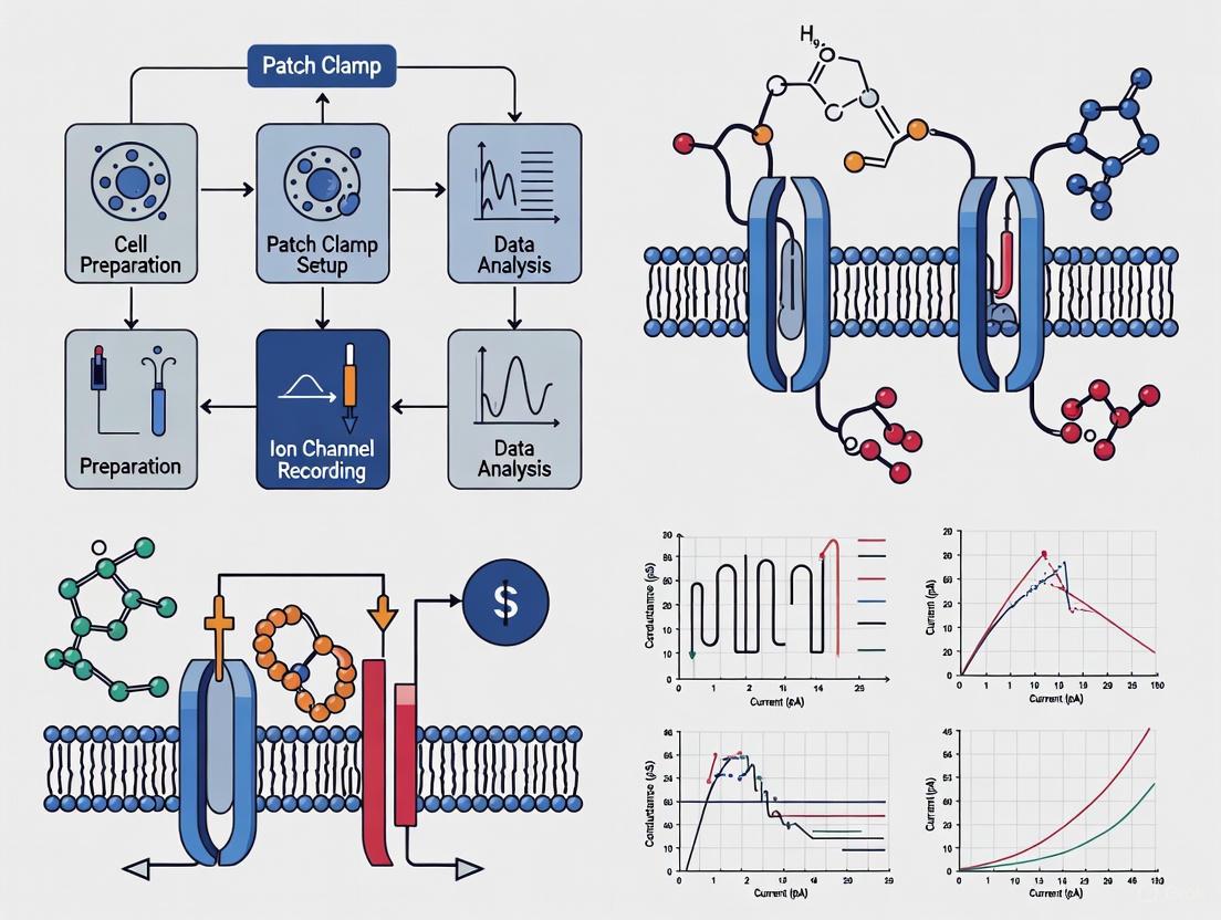
Application Note: Protocol for High-Throughput Screening of ENaC Modulators Using APC
Background and Principle
The epithelial sodium channel (ENaC) is crucial for sodium absorption in lung and kidney epithelia and represents a potential drug target for various renal and pulmonary disorders, including cystic fibrosis-like disease [7]. This protocol establishes a robust method for high-throughput screening of ENaC modulators using automated patch clamp technology, enabling the identification of novel activators and inhibitors with potential therapeutic implications.
Materials and Reagents
Table 3: Research Reagent Solutions for ENaC Screening
| Reagent/Material | Specification | Function/Application |
|---|---|---|
| HEK293 cell line | Stably transfected with human αβγ-ENaC | Heterologous expression of human ENaC |
| Enzymatic cell-detachment solution | Trypsin/EDTA or Accutase | Preparation of single-cell suspensions |
| Amiloride | 10-100 µM | Positive control for ENaC inhibition |
| γ-inhibitory peptide | Specific peptide inhibitor | Control for γ-ENaC subunit inhibition |
| S3969 | Small molecule ENaC activator | Positive control for ENaC activation |
| Chymotrypsin | Serine protease | Prototypical protease for proteolytic ENaC activation |
| Extracellular solution | Standard physiological salt solution | Bath solution for APC recordings |
| Intracellular solution | Low Na+ pipette solution | Pipette solution for whole-cell configuration |
Step-by-Step Protocol
Cell Preparation and Recovery
- Cell Detachment: Harvest hENaC-HEK293 cells using a standard enzymatic cell-detachment procedure to prepare single-cell suspensions.
- Recovery Phase: Resuspend detached cells in complete cell culture medium and incubate for 1-2 hours at 37°C. This recovery period reduces partial proteolytic ENaC activation caused by the detachment procedure, enhancing the sensitivity for detecting novel activators.
- Cell Quality Assessment: Verify cell viability (>85%) and single-cell dispersion before APC experiments.
Automated Patch Clamp Recording
- System Setup: Initialize the SyncroPatch 384PE or comparable APC system according to manufacturer specifications. Prime appropriate intracellular and extracellular solutions.
- Cell Positioning: Dispense cell suspension onto the patch clamp plate; allow cells to be guided by gentle suction onto micron-sized apertures.
- Whole-Cell Configuration: Establish whole-cell configuration by applying gentle suction and voltage pulses as needed. Monitor seal resistance, with gigaohm seals representing the quality standard.
- ENaC Current Recording: Apply voltage protocols appropriate for ENaC characterization (typically -100 mV to +100 mV ramp protocols). Record amiloride-sensitive currents as the definitive indicator of ENaC-mediated conduction.
- Compound Application: Using integrated fluidics, apply test compounds, controls, and reference molecules. Include amiloride (10-100 µM) as an inhibition control and S3969 as an activation control in each experiment.
- Data Acquisition: Continuously record currents throughout compound application, ensuring adequate time for compound effect stabilization (typically 2-5 minutes).
Data Analysis and Quality Control
- Current Normalization: Normalize ENaC currents to cell capacitance to account for variations in cell size.
- Amiloride Sensitivity: Calculate the percentage of amiloride-sensitive current relative to total current. Reject experiments with <70% amiloride sensitivity.
- Dose-Response Analysis: For active compounds, generate concentration-response curves using 4-6 concentrations with appropriate replicates (n≥4 per concentration).
- Statistical Analysis: Determine EC50/IC50 values using nonlinear regression analysis (e.g., Hill equation). Report values as mean ± SEM.
Technical Notes and Applications
- The enzymatic cell-detachment protocol inherently causes partial proteolytic ENaC activation. The recovery phase is crucial for identifying novel ENaC activators mimicking proteolytic channel activation.
- This APC-based screening method successfully identifies both inhibitors and activators of ENaC, demonstrating the system's utility for comprehensive drug discovery campaigns.
- The protocol achieves high success rates of APC recordings with amiloride-inhibitable ENaC currents, enabling reliable high-throughput screening for novel ENaC modulators with potential therapeutic applications in cystic fibrosis and other respiratory diseases [7].
Emerging Technologies and Future Directions
Artificial Intelligence in Ion Channel Research
Artificial intelligence is transforming ion channel drug discovery through multiple applications. Deep learning frameworks integrating 1D convolutional neural networks (1DCNN), bidirectional long short-term memory (BiLSTM), and attention mechanisms can classify ion channel kinetics from whole-cell recordings with 97.58% accuracy, enabling automated analysis of complex electrophysiological data [8]. These AI tools facilitate high-content screening of endogenous ion channel effects in disease models such as Alzheimer's, where they can identify voltage-dependent inhibitory effects of memantine on endogenous channels and antagonistic interactions with calcium ions [8]. For variant classification, protein language models (pLMs) like the MissENSE ION (MissION) classifier achieve ROC-AUC scores of 0.925 in predicting GOF/LOF effects of missense variants, significantly outperforming previous models [3]. These computational approaches are particularly valuable for classifying variants of unknown significance (VUS) in clinical genetics, enhancing diagnostic accuracy and therapeutic selection [3].
Structural Biology and Computational Simulations
Recent advances in cryo-electron microscopy (cryo-EM) have generated an explosion of high-resolution ion channel structures, enabling structure-based drug design [6] [4]. These structures are increasingly used for virtual screening of focused and ultralarge libraries, with AI-assisted protein design accelerating the identification of novel ion channel ligands [6] [4]. In a groundbreaking development, researchers have achieved atom-by-atom computational simulation of ion currents that quantitatively match experimental patch-clamp data [9]. These simulations revealed that potassium ions line up in the channel "like pearls on a string" - packed side by side rather than separated by water molecules as previously assumed - settling a decades-long scientific debate about the mechanism of potassium channel selectivity and conduction [9]. This atomic-level precision opens new avenues for studying drug interactions and designing more effective ion channel modulators.
Multi-Electrode Arrays and Network-Level Screening
Beyond single-channel recording, multi-electrode array (MEA) systems enable interrogation of collective behavior in neuronal or cardiac networks [1]. These platforms detect synchronized bursts, oscillations, and propagation patterns that define network excitability - features inaccessible to single-cell patch recordings [1]. Similarly, impedance-based systems like the CardioExcyte 96 measure changes in electrical resistance as cardiomyocytes contract, translating mechanical beating into dynamic impedance waveforms that capture both electrophysiological activity and contractility [1]. These network-level approaches bridge the gap between cellular electrophysiology and whole-organ physiology, creating a continuum from ion channel gating to network rhythmogenesis that enhances predictive validity for complex physiological and toxicological responses.
Visualization of Workflows
Ion Channel Drug Screening Workflow
Channelopathy Analysis Pipeline
Patch clamp electrophysiology stands as a foundational technique in cellular biophysics and pharmacology, providing direct insight into ion channel function and neuronal excitability. Originally developed by Neher and Sakmann in 1976 to study single ion channel currents, the technique was later improved to include the "whole-cell" configuration by Hamill and colleagues in 1981 [10]. This methodology has since become a gold standard for ion channel research, enabling scientists to understand how ion channels behave in both normal and disease states and how different drugs, ions, or other analytes can modify these conditions [11]. The core principle involves using a glass microelectrode to form a tight seal (typically >1 GΩ) on the cell membrane, allowing researchers to either control the membrane potential to measure ionic currents (voltage clamp) or control the current to measure changes in membrane potential (current clamp) [11] [10]. For drug development professionals, particularly in cardiac and neurological fields, patch clamp electrophysiology offers unparalleled precision for screening compounds that modulate ion channel activity, thereby accelerating the identification of potential therapeutic agents while assessing cardiac safety risks such as hERG channel blockade [12] [13].
Fundamental Principles: Voltage Clamp vs. Current Clamp
The patch clamp technique operates primarily in two fundamental modes: voltage clamp and current clamp, each serving distinct but complementary purposes in electrophysiological investigations. Understanding the principle, applications, and technical requirements of each mode is crucial for designing appropriate experiments and accurately interpreting results in ion channel drug screening research.
Voltage Clamp Mode
The voltage clamp technique is designed to maintain (or "clamp") the cell's membrane potential at a predetermined value set by the experimenter while measuring the ionic currents that flow across the membrane in response to that voltage [11]. This is achieved through a negative feedback circuit in the patch clamp amplifier that injects current equal in magnitude but opposite in sign to the current flowing through the membrane ion channels, thereby maintaining a constant membrane potential [14]. This technique is particularly valuable for studying the kinetic properties of voltage-gated ion channels, including their activation, inactivation, and deactivation characteristics, as well as for investigating the effects of pharmacological compounds on these parameters [11] [15]. In drug screening applications, voltage clamp enables researchers to construct concentration-response relationships for compound effects on specific ion channels by measuring current amplitudes at various holding potentials while applying different drug concentrations [15]. The technique is also indispensable for cardiac safety assessment, where standardized voltage protocols are used to quantify compound-induced block of hERG potassium channels, a common mechanism underlying drug-induced QT prolongation and Torsade de Pointes arrhythmia [12].
Current Clamp Mode
In contrast, current clamp mode allows the membrane potential to vary freely while the experimenter controls the amount of current injected into the cell through the recording electrode [14]. This configuration is ideal for investigating the electrogenic properties of cells, including resting membrane potential, synaptic potentials, receptor potentials, and action potential firing patterns [10]. In current clamp, the amplifier functions as a current source, delivering precisely defined current steps or waveforms while recording the resulting changes in membrane voltage [14]. This mode is particularly useful for studying cellular excitability and how pharmacological agents alter action potential generation and propagation, making it valuable for neuropharmacology and cardiotoxicity screening [16] [15]. For drug development researchers, current clamp recordings can reveal how compound-mediated modulation of specific ion channels translates to functional changes in cellular electrical activity, providing critical insights into both therapeutic potential and safety profiles.
Comparative Analysis
Table 1: Comparative Analysis of Voltage Clamp and Current Clamp Configurations
| Parameter | Voltage Clamp | Current Clamp |
|---|---|---|
| Controlled Variable | Membrane potential | Injected current |
| Measured Variable | Ionic currents | Membrane potential |
| Primary Applications | Ion channel kinetics, pharmacology, conductance measurements | Cellular excitability, action potential properties, synaptic integration |
| Typical Measurements | Current-voltage (I-V) relationships, activation/inactivation time constants, reversal potentials | Resting membrane potential, action potential threshold/amplitude/duration, firing frequency |
| Drug Screening Utility | Direct assessment of compound effects on specific ion channels | Functional assessment of how channel modulation affects cellular output |
| Technical Considerations | Requires series resistance compensation for accurate voltage control; capacitance compensation critical | Stable resting potential essential for meaningful data; bridge balance important for accurate potential measurement |
Figure 1: Patch Clamp Configurations and Their Applications in Drug Screening Research
Essential Equipment and Reagents
Establishing a reliable patch clamp electrophysiology setup requires careful selection of specialized equipment and reagents optimized for maintaining cellular health and ensuring signal fidelity. The core system consists of multiple integrated components that must work in concert to achieve the low-noise environment necessary for high-quality recordings.
Core Instrumentation
The patch clamp amplifier serves as the central electronic component, converting minute electrical signals from the pipette (on the order of picoamperes) into measurable voltage outputs [14]. Modern amplifiers provide critical capabilities for artifact management, including capacitance neutralization to counteract transient current artifacts from cell membrane capacitance and series resistance compensation to correct for voltage errors caused by pipette tip resistance [14]. The amplifier must seamlessly transition between voltage-clamp and current-clamp modes to support diverse experimental paradigms [14]. The mechanical stability of the system is equally critical, with high-precision micromanipulators enabling nanometer-scale movement of the patch pipette toward the cell membrane for successful seal formation [14]. Both hydraulic/mechanical and motorized/piezoelectric manipulators are used, with the latter providing digital control for highly repeatable, programmed movements preferred in standardized screening applications [14]. Vibration isolation via air tables or specialized platforms is non-negotiable for protecting the fragile gigaohm seal from environmental mechanical noise, while Faraday cages shield the preparation from electromagnetic interference that could compromise signal quality [14].
Research Reagent Solutions
Table 2: Essential Research Reagents for Patch Clamp Electrophysiology
| Reagent Category | Specific Examples | Function and Importance |
|---|---|---|
| Extracellular Solutions | Artificial Cerebrospinal Fluid (aCSF): 126 mM NaCl, 2.5 mM KCl, 1.25 mM NaH₂PO₄, 26 mM NaHCO₃, 12.5 mM D-glucose, 1 mM MgSO₄, 2 mM CaCl₂ [10] | Maintains physiological ionic environment; provides energy source; buffers pH when bubbled with carbogen (95% O₂/5% CO₂) |
| Intracellular Solutions | Potassium Gluconate-based: 126 mM K-gluconate, 4 mM KCl, 10 mM HEPES, 0.3 mM EGTA, 4 mM ATP-Mg²⁺, 0.3 mM GTP-Na₂, 10 mM phosphocreatine [10] | Controls intracellular ionic environment; provides energy substrates (ATP, GTP); buffers calcium (EGTA) and pH (HEPES) |
| Ion Channel Blockers | Tetrodotoxin (TTX, 300 nM) for voltage-gated sodium channels; Tetraethylammonium (TEA) for potassium channels; Cs⁺ for internal K⁺ channel blockade [15] | Isolates specific current components by blocking unwanted conductances; essential for studying individual channel types in mixed native systems |
| Cation Substitutions | Cs⁺-based internal solutions (135 mM CsF, 10 mM NaCl, 5 mM HEPES) [15] for K⁺ current suppression; TEA-Cl in external solutions | Enables isolation of specific currents (e.g., Na⁺ or Ca²⁺ currents) by eliminating confounding K⁺ conductances |
| Pharmacological Tools | Specific toxins, channel modulators, and test compounds for screening campaigns | Elucidate drug-channel interactions; establish concentration-response relationships; determine mechanism of action |
Pipette Fabrication and Setup Optimization
The patch pipette itself represents a critical interface between the electronic measurement system and the biological specimen, with its fabrication constituting one of the most technically demanding aspects of the technique [14]. Pipettes are typically pulled from borosilicate or quartz glass capillaries using specialized heated pullers, with the resulting taper angle and tip diameter determining the pipette resistance [14]. For whole-cell configuration, lower resistance pipettes (2-5 MΩ) are preferred to minimize series resistance and facilitate membrane rupture, while higher resistance pipettes (5-10 MΩ) are used for single-channel recordings to form higher resistance seals with reduced tip noise [14]. The composition of the internal pipette solution must be meticulously controlled for osmolarity (typically 280-310 mOsm for mammalian cells), pH (buffered to 7.2-7.4 with HEPES or Tris), and ionic composition tailored to the channels under study [14]. Inclusion of energy substrates like ATP and GTP is often necessary to maintain cell viability and metabolic function during longer recordings, as these cofactors are essential for the regulation and modulation of many ion channels [14] [10].
Standardized Protocols for Drug Screening Applications
Robust and reproducible patch clamp protocols are essential for reliable ion channel drug screening, particularly in regulatory contexts such as hERG channel safety assessment. Standardized methodologies help minimize inter-laboratory variability and ensure consistent data quality across different sites and operators.
hERG Channel Safety Screening Protocol
The hERG potassium channel has become a critical focus in cardiac safety pharmacology due to its association with drug-induced QT prolongation and potentially fatal Torsade de Pointes arrhythmia [12]. Recent multi-laboratory comparisons using standardized protocols have established best practices for hERG screening that align with ICH S7B Q&A 2.1 recommendations [12]. The standard external solution for these assays contains (in mM): 130 NaCl, 5 KCl, 1 MgCl₂·6H₂O, 1 CaCl₂·2H₂O, 10 HEPES, 12.5 dextrose; pH adjusted to 7.4 with 5 M NaOH; ~280 mOsm/L [12]. The internal solution consists of (in mM): 120 K-gluconate, 20 KCl, 10 HEPES, 5 EGTA, 1.5 MgATP; pH adjusted to 7.3 with 1 M KOH; ~280 mOsm/L [12]. Experiments are conducted using the manual whole-cell patch clamp method at near-physiological temperature (35-37°C) to better approximate clinical conditions [12]. Each laboratory tests at least four concentrations that yield good coverage of the concentration-inhibition relationship unless solubility limits are reached, with systematic verification of drug exposure to cells to account for potential compound loss in perfusion systems [12]. This standardized approach has revealed that hERG block potency values within approximately 5-fold of each other should not be considered different, as these values fall within the natural data distribution of the hERG assay, highlighting the importance of establishing laboratory-specific safety margin thresholds [12].
Voltage-Gated Sodium Channel Protocol
For screening compounds against voltage-gated sodium channels (NaV), specialized protocols enable isolation of specific channel subtypes relevant to pain research and neurological disorders [15]. In dorsal root ganglion (DRG) neurons, the bath solution for sodium channel recordings typically contains (in mM): 30 NaCl, 25 D-glucose, 1 MgCl₂, 1.8 CaCl₂, 90 TEA-Cl, 5 CsCl, and 5 HEPES at pH 7.4, while the pipette internal solution contains: 135 CsF, 10 NaCl, and 5 HEPES at pH 7.4 [15]. The addition of 300 nM TTX and selection of neurons based on diameter (<25 μm) enables discrimination between TTX-resistant (TTX-R) and TTX-sensitive (TTX-S) NaV channels [15]. Cells are activated by a 100-ms step depolarization to -10 mV from a holding potential of -80 mV for NaV currents [15]. For specific NaV1.8 channel voltage-clamp recording, DRG neurons are held at -70 mV to inactivate NaV1.9 channels, while for NaV1.9 channels, neurons are activated by a 100-ms step depolarization to -40 mV from a holding potential of -110 mV [15]. These specialized voltage protocols allow researchers to isolate specific sodium channel subtypes for pharmacological characterization, facilitating the development of more targeted analgesics and neurological therapeutics.
Automated High-Throughput Patch Clamp
To address the throughput limitations of manual patch clamp, several automated electrophysiology platforms have been developed that significantly increase screening capacity while maintaining acceptable data quality [17] [16] [13]. These systems can be divided into three main categories: automated glass pipette-based patch clamp, micro-fabricated planar electrode-based patch clamp, and automated two-electrode voltage clamp (TEVC) on Xenopus oocytes [13]. The planar patch clamp approach, exemplified by systems such as Q-Patch, IonWorks, and PatchXpress, utilizes microfabricated silicon or plastic-based planar arrays with micron-size holes that allow tight seal formations with suspended cells [13]. These systems offer varying degrees of throughput, from 150 data points per day (NPC-16) to 3000 (IonWorks HT), with significant reductions in compound consumption due to small recording chamber volumes [13]. However, automated systems currently face limitations in studying primary cells, tissue slices, and differentiated cells derived from iPSCs or ESCs due to their requirement for uniform suspension cells, making manual patch clamp still necessary for these more physiologically relevant but heterogeneous preparations [13]. Recent advancements in automated high-throughput patch clamp have enabled simultaneous voltage-clamp/current-clamp analysis of freshly isolated neurons, providing both detailed ion channel characterization and information about cellular excitability in a more efficient workflow [16].
Figure 2: Standardized Drug Screening Workflow Using Patch Clamp Electrophysiology
Advanced Applications and Emerging Technologies
The field of patch clamp electrophysiology continues to evolve with technological advancements that enhance throughput, data analysis capabilities, and physiological relevance. These innovations are particularly impactful for ion channel drug screening, where traditional limitations of manual patch clamp are being addressed through automation and computational approaches.
Artificial Intelligence in Ion Channel Kinetics Analysis
Recent breakthroughs in artificial intelligence are revolutionizing the analysis of patch clamp data, addressing significant challenges in recording acquisition and interpretation [8]. Advanced machine learning frameworks now enable automated classification of ion channel kinetics from whole-cell recordings, integrating anomaly detection to exclude recordings incompatible with typical ion channel behaviors followed by multi-class classification using deep learning models combining 1D convolutional neural networks (1DCNN), bidirectional long short-term memory (BiLSTM), and attention mechanisms [8]. These systems have demonstrated remarkable classification accuracy (97.58% in classifying 124 test datasets into six categories based on ion channel kinetics), significantly accelerating the analysis process while reducing operator bias [8]. In practical drug screening applications, such as Alzheimer's disease drug development, AI frameworks can identify voltage-dependent inhibitory effects of compounds like memantine on endogenous channels and reveal antagonistic interactions among potassium, magnesium, and calcium ion channels [8]. Similarly, for nanomatrix-induced neuronal differentiation, AI-based classification validates the functional properties of differentiated neurons by evaluating peak current density and inward/outward channel dynamics, providing critical quality control for cell-based therapies [8]. These computational advances represent a paradigm shift in electrophysiological data analysis, enabling more efficient and standardized evaluation of compound effects on ion channel function.
High-Throughput Neuronal Analysis
The development of automated high-throughput patch clamp approaches has enabled the simultaneous and unbiased analysis of acutely dissociated neurons in their native state, addressing significant limitations of traditional manual patch clamp [17] [16]. These systems utilize robotic technologies to streamline the entire experimental process, from cell preparation to data analysis, with protocols requiring 6-18 hours including cell preparation, experimental execution, and analysis of generated data [17]. To manage the large and complex datasets resulting from this methodology, researchers have developed open-source software with easy-to-use graphical interfaces that fit data from each neuron with appropriate biophysical equations to functionally characterize individual neurons [17]. This automated approach enables comprehensive assessment of neuronal biophysics, including voltage-gated sodium channel excitability, action potential properties, and pharmacological responses across large neuronal populations [16]. The methodology supports diverse applications ranging from fundamental assessment of neuronal biophysics to drug development, particularly for neurological disorders where compound effects on native neuronal excitability are more clinically relevant than effects on isolated channels expressed in heterologous systems [17] [16]. The unbiased nature of this automated selection process also helps overcome the selection bias inherent in manual patch clamp, where researchers might unconsciously choose cells based on specific morphological characteristics [16].
Integrated Voltage-Clamp/Current-Clamp Methodologies
Innovative approaches that combine voltage-clamp and current-clamp recordings in the same experimental session provide more comprehensive functional characterization of both ion channel properties and cellular excitability [16]. This integrated methodology is particularly valuable for drug screening, as it enables researchers to directly correlate compound effects on specific ion channels (measured under voltage clamp) with resulting changes in cellular output (measured under current clamp) [16]. For example, in studies of dorsal root ganglion neurons, combined voltage-clamp/current-clamp analysis has revealed how modulation of specific voltage-gated sodium channels translates to altered action potential generation and firing patterns, providing critical insights for pain therapeutic development [16]. The recent development of high-throughput systems capable of this combined analysis addresses the traditional trade-off between detailed ion channel characterization and functional assessment of excitability, offering a more complete picture of compound effects in a single efficient workflow [16]. These technological advances are particularly important for the Comprehensive in vitro Proarrhythmia Assay (CiPA) initiative, which aims to improve cardiac safety assessment through more integrated evaluation of compound effects on multiple cardiac ion channels and resultant changes in cellular electrophysiology [12].
Troubleshooting and Data Quality Considerations
Achieving reliable and reproducible patch clamp data requires careful attention to potential technical pitfalls and implementation of appropriate quality control measures. Even with standardized protocols, several factors can significantly impact data quality and interpretation in ion channel drug screening assays.
Technical Challenges and Solutions
The formation of a stable gigaohm seal (typically >1 GΩ resistance) represents the foundational technical requirement for quality patch clamp recordings, as this high-resistance connection minimizes current leakage and ensures measured currents flow predominantly through ion channels [14]. The sealing process relies on careful pressure management, beginning with gentle pipette movement toward the cell while applying positive pressure inside the pipette to keep the tip clean from debris, followed by pressure release and application of mild continuous negative pressure (suction) once the pipette contacts the cell membrane to achieve the characteristic sharp rise in resistance [14]. Series resistance compensation is another critical consideration, particularly in whole-cell configuration, where uncompensated resistance introduces voltage errors that cause the actual membrane potential to deviate from the command potential, especially when large currents are flowing [14]. Proper compensation improves voltage control and the accuracy of kinetic measurements, though it must be applied judiciously to prevent oscillatory feedback that compromises recording integrity [14]. Additional technical challenges include maintaining stable recordings over time, particularly in whole-cell configuration where intracellular contents may be dialyzed by the pipette solution, potentially affecting ion channel function and cellular health during longer recordings [13]. Careful attention to solution composition, including inclusion of ATP and GTP as energy sources, can help maintain cell viability and metabolic function throughout extended recording sessions [14] [10].
Data Variability and Reproducibility
Understanding and managing data variability is particularly crucial for ion channel drug screening, where decisions about compound advancement may hinge on relatively small differences in potency measurements [12]. Recent multi-laboratory comparisons of hERG data generated using standardized protocols have revealed that hERG block potency values within approximately 5-fold of each other should not be considered different, as these values fall within the natural data distribution of the hERG assay [12]. These findings highlight the importance of establishing laboratory-specific safety margin thresholds that account for systematic data differences rather than relying solely on literature-derived values [12]. Sources of variability include differences in recording temperature, stimulation frequencies, voltage waveforms, and drug exposure to cells, underscoring the importance of rigorous protocol standardization and exposure verification [12]. For automated patch clamp systems, additional considerations include cell quality uniformity and the limitation of studying only suspension-adapted cell types, which may not fully recapitulate the physiological context of native cells [13]. Implementation of appropriate quality control measures, including regular validation with reference compounds, careful monitoring of seal quality and series resistance, and verification of compound exposure concentrations, helps ensure the reliability and reproducibility of patch clamp data for drug screening applications [12] [13].
The patch-clamp technique, first developed by Erwin Neher and Bert Sakmann in the late 1970s, revolutionized the study of ion channels by enabling researchers to measure the tiny electrical currents flowing through single ion channel proteins [18] [19]. This groundbreaking work, which earned them the Nobel Prize in Physiology or Medicine in 1991, provided unprecedented insight into the fundamental mechanisms of electrical signaling in excitable cells and has since become an indispensable tool in basic research and drug discovery [18] [19]. For ion channel drug screening research, understanding the distinct advantages and applications of the four core patch-clamp configurations is essential for designing appropriate experiments and correctly interpreting compound effects on channel function.
Each configuration offers unique experimental access to the ion channel protein, enabling researchers to address specific pharmacological questions. The following sections provide detailed application notes and experimental protocols for the whole-cell, cell-attached, inside-out, and outside-out techniques, framed within the context of modern ion channel drug discovery.
Whole-Cell Configuration
The whole-cell configuration allows researchers to record the integrated activity of all ion channels across the entire cell membrane, providing crucial information about total ionic currents and their impact on cellular excitability [20] [19]. This configuration is established by forming a gigaohm seal between the patch pipette and cell membrane, followed by application of brief suction to rupture the membrane patch, thus establishing electrical and chemical continuity between the pipette interior and the cell cytoplasm [21] [18].
Applications in Drug Screening
Whole-cell recording is particularly valuable in secondary screening and lead optimization phases where detailed characterization of compound effects on ion channel function is required. It enables assessment of a compound's effects on action potential morphology in electrically excitable cells, including stem cell-derived cardiomyocytes used in safety pharmacology (CiPA initiative) [22] [1]. Voltage-clamp experiments allow precise measurement of compound affinity (IC50 values) and kinetics for voltage-gated ion channels, while current-clamp recordings reveal how compounds affect neuronal or cardiac excitability [1] [20]. The configuration also facilitates study of intracellular messenger-mediated channel regulation when compounds are included in the pipette solution [21].
Table 1: Key Applications of Whole-Cell Configuration in Ion Channel Drug Discovery
| Application | Measurement | Relevance to Drug Discovery |
|---|---|---|
| Cardiac Safety Pharmacology | Action potential parameters, hERG channel blockade | Assessment of proarrhythmic risk (CiPA panel) [22] |
| Mechanism of Action Studies | Current-voltage relationships, activation/inactivation kinetics | Determining state-dependent binding (e.g., resting, inactivated) [1] |
| Neuropharmacology | Neuronal excitability, firing patterns | Evaluation of potential anticonvulsants, analgesics [1] |
| Concentration-Response Analysis | IC50/EC50 values | Compound potency ranking for lead optimization [1] |
Experimental Protocol
Cell Preparation: Use adherent or suspended cells expressing the target ion channel. For primary cells or stem cell-derived neurons/cardiomyocytes, ensure appropriate differentiation and homogeneity [13] [23].
Pipette Solution: Prepare an intracellular-like solution containing (in mM): 140 KCl, 1 MgCl2, 10 EGTA, 10 HEPES, pH 7.2-7.4 (adjusted with KOH). For specific experiments, include ATP (2-5 mM) to prevent "run-down" of certain channels [19].
Pipette Preparation: Pull borosilicate glass capillaries to resistance of 2-5 MΩ. Fire-polish tips to optimize seal formation [18] [11].
Seal Formation: Approach the cell with positive pressure applied to the pipette. Upon contact, release pressure and apply gentle negative suction (approximately -20 to -50 mmHg) to form a gigaohm seal (>1 GΩ) [18] [19].
Whole-Cell Access: Apply brief, strong suction pulses or use zap function to rupture the membrane patch. Monitor for sudden increase in capacitive transients indicating whole-cell access [18] [11].
Series Resistance Compensation: After breakthrough, compensate for series resistance (typically 60-80%) to improve voltage control and temporal resolution [20].
Compound Application: Perfuse compounds using a rapid application system. For concentration-response curves, apply increasing concentrations with washout periods between applications [1].
Cell-Attached Configuration
In the cell-attached configuration, the pipette forms a tight seal with the cell membrane, but the patch remains intact, preserving the intracellular environment and allowing observation of single-channel activity without disrupting cellular integrity [21] [19]. This method is particularly valuable for studying ion channels that are modulated by intracellular second messengers or that exhibit "run-down" when the intracellular content is dialyzed [21].
Applications in Drug Screening
The cell-attached configuration excels in several specialized screening applications. It enables assessment of ligand-gated ion channels by including receptor agonists in the pipette solution, allowing observation of single-channel properties without whole-cell disruption [21] [19]. It is ideal for studying channels modulated by metabotropic receptors or intracellular second messengers, as the intact cytoplasm preserves native signaling pathways [21]. The configuration also facilitates investigation of compounds that might alter channel open probability, mean open time, or conductance without dialysis of intracellular components [21].
Table 2: Cell-Attached Configuration: Advantages and Limitations in Drug Screening
| Advantages | Limitations |
|---|---|
| Preserves intracellular environment and signaling pathways | Inability to control intracellular solution composition |
| Prevents "run-down" of sensitive channels | Membrane potential must be estimated |
| Allows study of second messenger systems | Only one drug concentration per patch |
| Stable recording configuration | Challenging for low-abundance channels |
| Minimal disturbance to cell physiology | Limited to single-channel analysis |
Experimental Protocol
Pipette Solution: Prepare an extracellular-like solution. For ligand-gated channels, include the agonist at the desired concentration. For isolation of specific currents, include appropriate channel blockers [21].
Pipette Preparation: Use pipettes with slightly higher resistance (4-6 MΩ) than for whole-cell to optimize single-channel recording [21].
Seal Formation: Approach the cell with positive pressure. Upon contact, release pressure and apply gentle negative suction to form a gigaohm seal [18].
Voltage Determination: Estimate membrane potential by rupturing the patch at the end of the experiment or by using physiological assumption (e.g., -70 mV for neurons) [21].
Single-Channel Recording: Record channel activity at various holding potentials. For drug testing, include compound in pipette solution before sealing [21] [19].
Data Analysis: Analyze single-channel parameters: amplitude, open probability, mean open and closed times, burst duration [21].
Inside-Out Configuration
The inside-out configuration involves excising a patch of membrane such that the intracellular surface faces the bath solution, enabling precise control of the environment at the cytoplasmic side of the channel [21] [19]. This is achieved by forming a cell-attached patch and then rapidly withdrawing the pipette, exposing the cytoplasmic surface to the bath solution [21] [18].
Applications in Drug Screening
This configuration offers unique advantages for specific screening applications. It allows direct application of intracellular messengers (Ca²⁺, cAMP, ATP) to study their effects on channel modulation, enabling mechanistic studies of compounds that act through intracellular signaling pathways [21] [19]. The configuration is ideal for identifying compounds that bind to the intracellular domain of ion channels, as drugs can be directly applied to the cytoplasmic side while monitoring channel activity [19]. It also facilitates study of phosphorylation-dependent channel regulation by including kinases/phosphatases in the bath solution [21].
Experimental Protocol
Pipette Solution: Use an extracellular-like solution. For specific experiments, include channel blockers to isolate currents of interest [19].
Bath Solution: Prepare an intracellular-like solution that can be rapidly exchanged during experiments [21].
Seal Formation: Establish a cell-attached configuration as described previously [18].
Patch Excision: Rapidly withdraw the pipette from the cell. The membrane will reseal, forming a vesicle that can be opened by briefly exposing the tip to air or a low-calcium solution [21] [19].
Solution Exchange: Utilize a rapid perfusion system to change the bath solution composition while recording channel activity [21].
Compound Application: Apply drugs or intracellular messengers to the bath solution while recording from the excised patch [19].
Outside-Out Configuration
The outside-out configuration is formed by transitioning from the whole-cell mode and then slowly withdrawing the pipette, causing the membrane to reform as a patch with the extracellular surface facing the bath solution [21] [19]. This configuration is particularly useful for studying ligand-gated ion channels while maintaining control over the intracellular solution composition [19].
Applications in Drug Screening
The outside-out configuration provides specific benefits for pharmacological studies. It enables rapid solution exchange for studying fast-desensitizing ligand-gated ion channels (e.g., GABAₐ, nicotinic acetylcholine receptors), as compounds can be applied and washed out quickly from the extracellular surface [21] [19]. The configuration allows construction of complete concentration-response relationships on a single patch, improving data consistency and efficiency [13]. It is also valuable for studying the effects of intracellular modulators on ligand-gated channels while maintaining control of the pipette solution composition [19].
Experimental Protocol
Pipette Solution: Use an intracellular-like solution, similar to whole-cell experiments [19].
Establish Whole-Cell Configuration: Follow the whole-cell protocol to achieve rupture of the membrane patch [18].
Patch Formation: Slowly withdraw the pipette from the cell. The membrane will tear and reseal into an outside-out configuration [21] [19].
Solution Verification: Confirm patch orientation by applying known agonists to the bath and verifying expected channel response [19].
Rapid Perfusion: Use a fast perfusion system (exchange time < 100 ms) for applying agonists and compounds [21].
Concentration-Response Curves: Apply increasing concentrations of test compounds to a single patch, with washout between applications [13].
The Scientist's Toolkit: Essential Materials for Patch-Clamp Experiments
Successful patch-clamp experimentation requires specialized equipment and reagents. The following table details essential components of a patch-clamp setup for ion channel drug discovery research.
Table 3: Essential Research Reagent Solutions and Materials for Patch-Clamp Electrophysiology
| Item | Function/Application | Examples/Specifications |
|---|---|---|
| Patch Pipettes | Formation of seal with cell membrane | Borosilicate glass capillaries, 1-5 MΩ resistance [18] [11] |
| Intracellular Solution | Mimics cytoplasmic environment | K-gluconate or KCl-based, with ATP, GTP, EGTA [19] |
| Extracellular Solution | Mimics physiological extracellular fluid | Ringer's, Hanks', or artificial cerebrospinal fluid [19] |
| Channel Blockers | Isolation of specific currents | Tetrodotoxin (Na⁺), Tetraethylammonium (K⁺), Cd²⁺ (Ca²⁺) [21] |
| Enzymes | Tissue dissociation for primary cells | Trypsin, papain, collagenase for cell isolation [13] |
| Perfusion System | Application of test compounds | Gravity-fed or automated systems with rapid exchange [11] |
| Vibration Isolation Table | Mechanical stability for seal formation | Anti-vibration tables essential for gigaohm seals [18] |
| Faraday Cage | Reduces electrical interference | Enclosure grounded to minimize noise [24] |
Technological Advances: Automated Patch-Clamp Systems
Traditional manual patch-clamp techniques, while providing the highest quality data, are labor-intensive and low-throughput, creating bottlenecks in drug discovery pipelines [24] [13]. The development of automated patch-clamp (APC) systems has revolutionized ion channel screening by enabling higher throughput while maintaining electrophysiological fidelity [24] [1].
These systems replace the glass pipette with planar substrates containing micro-fabricated apertures, allowing cells to be positioned automatically by suction and enabling parallel recording from multiple cells [24] [1]. Modern APC platforms range from medium-throughput systems (Patchliner, QPatch) capable of 8-48 parallel recordings to high-throughput systems (SyncroPatch 384PE, Qube) capable of 384 simultaneous recordings [24] [1].
Table 4: Comparison of Automated Patch-Clamp Platforms for Drug Screening
| Platform | Throughput (data points/day) | Seal Resistance | Key Applications in Drug Discovery |
|---|---|---|---|
| QPatch (Sophion) | 250-3,000 | GΩ | Secondary screening, cardiac safety [24] |
| Patchliner (Nanion) | 250-500 | GΩ | Lead optimization, mechanistic studies [24] |
| SyncroPatch 384PE (Nanion) | 20,000-38,000 | GΩ | High-throughput primary screening [24] |
| IonWorks (Molecular Devices) | 3,000-6,000/hour | 50-100 MΩ | Early screening, structure-activity relationships [24] |
| Qube (Sophion) | 30,000/24 hours | GΩ | Ultra-high-throughput screening [24] |
The four core patch-clamp configurations each offer unique experimental access to ion channels, enabling comprehensive pharmacological characterization throughout the drug discovery process. The whole-cell configuration provides information about integrated cellular responses, while the cell-attached method preserves intracellular integrity for studying native channel regulation. The inside-out and outside-out configurations enable precise control over the environments on either side of the membrane for mechanistic studies.
In modern ion channel drug discovery, these techniques are increasingly complemented by automated patch-clamp platforms that provide the throughput necessary for screening compound libraries while maintaining electrophysiological rigor. The strategic selection of appropriate patch-clamp configurations, based on the specific research question and stage of drug development, remains essential for generating high-quality data that reliably predicts therapeutic potential and safety profiles of novel ion channel modulators.
The patch clamp technique, developed in the late 1970s by Erwin Neher and Bert Sakmann (who received the Nobel Prize in Physiology or Medicine in 1991 for this work), represents the gold standard methodology for analysis of excitable cells and ion channel function [25] [26]. This powerful technique provides direct, real-time measurement of ion channel activity at the single-channel or whole-cell level, offering unparalleled insight into the biophysical and pharmacological properties of ion channels [27] [26]. Manual patch clamp electrophysiology has fundamentally advanced our understanding of cellular excitability, neuronal signaling, and cardiac electrophysiology, forming an essential foundation for ion channel drug discovery research [1] [27]. Despite the emergence of automated high-throughput systems, manual patch clamp remains indispensable for specific applications requiring maximal experimental flexibility, data quality, and investigation of complex primary cells [13] [28].
The technique's enduring value lies in its ability to provide high-information content that is difficult to obtain through other methods. Manual patch clamp allows researchers to record from specific subcellular domains and organelles, study ion channels in their native physiological contexts, and perform sophisticated experimental protocols that require real-time intervention and adjustment [27]. This application note examines the technical foundations, methodological approaches, and continuing relevance of manual patch clamp electrophysiology within modern drug screening paradigms, with particular emphasis on its role in target validation and detailed mechanistic studies of ion channel modulators.
Technical Foundations and Configurations
The fundamental principle of patch clamp electrophysiology involves forming a high-resistance seal (gigaohm seal or "gigaseal") between a glass micropipette and a cell membrane, enabling the precise measurement of ionic currents flowing through channel proteins [25] [26]. This intimate connection allows researchers to either control the membrane voltage and measure resulting currents (voltage-clamp mode) or inject current and record changes in membrane potential (current-clamp mode) [25]. The versatility of the technique is demonstrated through multiple configurations, each optimized for specific experimental questions.
Table 1: Patch Clamp Configurations and Their Experimental Applications
| Configuration | Technical Approach | Primary Applications | Advantages | Limitations |
|---|---|---|---|---|
| Cell-Attached | Pipette sealed to intact cell membrane | Studying single channel activity with intact intracellular environment [25] | Minimal cellular disturbance; intracellular mechanisms remain functional [25] | Limited access to intracellular environment; one drug concentration per patch [25] |
| Whole-Cell | Membrane patch ruptured after seal | Recording macroscopic currents from entire cell [25] | Better electrical access to cell interior; suitable for studying pharmacological effects [25] | Dialysis of intracellular contents over time [25] |
| Inside-Out | Patch excised with cytoplasmic face exposed | Studying channels activated by intracellular ligands [25] | Direct access to intracellular surface; controlled intracellular environment [13] [25] | Technically challenging; membrane vesicle formation [25] |
| Outside-Out | Patch excised with extracellular face exposed | Studying ligand-gated channels isolated from cell [13] | Controlled extracellular environment; multiple drug concentrations on same patch [13] | Technically challenging; may contain multiple channels [25] |
The manual patch clamp setup requires specialized equipment including a vibration-isolation table, micromanipulator, microscope, amplifier, digitizer, and data acquisition software [10]. The experimental process demands considerable technical expertise, as establishing high-quality gigaseals requires fine motor control and visual feedback to carefully lower the pipette onto the cell membrane while applying gentle suction [25] [26]. A typical skilled electrophysiologist requires approximately 10-15 minutes to assess a single cell, resulting in fundamental throughput limitations for drug screening applications [29].
Diagram 1: Manual patch clamp experimental workflow. The process requires multiple precise technical steps from preparation through recording, with configuration selection dependent on experimental goals.
The Manual Patch Clamp Methodology
Equipment and Setup Requirements
A complete manual patch clamp system requires several specialized components that collectively enable high-fidelity electrophysiological recordings. The core components include:
- Vibration Isolation System: An air table or other vibration damping system is essential to prevent mechanical disturbances that would disrupt the fragile seal formation [10].
- Microscope with Visualization Capabilities: An inverted microscope equipped with differential interference contrast (DIC) or phase contrast optics is typically used for visualizing cells during approach and seal formation [10]. Fluorescence capabilities are increasingly important for identifying specific cell types or monitoring fluorescent indicators.
- Micromanipulator: A high-precision manipulator allowing fine movement of the patch pipette in three dimensions with minimal drift [10].
- Patch Clamp Amplifier and Digitizer: Specialized electronic instrumentation for controlling membrane potential, measuring currents, and converting analog signals to digital format [10]. Companies such as Molecular Devices, HEKA, and ALA Scientific Instruments manufacture amplifiers specifically designed for patch clamp applications.
- Pipette Puller: A specialized instrument for fabricating glass micropipettes with consistent tip diameters and geometries suitable for patch clamping [10].
- Perfusion System: A method for controlling and changing the extracellular solution bath during experiments, essential for drug application studies [10].
Standardized Solutions for Electrophysiology
The composition of intracellular and extracellular solutions is critical for successful patch clamp experiments, as these solutions determine the ionic gradients and electrochemical driving forces that govern channel behavior [10]. Specific solution compositions vary depending on the experimental goals, but standard recipes have been established for common applications.
Table 2: Standard Patch Clamp Solution Compositions
| Component | Artificial Cerebrospinal Fluid (aCSF) [10] | Potassium Gluconate Internal Solution [10] | Physiological Function |
|---|---|---|---|
| NaCl | 126 mM | - | Maintains physiological extracellular sodium concentration |
| KCl | 2.5 mM | 4 mM | Sets resting membrane potential |
| K-Gluconate | - | 126 mM | Primary intracellular cation source |
| NaHCO₃ | 26 mM | - | pH buffering in extracellular environment |
| HEPES | - | 10 mM | Intracellular pH buffering |
| Glucose | 12.5 mM | - | Energy source for cells |
| MgSO₄ | 1 mM | - | Co-factor for enzymatic processes |
| CaCl₂ | 2 mM | - | Maintains physiological calcium signaling |
| EGTA | - | 0.3 mM | Calcium chelation for controlling intracellular Ca²⁺ |
| ATP-Mg²⁺ | - | 4 mM | Cellular energy source |
| GTP-Na₂ | - | 0.3 mM | G-protein coupling support |
| Phosphocreatine | - | 10 mM | Energy buffer system |
Solution osmolarity and pH must be carefully adjusted to match physiological conditions, typically around 300 mOsm and pH 7.3-7.4. For specific ion channel studies, solutions may be modified to isolate particular currents, such as replacing potassium with cesium to block potassium currents when studying sodium or calcium channels.
Step-by-Step Experimental Protocol
The following protocol outlines the standard procedure for whole-cell patch clamp recording, which is the most common configuration for drug screening applications:
Preparation of Patch Pipettes:
- Use borosilicate glass capillaries with outer diameter of approximately 1.5 mm.
- Pull pipettes using a multi-stage puller program to achieve tip diameters of 1-2 μm and resistances of 3-6 MΩ when filled with standard intracellular solution.
- Fire-polish pipette tips if necessary to smooth the surface and improve seal formation.
Solution Preparation and Cell Placement:
- Filter intracellular solution through 0.22 μm syringe filter to remove particulates.
- Fill the recording chamber with appropriate extracellular solution (aCSF).
- Place cells (primary neurons, cardiomyocytes, or cell lines) in the recording chamber and allow to settle.
- Continuously perfuse with oxygenated extracellular solution at a rate of 1-2 mL/min.
Pipette Placement and Gigaseal Formation:
- Fill pipette with filtered intracellular solution, avoiding bubbles.
- Attach pipette to holder and apply slight positive pressure to prevent tip contamination.
- Lower pipette into solution while monitoring resistance with test pulses.
- Approach the cell surface while maintaining positive pressure.
- Upon contact (indicated by a small increase in resistance), release positive pressure and apply gentle negative pressure to form a gigaseal (>1 GΩ resistance).
Whole-Cell Configuration:
- After stable gigaseal formation, apply additional brief pulses of negative pressure or a high-voltage electrical zap to rupture the membrane patch.
- Monitor the appearance of capacitive transients indicating whole-cell access.
- Compensate for series resistance and capacitive transients using amplifier circuitry.
Data Acquisition:
- Implement appropriate voltage or current protocols for the ion channel or cellular property being studied.
- For drug screening applications, record stable baseline activity, then apply compounds via perfusion system.
- Monitor changes in current amplitude, kinetics, or voltage-dependence in response to drug application.
This protocol requires considerable practice to master, with skilled electrophysiologists typically requiring months to years of training to consistently produce high-quality results across different cell types [26].
The Research Toolkit: Essential Reagents and Materials
Successful manual patch clamp experimentation requires access to specialized reagents and materials that ensure experimental reproducibility and data quality. The following research reagents represent essential components of the patch clamp toolkit:
Table 3: Essential Research Reagents for Manual Patch Clamp
| Reagent/Material | Function | Application Notes |
|---|---|---|
| Borosilicate Glass Capillaries | Fabrication of patch pipettes | Standard outer diameter of 1.5 mm; compatible with most pipette pullers |
| Enzymes for Cell Isolation | Tissue dissociation for primary cells | Collagenase, trypsin, or papain for isolating neurons or cardiomyocytes [29] [28] |
| Ion Channel Modulators | Positive and negative controls for experiments | Tetrodotoxin (TTX) for sodium channels, nifedipine for calcium channels [29] [28] |
| Metabolic Supplements | Maintaining cell health during recording | ATP, GTP, phosphocreatine in internal solution [10] |
| Calcium Chelators | Controlling intracellular calcium concentration | EGTA or BAPTA for buffering intracellular Ca²⁺ levels [10] |
| Protease Inhibitors | Preventing channel degradation | Particularly important for primary cell experiments |
The quality and consistency of these reagents directly impact experimental success rates and data reliability. For drug screening applications, compound libraries must be prepared in appropriate vehicle solutions that do not interfere with electrophysiological measurements, with DMSO concentrations typically kept below 0.1% to avoid nonspecific effects on channel function.
Throughput Limitations and Technical Challenges
The manual patch clamp technique faces significant constraints that limit its application in large-scale screening efforts. A direct comparison with automated systems highlights these fundamental throughput differences:
Diagram 2: Comparison of manual and automated patch clamp approaches. Manual patch clamp offers high-information content and flexibility with compatible cell types but suffers from severely limited throughput compared to automated systems.
The technical challenges of manual patch clamp extend beyond throughput limitations. The technique requires significant technical expertise that typically takes months to years to develop, creating a substantial barrier to entry for research programs [26]. Additionally, manual patch clamp experiments are susceptible to selection bias, as researchers may unconsciously select cells based on morphological characteristics that may not represent the overall population [29]. The labor-intensive nature of the technique also makes it expensive on a per-data-point basis, despite the relatively low cost of individual equipment components compared to automated systems.
Applications in Ion Channel Drug Discovery Research
Despite its limitations, manual patch clamp remains essential for specific applications within the ion channel drug discovery pipeline. The technique provides critical information that cannot be easily obtained through high-throughput methods:
Target Validation and Mechanistic Studies
Manual patch clamp enables detailed investigation of ion channel behavior in physiologically relevant contexts, including native cells and subcellular compartments. This capability is particularly valuable for target validation studies, where understanding channel function in native environments informs decisions about therapeutic targeting [13]. The ability to perform simultaneous voltage-clamp and current-clamp recordings from the same cell provides unique insights into how channel modulators affect both biophysical properties and overall cellular excitability [29].
Safety Pharmacology
Manual patch clamp remains important for comprehensive cardiac safety assessment, particularly for evaluating effects on action potential morphology and duration in native cardiomyocytes [28] [26]. While automated systems can screen for hERG channel blockade, manual patch clamp provides more physiologically complete assessment of proarrhythmic risk through measurement of integrated responses in genuine cardiac cells [26].
Investigation of Complex Cellular Models
The flexibility of manual patch clamp makes it ideally suited for studying ion channels in complex cellular preparations that are not amenable to automated systems, including:
- Primary neurons with extensive processes and heterogeneous channel expression [29]
- Native cardiomyocytes with complex geometry [28]
- Subcellular compartments such as dendrites, nerve terminals, and lysosomes [27] [22]
- Tissue slices maintaining native architecture and connectivity [13]
These applications leverage the key advantage of manual patch clamp: the ability to visually select specific cells or cellular compartments and adapt experimental protocols based on real-time observations.
Manual patch clamp electrophysiology continues to occupy a critical niche in ion channel drug discovery despite the advent of automated high-throughput systems. Its unparalleled data quality, experimental flexibility, and compatibility with complex native cells make it indispensable for target validation, mechanistic studies, and specialized safety pharmacology applications. While throughput limitations restrict its use in primary screening, the high-information content derived from manual patch clamp experiments provides fundamental insights that guide and interpret large-scale screening efforts. The technique remains a cornerstone of ion channel research, bridging molecular biology and integrated physiological function through direct observation of electrical signaling at the cellular level. As drug discovery efforts increasingly target complex channelopathies and specialized cell types, the manual patch clamp's ability to provide detailed electrophysiological characterization in physiologically relevant contexts ensures its ongoing value to the field.
High-Throughput Revolution: Automated and Population Patch Clamp (PPC) Platforms
Automated patch clamp (APC) technology has revolutionized ion channel research and drug discovery, transforming a traditionally low-throughput, skill-intensive technique into a robust, industrial-scale screening method. Since its development at the turn of the millennium, APC has become an integral element in ion channel research and drug development pipelines, overcoming the critical bottleneck posed by manual patch clamp (MPC) investigations [30] [31]. Ion channels represent the second-largest category of pharmacologically targetable proteins after G protein-coupled receptors, with approximately 15-18% of small molecule drugs targeting these crucial cellular gatekeepers [2]. The evolution of APC platforms has democratized access to high-quality electrophysiological data, enabling rapid screening of compound libraries against ion channel targets with implications for cardiovascular safety, neurological disorders, chronic pain, and myriad other therapeutic areas [31] [2]. This application note delineates established APC methodologies and protocols that have matured into indispensable tools for industrial-scale screening campaigns.
Quantitative Performance of APC Platforms
The transition from MPC to APC systems has yielded exponential increases in data output while maintaining the gold standard data quality required for informed decision-making in drug discovery programs.
Throughput Comparison: Manual vs. Automated Patch Clamp
| Method | Data Points Per Day | Technical Skill Requirement | Primary Use Cases |
|---|---|---|---|
| Manual Patch Clamp (MPC) | ~20-40 [31] | High (months of training) [32] | Detailed single-cell investigations, specialized preparations [22] [32] |
| Medium-Throughput APC | 250-500 [31] | Moderate | Secondary screening, lead optimization [31] |
| High-Throughput APC | 3,000-5,000 [31] | Low to moderate | Primary screening, safety pharmacology [31] |
Performance Metrics of Contemporary APC Platforms
| Platform | Recording Sites | Typical Seal Resistance | Special Features |
|---|---|---|---|
| SyncroPatch 384 | 384 [33] | >1 GΩ (with seal enhancer) [31] | Online internal perfusion, temperature control [31] |
| QPatch family | 8/16/48 [31] | >1 GΩ [31] | Multiple compound additions, washout capability [31] |
| PatchLiner | 16 [31] | >1 GΩ (with seal enhancer) [31] | Offline internal perfusion, temperature control [31] |
| IonFlux HT | 64 [31] | ~100 MΩ [31] | Microfluidic solution delivery, parallel assays [31] [32] |
Established APC Applications in Drug Development
APC technology has matured to address multiple critical phases of the drug discovery and development pipeline, with particularly strong penetration in safety pharmacology and ion channel-targeted screening.
Cardiac Safety Assessment
The implementation of APC systems has revolutionized cardiac safety testing, particularly for assessing hERG channel inhibition and its associated risk of drug-induced QT interval prolongation and fatal arrhythmias [34] [35]. The Comprehensive In vitro Proarrhythmia Assay (CiPA) initiative has further expanded APC utilization to include testing pharmaceuticals across a panel of cardiac ion channels in human cardiomyocytes [22] [35]. This integrated approach combines APC data with in silico modeling to more accurately predict clinical cardiac risk [35].
High-Throughput Screening of Native Cardiomyocytes
Recent methodological advances have enabled APC recordings from native cardiomyocytes, which better reflect in vivo cellular physiology compared to heterologous expression systems [28]. A 2022 study demonstrated robust recordings of action potentials, L-type calcium currents (I({Ca,L})), and inward rectifier potassium currents (I({K1})) from isolated swine atrial and ventricular cardiomyocytes using a fixed-well 384-well APC platform [28]. The patching success rate was reported at 13.9 ± 1.7% with seal quality parameters stable throughout experiments [28]. This approach enables detailed pharmacological profiling, as demonstrated by concentration-dependent inhibition of I(_{Ca,L}) by nifedipine (EC~50~ of 6.08 ± 1.14 nM in atrial myocytes and 3.41 ± 0.71 nM in ventricular myocytes) [28].
Epithelial Sodium Channel (ENaC) Screening
APC has been successfully implemented for identifying novel modulators of the epithelial sodium channel (ENaC), a therapeutic target for hypertension, cystic fibrosis, and other pulmonary and renal disorders [33]. A standardized APC protocol using HEK293 cells stably transfected with human αβγ-ENaC confirmed functional expression through amiloride-inhibitable currents and detected both inhibitory and stimulatory effects using a γ-inhibitory peptide and the small molecule ENaC activator S3969 [33]. The methodological optimization included addressing partial proteolytic ENaC activation caused by enzymatic cell-detachment through prolonged incubation recovery periods, enhancing the detection sensitivity for novel activators [33].
Detailed Experimental Protocols
Protocol 1: High-Throughput Screening of hENaC Modulators
Cell Line: HEK293 cells stably transfected with human α-, β-, and γ-ENaC subunits (Charles River, Catalog Number CT6259) [33].
Culture Conditions:
- Medium: DMEM/GlutaMAX supplemented with 10% fetal bovine serum, penicillin (100 U/ml), streptomycin (100 µg/ml) [33]
- Selection antibiotics: Hygromycin B (0.02 mg/ml), Zeocin (0.1 mg/ml), Geneticin (0.5 mg/ml) [33]
- Additive: Amiloride (50 µM) in culture medium to prevent sodium overload [33]
- Environment: 5% CO~2~, 37°C [33]
Cell Preparation:
- Use standard enzymatic cell-detachment procedure with TrypLE Express to prepare single cell suspensions [33]
- For recovery from proteolytic activation: Incubate suspended cells in cell culture medium for extended period (protocol detail optimized experimentally) [33]
- Resuspend cells at appropriate density for APC system (typically 1-5 × 10^6 cells/mL)
APC Recording Conditions:
- System: SyncroPatch 384 [33]
- Voltage protocol: Optimized for ENaC current measurements
- Solutions: Standard extracellular and intracellular solutions for sodium current recordings
- Compound application: Sequential additions of test compounds with washout steps
Validation:
- Confirm ENaC specificity with amiloride (10 µM) inhibition [33]
- Test known modulators: γ-inhibitory peptide (Acetyl-RFSHRIPLLIF-Amide) and S3969 activator [33]
- Include chymotrypsin as positive control for proteolytic activation [33]
Protocol 2: Native Cardiomyocyte Electrophysiology Profiling
Cell Isolation:
- Source: Swine or other mammalian atrial and ventricular tissue [28]
- Enzyme solution: Collagenase-based digestion protocol
- Yield: Approximately 7,200-8,790 viable cardiomyocytes per heart [28]
APC Recording Setup:
- System: Fixed-well 384-well APC platform with borosilicate-glass base [28]
- Cell attachment: Allow cells to settle via gravity and suction pressure [28]
- Success rate expectation: 13.9 ± 1.7% achieving >100 MΩ seal and whole-cell configuration [28]
Experimental Sequence from Single Cell:
- L-type Calcium Current (I(_{Ca,L}))
- Voltage protocol: Depolarizing ramp to inactivate Na+ channels followed by test pulse [28]
- Pharmacological validation: Nifedipine concentration response (1 nM - 5 µM) [28]
- Expected results: Larger current density in ventricular vs. atrial myocytes (-8.65 ± 1.2 pA/pF vs. -4.29 ± 0.17 pA/pF at +10 mV) [28]
- Voltage protocol: Depolarizing ramp to inactivate Na+ channels followed by test pulse [28]
Action Potential Recording
- Configuration: Current-clamp mode
- Stimulation: Square-wave current pulses at threshold intensity
- Parameters analyzed: AP duration at 50% repolarization (APD~50~), resting membrane potential, action potential morphology [28]
Inward Rectifier Currents
The Scientist's Toolkit: Essential Research Reagents and Materials
| Reagent/Cell Line | Function/Application | Example Sources/Compositions |
|---|---|---|
| Stable Cell Lines | Heterologous expression of target ion channels | hENaC-HEK293 (Charles River CT6259) [33]; hNav1.5, hNav1.7 cell lines [22] |
| Native Cardiomyocytes | Physiologically relevant ion channel studies | Freshly isolated from animal hearts [28] |
| Selection Antibiotics | Maintenance of stable cell lines | Hygromycin B, Zeocin, Geneticin [33] |
| Enzymatic Dissociation Agents | Preparation of single-cell suspensions | TrypLE Express, collagenase, dispase II [33] |
| Reference Agonists/Antagonists | Assay validation and controls | Amiloride (ENaC blocker) [33]; Nifedipine (Ca~V~1.2 blocker) [28]; Carbachol (I(_{K,ACh}) activator) [28] |
| Seal Enhancers | Improve success rate of GΩ seal formation | High Ca~2+~ solutions (40 mM); fluoride-based solutions [31] [2] |
| Ion Channel Modulators | Tool compounds for pharmacological characterization | S3969 (ENaC activator) [33]; γ-inhibitory peptide (ENaC inhibitor) [33] |
Workflow and Data Analysis
APC Experimental Workflow
Ion Channel Drug Screening Cascade
Automated patch clamp technology has unequivocally matured into an indispensable platform for industrial-scale ion channel screening. The methodologies and protocols detailed herein demonstrate robust applications across diverse screening scenarios, from high-throughput primary compound screening to detailed pharmacological characterization of lead compounds. As APC systems continue to evolve with improved affordability, accessibility, and capabilities for studying native cells [28] [32], their implementation is expected to expand further in both industrial and academic settings. The integration of APC data with other technologies such as stem cell biology, optogenetics, and in silico modeling represents the next frontier in ion channel drug discovery, promising more physiologically relevant screening outcomes and accelerated development of novel therapeutics targeting this crucial protein class [2].
Population Patch Clamp (PPC) is a groundbreaking high-throughput planar array electrophysiology technique that represents a significant evolution in ion channel screening. This method enables simultaneous recording of ionic currents from populations of cells under voltage clamp within a single well, summing the whole-cell currents from multiple cells to generate an ensemble current reading [36] [37]. For drug discovery pharmacologists, PPC delivers substantially greater speed and precision compared to conventional patch clamp methods, addressing critical bottlenecks in ion channel drug screening programs [36].
The technology was developed to overcome the limitations of traditional approaches, particularly their moderate consistency and throughput, which rendered impractical the functional measurement of large numbers of ion channel ligands or mutant channel genes [37]. By modifying planar patch clamp substrates and amplifiers in instruments like the IonWorks system, PPC achieves unprecedented success rates exceeding 95% per recording attempt while providing markedly improved data consistency [37]. This breakthrough allows direct electrophysiological recording of thousands of ensemble ionic currents per day—a throughput level essential for screening directed compound libraries against ion channel targets in modern drug discovery pipelines [37].
Fundamental Principles
The PPC methodology builds upon conventional planar array electrophysiology but introduces a crucial innovation: instead of recording from individual cells in separate apertures, a single voltage-clamp amplifier simultaneously captures signals from multiple cells within the same well, each sealed to a separate aperture in the planar substrate [36] [37]. This ensemble approach effectively averages currents across a cell population, producing more consistent data while maintaining the physiological relevance and information content of traditional electrophysiology.
The technique utilizes a modified PatchPlate substrate and amplifiers specifically engineered for population measurements. When implemented in a 384-well format with parallel recording capabilities, this system enables the direct electrophysiological recording of thousands of data points daily—a throughput previously unattainable with conventional electrophysiological methods [37]. The procedure incorporates sophisticated subtraction methods that correct for expected signal distortions, reliably producing data that align with established patch-clamp studies while dramatically increasing throughput [37].
Comparative Advantages
The transition from conventional to automated patch clamp technologies, and subsequently to PPC, represents a paradigm shift in ion channel screening. The table below quantifies the key advantages of PPC over these established methods:
Table 1: Comparative Analysis of Patch Clamp Technologies
| Feature | Conventional Patch Clamp | Automated Patch Clamp | Population Patch Clamp (PPC) |
|---|---|---|---|
| Throughput | Low (single cells) [38] | Medium (parallel cells) [38] | High (384-well parallel) [37] |
| Data Consistency | High (expert-dependent) [37] | Variable (system-dependent) | >95% success rate [37] |
| Recording Type | Single cell | Single cell | Ensemble cell population [36] [37] |
| Applications | Basic research, detailed characterization | Secondary screening, safety pharmacology | Primary screening, directed libraries [37] |
| Daily Data Points | 10-100 [38] | ~10,000 (IonWorks HT) [38] | Thousands of ensembles [37] |
Figure 1: PPC Performance Advantage Pathway. Population Patch Clamp simultaneously optimizes multiple key screening metrics that are typically mutually exclusive in conventional approaches.
Beyond the quantitative advantages detailed in Table 1, PPC provides unique access to challenging targets that have traditionally proven difficult to screen with standard planar array electrophysiology. This includes constitutively active channels and slow-ligand gated channels such as SK/IK channels, thereby expanding the druggable ion channel space for pharmaceutical development [36]. The technology also supports sophisticated experimental designs including ion channel assay duplexing and modulator assays, approaches that further enhance its utility in complex screening paradigms [36].
Application Notes and Experimental Protocols
PPC Screening Protocol for Compound Profiling
The following section provides a detailed methodology for implementing PPC in ion channel drug discovery campaigns, with particular emphasis on voltage-gated ion channel targets. This protocol has been optimized for screening directed compound libraries while maintaining data quality comparable to conventional electrophysiology.
Table 2: Key Research Reagent Solutions for PPC Screening
| Reagent/Material | Function | Specification Notes |
|---|---|---|
| Cell Line | Ion channel expression | Heterologous system (e.g., HEK, CHO) with stable target expression |
| PatchPlate PPC | Planar substrate | Multi-aperture wells for ensemble recordings |
| Extracellular Solution | Bath solution | Matches physiological extracellular ionic composition |
| Intracellular Solution | Pipette solution | Mimics cytoplasmic environment for whole-cell configuration |
| Reference Compounds | assay controls | Known agonists/antagonists for system validation |
| Test Compounds | Investigation | Directed library compounds in DMSO stocks |
Day 1: Cell Preparation
- Cell Harvesting: Gently detach cells expressing the target ion channel using enzymatic dissociation (e.g., Accutase). Avoid overtrypsinization which can damage membrane proteins critical for seal formation.
- Cell Resuspension: Centrifuge cells (1000 rpm for 5 minutes) and resuspend in appropriate extracellular solution at optimal density (typically 1-2×10^6 cells/mL). Maintain cell suspension at room temperature with gentle agitation until use.
- Quality Control: Assess cell viability using trypan blue exclusion, targeting >90% viability for optimal seal formation.
Day 1: PPC Recording
- System Priming: Prime the PPC instrument (e.g., IonWorks with modified PPC substrate) with appropriate intracellular and extracellular solutions according to manufacturer specifications.
- Plate Preparation: Dispense cell suspension into PPC plate wells using integrated fluidics, allowing cells to settle onto apertures (approximately 5-10 minutes).
- Seal Formation: Apply gentle suction to each well to form gigaseals simultaneously across multiple apertures within the same well. Monitor seal resistance automatically.
- Whole-Cell Configuration: Establish whole-cell access through automated perforated patch or gentle rupture protocols.
- Compound Addition: Using integrated liquid handling, add reference controls or test compounds (typically 3-5 concentrations for dose-response studies) with appropriate mixing.
- Voltage Protocol Application: Apply appropriate voltage protocols to activate the target ion channel (e.g., step to +20 mV for 200 ms from -80 mV holding potential for depolarization-activated channels).
- Ensemble Current Recording: Record summed whole-cell currents from multiple cells within each well simultaneously. The system typically employs subtraction methods to correct for anticipated distortions [37].
- Data Acquisition: Collect ensemble currents with appropriate sampling rates (typically 10-50 kHz) and filtering.
Data Analysis
- Current Normalization: Normalize ensemble current amplitudes to baseline or control conditions.
- Dose-Response Fitting: Fit concentration-response data to appropriate models (e.g., Hill equation) to determine IC50/EC50 values.
- Quality Control: Apply pre-defined quality criteria based on seal resistance, current stability, and control compound responses.
Figure 2: PPC Experimental Workflow. Key quality control checkpoints (yellow) ensure data integrity throughout the screening process.
Protocol for Native Cell Applications
While PPC is extensively utilized in heterologous expression systems, its application in native systems provides unique insights into endogenous ion channel function in more physiologically relevant contexts. The following protocol adapts PPC for primary neuronal cultures, enabling investigation of endogenous voltage-gated potassium (Kv) and sodium (Nav) channels [39].
Primary Culture Preparation
- Cell Isolation: Isbrate cortical neurons from embryonic or postnatal rats (E18-P2) using enzymatic and mechanical dissociation.
- Culture Maintenance: Plate cells on poly-D-lysine coated surfaces at appropriate density (50,000-100,000 cells/cm²) in neuronal culture medium supplemented with B27 and growth factors.
- Maturation Monitoring: Allow neuronal maturation for 10-21 days in vitro, monitoring expression of neuronal markers (e.g., MAP2, NeuN) and electrophysiological activity development through microelectrode array recordings.
PPC Recording from Native Neurons
- Cell Harvesting: Gently harvest mature primary neurons using mild enzymatic treatment optimized for sensitive native cells.
- PPC Configuration: Utilize systems capable of handling native cells (e.g., SyncroPatch 384PE) with appropriate settings for smaller cell sizes and more delicate membranes.
- Current Isolation: Apply specific voltage protocols to isolate different current components:
- For Kv channels: Step depolarizations from -80 mV to +60 mV in 10 mV increments from holding potential -90 mV.
- For Nav channels: Step depolarizations from -100 mV to +30 mV in 5 mV increments from holding potential -120 mV.
- Pharmacological Validation: Include specific channel blockers (e.g., tetrodotoxin for Nav channels, tetraethylammonium for Kv channels) to confirm current identity and specificity.
This approach enables investigation of endogenous ion channels in their native environment with significantly improved throughput compared to manual approaches, while capturing the natural subunit composition and regulatory environment of primary systems [39].
Implementation Considerations
System Requirements and Optimization
Successful implementation of PPC technology requires careful attention to several critical parameters that differ from conventional patch clamp approaches. The table below outlines key considerations for establishing a robust PPC screening platform:
Table 3: PPC Implementation and Optimization Guide
| Parameter | Consideration | Optimization Guidance |
|---|---|---|
| Cell Quality | Higher viability requirements | Maintain >90% viability; optimize dissociation protocols |
| Cell Density | Critical for multiple seals per well | Titrate for optimal single-cell per aperture occupancy |
| Solution Composition | Impacts seal success | Adjust divalent cations; optimize osmolarity |
| Temperature | Affects channel kinetics and seal stability | Standardize at physiological (35-37°C) or room temperature |
| Timing | Throughput vs. data quality balance | Balance recording length with stability requirements |
Applications in Drug Discovery Pipeline
PPC technology has transformed ion channel screening throughout the drug discovery pipeline. In primary screening, PPC enables the functional evaluation of large compound libraries against ion channel targets that were previously intractable to high-throughput approaches [37]. For secondary screening and lead optimization, PPC provides detailed pharmacological profiling (IC50/EC50, kinetics, use-dependence) at scales sufficient for structure-activity relationship (SAR) campaigns.
In safety pharmacology, PPC has become particularly valuable for hERG channel screening, where regulatory requirements demand comprehensive assessment of compound effects on this critical cardiac ion channel [38]. The technology's ability to generate high-quality data at scale makes it ideal for the thorough safety profiling required by regulatory agencies for new chemical entities.
Furthermore, PPC shows growing promise in investigating the function of large numbers of ion channel mutants, enabling functional proteomics and disease mechanism studies at unprecedented scale [37]. This application is particularly relevant for channelopathies—diseases caused by ion channel dysfunction—where high-throughput functional characterization of genetic variants can accelerate both target validation and personalized therapeutic approaches.
Population Patch Clamp technology represents a transformative advancement in ion channel electrophysiology, successfully addressing the critical trade-off between data quality and throughput that has long constrained ion channel drug discovery. By enabling ensemble recordings from multiple cells simultaneously while maintaining the physiological relevance and information content of conventional patch clamp, PPC provides researchers with a powerful tool for primary screening and detailed compound characterization.
The robust protocols and implementation frameworks outlined in this article provide a foundation for researchers to leverage PPC technology across the drug discovery pipeline, from initial target validation to safety pharmacology. As ion channels continue to represent important therapeutic targets for neurological, cardiovascular, and metabolic diseases, PPC stands as an essential technology for accelerating the development of novel ion channel modulators with improved efficacy and safety profiles.
Ion channels represent crucial targets for therapeutic intervention, implicated in a wide range of disorders from epilepsy to cardiac arrhythmia. The patch clamp technique, first developed by Neher and Sakmann in the 1970s, remains the gold standard for studying ion channel function and pharmacology due to its unparalleled ability to directly measure ionic currents with high temporal resolution [40]. This technique has evolved significantly from its origins as a manually intensive, low-throughput method to include automated platforms that now enable medium-to-high throughput screening essential for modern drug discovery programs [1].
The application workflows spanning from initial compound screening to mandatory cardiac safety testing represent a critical pathway in pharmaceutical development. This pathway has become increasingly important as regulatory frameworks recognize the value of high-quality nonclinical data for predicting clinical cardiac risk [12] [41]. The integration of patch clamp electrophysiology throughout this workflow provides the direct biophysical insight necessary to understand compound effects on ion channel function, enabling researchers to make informed decisions at each stage of drug development.
The Scientist's Toolkit: Essential Research Reagent Solutions
Successful execution of patch clamp experiments requires specific reagents and materials optimized for electrophysiological applications. The table below details key research reagent solutions essential for ion channel drug screening workflows.
Table 1: Essential Research Reagents for Patch Clamp Electrophysiology
| Reagent/Material | Function/Purpose | Example Specifications |
|---|---|---|
| Cell Lines | Heterologous expression of target ion channels for consistent screening | CHO hERG DUO cells [42]; HEK293 expressing hERG1a [12] |
| Cell Culture Medium | Supports growth and maintenance of expression cells | HAM's F-12 + Glutamax with FBS Gold and selection antibiotics [42] |
| Extracellular Solution | Maintains physiological ionic environment during recordings | 130 mM NaCl, 5 mM KCl, 1 mM MgCl₂, 1 mM CaCl₂, 10 mM HEPES, 12.5 mM dextrose; pH 7.4 [12] |
| Intracellular (Pipette) Solution | Controls intracellular ionic composition during whole-cell recordings | 120 mM K-gluconate, 20 mM KCl, 10 mM HEPES, 5 mM EGTA, 1.5 mM MgATP; pH 7.3 [12] |
| Serum-Free Medium | Used during cell harvesting to prevent serum-induced channel blockade | CHO-S-SFM I with HEPES, trypsin inhibitor, penicillin/streptomycin [42] |
Experimental Protocols: Standardized Methods for Cardiac Ion Channel Screening
Automated Patch Clamp Protocol for hERG Channel Screening
The following protocol outlines a standardized approach for assessing compound effects on hERG potassium channels using automated patch clamp systems, adapted for medium-throughput instrumentation such as the QPatch or Patchliner platforms [42].
Materials and Equipment:
- Automated patch clamp system (e.g., QPatch, Patchliner, SyncroPatch)
- CHO or HEK293 cell line stably expressing hERG channels
- Extracellular and intracellular solutions (see Table 1)
- Compound plates with test articles dissolved in DMSO
- Quality control criteria: Seal resistance >1 GΩ, cell viability >80%
Step-by-Step Procedure:
Cell Preparation:
- Culture hERG-expressing cells in appropriate medium with selection antibiotics
- Harvest cells at 70-80% confluence using enzymatic dissociation
- Resuspend cells in serum-free medium at optimal density (0.5-2 × 10^6 cells/mL)
System Preparation:
- Prime fluidics with extracellular solution
- Load compound plates following manufacturer's specifications
- Calbrate pressure and voltage systems according to platform requirements
Experimental Setup:
- Set recording temperature to 35-37°C (near physiological)
- Configure voltage protocol: -80 mV holding potential, steps to +20 mV for 2 sec, then to -50 mV for 2 sec to record tail currents
- Set sampling rate at 10-50 kHz with appropriate filtering (2-10 kHz Bessel)
Recording Protocol:
- Establish whole-cell configuration using platform-specific sequences
- Record baseline hERG current for 3-5 minutes to ensure stability
- Apply test compounds in increasing concentrations (typically 4-8 concentrations)
- Maintain each concentration for 5-10 minutes to reach steady-state block
- Include positive (e.g., 1 μM E-4031) and negative controls in each run
Data Analysis:
- Measure tail current amplitude at each concentration
- Normalize currents to baseline values
- Generate concentration-response curves using nonlinear regression
- Calculate IC₅₀ values with 95% confidence intervals
Manual Patch Clamp Protocol for Regulatory Studies
For regulatory submissions and detailed mechanistic studies, manual patch clamp remains essential. The following protocol reflects best practices from the HESI multi-laboratory study and ICH S7B Q&A 2.1 recommendations [12] [41].
Materials and Equipment:
- Manual patch clamp rig with amplifier, micromanipulators, and vibration isolation
- Borosilicate glass capillaries for pipette fabrication
- Perfusion system with temperature control (35-37°C)
- Drug application system (gravity-fed or perfusion)
Step-by-Step Procedure:
Pipette Preparation:
- Pull borosilicate glass to resistance of 1-3 MΩ
- Fire-polish to optimize seal formation
- Fill with filtered intracellular solution
Cell Preparation:
- Plate cells on coverslips at appropriate density
- Use within 24-48 hours after plating
- Maintain in extracellular solution during recordings
Giga-seal Formation:
- Position pipette approaching cell with positive pressure
- Contact cell membrane and release pressure
- Apply gentle suction to achieve >1 GΩ seal
Whole-Cell Configuration:
- Apply additional suction or voltage zap to rupture membrane
- Monitor access resistance (<10 MΩ preferred)
- Compensate series resistance (70-80%)
Drug Application:
- Establish stable baseline recording (3-5 minutes)
- Apply compounds via perfusion system with complete solution exchange
- Record until steady-state block achieved (typically 5-10 minutes per concentration)
- Include washout period when possible
Quality Control:
- Monitor seal stability throughout experiment
- Track access resistance for consistency
- Verify liquid junction potential correction
Application Workflow: Integrating Screening and Safety Assessment
The pathway from initial compound screening to definitive cardiac safety testing involves multiple stages with progressively more rigorous electrophysiological assessment. The workflow diagram below illustrates this integrated approach.
Figure 1: Integrated workflow for ion channel drug screening and cardiac safety assessment
Workflow Stage Descriptions
Primary Screening: Initial high-throughput screening using fluorescence-based assays (FLIPR, FMP) to identify potential modulators from large compound libraries [1]. This stage prioritizes compounds for more rigorous electrophysiological characterization.
Secondary Screening: Medium-throughput assessment using automated patch clamp systems to confirm activity and provide initial potency estimates [42]. Platforms such as SyncroPatch 384PE enable hundreds of parallel recordings, balancing throughput with data quality.
Hit Confirmation: Detailed characterization using manual patch clamp to verify compound effects and investigate mechanism of action [12]. This stage provides high-quality data for structure-activity relationship (SAR) studies.
Lead Optimization: Iterative compound modification and testing to improve potency, selectivity, and drug-like properties while minimizing off-target effects [42]. Electrophysiology data guides medicinal chemistry efforts.
Cardiac Safety Assessment: Comprehensive evaluation using standardized protocols following ICH S7B and E14 guidelines [12] [41]. This includes hERG channel testing and assessment of other cardiac ion channels (Nav1.5, Cav1.2) within the CiPA (Comprehensive in vitro Proarrhythmia Assay) initiative.
Regulatory Submission: Compilation of GLP-compliant data for regulatory review, incorporating the updated ICH E14/S7B Q&As that allow use of nonclinical data to support clinical QTC risk assessment [41].
Quantitative Data Analysis and Variability Assessment
Recent multi-laboratory studies have provided crucial insights into the reproducibility and variability of hERG assay data, which directly impacts safety margin calculations and risk assessment.
Table 2: hERG Assay Variability from Multi-Laboratory Study (2025 HESI)
| Parameter | Finding | Regulatory Implication |
|---|---|---|
| Overall hERG IC₅₀ Variability | ~5-fold difference between laboratories | Values within 5X should not be considered different [12] |
| Within-Laboratory Reproducibility | Most labs within 1.6X on retesting | Supports internal consistency in lead optimization [41] |
| Systematic Inter-Lab Differences | Observed in 1 of 5 laboratories | May require lab-specific safety margins [43] |
| Impact of Standardized Protocols | Reduced but did not eliminate variability | Supports ICH S7B Q&A 2.1 best practices [12] |
| Key Variability Factors | Cell lines, drug delivery, temperature control | Standardization improves comparability [12] |
The observed ~5-fold variability in hERG block potency measurements has significant implications for cardiac safety assessment. According to the recent HESI-coordinated study, hERG block potency values within 5-fold of each other represent natural assay distribution rather than true pharmacological differences [12]. This variability must be incorporated into safety margin calculations when using hERG data to predict clinical QTC prolongation risk.
Regulatory Framework and Future Directions
The regulatory landscape for cardiac safety testing has evolved significantly with recent updates to ICH E14 and S7B guidelines. The current framework enables more efficient integration of nonclinical data into clinical risk assessment, potentially reducing the need for dedicated thorough QTC studies in certain cases [41].
Emerging Technologies and Approaches
Stem Cell-Derived Cardiomyocytes: These cells replicate human cardiac electrophysiology with remarkable accuracy, enabling direct observation of action potential morphology and drug-induced arrhythmogenic risk [1] [44].
Optical Electrophysiology: New light-based techniques using voltage-sensitive dyes and optogenetic actuators enable higher throughput screening while maintaining pharmacological relevance [45].
Organellar Ion Channel Screening: Growing interest in mitochondrial and lysosomal ion channels has driven development of specialized assays for these targets implicated in neurodegenerative diseases [4].
The field continues to advance with improved technologies, standardized protocols, and better understanding of assay variability. These developments support more predictive safety assessment and efficient drug discovery while maintaining rigorous cardiac safety standards.
Expanding Horizons: Screening Native Cells, iPSC-Derived Neurons, and Cardiomyocytes
The field of ion channel drug discovery is undergoing a significant transformation, driven by the integration of more physiologically relevant human-based models. While recombinant systems remain a robust de-risking strategy, they often neglect intricate intracellular interactions [46]. The advent of human induced pluripotent stem cell (iPSC)-derived cells, particularly cardiomyocytes (iPSC-CMs) and neurons (iPSC-neurons), provides an unprecedented opportunity to study ion channel function and pharmacology in a more native context [46] [47] [48]. This shift is coupled with technological advancements in automated patch clamp (APC) systems, which are overcoming the traditional throughput limitations of manual techniques and enabling the high-quality electrophysiological characterization essential for drug screening [22] [49]. This application note details the methodologies and quantitative data for screening these advanced cellular models, framing them within the broader context of modern ion channel research and safety pharmacology.
Electrophysiological Profiling of iPSC-Derived Cardiomyocytes
Advantages of Perforated Patch Clamp Configurations
iPSC-derived cardiomyocytes are widely used in drug discovery due to their close resemblance to native cardiomyocytes [46]. However, the standard whole-cell (WC) patch clamp configuration can hinder accurate action potential measurements because cytoplasmic components are "washed out," which alters channel activities and disrupts Ca²⁺ buffering systems. This often results in recorded action potentials that are very short, especially during early repolarization [46].
- Solution: The perforated patch clamp technique on an automated APC platform, using nystatin as a pore-forming agent, preserves the cell's native intracellular environment.
- Validation: Application of the intracellular voltage-gated sodium channel (VGSC) blocker QX-314 via internal solution exchange completely inhibits VGSC currents in WC configuration but has minimal effect in the perforated configuration, confirming the integrity of the recording [46].
- Key Finding: Action potential duration at 30% repolarization (APD30) values were between 3.5 and 1.6 times longer in perforated patch clamp recordings compared to WC configurations, depending on the maturation time of the cardiomyocytes (8 to 21 days in vitro) [46]. Voltage clamp recordings attributed this change to larger voltage-gated calcium channel (VGCC) currents when the intracellular environment is preserved [46].
Quantitative Electrophysiology of Commercial iPSC-CM Models
Commercial iPSC-CM populations, such as iCell Cardiomyocytes and atrial/ventricular Pluricytes, are a mixture of spontaneously and electrically active cells. A single-cell patch-clamp and RT-qPCR study revealed their heterogeneous nature, combining traits of adult cardiomyocyte subtypes [50].
Table 1: Single-Cell Ion Channel Expression and Electrophysiology of Commercial iPSC-CMs vs. Primary Cells
| Cell Model / Parameter | iCell Cardiomyocytes (Mixed) | Atrial Pluricytes | Ventricular Pluricytes | Primary Human Atrial/Ventricular CMs |
|---|---|---|---|---|
| Spontaneous Activity | Present in a subset of cells | Information missing | Information missing | Specific to nodal cells |
| Key Ion Channel Transcripts | Combination of nodal (HCN4), atrial (KCNA5), and ventricular (SCN5A, KCNJ2) markers [50] | Trends towards atrial specificity | Trends towards ventricular specificity | Distinct, chamber-specific expression patterns [50] |
| Phenotype Interpretation | Immature, mixed subtype traits [50] | Trend towards chamber specificity | Trend towards chamber specificity | Mature, distinct subtype identity [50] |
| Utility for Drug Screening | Analysis of multiple cardiac ion channels in a near-native environment [50] | Promising for chamber-specific investigation [50] | Promising for chamber-specific investigation [50] | Gold standard but low availability |
Experimental Protocol: Automated Perforated Patch Clamp of iPSC-CMs
This protocol is adapted from assays developed for high-throughput APC platforms [46].
- Cell Preparation: Use hiPSC-CMs matured between 8 and 21 days in vitro. Ensure cells are properly dissociated into a single-cell suspension compatible with the APC system.
- Pore-Forming Agent: Prepare the internal solution containing nystatin as the pore-forming agent for perforated patch recordings.
- APC Setup: Load the cell suspension and solutions onto the automated patch clamp system (e.g., a platform capable of perforated patch clamp).
- Seal Formation and Perforation: The system will sequentially perform seal formation, followed by membrane perforation until a stable electrical access is achieved.
- Quality Control: Monitor access resistance. Validate the perforated configuration by verifying the lack of effect of intracellular blockers like QX-314 if applied.
- Action Potential Recording: Initiate current-clamp protocols to record action potentials.
- Voltage-Clamp Recording: Switch to voltage-clamp mode to isolate and record specific ionic currents, such as VGCC currents.
Diagram 1: Automated Perforated Patch Clamp Workflow for iPSC-CMs.
Functional Characterization of iPSC-Derived Neurons
Maturation Classification Using Automated Patch Clamp
Human iPSC-derived neurons are powerful models for neurological diseases, but variability in electrophysiological maturity has been a challenge. A high-throughput APC workflow has been established to classify neuronal maturity functionally [49].
Table 2: Electrophysiological Classification of iPSC-Derived Neuron Maturity
| Maturity Type | Action Potential (AP) Firing Profile | Key Characteristics |
|---|---|---|
| T1 | No action potential | Immature phenotype. |
| T2 | 1 action potential | Early stage of excitability. |
| T3 | 2 action potentials | Intermediate maturity. |
| T4 | >2 action potentials (multiple firing) | Highest maturity level; shows significantly higher NaV current density, more hyperpolarized resting membrane potential, and larger capacitance [49]. |
This classification allows researchers to select electrophysiologically comparable neurons, reducing variability and improving the fidelity of disease models [49]. The proportion of highly mature T4 neurons increases with days in vitro (DIV).
Profiling Sensory and Motor Neurons for Disease Modeling
iPSC-derived sensory and motor neurons provide humanized models for pain and motor neuron disease research.
- Sensory Neurons: Commercial iCell Sensory Neurons show a robust phenotype, with >80% expressing sensory neuron markers (BRN3A+/UCHL1+). RNASeq confirms expression of key channels like NaV1.7 (SCN9A), TRPV1, and P2RX3 [47]. Electrophysiological recordings between 28-56 DIV show waveforms typical of human dorsal root ganglion neurons, including a broad action potential with a 'hump' upon repolarization. The selective NaV1.7 inhibitor PF-05089771 (100 nM) inhibits repetitive firing, confirming functional channel expression [47].
- Motor Neurons: Commercially available iPSC-derived motor neurons express key biomarkers and functional voltage-gated ion channels (Na⁺, K⁺, Ca²⁺) [48]. However, they often exhibit high input resistance and depolarized resting membrane potentials, suggesting incomplete electrical maturation. Their functionality is confirmed by progressive increases in repetitive firing capacity and action potential kinetics over time [48].
Experimental Protocol: High-Throughput Profiling of iPSC-Neurons on APC
This protocol enables the sequential recording of multiple parameters from the same neuron on platforms like Sophion's Qube or QPatch [49].
- Cell Preparation: Dissociate iPSC-derived neurons (e.g., iCell GlutaNeurons) to a single-cell suspension optimized for APC systems, aiming for high viability.
- APC Assay Setup: Load cells onto the 384-site APC platform. A robust success rate of ~60% whole-cell recording is achievable.
- Sequential Electrophysiology Protocol:
- Voltage-Gated Currents: Record NaV and KV currents in voltage-clamp mode.
- Excitability: Switch to current-clamp mode and apply a current-step protocol to evoke action potentials. Classify the cell into T1-T4 based on the number of APs fired.
- Ligand-Gated Currents: Apply agonists (e.g., for AMPA, NMDA, GABA receptors) to record postsynaptic currents in the same cell.
- Data Correlation: Correlate maturity type (T1-T4) with parameters like NaV current density, resting membrane potential, and ligand-gated current amplitude.
Diagram 2: High-Throughput Functional Classification of iPSC-Neurons.
The Scientist's Toolkit: Essential Research Reagents and Materials
Table 3: Key Reagents and Materials for iPSC-Based Electrophysiology
| Item | Function / Application | Example / Note |
|---|---|---|
| iPSC-Derived Cardiomyocytes | In vitro model for cardiotoxicity screening, proarrhythmia assessment (CiPA), and disease modeling. | iCell Cardiomyocytes [50], Pluricytes (atrial/ventricular) [50]. |
| iPSC-Derived Neurons | In vitro model for neurological diseases, pain research, and neuropharmacology. | iCell GlutaNeurons [49], iCell Sensory Neurons [47]. |
| Automated Patch Clamp (APC) Systems | High-throughput electrophysiology screening; enables complex sequential protocols and maturity classification. | Sophion Qube384, QPatch [49]. |
| Perforated Patch Clamp Reagents | Enables action potential recording with preserved intracellular content, preventing "wash-out". | Nystatin [46]. |
| Selective Ion Channel Modulators | Pharmacological validation of specific ion channel function in native cellular environments. | NaV1.7 inhibitor: PF-05089771 [47]. Intracellular NaV blocker: QX-314 [46]. |
| GMP-compliant Differentiation Kits | Generation of clinical-grade cells under standardized, xenofree conditions for translational research. | StemMACS CardioDiff Kit XF [51]. |
| Cell Purification Systems | Ensures a pure population of differentiated cells, critical for safety and consistency in applications. | RNA-switch technology with microRNAs (e.g., miR-1 for CMs, miR-302 for iPSCs) [51]. |
The integration of human iPSC-derived cardiomyocytes and neurons with advanced electrophysiological platforms is undeniably expanding the horizons of ion channel drug discovery. The move towards perforated patch clamp assays addresses key limitations of traditional whole-cell methods, providing more physiologically relevant action potential and ionic current data from cardiomyocytes [46]. Simultaneously, the use of high-throughput automated patch clamp to classify neuronal maturity and profile disease-specific phenotypes, such as in a frontotemporal dementia model, brings new robustness and statistical power to functional studies in neuroscience [49]. As these models continue to improve through GMP-compliant generation [51] and a deeper understanding of their subtype-specific properties [50], they will increasingly become the standard for de-risking drug candidates and modeling human disease, ultimately improving the translation of preclinical findings to clinical success.
Optimizing Assay Quality: Tackling Series Resistance, Cell Dialysis, and Seal Integrity
Managing Series Resistance and Capacitance Compensation for Accurate Voltage Control
In patch-clamp electrophysiology for ion channel drug screening, the accuracy of voltage control is paramount for generating reliable, high-quality data. Series resistance (Rₛ) and cell capacitance are two fundamental physical properties that, if not properly managed, introduce significant errors in voltage-clamp measurements [52] [53]. Rₛ is the sum of all resistances between the amplifier and the cell's interior, primarily composed of the pipette resistance and the seal at the membrane interface [53]. In whole-cell voltage-clamp configurations, the desired command potential (Vcmd) does not translate directly to the actual membrane potential (Vm) due to the voltage drop across this Rₛ. The relationship is defined by Ohm's Law: Vm = Vcmd - (I × Rₛ), where I is the total membrane current [54]. Consequently, an uncompensated Rₛ leads to a lower-than-intended Vm, reduced temporal resolution, and a slower clamp settling time, which can distort the kinetics of rapid ionic currents, such as those from voltage-gated sodium channels [52] [54]. For ion channel drug discovery, where the goal is to accurately characterize the potency and mechanism of action of novel compounds, these errors can lead to incorrect pharmacological classifications and hinder the development of safe therapeutics [55] [56].
Table 1: Common Voltage-Clamp Errors Arising from Series Resistance and Capacitance
| Error Type | Cause | Impact on Measurement |
|---|---|---|
| Voltage Drop Error | Voltage drop across the uncompensated Rₛ [52] [53]. | The actual membrane potential is less negative (for outward currents) or more negative (for inward currents) than the command potential [54]. |
| Reduced Temporal Resolution | The Rₛ and cell membrane capacitance (Cm) form a low-pass RC filter [54]. |
Slows the charging of the cell membrane, filtering out fast current transients and distorting current kinetics [57]. |
| Inaccurate Current Amplitude | The voltage error leads to an incorrect driving force for ions [53]. | Measured peak current amplitudes are inaccurate, affecting the calculation of channel density and drug-blockade potency (IC₅₀). |
Technical Background: Fundamentals of Compensation
The Whole-Cell Circuit Model
The electrical equivalent circuit of a patched cell is fundamental to understanding compensation. In this model, the cell membrane is represented by a capacitor (Cm, the membrane capacitance) in parallel with a resistor (Rm, the membrane resistance). The Rₛ is in series with this parallel combination [52] [58]. When the clamp amplifier injects current to change the membrane voltage, this current must first charge Cm through Rₛ. The time constant (τ) of this charging is given by τ = Rₛ × Cm [54]. A large Rₛ or Cm results in a slow τ, which limits the bandwidth of the recording and makes it impossible to faithfully clamp rapid current changes.
Principles of Electronic Compensation
Modern patch-clamp amplifiers incorporate electronic circuits to compensate for these inherent properties.
- Capacitance Compensation: This circuit injects current to rapidly charge C
m, effectively "canceling out" the capacitive transient current that occurs at the beginning of a voltage step. This allows the experimenter to clearly observe the ensuing ionic currents [54]. - Series Resistance Compensation: This is a positive feedback circuit. It monitors the current (I) being passed and adds an additional voltage component to the command signal that is proportional to I × Rₛ. This compensates for the voltage drop across the uncompensated Rₛ [57] [53]. However, because it uses positive feedback, if set too high, it can cause instability and oscillations in the clamp, potentially rupturing the cell [53] [54].
The following diagram illustrates the core feedback mechanism of the voltage clamp and where series resistance introduces error.
Diagram 1: The voltage-clamp feedback loop and the point of series resistance error.
Application Note: A Practical Protocol for Rₛ and Cₘ Compensation
This protocol is designed for researchers performing whole-cell voltage-clamp experiments on isolated cells to characterize ion channel modulators. The goal is to achieve optimal compensation to maximize voltage control accuracy while maintaining a stable recording.
Pre-requisites and Initial Setup
- Cell Preparation: Use a standardized cell line (e.g., HEK293 or CHO) stably expressing the target ion channel [55] [56].
- Electrode Fabrication: Pull borosilicate glass pipettes to a resistance of 2-4 MΩ when filled with appropriate intracellular solution. Lower pipette resistance helps minimize Rₛ [53].
- Seal Formation: Establish a GΩ seal. After achieving the whole-cell configuration, allow 2-3 minutes for the cell interior to equilibrate with the pipette solution, which often leads to a slight decrease in Rₛ.
- Initial Measurements:
- Record the initial membrane potential in I=0 mode (current clamp).
- Note the pipette resistance (from the amplifier readout before break-in).
- Switch to voltage-clamp mode and hold the cell at a potential close to its resting potential (e.g., -70 mV to -80 mV).
Step-by-Step Compensation Procedure
Measure Whole-Cell Parameters:
- Apply a small, hyperpolarizing voltage step (e.g., -5 or -10 mV from the holding potential).
- The resulting current transient is used to calculate C
mand Rₛ. Most modern amplifiers have an "Auto" or "Manual" function to do this.
Compensate for Cell Capacitance (C
m):- Use the amplifier's C
mcompensation controls. Activating this will cancel the large, fast capacitive transient at the beginning and end of the voltage step. - Adjust the compensation until the capacitive transient is minimized, leaving a flat current trace during the step. This process also provides a readout of the cell's C
mand Rₛ.
- Use the amplifier's C
Compensate for Series Resistance (Rₛ`):
- Engage the Rₛ compensation circuit. Begin by applying a low level of compensation (e.g., 40-50%).
- Gradually increase the percentage of compensation while monitoring the current trace for stability.
- Stability Check: If you observe oscillations (a high-frequency "ringing" in the current trace), immediately reduce the compensation level. Sophion amplifiers, for instance, feature automatic clip detection to temporarily disable compensation and prevent cell loss during such oscillations [54].
- Aim for the highest level of stable compensation, typically 70-80% for manual patching [53]. Advanced systems like the Sophion QPatch can utilize a "fast" Rₛ compensation algorithm, sacrificing simultaneous C
mcompensation for a much faster time constant (theoretically down to 50 µs), which is crucial for resolving Na⁺ channel kinetics [54].
Application of Leak Subtraction (Optional but Recommended):
- For precise measurement of small ionic currents, use a leak subtraction protocol (e.g., P/N). This protocol applies small, scaled hyperpolarizing steps and subtracts the linear leak and residual capacitive currents from the test response.
Quality Control and Data Acceptance Criteria
For data to be considered valid in a drug screening context, the following criteria should be met and recorded for each cell [53]:
- Stable Rₛ: The initial Rₛ should be ≤ 20 MΩ and remain stable (variation < 20%) throughout the experiment.
- Membrane Resistance (R
m): Rmshould be significantly higher than Rₛ (ideally, Rₛ should be less than 10% of Rm). - Holding Current: The holding current at the set potential should be stable and within an acceptable range (e.g., ± 100 pA), indicating a healthy, non-leaky cell.
Table 2: Troubleshooting Common Compensation Problems
| Problem | Potential Cause | Solution |
|---|---|---|
| Unstable Oscillations | Rₛ compensation set too high [53] [54]. | Reduce the percentage of Rₛ compensation. Ensure the pipette is not clogged. |
| Inability to Compensate Capacitance | Poor seal, dirty amplifier headstage, or pipette tip clogging. | Check seal quality. Clean or replace headstage. Use a new pipette with a more gradual taper. |
| Gradual Increase in Rₛ During Recording | Membrane resealing around the pipette tip [53]. | Apply gentle negative pressure. If Rₛ continues to increase, terminate the recording. |
| Large Voltage Error with Small Currents | Very high Rₛ [54]. | Use a lower resistance pipette. Focus on achieving a lower initial Rₛ during patching. |
The Scientist's Toolkit: Essential Reagents and Materials
Table 3: Key Research Reagent Solutions for Patch-Clamp Electrophysiology
| Item | Function/Description | Example Application |
|---|---|---|
| Borosilicate Glass Capillaries | For fabricating recording pipettes. Low impurity content ensures stable, low-noise recordings. | Standard for all whole-cell patch-clamp recordings. |
| Intracellular/ Pipette Solution | Mimics the cytoplasmic composition. Contains K⁺ or Cs⁺ as charge carriers, ATP, and buffers like EGTA or HEPES. | A K⁺-based solution for studying K⁺ channels; a Cs⁺-based solution to block K⁺ currents when studying Na⁺ or Ca²⁺ channels. |
| Extracellular/ Bath Solution | Mimics the physiological extracellular environment (e.g., Hank's Balanced Salt Solution, artificial cerebrospinal fluid). | Used to bathe cells during experimentation. Can be rapidly exchanged for drug application [55]. |
| Ion Channel Cell Line | A mammalian cell line (e.g., HEK293, CHO) stably or transiently expressing the recombinant ion channel of interest. | Essential for target-specific drug screening campaigns [55] [56]. |
| Fluorescent Dyes (e.g., ACMA) | A membrane-permeable, pH-sensitive dye used in fluorescence-based flux assays as a surrogate for electrophysiology [59]. | In flux assays, it quenches upon protonation, allowing indirect measurement of ion channel activity in liposomes or cells [59]. |
| Ionophores (e.g., Valinomycin) | A K⁺-selective ionophore used to create a channel-independent path for K⁺ conduction. | Used as a control in K⁺ flux assays to induce maximum K⁺ efflux and calibrate the signal [59]. |
Integration with Drug Screening Workflows
Managing Rₛ and capacitance is not an isolated task but a critical component integrated into larger ion channel screening campaigns. High-throughput screening (HTS) often employs fluorescence-based assays (e.g., membrane potential dyes, thallium flux) to screen millions of compounds due to their lower cost and higher speed [55] [56] [59]. However, these assays provide indirect measures of ion channel function and can be influenced by non-specific compound effects.
The patch-clamp technique, particularly automated patch-clamp (APC) platforms, remains the gold standard for secondary screening and lead optimization because it provides a direct, real-time, and quantitative measurement of ion channel function and compound effects [56]. In APC systems, the principles of Rₛ and capacitance compensation are built into the platform's software and hardware. For example, Sophion's amplifiers utilize patented algorithms to provide up to 100% Rₛ compensation automatically, which is crucial for achieving high data quality and throughput in a industrial screening environment [54].
The following diagram places the technical management of series resistance within the broader context of a drug discovery pipeline.
Diagram 2: The role of series resistance management in an ion channel drug screening workflow.
In the field of ion channel drug screening, the patch clamp technique remains the gold standard for evaluating the functional properties of ion channels with high fidelity [1]. However, conventional whole-cell patch clamp configurations introduce significant technical artifacts that can compromise data quality and experimental longevity. The process of cell dialysis, wherein the pipette solution mixes with and dilutes the intracellular milieu, leads to the washout of essential cytoplasmic components [60]. This phenomenon subsequently causes "run-down," a time-dependent loss of ion channel function that particularly affects channels regulated by intracellular second messengers and phosphorylation states [60] [61]. These limitations present substantial obstacles for drug discovery research that requires stable, prolonged recordings to accurately characterize compound effects on ion channel kinetics.
The perforated patch clamp technique represents a sophisticated methodological advancement that directly addresses these challenges. By preserving the cell's native cytoplasmic environment while providing electrical access, this approach maintains the integrity of intracellular signaling cascades and significantly reduces channel run-down [60] [61]. This application note details the implementation of perforated patch techniques within the context of modern ion channel drug discovery, providing validated protocols and analytical frameworks to enhance the quality and reliability of electrophysiological data in screening environments.
Technical Foundation: Mechanisms and Advantages
Fundamental Principles of the Perforated Patch Technique
The perforated patch technique differs fundamentally from conventional whole-cell recording by avoiding complete membrane rupture. Instead, the patch of membrane beneath the pipette is permeabilized using pore-forming antibiotics such as nystatin or amphotericin B [61]. These agents form small pores in the membrane that permit the passage of monovalent ions, thereby establishing electrical continuity between the pipette interior and the cell cytoplasm [60]. However, these pores exclude larger molecules including secondary messengers, proteins, and other essential cytoplasmic components [61]. This selective permeability maintains the intracellular biochemical environment nearly intact, preventing the dilution of critical cellular constituents that occurs in traditional whole-cell configurations [62].
The preservation of cytoplasmic content has profound implications for ion channel stability. Numerous ion channel families, including potassium channels, nucleotide-gated channels, and calcium-dependent channels, require intact intracellular signaling systems for normal function [61]. In conventional whole-cell recordings, the washout of intracellular regulators leads to progressive deterioration of channel function—the phenomenon known as run-down [60]. The perforated patch approach effectively eliminates this problem by maintaining the concentration of essential intracellular messengers such as ATP, Ca²⁺, and cyclic nucleotides at physiological levels, thereby enabling extended recordings with stable baseline activity [61] [62].
Comparative Analysis of Patch Clamp Configurations
Table 1: Key characteristics of major patch clamp configurations
| Configuration | Intracellular Access | Cytoplasmic Preservation | Stability/Duration | Primary Applications in Drug Discovery |
|---|---|---|---|---|
| Cell-attached | None | Complete | Limited | Single-channel recording, spontaneous activity without disturbing intracellular environment |
| Conventional whole-cell | Complete | None (complete dialysis) | Moderate (prone to run-down) | Rapid solution exchange, controlled intracellular environment |
| Inside-out | Direct access to intracellular face | Partial | Variable | Direct application of drugs to intracellular domain, ligand-gated channel studies |
| Outside-out | Controlled extracellular environment | Partial | Variable | Single-channel studies of ligand-gated receptors with controlled extracellular environment |
| Perforated patch | Electrical access only | Excellent | Excellent (minimal run-down) | Prolonged recordings of second messenger-regulated channels, cardiac and neuronal action potentials |
Table 2: Advantages and limitations of perforated patch technique
| Parameter | Advantages | Limitations |
|---|---|---|
| Cytoplasmic integrity | Preserves intracellular messengers, phosphorylation states, and metabolic components | Limited control over intracellular solution composition |
| Recording stability | Minimal channel run-down; suitable for prolonged experiments | Higher access resistance compared to conventional whole-cell |
| Technical considerations | Avoids dialysis-induced artifacts | More challenging setup; longer time to establish access |
| Signal quality | Maintains physiological channel kinetics | Reduced signal-to-noise ratio due to higher series resistance |
| Pharmacological studies | Ideal for studying modulation by endogenous signaling pathways | Cannot introduce substances via pipette solution |
Visualizing the Perforated Patch Technique
The following diagram illustrates the key structural and functional differences between conventional whole-cell and perforated patch configurations:
Diagram 1: Mechanism comparison between conventional whole-cell and perforated patch techniques. The perforated patch method maintains cytoplasmic integrity through selective membrane permeabilization, preventing the run-down commonly observed in conventional whole-cell recordings.
Research Reagent Solutions: Essential Materials for Perforated Patch Experiments
Table 3: Key reagents for perforated patch clamp experiments
| Reagent Category | Specific Examples | Function and Application Notes |
|---|---|---|
| Pore-forming antibiotics | Nystatin, Amphotericin B | Create electrical access while maintaining cytoplasmic integrity; typically prepared as concentrated stock solutions in DMSO or methanol |
| Pipette solution components | K-gluconate, KCl, HEPES, MgATP | Establish appropriate ionic gradients and buffering capacity while excluding permeable intracellular molecules |
| Cell preparation reagents | TrypLE Express, Enzymatic cell detachment cocktails | Generate single-cell suspensions while preserving membrane integrity and ion channel function |
| Channel modulators | S3969 (ENaC activator), γ-inhibitory peptide | Reference compounds for validating technique efficacy and channel functionality |
| External bath solutions | NaCl, KCl, CaCl₂, HEPES, dextrose | Maintain physiological extracellular environment during recordings |
Detailed Experimental Protocol: Implementing Perforated Patch Techniques
Solution Preparation and Pipette Formulation
The foundation of successful perforated patch recording lies in the careful preparation of solutions. The pipette solution should contain an appropriate ionic basis for the channels under investigation, typically consisting of (in mM): 110 CsCl, 2 MgSO₄, 25 HEPES, 1 EGTA, 1 Na₂ATP, and 50 mannitol, pH adjusted to 7.4 [62]. The antibiotic stock solution must be prepared fresh daily by dissolving nystatin or amphotericin B in dimethyl sulfoxide (DMSO) to a concentration of 50-100 mg/mL. This stock is then sonicated briefly and added to the pipette solution at a final concentration of 200-400 µg/mL [61] [62]. The final solution should be protected from light and used within 2-3 hours of preparation. The external bath solution should mirror physiological conditions, typically containing (in mM): 130 NaCl, 5 KCl, 1 MgCl₂·6H₂O, 1 CaCl₂·2H₂O, 10 HEPES, 12.5 dextrose; pH adjusted to 7.4 with NaOH [12].
Cell Preparation and Sealing Methodology
Cell preparation techniques significantly impact seal success rates. For recombinant cell lines, use gentle enzymatic detachment protocols with reagents such as TrypLE Express rather than traditional trypsin-EDTA, which can proteolytically damage ion channels of interest [33]. After detachment, incubate cells in culture medium for 1-2 hours to allow recovery of surface proteins [33]. Patch pipettes should be fabricated from borosilicate glass with resistances of 2-3 MΩ when filled with the antibiotic-containing solution [62]. After obtaining a gigaseal (resistance >1 GΩ), monitor the access resistance continuously. The formation of electrical access typically occurs within 5-15 minutes after seal formation as evidenced by the appearance of capacitive transients in response to test pulses [61]. Access resistance stabilizes between 10-30 MΩ when the perforation process is complete and recordings can commence.
Data Acquisition and Quality Control
Once electrical access is established, implement rigorous quality control measures. Series resistance should be monitored throughout the experiment and compensated appropriately (typically 80-90%) to minimize voltage errors [60]. Recordings should be performed at physiological temperature (35-37°C) to ensure native channel behavior [12]. For drug screening applications, establish stable baseline recordings for at least 3-5 minutes before compound application to verify channel stability. Solution exchange systems should be calibrated to ensure rapid and complete application of test compounds, with exchange times typically under 100 milliseconds for accurate kinetic studies [1]. The exceptional stability of perforated patch recordings enables extended compound application times necessary for studying slow-acting modulators or use-dependent channel blockers.
Applications in Ion Channel Drug Discovery and Safety Pharmacology
Enhancing Cardiac Safety Assessment
The perforated patch technique provides significant advantages for cardiac safety pharmacology, particularly in the assessment of hERG channel blockade and proarrhythmic risk. The Comprehensive in vitro Proarrhythmia Assay (CiPA) initiative has highlighted the importance of evaluating drug effects on multiple cardiac ion channels under physiological conditions [22] [12]. The maintained intracellular environment in perforated patch configurations preserves native phosphorylation states that critically regulate hERG channel function, leading to more clinically relevant assessment of compound effects [63]. Multi-laboratory validation studies using standardized patch clamp protocols have demonstrated that incorporating physiological recording conditions reduces inter-laboratory variability and improves translational predictivity [12].
Advancing Neuroscience Drug Discovery
Perforated patch techniques have revolutionized neuroscience drug discovery by enabling stable recording of neuronal action potential firing patterns and synaptic transmission without the progressive deterioration characteristic of conventional whole-cell recordings [60]. This is particularly valuable for studying G-protein coupled receptor signaling cascades that modulate neuronal excitability and neurotransmitter release. The preservation of intracellular second messenger systems allows for accurate evaluation of compound effects on native receptor-channel interactions in recombinant systems, primary neurons, and human induced pluripotent stem cell (iPSC)-derived neuronal models [1]. For pain research targeting channels such as Nav1.7, Nav1.8, and TRPV1, the maintained intracellular environment enables prolonged compound testing that more accurately reflects therapeutic exposure conditions [4].
Integrated Workflow for Drug Screening Applications
The following diagram outlines a comprehensive workflow for implementing perforated patch techniques in ion channel drug discovery:
Diagram 2: Comprehensive workflow for perforated patch screening. This integrated approach ensures reliable implementation of perforated patch techniques for ion channel drug discovery applications.
The perforated patch technique represents a sophisticated electrophysiological approach that directly addresses the critical limitations of conventional whole-cell recording methods. By preserving native cytoplasmic composition and signaling cascades, this method enables more physiologically relevant and stable characterization of ion channel function, particularly for channels regulated by intracellular messengers. The protocols and methodologies outlined in this application note provide a robust framework for implementing perforated patch techniques in drug discovery environments, enhancing the quality and translational relevance of ion channel screening data. As the field continues to advance toward more complex cellular models, including stem cell-derived cardiomyocytes and neurons, the perforated patch approach will play an increasingly vital role in bridging the gap between recombinant systems and native tissue physiology.
In patch clamp electrophysiology, the formation of a high-resistance seal, known as a gigaohm seal (typically >1 GΩ), is the fundamental technical prerequisite for obtaining high-fidelity recordings of ion channel activity [14]. This electrical seal between the cell membrane and the glass pipette minimizes current leakage, ensuring that measured currents accurately represent ion flow through channels rather than artifact [14]. For research focused on ion channel drug screening, consistent achievement of gigaohm seals is not merely advantageous but essential for generating reliable, reproducible pharmacological data [24]. This application note details optimized protocols for pipette preparation, solution composition, and practical techniques to maximize seal success rates, framed within the context of modern drug discovery pipelines.
Pipette Preparation and Fabrication
The patch pipette serves as the primary physical and electrical interface with the cell, and its properties profoundly influence seal formation.
Glass Selection and Pulling Parameters
The choice of glass capillary is a critical first step. Borosilicate glass is widely used for its excellent dielectric properties and low electrical noise characteristics [24] [14]. For specialized applications requiring the lowest noise, such as single-channel recordings, quartz glass is an alternative, though it is more expensive and requires specialized pullers [24].
Pipettes are fabricated using a heated puller, and the resulting tip geometry must be tailored to the specific recording configuration:
- Whole-Cell Configuration: Utilizes low-resistance pipettes (2–5 MΩ) to minimize series resistance and facilitate membrane rupture for intracellular access [14].
- Single-Channel Recordings: Requires higher-resistance pipettes (5–10 MΩ) to form high-resistance seals and reduce pipette tip noise [14].
Tip Polishing and Conditioning
Fire polishing, the process of gently heating the pipette tip to smooth sharp edges, is a standard practice that promotes seal formation by creating a smoother surface for membrane contact [64]. Furthermore, chemical cleaning of pipettes is highly beneficial. One effective protocol involves cleaning pipettes for 1 hour in a solution of 0.1 M KMnO4 and 2.5 mM NaOH, followed by washing with a solution of 7.5% H2O2 and 2.5 mM H2SO4 to remove organic contaminants [65]. For theta tube application pipettes used in fast perfusion, the internal septum wall can be thinned from ~10 µm to ~3 µm by carefully filling the tip with 10% HF in absolute ethanol for 20 minutes, which can significantly improve solution exchange times [65].
Table 1: Patch Pipette Specifications for Different Recording Configurations
| Recording Configuration | Target Pipette Resistance | Primary Glass Type | Key Purpose |
|---|---|---|---|
| Whole-Cell | 2 - 5 MΩ | Borosilicate | Minimize series resistance for whole-cell access [14] |
| Single-Channel | 5 - 10 MΩ | Borosilicate or Quartz | Facilitate high-resistance seals and reduce tip noise [14] |
| Fast Solution Exchange | 10 - 15 MΩ | Borosilicate Theta Tube | Enable rapid agonist application for ligand-gated channels [65] |
Figure 1: Workflow for fabricating and preparing patch pipettes, including critical quality control steps.
Solution Composition and Optimization
The ionic and chemical environment at the pipette-cell interface is a major determinant of seal success and longevity.
Ionic Composition and pH
Solution composition directly impacts seal quality. It is generally accepted that divalent cations (Mg²⁺, Ca²⁺) in the recording solutions promote seal formation, potentially by enhancing salt bridge formation between the membrane and glass [64]. However, the absence of K⁺ in the pipette solution has been reported to be beneficial for seal formation, whereas high concentrations (e.g., 100 mM) can be detrimental [64]. Maintaining physiological osmolarity (280-310 mOsm for mammalian cells) is critical to prevent osmotic stress that compromises cell health and seal stability [14]. The pH of the solutions must be rigorously buffered, typically to pH 7.2-7.4 using HEPES or Tris buffers, as lower pH (e.g., 4.5) can inhibit seal formation [64].
Redox Modulation and Seal Enhancers
Recent evidence demonstrates that redox state significantly affects seal integrity. The addition of reducing agents to the external bath solution markedly enhances both the success of gigaohm seal formation and its longevity, particularly during strong hyperpolarizing voltages [64].
- Effective Reducing Agents: Dithiothreitol (DTT) and Tris-(2-Carboxyethyl)phosphine hydrochloride (TCEP) have been shown to improve seal success in heterologous cells (HEK, LM) and primary cultures like Dorsal Root Ganglion (DRG) neurons [64].
- Concentration: DTT is effective from stock solutions of 200 mM, diluted to working concentration on the day of experiment [64].
- Opposing Effect: Conversely, the oxidizing agent Hydrogen Peroxide (H2O2) appears to have a detrimental effect on seal integrity [64].
For automated planar patch clamp systems, specialized seal-enhancing solutions have been developed to overcome the challenge of F⁻ interference from traditional CaF₂-based enhancers [66]. Sophion's patented solutions (WO2018100206A1) represent a significant advancement for high-throughput screening applications [66].
Table 2: Key Solution Components for Optimizing Gigaohm Seals
| Solution Component | Recommended Concentration/Type | Function in Seal Formation |
|---|---|---|
| Divalent Cations (Ca²⁺, Mg²⁺) | 1 - 2 mM | Promote salt bridge formation between membrane and glass [64] |
| pH Buffer (HEPES) | 10 mM, pH 7.2 - 7.4 | Maintains physiological pH for optimal channel function and seal formation [65] [64] |
| Osmolarity | 280 - 310 mOsm | Matches physiological conditions to prevent osmotic stress [14] |
| Reducing Agents (DTT, TCEP) | Varies (e.g., from 200 mM DTT stock) | Enhances seal formation and longevity by modulating redox state [64] |
| ATP/GTP | Often included in internal solution | Maintains cell viability and metabolic function during long recordings [14] |
Best Practices for Seal Formation and Stabilization
The Sealing Procedure
Achieving a gigaohm seal is a mechanical process requiring precise pressure management.
Table 3: Step-by-Step Pressure Management for Gigaohm Seal Formation
| Stage | Action | Electrical Result | Purpose |
|---|---|---|---|
| Approach | Move pipette toward cell; apply mild positive pressure inside pipette. | Baseline pipette resistance (MΩ). | Keeps pipette tip clean from debris [14]. |
| Contact | Pipette gently touches the cell membrane. | Small, immediate increase in resistance. | Confirms physical contact with the cell surface [14]. |
| Seal Formation | Release positive pressure; apply mild, continuous negative pressure (suction). | Sharp rise in resistance to >1 GΩ. | Forms the high-resistance electrical seal, eliminating background noise [14]. |
Mechanical and Environmental Stability
The physical setup is paramount. Vibration isolation using an air-table or active isolation platform is non-negotiable to prevent disruption of the fragile seal [14]. The entire apparatus—microscope, stage, and manipulators—must be firmly mounted on this isolated platform. A Faraday cage is equally essential to shield the sensitive recording area from external electromagnetic interference, which contributes to background noise [14]. Micromanipulators, whether mechanical or motorized, must provide nanometer-scale precision for controlled movement toward the cell membrane [14].
The Scientist's Toolkit: Essential Research Reagents and Materials
Table 4: Key Reagents and Materials for Patch Clamp Electrophysiology
| Item | Function/Description |
|---|---|
| Borosilicate Glass Capillaries | Standard material for fabricating patch pipettes; offers good dielectric properties and low noise [14]. |
| Sutter P-97 Pipette Puller | A widely used instrument for reproducible pipette fabrication [64]. |
| Dithiothreitol (DTT) | Reducing agent added to external bath solution to promote seal formation and integrity [64]. |
| HEPES Buffer | Standard pH buffer for internal and external solutions to maintain physiological pH [65] [64]. |
| Seal-Enhancing Solutions (Planar Patch) | Proprietary solutions (e.g., Sophion) used in automated patch clamp to promote seal formation on chips [66]. |
| ATP/GTP | Cofactors added to internal solution to maintain cell viability and ion channel function during whole-cell recordings [14]. |
Figure 2: Logical relationship between core experimental factors required to achieve a stable gigaohm seal and the ultimate goal of generating high-quality data for ion channel drug screening.
The reliable achievement of gigaohm seals is a cornerstone of rigorous patch clamp electrophysiology, especially in industrial drug screening where data quality and throughput are paramount. Success hinges on a multifaceted approach: the meticulous preparation of pipettes, the careful optimization of internal and external solutions—now with the recognized benefit of reducing agents—and the disciplined application of technique within a stable mechanical environment. By adhering to these detailed protocols for pipette fabrication, solution optimization, and seal stabilization, researchers can significantly enhance the success and reproducibility of their electrophysiological recordings, thereby accelerating the reliable characterization of ion channel modulators.
Patch clamp electrophysiology remains the gold standard for directly measuring ion channel activity, providing unparalleled insight into the biophysical and pharmacological properties of these critical drug targets [13]. The technique's versatility stems from its multiple configurations, each enabling a unique experimental access point to the ion channel protein. Selecting the appropriate configuration is paramount for designing assays that accurately answer specific biological questions in drug discovery, from initial high-throughput safety screening to detailed mechanistic studies of lead compounds [1]. This guide details the primary patch clamp configurations, their applications in ion channel drug screening research, and protocols for their implementation, with a particular emphasis on the context of cardiac safety pharmacology and neuronal target validation.
Comparison of Patch Clamp Configurations
The table below summarizes the key technical attributes and primary applications of the major patch clamp configurations, providing a quick-reference guide for experimental design.
Table 1: Comparative Overview of Patch Clamp Configurations for Drug Screening
| Configuration | Technical Complexity | Primary Applications in Drug Screening | Seal Resistance (GΩ) | Throughput Potential |
|---|---|---|---|---|
| Cell-Attached | Intermediate | Studying ligand-gated channels; single-channel kinetics [13] | >1 [14] | Low |
| Whole-Cell | Intermediate to High | Compound affinity (IC50) screening; cardiac safety (hERG) assessment [12] [13] | >1 [14] | Medium (Manual), High (Automated) [1] |
| Inside-Out | High | Examining modulation by intracellular ligands [13] | >1 [14] | Low |
| Outside-Out | High | Studying ligand-gated channels with full solution control [13] | >1 [14] | Low |
| Loose Patch | Low | Rapid screening of multiple membrane areas on the same cell [13] | <1 (MΩ range) | Medium |
Detailed Configuration Analysis and Protocols
Whole-Cell Configuration
Application and Rationale
The whole-cell configuration is the workhorse of ion channel drug screening, particularly for assessing compound effects on voltage-gated ion channels. It provides electrical and chemical access to the cell interior, allowing for precise voltage control and the study of macroscopic currents representing the synchronized activity of thousands of channels [13]. This configuration is indispensable for cardiac safety pharmacology, where quantifying a compound's half-maximal inhibitory concentration (IC50) against the hERG potassium channel is a regulatory requirement to predict potential QT interval prolongation and Torsade de Pointes risk [12]. Furthermore, its use with physiologically relevant cells like human induced pluripotent stem cell (iPSC)-derived cardiomyocytes and neurons enables high-fidelity prediction of human clinical outcomes [1].
Step-by-Step Experimental Protocol
- Pipette Preparation: Pull borosilicate glass capillaries to a resistance of 2–5 MΩ for whole-cell recordings. Fire-polish the tips to improve seal quality. Prepare an internal pipette solution matching the intracellular ionic environment. A typical K⁺-based internal solution contains (in mM): 140 KCl, 10 HEPES, 5 EGTA, 1 MgCl₂, 5 MgATP; pH adjusted to 7.2–7.4 with KOH, and osmolarity approximately 10 mOsm less than the external solution [67] [14].
- Pipette Filling and Mounting: Back-fill the pipette with the filtered internal solution, ensuring no air bubbles are trapped at the tip. Gently tap the pipette to dislodge small bubbles. Mount the pipette securely into the holder [67].
- Seal Formation: Apply gentle positive pressure to the pipette and advance it toward the target cell. Monitor pipette resistance using a test pulse. Upon contact, indicated by a small increase in resistance, release the positive pressure and apply mild, continuous negative pressure (suction) to form a GΩ seal (>1 GΩ), achieving the "cell-attached" mode [67] [14].
- Achieving Whole-Cell Access: After seal formation, apply a brief, strong pulse of suction or a high-voltage electrical zap to rupture the membrane patch within the pipette tip. A successful rupture is indicated by a large, sudden increase in capacitive transient currents and access to the cell's resting membrane potential [67] [14].
- Data Acquisition and Compensation: In voltage-clamp mode, set the holding potential appropriate for the channel under study (e.g., -80 mV for hERG). Apply series resistance (Ra) compensation (typically 70-80%) to minimize voltage errors and use capacitance compensation to neutralize transient artifacts [14].
- Drug Application: Perfuse the cell with increasing concentrations of the test compound. For accurate IC50 determination, verify final drug concentrations in the bath via bioanalysis when possible, as compound loss can occur in perfusion systems [12].
Figure 1: Whole-cell patch clamp experimental workflow for drug application.
Cell-Attached Configuration
Application and Rationale
The cell-attached (or on-cell) configuration is ideal for studying the activity of single ion channels or channels modulated by metabotropic receptors without disturbing the intracellular environment [13]. Its key advantage is the preservation of native intracellular signaling and second messenger systems. In drug discovery, this configuration is used for detailed mechanistic studies, such as analyzing a compound's effect on single-channel kinetics—including open probability, mean open/closed times, and conductance—which provides a depth of mechanistic insight beyond simple block potency [13]. This is crucial for understanding how a modulator alters channel gating.
Step-by-Step Experimental Protocol
- Pipette Preparation: Use a pipette with a higher resistance (5–10 MΩ) suitable for single-channel recordings. The drug of interest is typically included in the pipette solution to ensure direct contact with the extracellular face of the channel [13].
- Seal Formation: Follow the same initial steps as the whole-cell protocol (positive pressure, approach, suction) to achieve a stable GΩ seal. The intracellular contents remain intact.
- Data Acquisition: Record single-channel currents at various holding potentials. The internal solution of the cell is not dialyzed, preserving native intracellular conditions.
- Data Analysis: Analyze traces to determine unitary current amplitude and gating kinetics. A key limitation is that only a single concentration of a compound can be tested per patch/experiment [13].
Excised Patch Configurations (Inside-Out & Outside-Out)
Application and Rationale
Excised patches allow for precise control over the solution environment on one or both sides of the membrane patch.
- Inside-Out Patch: This configuration is pulled from the cell-attached mode by rapidly withdrawing the pipette, exposing the intracellular channel face to the bath solution. It is particularly useful for studying channel modulation by intracellular ligands (e.g., Ca²⁺, ATP, kinases) and for establishing concentration-response curves for these ligands [13].
- Outside-Out Patch: Formed by pulling the pipette away from a whole-cell configuration, this method isolates a patch of membrane with the extracellular face exposed to the bath. It is excellent for studying ligand-gated ion channels (e.g., GABA-A, NMDA receptors) with rapid solution exchange, enabling the recording of a full concentration-response curve for a compound on a single patch [13].
Step-by-Step Experimental Protocol
- Inside-Out Patch Formation:
- Establish a GΩ seal in cell-attached mode.
- Rapidly withdraw the pipette from the cell. The patch of membrane attached to the pipette tip will vesiculate, and then the vesicle can be broken by briefly exposing the tip to air, exposing the intracellular surface to the bath solution [13].
- Outside-Out Patch Formation:
- First, establish the whole-cell configuration.
- Slowly withdraw the pipette from the cell. The membrane will tear and reseal, forming a patch with the extracellular surface facing the bath [13].
- Solution Control: For inside-out patches, the bath solution becomes the "intracellular" solution and can be rapidly exchanged to test the effects of different intracellular messengers on channel activity. For outside-out patches, the bath solution is the "extracellular" solution, allowing for fast application of drugs or neurotransmitters [13].
Research Reagent Solutions
The following table details essential materials and reagents required for successful patch clamp experiments in a drug screening context.
Table 2: Essential Reagents and Materials for Patch Clamp Drug Screening Assays
| Item | Function/Application | Key Considerations & Examples |
|---|---|---|
| Cell Lines | Provides the ion channel target for screening. | HEK293/CHO cells stably expressing hERG for cardiac safety [12]; Human iPSC-derived cardiomyocytes/neurons for physiological relevance [1]. |
| Internal Pipette Solution | Controls the intracellular ionic environment during whole-cell or inside-out recordings. | K⁺-based for K⁺ currents; Cs⁺-based (with TEA) to isolate Na⁺/Ca²⁺ currents; includes ATP/GTP for cell viability; osmolarity ~10 mOsm less than external [12] [67]. |
| External Bath Solution | Maintains physiological extracellular conditions. | Typical HEPES-buffered solution contains (in mM): 130 NaCl, 5 KCl, 1 MgCl₂, 1 CaCl₂, 10 HEPES, 12.5 dextrose; pH 7.4 [12]. Osmolarity must be checked and verified [67]. |
| Enzymes for Tissue Dissociation | Isolates primary cells or iPSC-derived cells for recording. | Collagenase type I for cardiomyocyte isolation; trypsin optimization is critical to avoid fragile membranes [68] [67]. |
| Reference Pharmacological Agents | Tool compounds for assay validation and control. | Known hERG blockers (e.g., Cisapride, Dofetilide) for positive control in cardiac risk assessment [12]. Tetrodotoxin (TTX) for blocking specific NaV channels. |
Configuration Selection Workflow
The diagram below outlines a logical decision process for selecting the optimal patch clamp configuration based on the primary biological or pharmacological question.
Figure 2: Decision workflow for selecting patch clamp configurations.
Beyond Electrophysiology: Validating Patch Clamp Data Against Other Screening Modalities
Ion channels are crucial membrane proteins that regulate fundamental physiological processes and represent significant drug targets. The study of ion channel function and modulation is a cornerstone of drug discovery and safety profiling. Two predominant technological approaches for investigating ion channels are patch clamp electrophysiology and fluorescence-based assays. This application note provides a detailed comparison of these methodologies, focusing on their information content, throughput capabilities, and practical applications within ion channel drug screening research. We present structured data comparisons, detailed experimental protocols, and visual workflows to guide researchers in selecting the appropriate technology for their specific screening needs.
Quantitative Comparison of Patch Clamp and Fluorescence-Based Assays
The following table summarizes the core characteristics of each method, highlighting the inherent trade-offs between information richness and screening capacity.
Table 1: Core Characteristics of Ion Channel Screening Assays
| Parameter | Manual Patch Clamp | Automated Patch Clamp (APC) | Fluorescence-Based Assays |
|---|---|---|---|
| Throughput | Very Low (a few cells/day) [18] | Medium-High (10 to 100x manual) [69] | Very High (384-/1536-well plates) [70] |
| Information Content | Direct, high-resolution current measurement; detailed kinetics; gold standard [56] [18] | Direct current measurement; good kinetic data [69] [28] | Indirect, surrogate measurement of ion channel activity (e.g., membrane potential or ion concentration) [70] |
| Temporal Resolution | Excellent (sub-millisecond) | Good (millisecond) [69] | Limited (seconds to minutes) [70] |
| Key Advantages | • Unbiased data• Single-channel recording possible• High sensitivity• Multiple configurations (whole-cell, inside-out, etc.) [18] | • Direct electrophysiology in a higher-throughput format• Good for variant functional characterization [69] [28] | • Amenable to ultra-high-throughput screening (uHTS)• Lower cost per sample• Less specialized equipment required [70] |
| Key Limitations | • Low throughput• High operator skill required• Labor-intensive [70] [18] | • Higher cost than fluorescence assays• Generally requires recombinant cell lines• Less consistency in voltage control than manual PC [69] | • Indirect measure prone to artefacts• Not ion-specific (for membrane potential dyes)• Dyes can be toxic or interfere with channels [70] |
Experimental Protocols
Protocol: Automated Patch Clamp Recording of L-type Calcium Current (ICa,L) in Native Cardiomyocytes
This protocol, adapted from modern APC applications, is suitable for high-throughput pharmacological screening on native cells [28].
Key Research Reagent Solutions:
- Cell Line: Freshly isolated swine atrial or ventricular cardiomyocytes [28].
- APC Platform: Fixed-well format APC system (e.g., SyncroPatch 384/768 PE) [69] [28].
- External Solution: Standard Tyrode's solution containing (in mM): NaCl 140, KCl 5, CaCl₂ 2, MgCl₂ 1, Glucose 10, HEPES 10 (pH 7.4 with NaOH). To isolate ICa,L, add Tetrodotoxin (TTX, 30 µM) to block Na⁺ channels and 4-Aminopyridine (4-AP, 2 mM) to block K⁺ channels.
- Internal (Pipette) Solution: Containing (in mM): CsCl 130, MgATP 5, EGTA 10, HEPES 10 (pH 7.2 with CsOH). CsCl blocks K⁺ currents.
- Pharmacological Agent: Nifedipine (a selective L-type Ca²⁺ channel blocker), prepared as a stock solution in DMSO and diluted in external solution to final concentrations (e.g., 1, 5, 25 nM, 5 µM).
Procedure:
- Cell Preparation: Isolate cardiomyocytes from swine heart tissue using a standard enzymatic digestion procedure. Keep cells in a calcium-tolerant, healthy state before the experiment [28].
- System Preparation: Prime the 384-well APC plate with the appropriate internal solution. Load the external solution and drug solutions into the system's reservoirs.
- Cell Seeding & Sealing: Dispense the cardiomyocyte suspension onto the APC plate. Allow cells to settle onto the patch-clamp apertures via gravity and application of gentle suction to achieve a Giga-ohm seal (>100 MΩ) [28].
- Whole-Cell Access: Apply gentle negative pressure to rupture the membrane patch and establish whole-cell configuration. Monitor capacitance and series resistance (Rseries) to ensure stable recording conditions [28].
- Current Recording:
- Hold the cell at -50 mV to inactivate Na⁺ channels.
- Apply a series of 200-ms depolarizing test pulses from -40 mV to +60 mV in 10-mV increments to elicit ICa,L.
- The peak inward current is typically observed at +10 mV [28].
- Pharmacological Testing: Perfuse the cell with increasing concentrations of nifedipine. Record ICa,L after each concentration has been applied for a sufficient period to reach equilibrium (e.g., 3-5 minutes).
- Data Analysis: Analyze the current density (pA/pF) by normalizing the current amplitude to the cell's capacitance. Plot the current-voltage (I-V) relationship and the concentration-response curve for nifedipine to calculate the half-maximal inhibitory concentration (IC₅₀).
The workflow for this protocol is illustrated below:
Protocol: Fluorescence-Based Membrane Potential Assay for Kᵥ11.1 (hERG) Channel Screening
This protocol is designed for high-throughput compound screening to identify modulators of the hERG channel, a critical anti-target in cardiac safety pharmacology [70] [56].
Key Research Reagent Solutions:
- Cell Line: HEK293 cells stably expressing the hERG potassium channel.
- Fluorescent Dye: Slow-response, redistribution-based potentiometric dye (e.g., a negatively charged oxonol dye) [70].
- Buffer: Assay-compatible physiological salt solution (e.g., HBSS).
- Equipment: High-throughput multimode plate reader (e.g., TECAN Spark) capable of fluorescence intensity measurements [70].
Procedure:
- Cell Culture and Plating: Culture hERG-HEK293 cells under standard conditions. Harvest and seed cells into 384-well assay plates at an optimal density (e.g., 20,000 cells/well). Incubate for 24-48 hours to achieve appropriate confluence.
- Dye Loading: On the day of the experiment, wash the cells with assay buffer. Incubate the cells with the membrane potential dye according to the manufacturer's instructions (typically 30-60 minutes at room temperature, protected from light).
- Baseline Recording: Place the plate in the plate reader and record the initial fluorescence (F_baseline) at the appropriate excitation/emission wavelengths.
- Channel Stimulation and Compound Addition:
- To screen for channel blockers, first add a positive control activator (e.g., RPR260243) to open hERG channels, inducing membrane hyperpolarization and a corresponding fluorescence change.
- Simultaneously with the activator, add the test compounds. Include control wells with a known hERG blocker (e.g., E-4031) and DMSO vehicle.
- Endpoint Measurement: After a defined incubation period (e.g., 15 minutes), measure the final fluorescence (F_final) from all wells.
- Data Analysis: Calculate the fluorescence change (ΔF = Ffinal - Fbaseline) or the normalized response. Compounds that block the hERG channel will inhibit the hyperpolarization-induced fluorescence change, resulting in a signal similar to the blocker control. The Z' factor should be calculated to validate assay robustness [70].
The workflow for this protocol is illustrated below:
The Scientist's Toolkit: Essential Research Reagents and Materials
Successful execution of ion channel screening assays requires a carefully selected set of reagents and tools. The following table details key materials for the featured experiments.
Table 2: Essential Research Reagent Solutions for Ion Channel Screening
| Item | Function/Description | Example Application |
|---|---|---|
| Native Cardiomyocytes | Freshly isolated heart cells; provide a physiologically relevant model for cardiac ion channel studies. | APC recordings of action potentials and L-type calcium currents [28]. |
| Stable Cell Lines (e.g., hERG-HEK293) | Recombinant cells consistently expressing a target ion channel; ensure uniform and reproducible responses. | Fluorescence-based screening for hERG channel modulators [70] [56]. |
| Voltage-Sensitive Dyes (e.g., Oxonols) | Fluorescent probes that change emission properties with membrane potential; enable indirect monitoring of channel activity. | High-throughput membrane potential assays in plate readers [70]. |
| Ion-Specific Fluorescent Dyes (e.g., Fluo-4 for Ca²⁺) | Probes that fluoresce upon binding specific ions; report changes in intracellular ion concentration due to channel flux. | Fluorescence-based flux assays for calcium or potassium channels [70] [56]. |
| Planar Patch Clamp Chips | Disposable substrates with microscopic apertures for cell sealing in APC systems; core consumable for automated electrophysiology. | High-throughput current recordings on platforms like SyncroPatch 384/768 [69] [28]. |
| Site-Specific Fluorophores (e.g., AlexaFluor 488) | Fluorescent dyes conjugated to cysteine-reactive groups (maleimide); used for labeling engineered cysteines in proteins for PCF. | Investigating conformational changes via Patch-Clamp Fluorometry [71]. |
The choice between patch clamp and fluorescence-based assays is not mutually exclusive but strategic. An integrated, tiered screening approach is widely adopted in ion channel drug discovery:
- Primary uHTS: Fluorescence-based assays are employed to rapidly screen large compound libraries (>1 million compounds) due to their ultra-high throughput and low cost per data point. Hits from this stage are identified for their potential to modulate the target [70] [56].
- Secondary Screening & Hit Validation: Automated Patch Clamp systems are used to reconfirm the activity of primary hits. This step provides direct electrophysiological evidence of modulation and removes false positives from the fluorescent assay [72] [69].
- Mechanistic & Safety Studies: Manual patch clamp is utilized for detailed follow-up studies on confirmed hits. It delivers gold-standard data on compound mechanism of action, kinetics, and potential off-target effects on other cardiac ion channels, which is crucial for lead optimization and safety pharmacology [56] [18].
In conclusion, patch clamp electrophysiology and fluorescence-based assays offer complementary value in ion channel drug screening. While fluorescence methods provide the necessary speed for initial library interrogation, patch clamp technologies—especially automated platforms—deliver the definitive functional data required for confident decision-making. The ongoing development of APC, particularly its expanding application to more physiologically relevant cells like native cardiomyocytes and human induced pluripotent stem cell-derived neurons, promises to further bridge the gap between throughput and biological relevance, ultimately accelerating the development of safer and more effective ion channel therapeutics [69] [28].
The convergence of high-throughput functional screening, high-resolution structural biology, and artificial intelligence is creating a powerful paradigm shift in ion channel drug discovery. Automated patch clamp (APC) electrophysiology provides direct, quantitative measurements of ion channel function and compound effects at unprecedented scale [24] [73]. Meanwhile, cryo-electron microscopy (cryo-EM) has emerged as a transformative structural biology technique, resolving complex membrane proteins like ion channels in multiple conformational states at near-atomic resolution [74] [75]. When these complementary datasets are integrated through machine learning (ML) and artificial intelligence (AI) frameworks, they create a synergistic pipeline that accelerates target validation, lead compound identification, and optimization [76] [77] [78]. This application note details protocols and strategies for combining these technologies to advance ion channel-targeted therapeutics.
The Integrated Workflow
The following diagram illustrates the synergistic integration of APC, Cryo-EM, and AI/ML within the drug discovery pipeline:
This integrated workflow creates a virtuous cycle where functional data from APC validates structural findings, structural insights inform compound design, and AI models predict new candidates for experimental testing.
Key Research Reagents and Platforms
Table 1: Essential Research Tools for Integrated Ion Channel Drug Discovery
| Tool Category | Specific Examples | Key Function | Application Notes |
|---|---|---|---|
| APC Platforms | SyncroPatch 384/768PE [79] [73], Patchliner [24] [79], QPatch [24] | High-throughput functional characterization of ion channels | SyncroPatch 768PE enables 768 parallel recordings with giga-seal quality [73] |
| Cryo-EM Systems | Titan Krios with Falcon 3 detector [74] | High-resolution structure determination of membrane proteins | Achieves 2.9Å resolution for complex structures [74] |
| AI/ML Platforms | AlphaFold2/3 [77] [75], GALILEO [78], Custom CNN models [76] | Protein structure prediction, virtual screening, data integration | AlphaFold has predicted 200+ million protein structures [75] |
| Cell Lines | CHO-Nav1.7 [73], HEK293, Stem cell-derived cardiomyocytes [24] [79] | Heterologous expression or endogenous ion channel studies | CHO-Nav1.7 enables high-success rate screening (79%) [73] |
| Voltage Protocols | CiPA step-ramp [79], Double-step [79] | Assess state-dependent compound effects | Double-step protocol reveals use-dependence [79] |
Experimental Protocols
High-Throughput APC for Nav Channel Screening
Objective: Reliable functional characterization of Nav1.7 channel activity and compound inhibition in high-throughput format [79] [73].
Materials:
- CHO-Nav1.7 stable cell line [73]
- SyncroPatch 384PE or 768PE system (Nanion Technologies) [79] [73]
- Extracellular solution: 140 mM NaCl, 4 mM KCl, 2 mM CaCl₂, 1 mM MgCl₂, 10 mM HEPES, 5 mM glucose, pH 7.4
- Intracellular solution: 140 mM CsF, 10 mM NaCl, 10 mM HEPES, 5 mM EGTA, pH 7.3
- Compound plates prepared in glass-coated plates [79]
Procedure:
- Cell Preparation: Culture CHO-Nav1.7 cells to 80% confluency in Ham's F-12 media with 10% FBS and selection antibiotics. Maintain at 37°C/5% CO₂, then shift to 32°C one day before recording. Harvest cells using enzyme-free dissociation buffer [73].
- System Configuration: Program SyncroPatch 768PE with the following voltage protocol [73]:
- Holding potential: -120 mV
- Step 1: 50-ms depolarization to -20 mV (measures peak current)
- Step 2: 5-ms repolarization to -80 mV
- Step 3: 50-ms depolarization to -20 mV (assesses use-dependence)
- Inter-sweep interval: 30 seconds (allows recovery from inactivation)
- Quality Control: Apply cells to wells and monitor for seal resistance >500 MΩ and peak current >500 pA. Exclude wells not meeting these criteria [73].
- Compound Testing: Pre-incubate compounds for 5-10 minutes in the recording chip. For use-dependence studies, apply multiple depolarizing pulses before and during compound application [79].
- Data Analysis: Calculate percentage inhibition from peak current amplitudes. Generate IC₅₀ curves using 4-parameter logistic fit with minimum 3 replicates per concentration [79].
Technical Notes:
- Maintain physiological temperature (35-37°C) for clinically relevant pharmacology [79]
- Use glass-coated compound plates to prevent compound adsorption during storage [79]
- Include reference compounds (tetracaine, lidocaine) for assay validation [79]
Cryo-EM Structure Determination of Ion Channel Complexes
Objective: Determine high-resolution structure of ion channels in complex with modulatory compounds to guide drug design [74] [75].
Materials:
- Purified ion channel protein (>0.5 mg/mL at >90% purity)
- Titan Krios cryo-electron microscope with Falcon 3 detector [74]
- Quantifoil R1.2/1.3 or R2/2 300-mesh gold grids
- Vitrobot Mark IV (Thermo Fisher Scientific)
Procedure:
- Sample Preparation: Purify target ion channel using affinity and size-exclusion chromatography. Add compound of interest at 10-100 µM concentration during purification for complex formation [74].
- Grid Preparation: Apply 3-4 µL protein sample to glow-discharged grids. Blot for 3-6 seconds at 100% humidity and plunge-freeze in liquid ethane using Vitrobot [74].
- Data Collection: Collect movies at 105,000x magnification (0.824 Å/pixel) with defocus range of -0.8 to -2.5 µm. Use 50-frame movies with total dose of 50 e⁻/Ų [74].
- Data Processing:
- Motion correction and CTF estimation using Relion or cryoSPARC [74]
- AI-powered particle picking using Topaz or custom CNN models [76]
- 2D classification to remove junk particles
- Ab initio reconstruction and heterogeneous refinement
- Non-uniform refinement and local resolution estimation
- Bayesian polishing and CTF refinement [74]
- Model Building: Use AI-based tools like DeepTracer for initial model building [76]. Iteratively refine in Coot and Phenix using real-space refinement [74].
Technical Notes:
- For small membrane proteins (<100 kDa), use nanobody or Fab fragments to improve particle size and alignment [75]
- Implement multi-body refinement to resolve flexible regions [74]
- Apply AI-based density modification tools with caution as they may distort ligand densities [76]
Data Integration and AI/ML Implementation
Machine Learning for Structure-Function Correlation
Objective: Build predictive models that correlate structural features from cryo-EM with functional data from APC to guide compound optimization [76] [77].
Workflow:
- Feature Extraction:
- Model Training: Use gradient boosting or graph neural networks to predict functional effects from structural features [77]. Train on paired datasets where both structure and function are available for multiple compounds.
- Virtual Screening: Apply trained models to ultra-large chemical libraries (10⁶⁰ compounds) to prioritize candidates for experimental testing [75].
Table 2: Quantitative APC Data for Nav1.7 Reference Compounds [79]
| Compound | IC₅₀ (μM) at 35°C | Use-Dependence | Clinical Relevance |
|---|---|---|---|
| Tetracaine | 12.5 ± 2.1 | Moderate | Local anesthetic |
| Lidocaine | 68.9 ± 10.3 | Strong | Antiarrhythmic, local anesthetic |
| Ranolazine | 125.4 ± 15.7 | Strong | Antianginal agent |
| GS967 | 0.8 ± 0.2 | Minimal | Late Na⁺ current inhibitor |
AI-Enhanced Cross-Technology Validation
Objective: Leverage AI to identify discrepancies between structural predictions and functional data, highlighting areas for further investigation [76] [78].
Implementation:
- Use Constellation AI platform (Model Medicines) to learn from protein-protein interactions and predict small molecule binding [78]
- Implement conditional generative adversarial networks to reconstruct phase information from cryo-EM diffraction data [76]
- Apply disentanglement methods for latent space interpretability in cryo-EM heterogeneity analysis [76]
The integration of automated patch clamp electrophysiology, cryo-electron microscopy, and artificial intelligence represents a transformative approach for ion channel drug discovery. This multi-dimensional strategy enables researchers to move beyond sequential experimentation to a parallel, iterative process where structural insights immediately inform functional studies and vice versa. As these technologies continue to advance—with improvements in APC throughput, cryo-EM resolution, and AI algorithm sophistication—their synergistic integration will become increasingly powerful for targeting previously "undruggable" ion channels and accelerating the development of novel therapeutics.
Application Note 1: CFTR Modulators in Cystic Fibrosis
Clinical Impact and Quantitative Outcomes
Cystic Fibrosis Transmembrane Conductance Regulator (CFTR) modulators represent a breakthrough in precision medicine for Cystic Fibrosis (CF). These small molecule drugs target the underlying protein defects caused by CFTR gene mutations, facilitating improved protein folding, trafficking, and function at the cell surface [80]. The transformative clinical success of these modulators is documented through multiple clinical trials and real-world studies, with significant improvements in both physiological parameters and patient quality of life [81] [82] [83].
Table 1: Clinical Efficacy Outcomes of Major CFTR Modulators
| Modulator Drug | Approved For | FEV1 Improvement (Absolute % Predicted) | Other Key Clinical Benefits | Patient Population Reached |
|---|---|---|---|---|
| Ivacaftor (Kalydeco) | G551D and other gating mutations [81] | Significant increase [81] | Decreased sweat chloride, increased weight gain, reduced exacerbation frequency, improved quality of life [81] | ~20% of CF patients [81] |
| Elexacaftor-Tezacaftor-Ivacaftor (Trikafta) | ≥1 F508del mutation [81] | ~14-15% [81] | Dramatic improvement in sweat chloride, nutritional status, exacerbation frequency, and quality of life; reduced need for supplemental O₂ and ventilation [81] | ~90% of CF patients [81] |
For patients with severe lung disease (FEV1 <40%), Eleaxacaftor-Tezacaftor-Ivacaftor (ETI) therapy led to a mean 15% increase in absolute FEV1% predicted, reduced the need for supplemental oxygen by 50%, noninvasive ventilation by 30%, and enteral tube feeding by 50% [81]. This dramatic efficacy has consequently altered the clinical trajectory for many patients with advanced disease, reducing the immediate need for lung transplantation and establishing a new benchmark for disease management [81] [84].
Beyond objective clinical metrics, qualitative studies reveal profound impacts on patients' lived experiences. Individuals report themes of stability, identity, potentiality, and hope [82]. The psychological burden of CF is lessened, allowing patients to re-envision their futures concerning career, relationships, and family planning [82]. Surveys confirm that patients on CFTR modulators maintain more positive outlooks on their future and current treatment plans compared to those not on modulators [85].
Experimental Protocol: In Vitro Assessment of CFTR Modulator Efficacy Using Manual Patch-Clamp
The following protocol details the use of manual patch-clamp electrophysiology to functionally characterize CFTR modulators in a stable cell line, a critical step in the drug development cascade.
Objective: To measure compound-induced potentiation or correction of CFTR channel function in a recombinant cell system.
Materials & Reagents:
- Cell Line: Fischer Rat Thyroid (FRT) cells or Human Embryonic Kidney (HEK293) cells stably expressing the mutant CFTR channel of interest (e.g., F508del) [80].
- Key Reagent Solution - CFTR Potentiator/Corrector: The investigational small molecule (e.g., Ivacaftor as a potentiator; Elexacaftor/Tezacaftor as correctors) [80].
- Key Reagent Solution - Forskolin: An adenylate cyclase activator used to increase intracellular cAMP levels, promoting CFTR channel activation [80].
- Key Reagent Solution - CFTR Inhibitor: CFTR-inh-172, a specific CFTR channel blocker, used to confirm that recorded currents are CFTR-specific.
Methodology:
- Cell Preparation: Culture FRT or HEK293 cells stably expressing the mutant CFTR channel. Plate cells onto glass coverslips at a suitable density 24-48 hours before recording to achieve 50-80% confluence.
- Electrode and Solution Preparation:
- Prepare patch pipettes with a resistance of 2-5 MΩ when filled with the internal solution.
- Standard Internal Solution (Pipette Solution): 120 mM KCl, 10 mM HEPES, 1 mM EGTA, 1 mM MgATP, pH 7.2 with KOH.
- Standard External Solution (Bath Solution): 140 mM NaCl, 5 mM KCl, 2 mM CaCl₂, 1 mM MgCl₂, 10 mM HEPES, 10 mM Glucose, pH 7.4 with NaOH.
- Whole-Cell Configuration: Establish the whole-cell patch-clamp configuration on a single cell. Maintain a holding potential of -30 mV.
- Current Recording and Drug Application:
- Baseline Recording: Record baseline CFTR currents.
- Forskolin Application: Perfuse the cell with external solution containing 10 μM Forskolin to fully activate CFTR channels. Observe and record the resulting current.
- Potentiator Application: In the continued presence of Forskolin, apply the investigational potentiator compound (e.g., 1 μM Ivacaftor). Record the change in current amplitude, which reflects direct potentiation of channel gating.
- Inhibition Control: Apply CFTR-inh-172 (10-20 μM) to the solution. The subsequent reduction in current confirms that the recorded current is mediated by CFTR channels.
- Data Analysis: Analyze the whole-cell current traces. The efficacy of a potentiator is quantified by the fold-increase in current amplitude after its application compared to the Forskolin-only current.
Diagram 1: Experimental workflow for the in vitro assessment of CFTR modulator function using manual patch-clamp electrophysiology.
Application Note 2: Nav1.8 as a Target in Pain Disorders
Therapeutic Rationale and Research Progress
The voltage-gated sodium channel Nav1.8, encoded by the SCN10A gene, is a promising, non-opioid target for pain management. It is primarily expressed in peripheral sensory neurons, including nociceptors, where it plays a critical role in the initiation and propagation of action potentials [22]. Its selective expression limits the potential for central nervous system side effects. Furthermore, Nav1.8 is known to be resistant to block by tetrodotoxin (TTX), a hallmark used to identify its currents electrophysiologically.
Human genetic studies have identified gain-of-function variants in Nav1.8 linked to increased pain perception, while loss-of-function variants are associated with diminished pain experience, providing strong genetic validation for its role in human pain pathways [22]. This has spurred significant drug discovery campaigns focused on developing selective Nav1.8 inhibitors.
Research in this field is technologically advanced, leveraging high-throughput automated patch-clamp (APC) systems to screen large compound libraries against human Nav1.8 (hNav1.8) [22]. The enthusiasm for this target was echoed in student feedback from a recent ion channel drug discovery workshop, where talks on "NaV1.7, NaV1.8 and NaV1.9 drug discovery campaigns" were cited as "particularly engaging and inspiring" [22].
Experimental Protocol: Automated Patch-Clamp Screening for Nav1.8 Inhibitors
This protocol outlines a standardized approach for medium-to-high-throughput screening and characterization of novel Nav1.8 blockers using automated patch-clamp systems, which are essential for profiling compound libraries and optimizing lead molecules.
Objective: To reliably measure the concentration-dependent block of hNav1.8 currents by novel compounds using an automated patch-clamp platform.
Materials & Reagents:
- Cell Line: HEK293 or CHO cell line stably expressing the human Nav1.8 α-subunit, ideally with an auxiliary β1-subunit to improve biophysical properties.
- Key Technology Solution - Automated Patch-Clamp System: Platforms such as the QPatch (Sophion) or SyncroPatch (Nanion) for parallel, high-quality electrophysiology recordings [22].
- Key Reagent Solution - Nav1.8 Activator: A standardized voltage-protocol to elicit Nav1.8 currents.
- Key Reagent Solution - Positive Control Inhibitor: A reference Nav1.8 blocker (e.g., PF-01247324) for assay validation.
Methodology:
- Cell Preparation: Harvest stably transfected cells using a standard enzymatic detachment procedure (e.g., Accutase) to create a single-cell suspension. Resuspend cells in the appropriate external recording solution at a density optimized for the specific APC system.
- Platform Setup:
- Program the voltage protocol on the APC system. A typical protocol includes:
- A holding potential of -100 mV.
- A series of depolarizing steps to -10 mV for 100 ms to activate Nav1.8 channels, repeated every 10-15 seconds.
- Design a compound addition regimen, typically starting with a vehicle control (e.g., 0.3% DMSO) followed by cumulative addition of the test compound at increasing concentrations.
- Program the voltage protocol on the APC system. A typical protocol includes:
- Run Execution: Load the cell suspension and compound plates onto the APC system. The run will proceed automatically, establishing seals, breaking into whole-cell mode, and executing the voltage protocol before and after each compound application.
- Data Collection and Analysis:
- The APC software will track the peak inward sodium current amplitude at each test concentration.
- Analysis involves normalizing the current amplitude at each concentration to the pre-compound (vehicle) baseline.
- Plot the normalized response against the logarithm of the compound concentration and fit the data with a Hill equation to calculate the half-maximal inhibitory concentration (IC₅₀).
Diagram 2: Automated workflow for screening and characterizing Nav1.8 inhibitors using an automated patch-clamp platform.
Research Reagent Solutions for Ion Channel Drug Discovery
Table 2: Essential Research Tools for Ion Channel Screening
| Research Tool | Function in Assay | Specific Examples |
|---|---|---|
| Stable Recombinant Cell Lines | Provides a consistent, high-expression source of the human ion channel target for screening. | hNav1.8-HEK293 cells [22]; hCFTR-FRT cells [80] |
| Validated Reference Compounds | Serves as positive control for assay validation and data normalization. | Ivacaftor for CFTR potentiation [80]; PF-01247324 for Nav1.8 block |
| Automated Patch Clamp (APC) Systems | Enables high-throughput, reproducible electrophysiology screening of compound libraries. | QPatch, Qube 384 (Sophion) [22]; SyncroPatch (Nanion) [7] |
| Standardized Electrophysiology Protocols | Ensures consistent experimental conditions and data comparability across labs, as per ICH S7B Q&A best practices. | FDA hERG assay protocol [12]; CiPA voltage protocols [12] |
Ion channels have long been regarded as challenging drug targets due to technical limitations in screening and a perception of inherent intractability. However, recent technological and methodological breakthroughs are systematically dismantling these barriers, transforming ion channel drug discovery into a tractable and productive endeavor. The integration of automated electrophysiology, advanced computational methods, and structural biology has created a powerful new paradigm for ion channel-targeted therapeutic development. This application note details the specific protocols and data supporting this paradigm shift, providing researchers with practical frameworks for implementing these approaches in drug screening pipelines. We focus particularly on the central role of patch clamp electrophysiology in validating ion channel modulators with unprecedented efficiency and precision.
Quantitative Advances in Ion Channel Screening Technologies
The evolution from manual patch clamp to automated high-throughput systems represents the most significant practical advancement in ion channel pharmacology. These platforms now provide the data density and reliability required for robust drug discovery campaigns while maintaining the gold standard fidelity of traditional electrophysiology.
Table 1: Comparison of Automated Patch Clamp (APC) Platforms
| Platform | Company | Parallel Recordings | Recording Configurations | Key Applications |
|---|---|---|---|---|
| SyncroPatch 384/768 PE | Nanion | 384 to 768 | Whole-cell, perforated patch | High-throughput compound screening [69] [1] |
| QPatch 16X/48X | Sophion | 16 to 48 | Whole-cell | Secondary screening & cardiac safety [69] |
| Patchliner | Nanion | 8 | Whole-cell, perforated patch, cell-attached | Detailed kinetic studies [69] [1] |
| IonWorks Barracuda | Molecular Devices | 384 | Perforated patch | Medium-throughput screening [69] |
The quantitative impact of these systems is demonstrated by specific performance metrics. Modern APC systems can achieve throughput of approximately 6,000 data points per day with the SyncroPatch 768PE, representing a 10- to 100-fold increase over manual methods [69] [86]. This scalability brings electrophysiological screening into parity with fluorescence-based assays while preserving direct functional measurement [1]. Quality control metrics remain rigorous, with success rates of approximately 70% for automated sequential patching demonstrated in systems like the patcherBot [86].
Integrated Experimental Framework for Ion Channel Drug Screening
The following workflow integrates anomaly detection with deep learning classification to create a robust pipeline for evaluating compound effects on ion channel kinetics, particularly useful for complex endogenous channel responses in disease-relevant cell models.
AI-Driven Ion Channel Analysis Workflow
Protocol: AI-Enhanced Ion Channel Kinetics Classification
This protocol enables high-accuracy classification of ion channel recordings for drug screening applications, achieving 97.58% accuracy on test datasets [8].
Materials and Equipment
- Automated or manual patch clamp system
- Cell lines expressing target ion channels
- Data acquisition software
- Python environment with TensorFlow/Keras and scikit-learn
Procedure
- Data Acquisition: Perform whole-cell patch clamp recordings under voltage-clamp conditions with appropriate voltage protocols.
- Data Preprocessing: Normalize current traces and segment recordings into consistent lengths.
- Anomaly Detection: Apply K-Nearest Neighbors (KNN) algorithm to exclude recordings incompatible with typical ion channel behavior.
- Model Training: Implement 1DCNN-BiLSTM-Attention architecture for spatiotemporal pattern recognition.
- Validation: Evaluate model performance on withheld test datasets using accuracy metrics.
Applications
- Alzheimer's disease drug screening: Reveal voltage-dependent inhibitory effects and antagonistic interactions [8].
- Nanomatrix-induced neuronal differentiation: Validate functional properties of differentiated neurons [8].
Structural Insights Enable Targeted Drug Design
Recent structural biology breakthroughs have identified previously unknown binding sites, enabling more selective ion channel modulation through rational drug design.
Table 2: Key Structural Discoveries Enabling Ion Channel Drug Discovery
| Ion Channel | Structural Insight | Drug Discovery Implications | Reference |
|---|---|---|---|
| BK Channels | Side-opening fenestrations in closed state | Enables selective targeting avoiding conserved pore region [87] | Nimigean et al., 2023 |
| TRPC5 | Lipid-displacement mechanism by Pico145 | Reveals allosteric regulation site for xanthine-based inhibitors [88] | University of Leeds, 2020 |
| MthK (BK analog) | Membrane-accessible fenestrations | Provides pathway for compounds to reach pore without channel opening [87] | Nimigean et al., 2023 |
Protocol: Structure-Based Screening for Ion Channel Modulators
This protocol utilizes cryo-EM structures and computational modeling to identify and optimize selective ion channel modulators.
Materials and Equipment
- High-resolution ion channel structures (PDB)
- Molecular docking software (AutoDock, Schrodinger)
- Virtual compound libraries
- Automated patch clamp system for validation
Procedure
- Target Identification: Identify unique structural features (e.g., BK channel fenestrations) absent in other ion channels.
- Binding Site Characterization: Define binding pocket dimensions, hydrophobicity, and electrostatic properties.
- Virtual Screening: Dock compound libraries against target site using flexible docking protocols.
- Hit Validation: Test top candidates using automated patch clamp systems.
- Compound Optimization: Iterate using structure-activity relationship (SAR) data.
Key Considerations
- Prioritize channels with unique structural features (e.g., BK fenestrations) for enhanced selectivity [87].
- Utilize conserved lipid-binding sites as allosteric modulation targets [88].
- Combine cryo-EM with molecular dynamics to understand gating mechanisms.
Essential Research Reagents and Solutions
Table 3: Key Research Reagent Solutions for Ion Channel Screening
| Reagent/Solution | Function | Application Notes | Reference |
|---|---|---|---|
| TRPC5 Inhibitors (Pico145) | Potent, selective TRPC5 channel blocker | Displaces bound lipids; useful for studying TRPC channel regulation [88] | University of Leeds, 2020 |
| hiPSC-Derived Cardiomyocytes | Physiologically relevant human cardiac models | Predict arrhythmogenic risk; study disease mechanisms [69] [1] | Multiple APC Studies |
| Perforated Patch Agents (Amphotericin B) | Forms pores without complete dialysis | Preserves intracellular signaling; maintains physiological responses [86] | Electrophysiology Protocols |
| BK Channel Tool Compounds | Block via fenestration access | Mechanistic probes for BK channel structure-function studies [87] | Nimigean Lab, 2023 |
| Nanomatrix Differentiation Substrates | Promotes neuronal differentiation | Generates functional neurons for disease modeling [8] | Alzheimer's/PD Research |
Integrated Discovery Pipeline
The convergence of automated electrophysiology, artificial intelligence, and structural biology creates a powerful integrated pipeline for ion channel drug discovery, effectively addressing historical challenges of throughput, selectivity, and mechanistic understanding.
Integrated Ion Channel Drug Discovery Pipeline
The perception of ion channels as 'difficult' drug targets is being fundamentally transformed by technological progress. Automated patch clamp systems provide unprecedented throughput while maintaining gold-standard data quality. Artificial intelligence frameworks enable accurate classification of complex ion channel kinetics. Structural biology revelations offer new avenues for selective modulation. Together, these advances establish a new reality where ion channel-targeted drug discovery is not only tractable but increasingly productive. Researchers adopting these integrated approaches can confidently pursue ion channel targets with the expectation of generating high-quality chemical matter for therapeutic development across a broad spectrum of diseases.
Conclusion
Patch clamp electrophysiology has successfully evolved from a specialized manual technique to a cornerstone of industrial ion channel drug discovery, underpinned by Automated and Population Patch Clamp technologies. Its role is expanding beyond classical screening into new frontiers, including the functional analysis of organellar ion channels and the integration with high-resolution cryo-EM and AI-driven virtual screening. As our understanding of channelopathies deepens and the repertoire of humanized iPSC models grows, patch clamp will remain indispensable for translating ion channel biology into the next generation of safe and effective clinical therapeutics, reaffirming its status as a critical and dynamic tool in biomedical research.
References
- 1. New Frontiers in Ion Channel Drug Screening [https://pharmafeatures.com/electrophysiology-reimagined-new-frontiers-in-ion-channel-drug-screening/]
- 2. Editorial: Targeting ion channels for drug discovery [https://www.frontiersin.org/journals/molecular-neuroscience/articles/10.3389/fnmol.2024.1414816/full]
- 3. Functional Effect Predictions For Ion Channel Missense ... [https://www.medrxiv.org/content/10.1101/2025.10.16.25337735v1.full]
- 4. Current trends and future opportunities for ion channel ... [https://www.ddw-online.com/the-current-trends-and-future-opportunities-for-ion-channel-drug-discovery-34155-202503/]
- 5. Genetic mutations in potassium ion channel target of new ... [https://source.washu.edu/2025/05/genetic-mutations-in-potassium-ion-channel-target-of-new-drug-development/]
- 6. Recent advances in the pharmacology of voltage-gated ion ... [https://www.sciencedirect.com/science/article/pii/S0031699725074988]
- 7. Automated patch-clamp recordings for detecting activators ... [https://pubmed.ncbi.nlm.nih.gov/40335709/]
- 8. Deep Learning‐Based Ion Channel Kinetics Analysis for ... [https://pmc.ncbi.nlm.nih.gov/articles/PMC12083860/]
- 9. scientists simulate ion channel currents with unprecedented [https://www.qmul.ac.uk/media/news/2025/science-and-engineering/se/electrophysiology-at-atomic-resolution-scientists-simulate-ion-channel-currents-with-unprecedented.html]
- 10. Patch clamp | Spikes and bursts [https://spikesandbursts.wordpress.com/patch-clamp/]
- 11. Patch Clamp Electrophysiology, Action Potential, Voltage ... [https://www.moleculardevices.com/applications/patch-clamp-electrophysiology]
- 12. Multi-laboratory comparisons of manual patch clamp hERG ... [https://www.nature.com/articles/s41598-025-15761-8]
- 13. A Comparison of the Performance and Application ... [https://pmc.ncbi.nlm.nih.gov/articles/PMC3549544/]
- 14. Patch-Clamp Techniques: Choosing Equipment and ... [https://www.labx.com/resources/patch-clamp-techniques-choosing-equipment-and-optimizing-experiments/5621]
- 15. Voltage-Clamp and Current-Clamp Recordings [https://bio-protocol.org/exchange/minidetail?id=3282620&type=30]
- 16. Article High-throughput combined voltage-clamp/current- ... [https://www.sciencedirect.com/science/article/pii/S2667237522002909]
- 17. High-throughput multiplex voltage-clamp/current ... - PubMed [https://pubmed.ncbi.nlm.nih.gov/40514421/]
- 18. What is the Patch-Clamp Technique? | Learn & Share [https://www.leica-microsystems.com/science-lab/life-science/the-patch-clamp-technique/]
- 19. Patch Clamp and Ion Channel Dynamics [https://www.intechopen.com/chapters/1206677]
- 20. Whole Cell Patch Clamp - an overview [https://www.sciencedirect.com/topics/biochemistry-genetics-and-molecular-biology/whole-cell-patch-clamp]
- 21. Cell Attached Patch Clamp - ScienceDirect.com [https://www.sciencedirect.com/topics/biochemistry-genetics-and-molecular-biology/cell-attached-patch-clamp]
- 22. Review: Academic Ion Channels Drug Discovery ... [https://metrionbiosciences.com/ion-channels-drug-discovery-workshop-2025/]
- 23. The Cell Electrophysiology Core [https://www.desu.edu/research/cell-electrophysiology-core]
- 24. Novel screening techniques for ion channel targeting drugs [https://pmc.ncbi.nlm.nih.gov/articles/PMC4850050/]
- 25. Patch clamp [https://en.wikipedia.org/wiki/Patch_clamp]
- 26. From manual to automated patch-clamp recording [https://www.sciencedirect.com/science/article/abs/pii/S0165993620303113]
- 27. “Patch Clamp Electrophysiology Methods and Protocols ... [https://pmc.ncbi.nlm.nih.gov/articles/PMC8380932/]
- 28. A modern automated patch-clamp approach for high ... [https://www.nature.com/articles/s42003-022-03871-2]
- 29. High-throughput combined voltage-clamp/current-clamp ... [https://pmc.ncbi.nlm.nih.gov/articles/PMC9939380/]
- 30. Advances in ion channel high throughput screening [https://pubmed.ncbi.nlm.nih.gov/38108110/]
- 31. Using automated patch clamp electrophysiology platforms ... [https://pmc.ncbi.nlm.nih.gov/articles/PMC5980593/]
- 32. Embracing Automated Patch Clamp for Affordable High- ... [https://cellmicrosystems.com/blog/revolutionizing-electrophysiology-embracing-automated-patch-clamp-for-affordable-high-throughput-solutions/]
- 33. Automated patch-clamp recordings for detecting activators ... [https://pmc.ncbi.nlm.nih.gov/articles/PMC12092551/]
- 34. Use of automated patch clamp in cardiac safety assessment [https://www.sciencedirect.com/science/article/pii/S1056871921001246]
- 35. The past, present and future of ion channel safety screening [https://www.nature.com/articles/d44224-023-00153-y]
- 36. Population patch clamp electrophysiology: a breakthrough ... [https://pubmed.ncbi.nlm.nih.gov/17882333/]
- 37. Population Patch Clamp Improves Data Consistency and ... [https://www.sciencedirect.com/science/article/pii/S2472555222084891]
- 38. Ion channel screening – automated patch clamp on the rise [https://www.sciencedirect.com/science/article/abs/pii/S174067490900002X]
- 39. Application of High-Throughput Automated Patch-Clamp ... [https://www.sciencedirect.com/science/article/pii/S2472555222065613]
- 40. : Unlocking the Secrets of Patch in... | The Scientist Clamp Ion Channels [https://www.the-scientist.com/eavesdropping-on-ion-channels-using-the-patch-clamp-technique-72140]
- 41. Multi-laboratory comparisons of manual patch clamp hERG ... [https://pubmed.ncbi.nlm.nih.gov/40819150/]
- 42. Early identification of hERG liability in drug discovery programs by... [https://pmc.ncbi.nlm.nih.gov/articles/PMC4151236/]
- 43. Variability of manual patch clamp hERG data generated ... [https://www.sciencedirect.com/science/article/abs/pii/S1056871925002217]
- 44. HESI Cardiac Safety Committee [https://hesiglobal.org/cardiac-safety/]
- 45. Optical electrophysiology for probing function and pharmacology of... [https://elifesciences.org/articles/15202]
- 46. Development of a perforated patch clamp assay for ... [https://www.sciencedirect.com/science/article/pii/S1056871925002229]
- 47. Society for Neuroscience 2025 [https://metrionbiosciences.com/society-for-neuroscience-2025/]
- 48. Electrophysiological characterization of sourced human ... [https://pubmed.ncbi.nlm.nih.gov/40131207/]
- 49. iPSC neuron maturation defined by automated patch clamp [https://sophion.com/ipsc-neuron-maturation-defined-by-apc/]
- 50. Ion Channel Expression and Electrophysiology of Singular ... [https://pmc.ncbi.nlm.nih.gov/articles/PMC8699770/]
- 51. Generation and purification of iPSC-derived cardiomyocytes ... [https://stemcellres.biomedcentral.com/articles/10.1186/s13287-025-04319-0]
- 52. Voltage clamp [https://en.wikipedia.org/wiki/Voltage_clamp]
- 53. #LabHacks: To compensate or not to ... - Scientifica - UK.COM [https://www.scientifica.uk.com/neurowire/labhacks-to-compensate-or-not-to-compensate-that-is-the-question]
- 54. Enhance acuracy with Sophion's 100% Rs Compensation [https://sophion.com/knowledge-center/our-technologies/series-resistance-compensation/]
- 55. Ion Channel Screening - Assay Guidance Manual - NCBI - NIH [https://www.ncbi.nlm.nih.gov/books/NBK100915/]
- 56. High throughput screening technologies for ion channels [https://www.nature.com/articles/aps2015108]
- 57. Series resistance compensation for whole-cell patch-clamp ... [https://pubmed.ncbi.nlm.nih.gov/10545359/]
- 58. What is voltage-clamp method? [https://www.moleculardevices.com/applications/patch-clamp-electrophysiology/what-voltage-clamp-method]
- 59. Ion Channel Flux Assays and Protocols [https://www.sigmaaldrich.com/US/en/technical-documents/protocol/research-and-disease-areas/cell-signaling/ion-channel-flux-assays-and-protocols?srsltid=AfmBOoocgxh9wNuSk_PTijhvhgICQICY0a8UxXXoT7ROghYRom-neboN]
- 60. Patch clamp techniques for investigating neuronal… [https://www.scientifica.uk.com/learning-zone/different-neuronal-electrophysiology-approaches]
- 61. Whole-cell recording using the perforated patch clamp ... [https://pubmed.ncbi.nlm.nih.gov/18998090/]
- 62. Whole Cell Patch Clamp - an overview [https://www.sciencedirect.com/topics/immunology-and-microbiology/whole-cell-patch-clamp]
- 63. Optimizing experimental designs for model selection of ion ... [https://pmc.ncbi.nlm.nih.gov/articles/PMC11904620/]
- 64. Reducing agents facilitate membrane patch seal integrity ... [https://pmc.ncbi.nlm.nih.gov/articles/PMC10761044/]
- 65. Achieving Maximal Speed of Solution Exchange for Patch ... [https://pmc.ncbi.nlm.nih.gov/articles/PMC3411769/]
- 66. Enhanced Seal Resistance in Automated Patch Clamp ... [https://metrionbiosciences.com/seal-resistance-poster/]
- 67. #LabHacks: 14 sharp tips for patch clamping [https://www.scientifica.uk.com/neurowire/14-sharp-tips-for-patch-clampers]
- 68. Whole-cell patch-clamp recording and parameters - PMC [https://pmc.ncbi.nlm.nih.gov/articles/PMC10133435/]
- 69. Development of automated patch clamp assays to ... [https://pmc.ncbi.nlm.nih.gov/articles/PMC10712320/]
- 70. Fluorescence‐Based HTS Assays for Ion Channel ... [https://pmc.ncbi.nlm.nih.gov/articles/PMC11648840/]
- 71. Patch-Clamp Fluorometry: Electrophysiology meets ... [https://pmc.ncbi.nlm.nih.gov/articles/PMC3984986/]
- 72. Using automated patch clamp electrophysiology platforms ... [https://pubmed.ncbi.nlm.nih.gov/38481119/]
- 73. High-throughput electrophysiological assays for voltage gated ... [https://journals.plos.org/plosone/article?id=10.1371/journal.pone.0180154]
- 74. Cryo-EM structures of apo-APC/C and APC/CCDH1:EMI1 ... [https://www.nature.com/articles/s41467-024-54398-5]
- 75. Structural bioinformatics for rational drug design - PMC [https://pmc.ncbi.nlm.nih.gov/articles/PMC11869865/]
- 76. Editorial: Breakthroughs in Cryo-EM with Machine ... [https://www.frontiersin.org/journals/molecular-biosciences/articles/10.3389/fmolb.2025.1739481/full]
- 77. Integrating artificial intelligence in drug discovery and early ... [https://biomarkerres.biomedcentral.com/articles/10.1186/s40364-025-00758-2]
- 78. Cryo-EM at the Forefront of AI Drug Discovery [https://modelmedicines.com/newsroom/cryo-em]
- 79. Best practices for Nav1.5 peak current in a high throughput ... [https://www.sciencedirect.com/science/article/abs/pii/S1056871921001787]
- 80. CFTR Modulators: The Changing Face of Cystic Fibrosis in ... [https://pmc.ncbi.nlm.nih.gov/articles/PMC7046560/]
- 81. CFTR Modulators to the Rescue of Individuals with Cystic ... [https://pmc.ncbi.nlm.nih.gov/articles/PMC8437116/]
- 82. Lived experiences of individuals with cystic fibrosis on CFTR ... [https://bmcpulmmed.biomedcentral.com/articles/10.1186/s12890-022-01825-2]
- 83. Beyond the lungs: How CFTR modulators benefit cystic ... [https://utswmed.org/medblog/cftr-modulators-cystic-fibrosis/]
- 84. The Impact of CFTR Modulators on Individuals with Cystic ... [https://www.upmcphysicianresources.com/news/090921-cftr-modulators]
- 85. Survey: Cystic Fibrosis Patients Receiving CFTR Modulators ... [https://respiratory-therapy.com/disorders-diseases/chronic-pulmonary-disorders/cystic-fibrosis/cystic-fibrosis-in-america-2019/]
- 86. Patch Clamp - an overview | ScienceDirect Topics [https://www.sciencedirect.com/topics/neuroscience/patch-clamp]
- 87. Discovery Opens Possibility of New Ion Channel-Targeting ... [https://gca.weill.cornell.edu/news/discovery-opens-possibility-new-ion-channel-targeting-drugs]
- 88. Understanding ion channel inhibition to open doors in drug ... [https://www.sciencedaily.com/releases/2020/11/201123085320.htm]