SNAC vs. Sodium Caprylate: Mechanisms and Applications as Oral Drug Absorption Enhancers
This article provides a comprehensive analysis of two prominent pharmaceutical absorption enhancers, SNAC (Salcaprozate sodium) and sodium caprylate.
SNAC vs. Sodium Caprylate: Mechanisms and Applications as Oral Drug Absorption Enhancers
Abstract
This article provides a comprehensive analysis of two prominent pharmaceutical absorption enhancers, SNAC (Salcaprozate sodium) and sodium caprylate. Targeting researchers and drug development professionals, we explore their foundational mechanisms of action, methodological applications in formulation design, common troubleshooting strategies for optimization, and comparative validation against other technologies. The review synthesizes current research to guide the selection and implementation of these excipients for improving the bioavailability of challenging oral therapeutics, particularly peptides and macromolecules.
Understanding the Science: How SNAC and Sodium Caprylate Enhance Permeability
Application Notes: Chemical and Functional Profiles
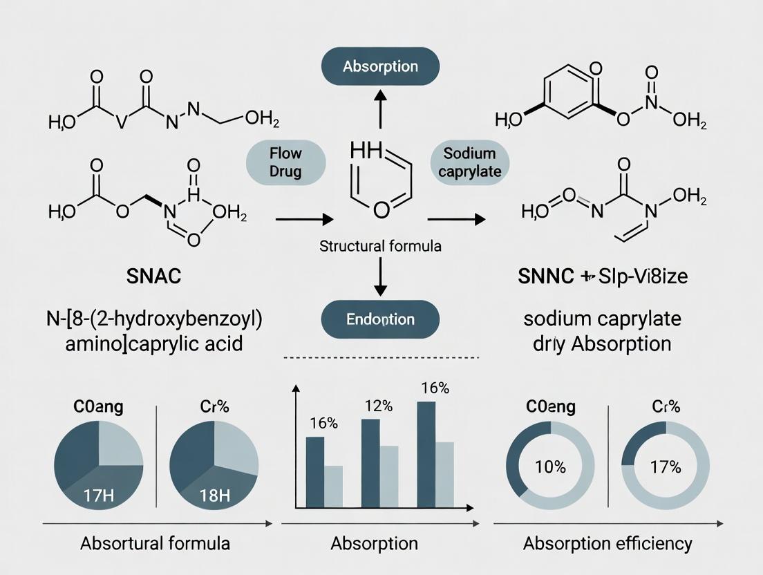
SNAC (N-(8-[2-hydroxybenzoyl]amino)caprylic acid) and sodium caprylate (sodium octanoate) are critical absorption enhancers under investigation for oral delivery of macromolecular drugs, peptides, and poorly permeable actives. Their mechanisms, while distinct, converge on transiently modulating gastrointestinal barriers.
Key Functional Comparison:
| Property | SNAC (Salcaprozate sodium) | Sodium Caprylate |
|---|---|---|
| Chemical Name | Sodium N-(8-[2-hydroxybenzoyl]amino)caprylate | Sodium octanoate |
| Molecular Formula | C₁₅H₁₉NNaO₄ | C₈H₁₅NaO₂ |
| Molecular Weight | 300.30 g/mol | 166.19 g/mol |
| Primary Structure | Acylated amino acid derivative (salicylic acid linked to 8-aminocaprylic acid) | Medium-chain fatty acid salt (C8) |
| Mechanistic Hypothesis | Non-covalent, pH-dependent transient interaction with drug & membrane components; may reduce intramolecular H-bonding in peptides. | Endogenous-like permeation enhancer; induces transient loosening of tight junctions via intracellular signaling (e.g., MLCK). |
| Key Applications | Oral semaglutide (Rybelso), oral heparin formulations. | Monoclonal antibody formulations, insulin delivery, cell culture additive (e.g., in CHO cell media). |
| Typical Use Conc. | 50-300 mM in preclinical/clinical studies. | 5-100 mM (dependent on application & toxicity profile). |
| Regulatory Status | FDA-approved as an excipient in a drug product (Rybelso). | Generally Recognized As Safe (GRAS) for food use; widely used pharmaceutical excipient. |
| Critical CMC Note | Hydrophobic; may require cosolvents for aqueous formulation at high conc. | Highly water-soluble; forms micelles at high concentrations (> ~400 mM). |
Experimental Protocols
Protocol 1: Assessing Transepithelial Electrical Resistance (TEER) Reduction in Caco-2 Monolayers
Objective: To quantify the transient disruption of intestinal epithelial tight junctions by sodium caprylate.
Materials:
- Caco-2 cells (passage 35-55)
- 12-well or 24-well Transwell inserts (polycarbonate, 0.4 µm pore)
- EVOM2 voltmeter with STX2 chopstick electrodes
- Hanks' Balanced Salt Solution (HBSS), pH 6.5 & 7.4
- Test solutions: Sodium caprylate (10, 25, 50 mM in HBSS, pH 6.5), SNAC (150 mM in HBSS, pH 6.5), Control (HBSS only)
- Recovery medium (complete DMEM)
Methodology:
- Culture Caco-2 cells on Transwell inserts until monolayers achieve TEER > 500 Ω·cm² (typically 21 days).
- Pre-equilibrate monolayers with HBSS (pH 6.5) for 20 min at 37°C. Measure initial TEER (T₀).
- Aspirate buffer. Apically apply 0.5 mL (24-well) of test or control solution. Basolaterally apply 1.5 mL of HBSS pH 7.4.
- Incubate at 37°C for 60 min. Measure TEER (T₆₀).
- Remove test solutions, wash apically 3x with HBSS pH 7.4. Replace both compartments with complete DMEM.
- Measure TEER at 2, 4, and 24 hours post-treatment to assess recovery (Tᵣ).
- Calculate: % TEER Reduction = [(T₀ - T₆₀) / T₀] * 100. Normalize to control.
Protocol 2: In Situ Single-Pass Intestinal Perfusion (SPIP) Study in Rodents
Objective: To determine the region-specific enhancement of peptide (e.g., insulin) absorption by SNAC.
Materials:
- Anesthetized rat (or mouse) model
- Perfusion buffer: Krebs-Ringer Buffer (KRB), pH adjusted to target intestinal segment (e.g., pH 6.8 for jejunum)
- Test solution: Insulin (e.g., 2 IU/mL) + 200 mM SNAC in KRB (pH 6.8)
- Control solution: Insulin alone in KRB (pH 6.8)
- Peristaltic pump, heating pad, surgical tools
- HPLC-MS system for quantitation
Methodology:
- Anesthetize animal and maintain at 37°C. Perform midline laparotomy.
- Isolate a 10 cm jejunal segment, cannulate proximally and distally.
- Flush segment with warm KRB (pH 6.8) to clear luminal contents.
- Perfuse test or control solution at a constant rate (e.g., 0.2 mL/min) for 90 min. Collect effluent from distal cannula at 10-min intervals.
- Collect serial blood samples from the jugular vein for plasma insulin analysis (HPLC-MS or ELISA).
- At termination, measure the exact length and radius of the perfused segment.
- Calculate: Apparent permeability (Pₐₚₚ) = [-Q * ln(Cₒᵤₜ/Cᵢₙ)] / (2πrL), where Q is flow rate, r is radius, L is length, C is concentration corrected for water transport.
Visualization: Pathways and Workflows
Title: Proposed SNAC-Mediated Permeation Enhancement Pathway
Title: TEER Assay Experimental Workflow
The Scientist's Toolkit: Key Research Reagent Solutions
| Reagent/Material | Supplier Examples | Critical Function in Research |
|---|---|---|
| Caco-2 Cell Line | ATCC (HTB-37), ECACC | Gold-standard in vitro model of human intestinal epithelium for permeability & TEER studies. |
| Transwell Permeable Supports | Corning, MilliporeSigma | Polycarbonate membranes for culturing polarized cell monolayers; enables separate apical/basolateral access. |
| EVOM2 Voltmeter with STX2 | World Precision Instruments | Precisely measures transepithelial electrical resistance (TEER) to quantify monolayer integrity. |
| Salcaprozate Sodium (SNAC) | MedChemExpress, Cayman Chemical | High-purity reference standard for formulation studies and mechanistic in vitro/in vivo experiments. |
| Sodium Caprylate (≥99%) | Sigma-Aldrich, Avantor | Critical for preparing stock solutions for permeation studies and as a cell culture supplement. |
| Hanks' Balanced Salt Solution (HBSS) | Gibco, Sigma-Aldrich | Physiological buffer for permeability assays; allows pH adjustment to simulate GI conditions. |
| Recombinant Human Insulin | Sigma-Aldrich, ProSpec | Model peptide drug for evaluating absorption enhancement in SPIP and in vivo studies. |
| MLC2 Antibody (Phospho S19) | Cell Signaling Technology | Detects phosphorylated myosin light chain 2 to investigate sodium caprylate's TJ mechanism via MLCK. |
Within the context of investigating SNAC (sodium N-[8-(2-hydroxybenzoyl) amino] caprylate) and sodium caprylate (C8) as oral absorption enhancers, understanding their core mechanistic pathways is paramount. These agents are known to improve the bioavailability of co-administered drugs, primarily through two distinct routes: transiently modulating the paracellular pathway or facilitating transcellular transport. This document details application notes and protocols for elucidating and differentiating these mechanisms, providing researchers with actionable methodologies.
Core Pathways and Quantitative Comparison
SNAC and sodium caprylate, while structurally related, exhibit differences in their primary mechanisms and potency. The following table summarizes key quantitative findings from recent studies.
Table 1: Comparative Mechanistic Data for SNAC and Sodium Caprylate
| Parameter | SNAC (Typical Experimental Range) | Sodium Caprylate (Typical Experimental Range) | Primary Assay/Model |
|---|---|---|---|
| Primary Enhancement Pathway | Transcellular (pH-dependent carrier/micelle) | Paracellular (Tight Junction Modulation) | Caco-2 TEER, Papp studies |
| Effective Concentration | 0.5% - 2.0% (w/v) | 5 mM - 100 mM | In vitro permeability models |
| TEER Reduction | Minimal (<20%) | Significant (30-70%) | Caco-2 monolayer TEER |
| Papp Increase (Marker Drug) | 2- to 10-fold (e.g., Heparin) | 5- to 50-fold (e.g., FD4, peptides) | Caco-2, rat intestinal perfusion |
| Onset of Action | Rapid (<5 min) | Rapid (<10 min) | TEER kinetics |
| Reversibility | Highly Reversible (≥90% in 60-120 min) | Reversible (≥80% in 90-180 min) | TEER recovery post-wash |
| Key Molecular Target | Membrane fluidization / micelle formation | Intracellular Ca2+ / MLCK pathway | Immunofluorescence, kinase assays |
Experimental Protocols
Protocol 1: Differentiating Pathways via TEER and Paracellular Flux
Objective: To determine whether an enhancer acts primarily via the paracellular pathway by measuring Transepithelial Electrical Resistance (TEER) and the permeability of paracellular markers.
Materials:
- Caco-2 cell monolayers (21-25 days post-seeding, TEER >300 Ω·cm²)
- Hanks' Balanced Salt Solution (HBSS), pH 6.5-7.4
- Test compounds: SNAC and Sodium Caprylate (stock solutions in buffer)
- Paracellular marker: Fluorescein isothiocyanate-dextran 4 kDa (FD4, 1 mg/mL)
- Transwell plate (12-well, 1.12 cm² insert area)
- Voltohmmeter (chopstick or electrode array)
- Fluorescence plate reader
Procedure:
- Baseline Measurement: Aspirate culture media from apical (AP) and basolateral (BL) compartments. Wash twice with warm HBSS (pH 6.8). Add fresh HBSS (pH 6.8) to both sides. Measure and record baseline TEER.
- Dosing: Remove apical buffer. Add pre-warmed test solutions (HBSS, pH 6.8) containing the absorption enhancer (e.g., 1% SNAC, 50 mM sodium caprylate) and FD4 to the AP compartment. Add fresh HBSS to the BL compartment.
- Kinetic Monitoring: Incubate at 37°C. Measure TEER at 15, 30, 60, and 120 minutes.
- Sampling: At 120 minutes, sample 200 µL from the BL compartment. Replace with fresh pre-warmed buffer.
- Analysis: Quantify FD4 fluorescence (Ex/Em: 492/518 nm). Calculate apparent permeability (Papp) using standard formulas. Normalize TEER values as % of baseline.
- Recovery Phase (Optional): Wash monolayers with enhancer-free buffer and monitor TEER for an additional 2-3 hours to assess reversibility.
Protocol 2: Assessing Transcellular Enhancement via Lipophilic Probe Uptake
Objective: To probe transcellular pathway enhancement by measuring the intracellular uptake of a lipophilic fluorescent probe.
Materials:
- Caco-2 monolayers or suspension
- HBSS buffers (pH 5.5, 6.5, 7.4)
- Test compounds
- Lipophilic probe: Nile Red or Coumarin 6 (1 µM stock in DMSO)
- Flow cytometer or fluorescence microscope
- Cell lysis buffer (for plate reader assay)
Procedure:
- Cell Preparation: Harvest Caco-2 cells or use monolayers on 24-well plates. Wash with appropriate pH buffer.
- pH-Dependent Incubation: Prepare test solutions at varying pH (e.g., 5.5, 6.5, 7.4) containing the enhancer (e.g., 1.5% SNAC) and the lipophilic probe. Incubate with cells for 30-60 min at 37°C.
- Termination & Washing: Aspirate solution. Wash cells 3x thoroughly with ice-cold buffer containing 0.1% BSA to remove surface-adherent probe.
- Quantification:
- Flow Cytometry: Detach cells, resuspend in buffer, and analyze fluorescence intensity.
- Microscopy/Plate Reader: Lyse cells with 1% Triton X-100. Measure fluorescence in the supernatant.
- Data Analysis: Compare intracellular fluorescence intensity with enhancer vs. control across different pH conditions. A pH-dependent increase with SNAC suggests a transcellular mechanism involving hydrophobic interactions or micelle formation.
The Scientist's Toolkit: Key Research Reagent Solutions
Table 2: Essential Materials for Mechanistic Studies
| Item | Function in Research | Example/Catalog Consideration |
|---|---|---|
| Caco-2 Cell Line | Gold standard in vitro model of human intestinal epithelium for permeability and TEER studies. | ATCC HTB-37; use passages 30-50 for consistency. |
| TEER Measurement System | Quantifies tight junction integrity; paracellular pathway indicator. | EVOM3 with chopstick electrodes (World Precision Instruments). |
| Paracellular Flux Markers | Non-absorbable tracers to quantify paracellular permeability. | FITC-Dextran 4 kDa (FD4) or Lucifer Yellow. |
| MLCK Inhibitor (ML-7) | Pharmacological tool to inhibit the myosin light chain kinase pathway, confirming its role in enhancer action. | Used to dissect sodium caprylate's mechanism. |
| Intracellular Calcium Chelator (BAPTA-AM) | Probes the role of Ca2+ signaling in tight junction modulation. | Incubate prior to sodium caprylate to block its effect. |
| Lipophilic Fluorescent Probes | Report on membrane fluidity and transcellular uptake. | Nile Red, Coumarin 6, or DiI. |
| HBSS Buffers (varying pH) | Simulate different gastrointestinal pH environments critical for studying pH-dependent mechanisms (e.g., SNAC). | Prepare with MES (pH 5.5-6.5) or HEPES (pH 7.4). |
Visualizations
Title: SNAC vs Caprylate: Enhancement Pathways
Title: TEER & Paracellular Flux Assay Workflow
Title: Caprylate: Ca2+-MLCK Pathway to TJ Opening
Within the ongoing thesis research on the absorption enhancers SNAC (sodium N-[8-(2-hydroxybenzoyl) amino] caprylate) and sodium caprylate, understanding their interactions with key gastrointestinal (GI) barriers is paramount. Both enhancers are postulated to improve oral bioavailability of macromolecular drugs (e.g., peptides) through multifaceted mechanisms. This document provides detailed application notes and protocols for studying these interactions, focusing on three primary barriers: the mucus layer, the intestinal epithelial tight junctions (TJs), and the enterocyte membrane. The goal is to establish standardized methods to quantify and differentiate the contributions of SNAC and sodium caprylate to each mechanism.
| Parameter | Typical Value/Range | Significance in Absorption Enhancement | Primary Assay |
|---|---|---|---|
| Mucus Layer Viscosity | ~10-1000 Pa·s (shear-dependent) | Reduced viscosity may enhance diffusion. | Rheology (ex vivo mucus) |
| Mucus Penetration Rate | Variable; e.g., 2-10x increase vs. control | Direct measure of enhancer effect on diffusion barrier. | Fluorescent Nanoparticle Tracking |
| Transepithelial Electrical Resistance (TEER) | Caco-2 monolayers: 300-600 Ω·cm² | Reduction indicates TJ modulation. | TEER Measurement |
| Paracellular Flux (4 kDa FD-4) | Apparent Permeability (Papp): ~0.5-2 x 10⁻⁶ cm/s (baseline) | Quantitative marker of TJ opening. | USsing Chamber/Transport Assay |
| Membrane Fluidity Increase | % change in anisotropy (r) | Increased fluidity may promote transcellular uptake. | Fluorescence Polarization (DPH probe) |
| Critical Micelle Concentration (CMC) - Sodium Caprylate | ~100-150 mM | Surfactant action relevant for membrane perturbation. | Conductivity/Surface Tension |
| Intracellular Calcium [Ca²⁺]ₙ | Baseline ~100 nM; spikes to >500 nM | Calcium signaling linked to TJ regulation. | Fluorometric Assays (Fluo-4 AM) |
Detailed Experimental Protocols
Protocol 3.1: Assessing Mucus-Penetrating Properties
Objective: To evaluate the effect of SNAC/sodium caprylate on the diffusion of model drugs through native intestinal mucus. Materials: Porcine intestinal mucus (commercial source), fluorescently-labeled dextran (40 kDa, FITC-labeled), test compounds (SNAC, sodium caprylate in HBSS, pH 6.8), transwell inserts (3.0 µm pores), plate reader. Procedure:
- Mucus Preparation: Thaw mucus on ice, homogenize gently. Aliquot 100 µL into the apical chamber of a transwell insert.
- Test Solution: Add 200 µL of HBSS containing the fluorescent probe (50 µg/mL) ± absorption enhancer (e.g., 10 mM) to the mucus layer.
- Incubation: Place insert into a receiver plate containing 800 µL of HBSS. Incubate at 37°C with mild orbital shaking.
- Sampling: At t=30, 60, 90, 120 min, sample 50 µL from the basolateral chamber and replace with fresh buffer.
- Analysis: Measure fluorescence (Ex/Em: 485/535 nm). Calculate the apparent permeability (Papp) and the enhancement ratio vs. control.
Protocol 3.2: Evaluating Tight Junction Modulation via TEER and Paracellular Markers
Objective: To quantify the temporal and reversible effects on TJ integrity in intestinal epithelial cell monolayers. Materials: Differentiated Caco-2 or HT29-MTX-E12 co-culture monolayers (21 days), EVOM3 voltmeter with chopstick electrodes, USsing chamber system, FITC-Dextran 4 kDa (FD-4). TEER Procedure:
- Baseline: Measure TEER of monolayers in HBSS.
- Treatment: Apically add HBSS containing test enhancer (e.g., 5-20 mM SNAC). Include control (buffer) and positive control (5 mM EDTA).
- Monitoring: Record TEER at 10, 20, 30, 45, 60 min post-treatment. Express as % of initial TEER.
- Recovery: Replace with fresh culture medium and monitor TEER for up to 24h to assess reversibility. Paracellular Flux Procedure (USsing Chamber):
- Mount monolayers in USsing chambers (37°C, carbogen gas).
- Add FD-4 (0.1 mg/mL) apically with/without enhancer. Sample (100 µL) from the basolateral side every 30 min for 2h.
- Analyze FD-4 concentration by fluorescence. Calculate Papp and flux enhancement ratio.
Protocol 3.3: Measuring Plasma Membrane Fluidity Changes
Objective: To detect enhancer-induced changes in the structural order of the epithelial cell plasma membrane. Materials: Caco-2 cells (or similar), fluorescent probe 1,6-diphenyl-1,3,5-hexatriene (DPH), polarization-compatible black microplates, fluorescence plate reader with polarizers. Procedure:
- Cell Preparation: Harvest cells, wash, and resuspend in buffer at ~1x10⁶ cells/mL.
- Probe Loading: Incubate cell suspension with 1 µM DPH for 30 min at 37°C in the dark.
- Treatment & Measurement: Aliquot cells into a microplate. Add test compounds directly. Using the plate reader, measure fluorescence intensity with polarizers parallel (Ivv) and perpendicular (Ivh) to the excitation plane.
- Calculation: Calculate anisotropy (r) = (Ivv - GIvh) / (Ivv + 2G*Ivh), where G is an instrument correction factor. Decreased anisotropy indicates increased membrane fluidity.
Visualization of Mechanisms & Workflows
Diagram 1: Workflow for Mucus Penetration Assays
Diagram 2: Proposed Mechanisms of SNAC/Caprylate on GI Barriers
The Scientist's Toolkit: Key Research Reagent Solutions
Table 2: Essential Materials for GI Barrier Interaction Studies
| Reagent/Material | Function/Application | Example Product/Source |
|---|---|---|
| Porcine Intestinal Mucus (Lyophilized) | Provides a physiologically relevant, native mucus barrier for ex vivo penetration studies. | Sigma-Aldrich (M2378) or in-house isolation. |
| Fluorescent Probes (FITC-Dextrans, 4 & 40 kDa) | Non-absorbable markers for quantifying paracellular (4kDa) and mucopenetration (40kDa) flux. | Thermo Fisher Scientific (D1844, D1845). |
| Caco-2 & HT29-MTX-E12 Cell Lines | Standard in vitro models for human intestinal epithelium and mucus-producing goblet cells, respectively. | ATCC (HTB-37, HTB-?) or ECACC. |
| EVOM3 Epithelial Voltohmmeter | Gold-standard instrument for accurate, non-destructive TEER measurements of cell monolayer integrity. | World Precision Instruments. |
| DPH (1,6-Diphenyl-1,3,5-hexatriene) | Hydrophobic fluorescent probe that intercalates into lipid bilayers for membrane fluidity measurements via anisotropy. | Cayman Chemical (15022). |
| Fluo-4 AM, Cell Permeant | Ratiometric calcium indicator dye to monitor intracellular Ca²⁺ signaling linked to TJ regulation. | Thermo Fisher Scientific (F14201). |
| USsing Chamber System | For precise measurement of active ion transport and passive molecular flux across tissue or cell monolayers. | Physiologic Instruments or Warner Instruments. |
| SNAC (Sodium N-[8-(2-hydroxybenzoyl) amino] caprylate) | The primary absorption enhancer of study; acts as a multi-functional carrier. | MedChemExpress (HY-109023) or custom synthesis. |
Historical Context and Landmark Studies in Absorption Enhancement
Application Notes: Framing SNAC and Sodium Caprylate within a Broader Thesis
The investigation of absorption enhancers is pivotal for advancing oral delivery of macromolecular drugs and poorly permeable compounds. The broader thesis positions SNAC (sodium N-[8-(2-hydroxybenzoyl) amino] caprylate) and sodium caprylate (C8) as archetypal, clinically validated enhancers with distinct yet complementary mechanisms. SNAC, central to the marketed oral semaglutide formulation, facilitates transcellular transport via noncovalent, pH-dependent complexation. Sodium caprylate, widely used in approved liquid formulations (e.g., oral solutions of antivirals), primarily enhances paracellular permeability through transient tight junction modulation. This thesis posits that their historical development pathway—from initial discovery in biochemical models to clinical application—provides a blueprint for rational enhancer design, balancing efficacy with localized, reversible epithelial interaction.
| Study (Year) | Enhancer Tested | Model System | Key Parameter Measured | Result (Mean ± SD or as reported) | Significance |
|---|---|---|---|---|---|
| Brayden et al. (2020) Adv Drug Deliv Rev | SNAC | Caco-2 monolayers | Apparent Permeability (Papp) of Semaglutide (10^-6 cm/s) | Control: 0.01 ± 0.002; +SNAC: 0.57 ± 0.11 | Demonstrated >50-fold increase in peptide permeability in vitro. |
| Buckley et al. (2008) Pharm Res | Sodium Caprylate | Rat jejunal perfusion | Effective Permeability (Peff) of Mannitol (10^-4 cm/s) | Control: 0.40 ± 0.05; +C8 (13.5 mM): 2.11 ± 0.30 | 5.3-fold increase, confirming reversible paracellular enhancement. |
| Dahlgren et al. (2019) Eur J Pharm Sci | SNAC | In vivo (Porcine) | Absolute Bioavailability of Semaglutide (%) | Oral (with SNAC): 0.8-1.0%; Subcutaneous: 100% (ref) | First large animal data correlating SNAC's in vitro effect to in vivo bioavailability. |
| Suzuki et al. (2016) Mol Pharm | Sodium Caprylate | Caco-2, HT29-MTX co-culture | TEER Reduction (% of baseline) | 10mM C8: ~40% reduction (reversible within 60 min washout) | Quantified rapid, reversible tight junction opening. |
| Leonard et al. (2021) J Control Release | SNAC | Caco-2 & In silico | Log P (Octanol/Water) of SNAC | ~2.5 (at pH 6.0) | Highlights SNAC's amphiphilic, membrane-partitioning property. |
Experimental Protocols
Protocol 1: In Vitro Permeability Assessment Using Caco-2 Monolayers (Adapted from Brayden et al.)
Objective: To quantify the absorption enhancement effect of SNAC on a macromolecule (e.g., peptide). Materials:
- Caco-2 cells (passage 35-55)
- Transwell plates (12-well, 1.12 cm², 0.4 μm pore polyester membrane)
- Hanks' Balanced Salt Solution (HBSS), 10 mM HEPES, pH 6.5 (donor) and 7.4 (receiver)
- Test peptide (e.g., semaglutide)
- SNAC stock solution (500 mM in buffer, pH adjusted to 6.5)
- LC-MS/MS system for analyte quantification
Methodology:
- Monolayer Preparation: Seed Caco-2 cells at 1x10^5 cells/cm². Culture for 21-28 days, changing media every 2-3 days. Use monolayers with Transepithelial Electrical Resistance (TEER) >500 Ω·cm².
- Pre-incubation: Equilibrate monolayers in pre-warmed transport buffers (HBSS-HEPES) for 20 min.
- Dosing Solution: Prepare donor solution (pH 6.5) containing the peptide (e.g., 1 mg/mL) with or without SNAC (e.g., 20 mM). Receiver phase is pH 7.4 buffer.
- Transport Assay: Aspirate buffers. Add 0.5 mL donor and 1.5 mL receiver. Incubate at 37°C, 60 rpm orbital shaking.
- Sampling: At t=0 and 120 min, sample 100 µL from receiver, replacing with fresh buffer. Sample donor at 120 min.
- Analysis: Quantify peptide concentration via LC-MS/MS. Calculate Papp:
Papp = (dQ/dt) / (A * C0), where dQ/dt is flux, A is membrane area, C0 is initial donor concentration. - Viability Check: Measure TEER pre- and post-experiment; confirm >80% viability via MTT assay.
Protocol 2: Rat Single-Pass Intestinal Perfusion (SPIP) for Paracellular Enhancers (Adapted from Buckley et al.)
Objective: To assess in situ the regional intestinal permeability enhancement by sodium caprylate. Materials:
- Male Sprague-Dawley rats (fasted overnight)
- Krebs-Ringer bicarbonate buffer (KRB), gassed with 95% O2/5% CO2
- Sodium caprylate, radiolabeled [¹⁴C]-Mannitol (paracellular marker)
- Surgical tools, peristaltic pump, fraction collector
- Liquid scintillation counter
Methodology:
- Surgical Preparation: Anesthetize rat. Expose a 10 cm jejunal segment via midline incision, cannulate proximally and distally. Flush segment with warm KRB.
- Perfusate Preparation: KRB containing [¹⁴C]-Mannitol (tracer) ± sodium caprylate (e.g., 13.5 mM). Maintain at 37°C and gas continuously.
- Perfusion: Perfuse segment at 0.2 mL/min for 90 min (steady-state). Collect effluent from distal cannula at 10-min intervals.
- Outlet Concentration: Measure [¹⁴C] activity in effluent samples via scintillation counting.
- Calculation: Determine effective permeability (Peff) using the parallel tube model:
Peff = [-Q * ln(Cout/Cin)] / (2πrL), where Q is flow rate, r is intestinal radius, L is length, Cin and Cout are corrected inlet/outlet concentrations. - Histology: Post-perfusion, fix intestinal segment for H&E staining to assess mucosal integrity.
The Scientist's Toolkit: Key Research Reagent Solutions
| Item | Function in Absorption Enhancer Research |
|---|---|
| Caco-2 Cell Line | Human colorectal adenocarcinoma cells; gold standard for in vitro model of intestinal epithelial permeability. |
| Transwell Plates | Permeable supports for growing cell monolayers, enabling separate access to apical and basolateral compartments. |
| TEER Measurement System (Volt/Ohm Meter) | Monitors tight junction integrity and monolayer confluence in real-time before/during permeability assays. |
| USsing Chamber System | Ex vivo apparatus for measuring ion and molecular flux across intact intestinal mucosal tissues under voltage-clamp conditions. |
| Sodium Caprylate (≥98% purity) | Medium-chain fatty acid salt; positive control for reversible, calcium-mediated tight junction opening. |
| SNAC (GMP-grade for in vivo) | Critical for preclinical/clinical correlation studies; ensures translational relevance of formulation data. |
| Fluorescein Isothiocyanate-Dextran (FD-4, 4 kDa) | Standard paracellular flux marker to quantify tight junction modulation. |
| LC-MS/MS with ESI Source | Essential for sensitive, specific quantification of peptides and enhancers in complex biological matrices. |
Visualizations
Diagram Title: Proposed Transcellular Pathway for SNAC-Peptide Permeation
Diagram Title: Sodium Caprylate Paracellular Enhancement Mechanism
Diagram Title: Tiered Experimental Workflow for Enhancer Validation
Key Physicochemical Factors Influencing Enhancer Performance
Within the context of investigating SNAC (salcaprozate sodium) and sodium caprylate as archetypal absorption enhancers for oral delivery, this document outlines the key physicochemical factors governing their performance. These medium-chain fatty acid salts enhance permeability primarily via transient mucosal interactions. Their efficacy is not intrinsic but is dictated by a precise interplay of molecular and formulation properties.
The performance of SNAC and sodium caprylate is contingent upon several interrelated factors. The following table summarizes the critical parameters and their optimal ranges or influences based on current research.
Table 1: Key Physicochemical Factors for SNAC and Sodium Caprylate Performance
| Factor | Description & Impact on Performance | Typical Optimal Range/Consideration |
|---|---|---|
| Critical Micelle Concentration (CMC) | Concentration at which self-assembly into micelles begins. Enhancer action is typically maximal near or below CMC, promoting monomer interaction with membranes. Above CMC, capacity for membrane perturbation may be reduced. | SNAC: ~1-10 mM (formulation-dependent). Sodium Caprylate: ~100-300 mM. Performance window is close to but often below CMC. |
| pKa / Ionization State | Dictates the proportion of unionized (caprylic acid) vs. ionized (caprylate) species. The unionized form promotes membrane partitioning, while the ionized form provides solubility. The pH-pKa relationship is critical. | pKa ~4.9 for caprylic acid. Optimal activity often near luminal pH where both species coexist (e.g., pH 4-6). |
| Lipophilicity (Log P) | Measure of partitioning into lipid bilayers. Higher log P of the unionized acid promotes membrane interaction but may reduce aqueous solubility. Fine balance is required. | Caprylic acid Log P ~3.0. Enhancers require sufficient lipophilicity for membrane interaction without precipitating. |
| Chelation Potential | Ability to bind luminal calcium ions (Ca²⁺). This can disrupt epithelial tight junctions by depleting extracellular calcium, a key regulator of junctional integrity. | SNAC has known Ca²⁺ chelation properties, contributing to its transient tight junction modulation. |
| Formulation pH | Governs the ionization state (via pKa) and chemical stability of the enhancer and API. Must be compatible with the physiological pH environment of the target absorption site. | Often adjusted to ~pH 4-6.5 to optimize the unionized fraction and enhancer stability. |
| Concentration | Must be sufficient to achieve effective local monomer concentration at the membrane interface without causing cytotoxicity or irreversible damage. Shows a sharp dose-response. | Typically 0.5-5% w/w in solid dosage forms; molar ratios to API are often >1:1. |
| Chain Length | For fatty acid-based enhancers, chain length (C8 for caprylate) balances lipophilicity and solubility. Shorter chains are less effective; longer chains are poorly soluble. | C8 (caprylate) and C10 (caprate) are most common for oral delivery. |
Experimental Protocols
Protocol 1: Determining the Critical Micelle Concentration (CMC)
Objective: To determine the CMC of SNAC or sodium caprylate in relevant buffer using the pyrene fluorescence probe method. Materials: Test compound (SNAC or sodium caprylate), pyrene (fluorescent probe), appropriate buffer (e.g., FaSSIF, pH 6.5), fluorometer, sonicator. Procedure:
- Prepare a stock solution of pyrene (6.0 x 10⁻⁵ M) in acetone. Evaporate aliquots in glass vials to form a thin film.
- Prepare a series of enhancer solutions in buffer across a broad concentration range (e.g., 0.1 mM to 500 mM).
- Add 2 mL of each enhancer solution to the pyrene-coated vials. Sonicate for 30 min and equilibrate overnight in the dark.
- Measure fluorescence emission spectra (λ_ex = 339 nm). Record the intensity ratio (I₁/I₃) of the first (373 nm) and third (384 nm) vibrational peaks.
- Plot the I₁/I₃ ratio against the logarithm of enhancer concentration. The inflection point (sharp change in slope) indicates the CMC.
Protocol 2: In Vitro Permeability Assessment in Caco-2 Monolayers
Objective: To evaluate the concentration-dependent enhancement effect on apparent permeability (P_app) of a model compound. Materials: Caco-2 cells, Transwell plates, model drug (e.g., FD4, heparin), test enhancer, HBSS transport buffer, LC-MS or suitable analytical instrument. Procedure:
- Culture Caco-2 cells on collagen-coated polycarbonate filters for 21-28 days until transepithelial electrical resistance (TEER) > 300 Ω·cm².
- Pre-equilibrate monolayers with transport buffer (pH 6.5-7.4) for 20 min.
- Control: Add fresh buffer to donor and receiver compartments. Test: Add buffer containing enhancer at sub-CMC and supra-CMC concentrations to the donor compartment (apical for oral absorption studies).
- Add model drug to donor compartment. Sample from receiver compartment at scheduled intervals (e.g., 30, 60, 90, 120 min), replacing with fresh buffer.
- Analyze samples for drug concentration. Calculate Papp using the equation: Papp = (dQ/dt) / (A * C₀), where dQ/dt is the steady-state flux, A is the membrane area, and C₀ is the initial donor concentration.
- Monitor TEER before and after experiment to assess monolayer integrity recovery.
Protocol 3: Calcium Chelation Assay
Objective: To quantify the calcium-binding capacity of the enhancer using a colorimetric assay. Materials: Test enhancer, calcium chloride solution, o-cresolphthalein complexone (CPC) assay kit, spectrophotometer. Procedure:
- Prepare a standard curve of free calcium (0-10 mM) using the CPC kit reagents.
- Prepare solutions containing a fixed concentration of calcium (e.g., 5 mM) and varying concentrations of the enhancer (0-50 mM).
- Incubate mixtures for 15 min at 37°C.
- Add CPC reagent to each sample and standard. Incubate for 10 min for color development.
- Measure absorbance at 575 nm. The chelated calcium will not react, leading to reduced color intensity.
- Calculate the percentage of calcium chelated relative to the control (calcium without enhancer).
Visualization
Diagram 1: SNAC/Caprylate Action on Intestinal Epithelium
Title: Mechanism of SNAC/Caprylate Enhancing Permeability
Diagram 2: Key Physicochemical Factors Workflow
Title: Interplay of Key Factors for Performance
The Scientist's Toolkit
Table 2: Essential Research Reagent Solutions
| Item | Function in Enhancer Research |
|---|---|
| Caco-2 Cell Line | Human colonic adenocarcinoma cell line forming polarized monolayers with tight junctions; gold standard for in vitro intestinal permeability prediction. |
| Snapwell/Transwell Inserts | Permeable supports for culturing epithelial cell monolayers, enabling apical-basolateral compartmentalization for transport studies. |
| Simulated Intestinal Fluids (SIF) | Biorelevant media (e.g., FaSSIF/FeSSIF) containing bile salts/phospholipids to mimic luminal conditions for CMC and solubility studies. |
| Fluorescent Probes (Pyrene, FD4, FITC-Dextran) | Pyrene: CMC determination via polarity-sensitive fluorescence. FD4/FITC-dextran: paracellular flux markers. |
| Transepithelial Electrical Resistance (TEER) Meter | Measures ohmic resistance across cell monolayers as a real-time, non-destructive indicator of tight junction integrity. |
| Calcium-Sensitive Dyes/Kits (e.g., Fluo-4, CPC Assay) | Quantify intracellular or extracellular calcium concentration changes to elucidate chelation-mediated enhancement mechanisms. |
| LC-MS/MS System | Essential for sensitive and specific quantification of low-permeability model drugs or peptides in permeability assay samples. |
| Physiological pH Buffers | MES (pH 5.5-6.7), HEPES (pH 7.0-8.0) for precise control of ionization state during experiments. |
Formulation in Practice: Integrating SNAC and Caprylate into Drug Products
Within the context of advancing the oral bioavailability of challenging Active Pharmaceutical Ingredients (APIs), formulation strategies such as co-processing, coating, and rational tablet design are critical. This research is situated within a broader thesis investigating Sodium Caprylate (C8) and SNAC (Salcaprozate Sodium) as permeability-enhancing absorption enhancers. These excipients can be strategically incorporated via the described formulation techniques to create robust, effective solid dosage forms for enhanced gastrointestinal delivery.
Application Notes & Protocols
Co-processing of API with Absorption Enhancers
Application Note: Co-processing involves the particle engineering of the API with the absorption enhancer (e.g., SNAC or C8) at a sub-particle level. This intimate association aims to ensure co-localization and simultaneous release in the gastrointestinal tract, which is crucial for the enhancer's mechanism of action (often involving transient loosening of tight junctions or membrane fluidization).
Protocol: Spray-Dry Co-processing of API with SNAC
- Objective: To produce a co-processed composite powder of a poorly permeable Model API and SNAC with uniform distribution.
- Materials: See "The Scientist's Toolkit" section.
- Methodology:
- Dissolve the Model API and SNAC in a suitable common solvent system (e.g., ethanol/water mixture) to achieve a target dry-weight ratio (e.g., 1:1 to 1:3 API:SNAC).
- Filter the solution through a 0.45 µm membrane filter to remove any undissolved particulates.
- Set spray-dryer parameters: Inlet temperature: 120°C; Outlet temperature: 70-75°C; Aspirator flow: 35 m³/hr; Feed pump rate: 5 mL/min; Nozzle size: 0.7 mm.
- Process the solution through the spray dryer. Collect the resulting powder in a dried collection vessel.
- Transfer the powder to a vacuum desiccator containing phosphorus pentoxide and dry for 24 hours to remove residual solvent.
- Characterize the powder for morphology (SEM), particle size distribution (laser diffraction), crystallinity (PXRD), and chemical homogeneity (DSC, FT-IR).
Table 1: Typical Characterization Data for Spray-Dried API/SNAC Composites
| API:SNAC Ratio | Mean Particle Size (D50, µm) | Yield (%) | Identified Solid State (PXRD) | Glass Transition Temp., Tg (°C) |
|---|---|---|---|---|
| 1:0 (Pure API) | 15.2 ± 3.1 | 88.5 | Crystalline | N/A |
| 1:1 | 8.5 ± 1.7 | 76.3 | Amorphous | 112.4 |
| 1:2 | 7.9 ± 1.5 | 72.8 | Amorphous | 105.7 |
| 1:3 | 9.1 ± 2.0 | 70.1 | Amorphous | 98.2 |
Title: Spray-Dry Co-processing Workflow
Functional Coating with Permeation Enhancers
Application Note: A polymer-based coating containing SNAC or C8 can be applied to tablets or multiparticulates. This strategy allows for targeted release in specific GI regions (e.g., duodenum/jejunum) where absorption enhancement is most effective, and can protect the enhancer from premature gastric degradation.
Protocol: Fluid Bed Coating of Tablets with SNAC-Eudragit L100-55 Film
- Objective: To apply an enteric polymer coating containing dispersed SNAC onto immediate-release core tablets.
- Materials: See "The Scientist's Toolkit" section.
- Methodology:
- Coating Dispersion Preparation: Dissolve Eudragit L100-55 (6.0% w/w) in isopropanol under stirring. Separately, disperse SNAC (2.0% w/w) and talc (1.0% w/w) in a small amount of isopropanol using a high-shear homogenizer. Combine the two dispersions and stir continuously.
- Coating Process: Load core tablets (500g) into the fluid bed coater (Wurster insert). Set parameters: Inlet temperature: 38°C; Product temperature: 32-35°C; Airflow: 70 CFM; Spray rate: 8-10 g/min; Atomization pressure: 1.5 bar.
- Spray the coating dispersion onto the fluidized tablets until a theoretical weight gain of 5% w/w is achieved.
- Dry the coated tablets in the chamber for 10 minutes at 35°C. Unload and condition at 25°C/60% RH for 24 hours.
- Characterize for coating uniformity (weight variation, thickness), SNAC content (assay via HPLC), and in-vitro drug release (USP Apparatus II, pH 1.2 for 2 hrs, then pH 6.8).
Table 2: Performance of SNAC-Containing Enteric Coatings
| Coating Type | Weight Gain (%) | SNAC Assay (% of Theory) | Drug Release at pH 1.2, 2h (%) | Drug Release at pH 6.8, 45min (%) |
|---|---|---|---|---|
| Eudragit L100-55 (Control) | 5.1 ± 0.2 | 0 | 0.8 ± 0.3 | 98.5 ± 2.1 |
| Eudragit L100-55 + 2% SNAC | 5.3 ± 0.3 | 95.7 ± 3.2 | 1.2 ± 0.5 | 99.1 ± 1.8 |
Tablet Design for Enhanced Absorption
Application Note: Tablet formulation involves the strategic selection of fillers, disintegrants, and compression parameters to ensure rapid disintegration and dissolution of both API and absorption enhancer, creating a high local concentration for effective permeation enhancement.
Protocol: Formulation and Compression of Rapid-Disintegrating Tablets
- Objective: To manufacture tablets ensuring rapid release of Model API and SNAC.
- Materials: See "The Scientist's Toolkit" section.
- Methodology:
- Blending: Weigh the co-processed API/SNAC powder (equivalent to 50 mg API), microcrystalline cellulose (MCC; 45% of tablet weight), and croscarmellose sodium (CCS; 5% of tablet weight). Blend in a V-blender for 15 minutes.
- Lubrication: Sieve magnesium stearate (1% of tablet weight) through a 250 µm sieve. Add to the blend and mix for an additional 2 minutes.
- Compression: Compress the final blend on a rotary tablet press using 8 mm round standard concave punches. Target hardness: 40-60 N; Target tablet weight: 250 mg.
- Evaluation: Test tablets for weight variation, hardness, friability (<0.8%), disintegration time (<3 minutes in 37°C water), and dissolution (USP Apparatus II, 50 mM phosphate buffer pH 6.8, 50 rpm).
Table 3: Critical Quality Attributes of Designed Tablets
| Attribute | Specification | Typical Result (n=10) |
|---|---|---|
| Average Weight | 250 mg ± 5% | 252 mg ± 4 mg |
| Hardness | 40 - 60 N | 52 N ± 5 N |
| Friability | ≤ 0.8% | 0.3% |
| Disintegration Time | ≤ 3 minutes | 65 ± 12 seconds |
| API Dissolution (Q45min) | ≥ 85% | 98.5 ± 1.5% |
| SNAC Release (Q10min) | ≥ 90% | 99.2 ± 0.8% |
Title: Tablet Manufacturing Process Flow
The Scientist's Toolkit: Key Research Reagent Solutions
| Item | Function / Relevance |
|---|---|
| SNAC (Salcaprozate Sodium) | Primary absorption enhancer. Acts via transient alteration of epithelial tight junctions and membrane fluidity to increase paracellular transport. |
| Sodium Caprylate (C8) | Endogenous medium-chain fatty acid salt used as a comparator absorption enhancer. Mechanism involves intracellular calcium mobilization and actomyosin contraction. |
| Eudragit L100-55 | Methacrylic acid-ethyl acrylate copolymer (1:1). Dissolves at pH >5.5. Used for enteric coating to target release to the duodenum. |
| Microcrystalline Cellulose (MCC) | Versatile, compressible diluent/filler. Provides good tablet hardness and rapid disintegration via wicking. |
| Croscarmellose Sodium (CCS) | Super-disintegrant. Swells rapidly upon contact with water, creating a disruptive force for rapid tablet breakup. |
| Magnesium Stearate | Lubricant. Reduces friction during tablet ejection but must be used sparingly to avoid hindering dissolution. |
| Talc | Anti-tacking agent. Prevents agglomeration of coated tablets during the fluid bed coating process. |
Title: Formulation-Enhancer Synergy Logic
Within the broader thesis investigating SNAC (N-(8-[2-hydroxybenzoyl]amino)caprylate) and sodium caprylate as oral absorption enhancers, defining and controlling Critical Process Parameters (CPPs) is paramount. The transition from promising in vitro results to a robust, commercial-scale dosage form (e.g., tablets, capsules) hinges on identifying the material attributes and process parameters that critically impact the Critical Quality Attributes (CQAs) of the final drug product. These CQAs include dissolution profile, stability of the absorption enhancer-drug complex, and ultimately, bioavailability. This document outlines application notes and protocols for determining CPPs in the context of manufacturing solid dosage forms containing SNAC/sodium caprylate.
Based on current literature and processing knowledge for lipid-based and enhancer-containing formulations, the following CPPs have been identified as highly influential. Quantitative data ranges are illustrative and must be established for a specific formulation during process development.
Table 1: Critical Process Parameters for Wet Granulation (A Common Method for SNAC Formulations)
| Unit Operation | Critical Process Parameter (CPP) | Target Range (Illustrative) | Impact on Critical Quality Attributes (CQAs) |
|---|---|---|---|
| Wet Granulation | Binder Solution Addition Rate | 5-15 mL/min/kg | Too fast: Uneven distribution, overwetting. Too slow: Poor agglomeration, fines. Affects granule size distribution (GSD), flow, and content uniformity. |
| Impeller Speed | 150-300 rpm | Controls densification and agglomerate growth. Directly impacts GSD, porosity, and subsequent dissolution. | |
| Granulation Endpoint (by amperometry or torque) | 5-15 A (system dependent) | Determines final granule moisture and density. Critical for tablet hardness, disintegration, and dissolution consistency. | |
| Drying (Fluid Bed) | Inlet Air Temperature | 40-60°C | Too high: Degradation of SNAC/drug, melt-back of caprylate. Too low: Prolonged process, microbial risk. Impacts chemical stability and residual moisture. |
| Drying Endpoint (Loss on Drying, LOD) | 1.0-2.5% w/w | Residual moisture critical for powder flow, compaction, and long-term chemical stability of the absorption enhancer complex. | |
| Milling | Mill Screen Size | 0.8-1.5 mm | Controls final particle size distribution of granules. Affects blend uniformity, tablet weight variation, and dissolution rate. |
| Blending | Blending Time | 10-30 min | Ensures homogeneous distribution of API, SNAC, and excipients. Over-blending: May cause segregation or granule attrition. Critical for content uniformity. |
| Tableting | Main Compression Force | 10-25 kN | Directly controls tablet hardness and tensile strength. Impacts disintegration time and dissolution profile—key for absorption enhancer release. |
| Pre-compression Force | 1-5 kN | Aids in deaeration, reducing capping risk. Improves weight uniformity for low-dose formulations. | |
| Turret Speed | 20-40 rpm | Affects dwell time and compaction mechanics, influencing tablet mechanical properties and potential for sticking. |
Experimental Protocols for CPP Determination
Protocol 3.1: Design of Experiments (DoE) for Granulation Optimization
Objective: To systematically evaluate the impact of granulation CPPs (binder addition rate, impeller speed, granulation time) on granule CQAs and identify the optimal design space.
Materials: Dry blend (API, SNAC, filler, disintegrant), binder solution (e.g., PVP in water).
Equipment: High-shear granulator with torque/power monitoring, sieves, analytical balances, laser diffraction particle size analyzer, tap density tester.
Methodology:
- DoE Design: Employ a central composite design (CCD) with three factors: Binder Addition Rate (X1), Impeller Speed (X2), and Wet Massing Time (X3). Define low and high levels for each (e.g., X1: 8 vs. 12 mL/min/kg).
- Execution: For each DoE run, charge dry powder into granulator. Start impeller and chopper. Add binder solution at the specified rate. After addition, continue wet massing for the specified time.
- In-process Monitoring: Record torque/power consumption curve throughout the process.
- Granule Analysis: Wet sieve the granules to determine particle size distribution. Dry a representative sample in an oven (40°C) to constant weight for bulk/tapped density and moisture content (LOD) analysis.
- Tableting & Evaluation: Compress granules from each run under standardized conditions. Evaluate tablets for hardness, disintegration time, and dissolution (USP Apparatus II, pH 6.8 buffer).
- Data Analysis: Use statistical software (e.g., JMP, Design-Expert) to perform multiple regression analysis. Generate response surface models to visualize the relationship between CPPs and CQAs (granule density, dissolution at 15 min). Define the design space where all CQAs meet acceptance criteria.
Protocol 3.2: Establishing the Compression Design Space
Objective: To determine the relationship between main compression force and tablet CQAs (hardness, disintegration, dissolution) for a fixed formulation blend.
Materials: Final blend (containing API, SNAC, excipients).
Equipment: Rotary tablet press instrumented with force sensors, tablet hardness tester, disintegration apparatus, dissolution tester.
Methodology:
- Compression Runs: Set the tablet press to a fixed turret speed and pre-compression force. Perform compression runs at a minimum of five different main compression forces, spanning a wide range (e.g., 8, 12, 16, 20, 24 kN).
- Sampling: Collect a representative sample of tablets (~50) from each compression run after the process stabilizes.
- Physical Testing: Measure tablet weight, thickness, and hardness (n=10). Perform disintegration tests (n=6).
- Performance Testing: Perform dissolution testing (n=6) on tablets from each force level. Key time points: 10, 20, 30, 45, 60 minutes.
- Analysis: Plot tablet tensile strength (derived from hardness and dimensions) vs. compression force. Plot dissolution profiles (e.g., % dissolved at 30 min) vs. compression force. Establish the acceptable range of compression force that yields tablets meeting all hardness, disintegration, and dissolution specifications.
Mandatory Visualizations
Diagram 1: CPP Impact Pathway on Bioavailability
Diagram 2: DoE Workflow for CPP Identification
The Scientist's Toolkit: Key Research Reagent Solutions
Table 2: Essential Materials for CPP Studies on Absorption Enhancer Formulations
| Item | Function / Relevance to CPP Studies |
|---|---|
| SNAC (N-(8-[2-hydroxybenzoyl]amino)caprylate) | The primary absorption enhancer. Its stability (heat, moisture) and compatibility with other ingredients are key CQAs influenced by CPPs like drying temperature and granulation moisture. |
| Sodium Caprylate | Alternative/complementary absorption enhancer. Its hygroscopicity and anionic nature make process parameters (humidity control, blending order) critical. |
| PVP K-30 (Polyvinylpyrrolidone) | Common binder in wet granulation. The concentration and method of its addition (CPP: binder solution rate) are vital for granule formation and drug release. |
| Microcrystalline Cellulose (MCC) | Universal filler/diluent with good compaction properties. Its grade and particle size can interact with granulation CPPs to affect flow and compressibility. |
| Croscarmellose Sodium | Superdisintegrant. Its performance can be sensitive to granulation wetness (CPP) and compaction force (CPP), impacting the crucial CQA of disintegration time. |
| pH 6.8 Phosphate Buffer | Standard dissolution medium for enteric-coated or neutral pH-release formulations common with absorption enhancers. Used to assess the dissolution profile CQA. |
| Torque/Power Probe | Equipment: In-line sensor for high-shear granulators. Used to objectively determine granulation endpoint (a key CPP), moving beyond subjective "hand-testing". |
| Near-Infrared (NIR) Spectroscopy Probe | Equipment: For real-time, in-line monitoring of moisture content and blend uniformity during granulation and drying, enabling advanced process control of CPPs. |
This document provides detailed application notes and protocols for oral peptide delivery, framed within a broader research thesis investigating sodium N-[8-(2-hydroxybenzoyl) amino] caprylate (SNAC) and sodium caprylate (C8) as absorption enhancers. The thesis posits that these medium-chain fatty acid derivatives facilitate oral bioavailability of peptides through distinct but complementary mechanisms: localized pH elevation and transient membrane permeabilization, without systemic absorption enhancement. The case of semaglutide (Rybelsus) serves as the primary validation model.
Table 1: Key Pharmacokinetic Parameters of Oral Semaglutide with SNAC vs. Subcutaneous Injection
| Parameter | Oral Semaglutide (14 mg with SNAC) | Subcutaneous Semaglutide (1.0 mg) | Notes |
|---|---|---|---|
| Bioavailability (F) | ~0.8-1.0% | 89% | Absolute bioavailability for oral form. |
| T_max (hours) | 1-2 | 1-3 | Rate of absorption is similar. |
| C_max (nmol/L) | ~16.5 | ~64.5 | Peak concentration is lower orally. |
| t½ (weeks) | ~1 | ~1 | Half-life is comparable, driven by albumin binding. |
| AUC_0-∞ (nmol·h/L) | ~1,940 | ~195,000 | Significant reduction in total systemic exposure. |
Table 2: Comparative Efficacy of Key Oral Peptide Formulations with Absorption Enhancers
| Peptide / Enhancer | Phase / Status | Primary Enhancer | Reported Bioavailability | Key Mechanism Postulated |
|---|---|---|---|---|
| Semaglutide / SNAC | Market (Rybelsus) | SNAC (300 mg) | ~1% | Localized pH↑, Monomer stabilization, Transient membrane interaction. |
| Octreotide / C8 | Phase III (MYCAPSSA) | Sodium Caprylate | ~0.5-1% | Tight junction modulation (↓ TER), Transcellular permeation. |
| Salmon Calcitonin / Eligen | Discontinued | SNAC / C8 variants | <1% | pH management and membrane fluidity alteration. |
| Insulin / C8 | Preclinical/Phase I | Sodium Caprylate | <1% (variable) | Paracellular enhancement via TJ opening. |
Detailed Experimental Protocols
Protocol 3.1: In Vitro Permeability Assessment Using Caco-2 Monolayers
Objective: To quantify the apparent permeability (P_app) of a peptide (e.g., semaglutide) in the presence of SNAC or sodium caprylate and elucidate the primary transport pathway.
Materials & Reagents:
- Caco-2 cells (passage 40-60)
- Transwell inserts (12-well, 1.12 cm², 0.4 µm pore)
- Hank's Balanced Salt Solution (HBSS), pH 6.5 (apical) & 7.4 (basolateral)
- Test peptide stock solution (e.g., 10 mM semaglutide in DMSO)
- Enhancer stock: 100 mM SNAC or Sodium Caprylate in HBSS, pH 6.5
- LC-MS/MS system for quantification
Procedure:
- Cell Culture: Seed Caco-2 cells at 100,000 cells/cm² on Transwell inserts. Culture for 21-25 days, changing media every 2-3 days, until Transepithelial Electrical Resistance (TEER) > 600 Ω·cm².
- Pre-incubation: Equilibrate monolayers with pre-warmed HBSS (pH 6.5 apical, 7.4 basolateral) for 30 min at 37°C.
- Dosing Solution Preparation: Prepare apical dosing solution in HBSS pH 6.5 containing:
- A: Peptide only (100 µM, control).
- B: Peptide (100 µM) + SNAC (10 mM).
- C: Peptide (100 µM) + Sodium Caprylate (10 mM).
- D: Enhancer only (for toxicity/TER control).
- Transport Experiment: Replace apical buffer with 0.5 mL dosing solution. Add 1.5 mL fresh HBSS pH 7.4 to basolateral chamber. Place plate on orbital shaker (37°C, 50 rpm).
- Sampling: At t=30, 60, 90, 120 min, sample 200 µL from basolateral chamber and replace with fresh buffer.
- TEER Monitoring: Measure TEER before and after experiment to assess monolayer integrity.
- Analysis: Quantify peptide concentration in basolateral samples via LC-MS/MS. Calculate Papp: Papp = (dQ/dt) / (A * C₀), where dQ/dt is flux rate, A is membrane area, C₀ is initial apical concentration.
Protocol 3.2: In Vivo Pharmacokinetic Study in Rodent Model
Objective: To evaluate the pharmacokinetic profile of an oral peptide formulation with enhancers in rats.
Materials & Reagents:
- Sprague-Dawley rats (fasted overnight, n=6/group)
- Test Formulations: 1) Peptide + SNAC/C8 in tablet/gelatin capsule, 2) Peptide alone, 3) SC injection control.
- Blood collection tubes (K2EDTA)
- Centrifuge, -80°C freezer
- Validated ELISA or LC-MS/MS assay for peptide quantification
Procedure:
- Dosing: Administer formulations orally by gavage (e.g., semaglutide 1 mg/kg + SNAC 300 mg/kg) or subcutaneously (0.1 mg/kg). Record exact time.
- Serial Blood Sampling: Collect ~200 µL blood from jugular vein or tail vein at pre-dose, 0.25, 0.5, 1, 2, 4, 8, 12, 24, and 48 hours post-dose.
- Sample Processing: Centrifuge blood at 4°C, 1500 x g for 10 min. Aliquot plasma and store at -80°C until analysis.
- Bioanalysis: Analyze plasma samples using a validated method (e.g., peptide-specific ELISA with cross-reactivity checks).
- PK Analysis: Use non-compartmental analysis (NCA) in software (e.g., Phoenix WinNonlin) to calculate AUC, Cmax, Tmax, t½, and bioavailability (F) relative to SC dose.
Visualizations
The Scientist's Toolkit
Table 3: Essential Research Reagent Solutions for Oral Peptide Delivery Studies
| Item | Function & Rationale | Example Product/Catalog |
|---|---|---|
| SNAC (Salcaprozate Sodium) | Gold-standard absorption enhancer for acidic environments; buffers local pH and interacts with membrane. Used as positive control. | MedChemExpress HY-101152; Sigma (custom synthesis). |
| Sodium Caprylate (C8) | Medium-chain fatty acid salt; modulates tight junctions for paracellular enhancement. Key comparator to SNAC. | Sigma C5035 (≥98%). |
| Caco-2 Cell Line | Human colorectal adenocarcinoma cell line; gold standard for predicting intestinal permeability in vitro. | ATCC HTB-37. |
| Transwell Permeable Supports | Polycarbonate membrane inserts for growing confluent, polarized cell monolayers for transport assays. | Corning 3460 (12-well, 0.4 µm). |
| EVOM3 Voltohmmeter | Measures Transepithelial Electrical Resistance (TEER) to quantify monolayer integrity and tight junction dynamics. | World Precision Instruments. |
| LC-MS/MS System | Essential for sensitive, specific quantification of peptides and enhancers in complex matrices (buffer, plasma). | e.g., Sciex Triple Quad 6500+. |
| GLP-1/Peptide Specific ELISA Kits | For high-throughput pharmacological endpoint assessment in plasma/serum (e.g., active semaglutide). | e.g., MilliporeSigma EZGLP1T-36K. |
| pH-Robust Dissolution Apparatus | To test formulation dissolution profiles under simulated gastric-to-intestinal pH gradients (USP II/IV). | Distek or Sotax systems. |
| Tricellulin & ZO-1 Antibodies | For immunofluorescence staining to visualize and quantify tight junction modulation by enhancers. | Invitrogen antibodies (e.g., 48-8400). |
| Simulated Gastric/Intestinal Fluids (SGF/SIF) | Biorelevant media for pre-clinical dissolution and stability testing of enteric formulations. | Biorelevant.com FaSSGF/FaSSIF-V2. |
Compatibility Assessment with Diverse API Classes
Application Note AN-2024-001
Context: This document is a contribution to a broader thesis investigating the use of SNAC (Sodium N-[8-(2-hydroxybenzoyl) amino] caprylate) and sodium caprylate as selective permeability enhancers for oral drug delivery. This note specifically addresses the methodological framework for assessing the in vitro compatibility of these absorption enhancers with a diverse array of Active Pharmaceutical Ingredient (API) classes.
The efficacy of co-formulation with permeation enhancers like SNAC and sodium caprylate is highly dependent on the physicochemical properties of the API. This protocol outlines a systematic approach to evaluate pre-formulation compatibility and in vitro permeability enhancement across four major API classes: Biologics (Peptides/Proteins), BCS Class III (High Solubility, Low Permeability), BCS Class IV (Low Solubility, Low Permeability), and Small Hydrophobic Molecules.
Research Reagent Solutions & Essential Materials
| Item | Function / Explanation |
|---|---|
| SNAC (≥98% purity) | Primary permeation enhancer; facilitates transcellular and paracellular transport via transient mucosal alteration. |
| Sodium Caprylate (≥99% purity) | Comparator enhancer; acts via intracellular chelation and tight junction modulation. |
| Caco-2 Cell Line (ATCC HTB-37) | Gold-standard in vitro model of human intestinal epithelium for permeability assessment. |
| HT29-MTX Cell Line | Goblet cell model for co-culture to incorporate mucus layer. |
| Hanks' Balanced Salt Solution (HBSS), pH 6.5 & 7.4 | Transport buffers simulating intestinal and serosal pH conditions. |
| Lucifer Yellow (457 Da) | Paracellular flux marker to validate tight junction integrity and enhancer activity. |
| TEER Measurement System (e.g., EVOM2) | Monitors transepithelial electrical resistance as a real-time indicator of barrier integrity. |
| LC-MS/MS System | For quantitative analysis of API concentrations in transport studies. |
| Differential Scanning Calorimetry (DSC) | Detects API-enhancer solid-state interactions (e.g., eutectic formation, amorphous conversion). |
Experimental Protocols
Protocol 3.1: Solid-State Compatibility Screening
Objective: To identify physical/chemical interactions between API and enhancer (SNAC/sodium caprylate) in the solid state.
- Sample Preparation: Prepare binary mixtures (1:1, 1:2 w/w API:Enhancer) by gentle geometric mixing. Include pure components as controls.
- DSC Analysis: Seal samples in perforated aluminum pans. Run from 25°C to 250°C at 10°C/min under N₂ purge.
- Data Interpretation: Shift, broadening, or disappearance of melting endotherms indicates interaction. Confirm with Powder X-ray Diffraction (PXRD).
Protocol 3.2:In VitroPermeability Assessment (Caco-2/HT29-MTX Co-culture)
Objective: To quantify the apparent permeability (Papp) enhancement of diverse APIs.
- Cell Culture: Seed Transwell inserts (0.4 µm pore, 12-well) with Caco-2:HT29-MTX cells at 9:1 ratio. Culture for 21-28 days until TEER > 400 Ω·cm².
- Dosing Solution Preparation: Dissolve API (at sub-saturating concentration) in HBSS (pH 6.5) with/without enhancer (SNAC or sodium caprylate at 5-10 mM). Filter sterilize (0.2 µm).
- Transport Experiment:
- Aspirate media from apical (AP, 0.5 mL) and basolateral (BL, 1.5 mL) compartments.
- Add pre-warmed dosing solution to AP. Add fresh HBSS (pH 7.4) to BL.
- Incubate at 37°C, 5% CO₂ on orbital shaker (50 rpm).
- Sample 100 µL from BL at t=30, 60, 90, 120 min, replacing with fresh buffer.
- Measure TEER pre- and post-experiment.
- Analytical & Calculation: Quantify API in samples via LC-MS/MS. Calculate Papp (cm/s): Papp = (dQ/dt) / (A * C₀), where dQ/dt is transport rate, A is insert area, C₀ is initial AP concentration.
Protocol 3.3: Cytotoxicity Assessment (MTT Assay)
Objective: To ensure enhancer concentrations used do not induce cytotoxicity.
- Seed cells in 96-well plates and incubate for 24h.
- Treat with enhancer solutions (1-20 mM in HBSS, pH 6.5) for 120 min (matching transport study duration).
- Replace with MTT reagent (0.5 mg/mL) in full media, incubate 3h.
- Solubilize formazan crystals with DMSO. Measure absorbance at 570 nm. Cell viability >80% is acceptable.
Data Presentation
Table 1: Summary of Expected Compatibility and Enhancement by API Class
| API Class | Exemplary API | Key Challenge | SNAC (10 mM) Expected Papp Fold-Change* | Sodium Caprylate (10 mM) Expected Papp Fold-Change* | Primary Compatibility Note |
|---|---|---|---|---|---|
| Biologic (Peptide) | Glucagon-like peptide-1 (GLP-1) | Enzymatic degradation, large size | 4.5 - 6.5 | 2.0 - 3.5 | SNAC offers superior protection from peptidases. |
| BCS Class III | Metformin | Passive permeability limit | 2.0 - 3.0 | 1.8 - 2.5 | Moderate enhancement via paracellular route. |
| BCS Class IV | Cyclosporine A | Solubility & permeability | 1.2 - 1.8 | 1.5 - 2.2 | Caprylate may improve micellar solubilization. |
| Small Hydrophobic | Dexamethasone | High logP, formulation | 1.0 - 1.3 | 1.0 - 1.4 | Minimal enhancement expected; already permeable. |
*Compared to control (API alone). Fold-change is system-dependent.
Table 2: Key Experimental Parameters for Permeability Assessment
| Parameter | Setting/Range | Rationale |
|---|---|---|
| Enhancer Concentration | 5 mM, 10 mM | Balances efficacy with safety margin from cytotoxicity. |
| Dosing pH (AP) | 6.5 | Simulates proximal small intestinal pH. Critical for SNAC protonation. |
| Incubation Time | 120 min | Ensures sufficient sampling points while maintaining monolayer integrity. |
| TEER Acceptance | >400 Ω·cm² (pre), >70% of initial (post) | Validates monolayer quality and non-detergent enhancer action. |
| Lucifer Yellow Papp | < 1.0 x 10⁻⁶ cm/s (control) | Confirms baseline tight junction integrity. |
Visualizations
Diagram 1: Compatibility Assessment Workflow
Diagram 2: SNAC vs. Caprylate Enhancement Pathways
Application Notes
Integration of Absorption Enhancers in Solid Oral Dosage Forms
The research on SNAC (Sodium N-[8-(2-hydroxybenzoyl)amino]caprylate) and sodium caprylate as absorption enhancers provides a critical framework for selecting modern dosage forms. These enhancers, which facilitate paracellular transport and inhibit intestinal efflux pumps, must be incorporated into formulations that maintain their stability and ensure precise co-localization with the API at the absorption site. Tablets offer high dose precision and stability but may present challenges in co-release kinetics. Capsules allow for flexible filling of powder blends or granules containing the enhancer complex. Solid dispersions, particularly those prepared via hot-melt extrusion (HME) or spray drying, represent an advanced strategy for poorly soluble drugs, where the absorption enhancer can be integrated into the amorphous matrix to concurrently enhance solubility and permeability.
Performance Matrix for Formulation Strategies
The efficacy of SNAC/sodium caprylate is highly dependent on the selected dosage form's ability to control release profiles, protect the enhancer from premature degradation, and ensure manufacturability. Key performance indicators include dissolution rate, stability under accelerated conditions, and in vivo bioavailability. The following table summarizes comparative data from recent studies integrating these enhancers.
Table 1: Comparative Analysis of Dosage Forms with SNAC/Sodium Caprylate
| Parameter | Direct Compression Tablet | Filled Hard Gelatin Capsule | Spray-Dried Solid Dispersion (in Capsule) |
|---|---|---|---|
| Typical Drug Load (%) | 5-70 | 1-50 | 10-30 (in dispersion carrier) |
| Enhancer Incorporation | Dry Blending | Dry Blending or Granulate | Molecularly Dispersed in Matrix |
| Dissolution T₉₀ (min) | 45-60 | 20-40 | 10-20 |
| Friability / Loss (%) | <1.0 | N/A | N/A |
| Content Uniformity RSD | ≤2.0% | ≤3.0% | ≤2.5% |
| Storage Stability (40°C/75% RH, 3 months) | Potential hydrolysis | Sensitive to moisture uptake | Requires desiccant; prone to amorphous re-crystallization |
| Relative Bioavailability (vs. Solution) | 1.2-1.5x | 1.3-1.6x | 1.8-2.5x |
Critical Factors for Selection
- Drug-Enhancer Ratio: For SNAC, a typical molar ratio of 1:1 to 1:10 (drug:enhancer) must be physically stabilized in the formulation.
- pH-Dependent Release: Both enhancers require microenvironmental pH control to remain in non-ionized form for efficacy; formulations may require enteric coating or pH-modifying excipients.
- Solid Dispersion Carrier Choice: Polymers like Vinylpyrrolidone-vinyl acetate copolymer (PVP-VA) or Polyethylene glycol-polyvinyl alcohol graft copolymer (PEG-PVA) are preferred for maintaining supersaturation and accommodating the enhancer.
Experimental Protocols
Protocol 1: Formulation of SNAC-Containing Immediate-Release Tablets
Objective: To manufacture tablets by direct compression for the evaluation of SNAC-mediated enhancement. Materials: API, SNAC, Microcrystalline Cellulose (diluent), Croscarmellose Sodium (disintegrant), Magnesium Stearate (lubricant). Procedure:
- Pre-blend the API and SNAC in a V-blender at a fixed molar ratio (e.g., 1:8) for 15 minutes.
- Add the diluent and disintegrant to the blender and mix for an additional 20 minutes.
- Add the lubricant and blend for 3-5 minutes.
- Compress the final blend using a rotary tablet press to a target hardness of 50-80 N.
- Evaluate tablets for weight variation, content uniformity, disintegration time (<15 min in 0.1N HCl), and dissolution profile.
Protocol 2: Preparation of Solid Dispersions via Spray Drying
Objective: To create an amorphous solid dispersion of a BCS Class II/IV drug with sodium caprylate. Materials: Drug, sodium caprylate, HPMC-AS (carrier polymer), Dichloromethane/Methanol (solvent system). Procedure:
- Dissolve the drug, sodium caprylate, and HPMC-AS in a 1:1 DCM:MeOH mixture at a total solid concentration of 2% w/v. Maintain a drug:enhancer:polymer ratio of 1:2:5.
- Feed the solution into a spray dryer (e.g., Büchi B-290) with the following parameters: inlet temperature 65°C, outlet temperature 40-45°C, aspiration rate 100%, feed rate 3 mL/min.
- Collect the dried powder in a cyclone separator and store in a desiccator over silica gel.
- Characterize the powder by Differential Scanning Calorimetry (DSC) and X-ray Powder Diffraction (XRPD) to confirm amorphous state.
- Fill the solid dispersion powder into size 3 hydroxypropyl methylcellulose (HPMC) capsules for testing.
Visualizations
Title: Dosage Form Selection Workflow
Title: SNAC Enhancement Mechanism
The Scientist's Toolkit: Research Reagent Solutions
Table 2: Key Materials for Formulation Development with Absorption Enhancers
| Item Name | Function & Relevance |
|---|---|
| SNAC (Pharmaceutical Grade) | Primary absorption enhancer; facilitates paracellular transport. Must be protected from moisture. |
| Sodium Caprylate | Alternative enhancer; modulates tight junctions. Used in buffer systems for permeability studies. |
| HPMC Capsules (Size 00-3) | Vegetarian, low-moisture capsules ideal for hygroscopic formulations containing enhancers. |
| PVP-VA (Soluplus) | A common graft copolymer carrier for solid dispersions; maintains supersaturation and can incorporate enhancers. |
| Enteric Coating Polymer (Eudragit L100-55) | For pH-dependent release, ensuring dosage form reaches the small intestine before releasing SNAC/drug. |
| Magnetic Stirrer with Heating | For preparing homogeneous organic/aqueous solutions prior to spray drying or solvent evaporation. |
| Mini-Scale Rotary Tablet Press | For preclinical and small-batch tablet formulation trials. Allows for compression parameter optimization. |
| Dissolution Apparatus (USP II) | Equipped with pH-change capability to simulate gastrointestinal transition and assess enhancer effect on release. |
| HPLC with CAD/ELSD Detector | For quantification of non-chromophoric enhancers like sodium caprylate and drug in dissolution/stability samples. |
Overcoming Challenges: Stability, Irritation, and Variability Issues
Mitigating Gastrointestinal Irritation and Toxicity Concerns
This application note details experimental protocols and mechanistic insights for assessing and mitigating gastrointestinal (GI) irritation and toxicity associated with absorption enhancers, specifically within the research context of SNAC (N-[8-(2-hydroxybenzoyl) amino] caprylate) and sodium caprylate. As these compounds disrupt epithelial barriers to promote drug absorption, a rigorous evaluation of their safety profile is paramount for viable oral dosage form development. This document provides standardized methods for in vitro and ex vivo evaluation, focusing on quantitative markers of irritation and cellular toxicity.
Table 1: In Vitro Cytotoxicity and Barrier Disruption Profiles
| Enhancer | Concentration Range Tested | Caco-2 IC50 (approx.) | TEER Reduction at 1 hr (5 mM) | LDH Release Increase (vs. Control) | Key Mitigation Strategy |
|---|---|---|---|---|---|
| SNAC | 0.1 - 20 mM | > 10 mM | 30-40% | 2.5-fold | Co-formulation with buffering agents; enteric coating. |
| Sodium Caprylate | 0.5 - 50 mM | ~8 mM | 50-60% | 4.0-fold | Use of lower, pulsed doses; combination with protective polymers (e.g., chitosan). |
| Control (Buffer) | N/A | N/A | <5% | 1.0-fold (baseline) | N/A |
Table 2: Markers of Irritation and Inflammation
| Assay / Marker | Enhancer & Condition | Result (vs. Untreated Control) | Implication for Irritation |
|---|---|---|---|
| IL-8 Secretion (ELISA) | SNAC (10 mM, 2h) | 3.8x increase | Moderate pro-inflammatory stimulus. |
| IL-8 Secretion (ELISA) | Na Caprylate (10 mM, 2h) | 7.5x increase | Strong pro-inflammatory stimulus. |
| Mucin Secretion (ex vivo) | Na Caprylate (5 mM) | 2.1x increase | Compensatory protective response. |
| Transepithelial Resistance Recovery | SNAC (5 mM, washout) | >90% in 4h | Reversible barrier disruption. |
Experimental Protocols
Protocol 1:In VitroAssessment of Cytotoxicity and Barrier Integrity
Objective: To quantify the concentration-dependent effects of SNAC/sodium caprylate on cell viability and epithelial monolayer integrity using the Caco-2 model.
Materials:
- Differentiated Caco-2 cell monolayers (21-23 days post-seeding)
- Test compounds: SNAC, sodium caprylate (prepared in transport buffer, pH 6.5-7.0)
- Transwell plates (e.g., 12-well, 1.12 cm² insert, 0.4 µm pore)
- Voltmeter for Transepithelial Electrical Resistance (TEER) measurement
- LDH Cytotoxicity Assay Kit
- MTT or WST-1 Cell Viability Reagents
- Microplate reader
Methodology:
- Monitor Barrier Integrity (TEER): Measure baseline TEER (Ω·cm²). Apply test solutions (0.1-20 mM) to the apical compartment. Monitor TEER at 15, 30, 60, and 120 minutes. Express data as % of baseline TEER.
- Assay Cytotoxicity (LDH Release): After 2-hour exposure, collect apical buffer. Centrifuge (250 x g, 5 min). Perform LDH assay per manufacturer's protocol. Calculate % cytotoxicity relative to a 100% lysis control.
- Assess Cell Viability (MTT/WST-1): Wash monolayers post-exposure. Add MTT/WST-1 reagent to the basolateral side. Incubate (2-4 h, 37°C). Measure absorbance (570 nm for MTT, 440 nm for WST-1). Express viability as % of untreated control.
- Recovery Assessment: After exposure, replace test solution with fresh culture medium. Monitor TEER at 4, 8, and 24 hours to assess monolayer recovery.
Protocol 2:Ex VivoEvaluation of Mucosal Irritation Using Ussing Chambers
Objective: To measure functional and immunological markers of irritation in intact intestinal tissue.
Materials:
- Rodent intestinal segments (typically jejunum) or human intestinal biopsies mounted in Ussing chambers.
- Krebs-Ringer bicarbonate buffer (oxygenated, 37°C)
- Test compounds in buffer
- Short-Circuit Current (Isc) measurement system
- ELISA kits for cytokines (e.g., IL-8, TNF-α)
- Mucin detection reagents (e.g., periodic acid-Schiff stain, ELISA)
Methodology:
- Tissue Mounting: Mount tissue between chamber halves, exposing mucosal (apical) and serosal (basolateral) sides.
- Baseline Measurement: Measure baseline Isc (µA/cm²) and potential difference (PD) for 15-20 minutes until stable.
- Enhancer Exposure: Add SNAC/sodium caprylate to the mucosal reservoir. Continuously monitor Isc/PD for 60-90 minutes.
- Sample Collection: Collect mucosal buffer at endpoint for cytokine analysis via ELISA. Assess tissue histology (H&E staining) or mucin secretion via specific assays.
- Data Analysis: Correlate changes in Isc (indicative of ion transport/flux changes) with cytokine release levels.
Protocol 3: Mechanistic Investigation via Tight Junction Protein Localization
Objective: To visualize the effect on tight junction (TJ) integrity using immunofluorescence.
Materials:
- Caco-2 monolayers grown on glass coverslips
- Ice-cold methanol or paraformaldehyde for fixation
- Permeabilization buffer (e.g., 0.1% Triton X-100)
- Blocking buffer (e.g., 1% BSA in PBS)
- Primary antibodies: anti-ZO-1, anti-occludin
- Fluorescently-labeled secondary antibodies
- DAPI stain
- Confocal fluorescence microscope
Methodology:
- Exposure & Fixation: Treat monolayers for 30-60 minutes. Wash with PBS and fix cells (15 min).
- Immunostaining: Permeabilize and block non-specific sites. Incubate with primary antibodies (overnight, 4°C), followed by secondary antibodies (1 h, RT).
- Imaging & Analysis: Counterstain nuclei with DAPI. Image using a confocal microscope (60-63x oil objective). Analyze ZO-1/occludin continuity and signal intensity at cell borders.
Visualizations
Title: Pathway from Enhancer Exposure to GI Irritation
Title: Safety Assessment and Mitigation Workflow
The Scientist's Toolkit: Research Reagent Solutions
Table 3: Essential Materials for Irritation & Toxicity Studies
| Item | Function in Context | Example/Notes |
|---|---|---|
| Differentiated Caco-2 Cells | Gold-standard in vitro model of human intestinal epithelium. Required for TEER, permeability, and baseline cytotoxicity assays. | Use passages 30-50; ensure full differentiation (TEER > 300 Ω·cm²). |
| Transwell Permeable Supports | Physical support for growing polarized epithelial monolayers for independent apical/basolateral access. | Polycarbonate membrane, 0.4 µm pore, various sizes. |
| Voltohmmeter (TEER) | Quantitative, non-destructive measurement of epithelial barrier integrity. | e.g., EVOM3 with STX2 chopstick electrodes. |
| LDH Cytotoxicity Assay Kit | Quantifies lactate dehydrogenase released upon plasma membrane damage (necrosis). | Colorimetric or fluorometric; high-throughput compatible. |
| Using Chamber System | Ex vivo system for measuring ion transport, permeability, and secretion in real-time using live tissue. | Critical for translational bridge between cell studies and in vivo. |
| Tight Junction Protein Antibodies | Immunofluorescence visualization of ZO-1, occludin, claudin localization to assess barrier disruption. | Validate for immunofluorescence in your model system. |
| Cytokine ELISA Kits (IL-8, TNF-α) | Quantify protein levels of key pro-inflammatory cytokines released upon epithelial irritation. | Use high-sensitivity kits for cell culture supernatants. |
| Mucin Detection Assay | Quantifies mucin secretion as a marker of compensatory mucosal protection. | Can use periodic acid-Schiff (PAS) stain or specific MUC2/MUC5AC ELISAs. |
Addressing Chemical and Physical Stability of the Formulation
Within the broader thesis investigating SNAC (Sodium N-[8-(2-hydroxybenzoyl) amino] caprylate) and sodium caprylate as intestinal absorption enhancers for oral macromolecule delivery, formulation stability is a critical, rate-limiting parameter. These enhancers, while effective at increasing permeability, can introduce chemical and physical instability to the active pharmaceutical ingredient (API) and the formulation matrix itself. This application note details protocols for assessing key stability parameters, grounded in current regulatory and scientific guidelines, to enable the development of viable dosage forms incorporating these enhancers.
Key Stability Challenges & Mechanisms
The incorporation of SNAC and sodium caprylate presents specific challenges:
- Chemical Instability: Elevated pH conditions (often required for enhancer solubility) can accelerate hydrolysis, deamidation, or oxidation of peptide/protein APIs. SNAC itself may undergo pH-dependent hydrolysis to salicylamide and caprylic acid.
- Physical Instability: Surfactant properties of these enhancers can promote protein unfolding, aggregation, and adsorption to surfaces. Sodium caprylate, as a fatty acid salt, can form micelles that interact with API structures.
- Solid-State Stability: In solid oral dosage forms (e.g., tablets), moisture uptake is critical due to the hygroscopic nature of these salts, potentially leading to chemical degradation and physical changes like loss of tablet hardness.
Table 1: Representative Stability Data for a Model Peptide (GLP-1 analogue) with SNAC in Tablet Formulation under ICH Conditions
| Stability Condition (ICH) | Time Point | % Potency Remaining (API) | % SNAC Remaining | Total Related Substances (%) | Physical Description (Tablet) |
|---|---|---|---|---|---|
| 25°C / 60% RH | Initial | 100.0 | 100.0 | 0.5 | White, intact |
| 1 month | 99.2 | 99.5 | 0.8 | White, intact | |
| 3 months | 98.1 | 98.0 | 1.5 | White, intact | |
| 6 months | 96.5 | 96.2 | 2.8 | Slight discoloration | |
| 40°C / 75% RH | 1 month | 97.8 | 97.0 | 2.1 | Slight softening |
| 3 months | 92.4 | 90.5 | 5.9 | Notable discoloration, tacky |
Note: Data is illustrative, based on simulated trends from current literature. RH = Relative Humidity.
Table 2: Key Degradation Products Identified for SNAC-Containing Formulations
| Degradant Compound | Proposed Formation Mechanism | Primary Stress Condition | Detection Method (HPLC) RRT |
|---|---|---|---|
| Salicylamide | Hydrolysis of SNAC amide bond | High pH, High Humidity | 0.32 |
| Caprylic Acid / Salt | Hydrolysis of SNAC ester & amide bonds | High pH, Thermal | 1.45 |
| Dimer/Oligomer of API | Disulfide scrambling / Aggregation | Oxidative, Thermal | 0.95 (Dimer) |
| API Deamidation Isoforms | Hydrolytic deamidation of asparagine residues | Elevated pH | Variant peaks |
RRT = Relative Retention Time
Detailed Experimental Protocols
Protocol 4.1: Forced Degradation (Stress Testing) of Solid Dosage Form with SNAC
Objective: To identify potential degradation pathways and products of the API and SNAC in the final formulation matrix. Materials: Tablet blend or finished tablets, controlled humidity chambers, thermal stability ovens, UV light chamber. Procedure:
- Thermal Stress: Place tablets in open glass vials in a dry oven at 80°C. Sample at 0, 3, 7, and 14 days.
- Humidity Stress: Place tablets on a petri dish in a stability chamber set at 40°C/75% RH. Sample at 0, 1, 2, and 4 weeks.
- Hydrolytic Stress (Solution): For mechanistic insight, prepare a solution of API + SNAC in buffers (pH 2.0, 4.5, 7.4, 9.0). Incubate at 40°C. Sample at 0, 24, 72, and 168 hours. Quench immediately on ice.
- Oxidative Stress: Expose tablet surface or a solution to 0.1-0.3% hydrogen peroxide at room temperature for 24 hours.
- Analysis: For all samples, analyze by:
- HPLC-DAD/MS for assay, purity, and degradant identification.
- Karl Fischer Titration for moisture content (solid samples).
- Dissolution Testing (USP Apparatus II) to monitor performance changes.
Protocol 4.2: Monitoring Physical Stability and Solid-State Interactions
Objective: To assess aggregation of API and physical form changes in the presence of SNAC/sodium caprylate. Materials: Formulation powder, Differential Scanning Calorimeter (DSC), X-Ray Powder Diffractometer (XRPD), Dynamic Light Scattering (DLS) instrument, Microflow Imaging (MFI). Procedure:
- Accelerated Aggregation Study:
- Prepare solution formulations with API and increasing molar ratios of sodium caprylate (e.g., 1:0, 1:5, 1:20).
- Agitate samples on a horizontal shaker at 25°C and 40°C.
- At time points (0, 1, 2, 4 weeks), analyze sub-visible particles via MFI and soluble aggregates via Size-Exclusion HPLC (SE-HPLC).
- Solid-State Compatibility (DSC/XPRD):
- Prepare intimate physical mixtures of API, SNAC, and excipients (1:1 w/w for binary mixes).
- Store mixtures in closed vials at 40°C/75% RH for 4 weeks.
- Analyze initial and stored samples by DSC (10°C/min ramp) and XRPD to detect melting point shifts, amorphous halo formation, or new crystalline peaks.
Protocol 4.3: Long-Term & Accelerated Stability Protocol per ICH Q1A(R2)
Objective: To establish a shelf-life for the clinical/commercial product. Materials: Final packaged dosage form (e.g., tablets in blister packs or HDPE bottles), stability chambers. Procedure:
- Batch Selection: Include a minimum of one pilot-scale batch (representative of final process).
- Storage Conditions:
- Long-Term: 25°C ± 2°C / 60% RH ± 5% RH. Minimum timepoint: 0, 3, 6, 9, 12, 18, 24 months.
- Accelerated: 40°C ± 2°C / 75% RH ± 5% RH. Timepoints: 0, 3, 6 months.
- Intermediate (if required at 40°C): 30°C ± 2°C / 65% RH ± 5% RH.
- Testing Frequency: Follow a stability-testing protocol covering:
- Chemical: Potency (HPLC), degradation products (HPLC), SNAC content, dissolution.
- Physical: Appearance, moisture, hardness/friability (tablets), particle size (powder).
- Performance: In vitro permeability assay (e.g., Caco-2 model) at 0, 6, 12, 24 months to confirm enhancer functionality is retained.
Visualization: Pathways & Workflows
Diagram Title: Stability Challenge Assessment Workflow
Diagram Title: SNAC Chemical Degradation Pathway
The Scientist's Toolkit: Research Reagent Solutions
Table 3: Essential Materials for Stability Studies of SNAC-Containing Formulations
| Item / Reagent Solution | Function / Rationale |
|---|---|
| SNAC (Pharmaceutical Grade) | The absorption enhancer of interest. High purity (>98%) is critical to minimize confounding variables in stability studies. |
| Sodium Caprylate (NF/Ph Eur Grade) | Comparator/alternative absorption enhancer. Its surfactant properties require specific monitoring for micelle-induced API instability. |
| Model Macromolecule APIs (e.g., Glucagon-like peptide-1 (GLP-1) analogues, Heparin, Insulin) | Representative therapeutic agents used to study the universal impact of enhancers on stability across different molecule classes. |
| Stability-Indicating HPLC/UPLC Method with Photodiode Array (PDA) and Mass Spectrometry (MS) Detection | Essential for separating and quantifying intact API, SNAC, and their degradants. MS is necessary for structural identification of unknown degradation products. |
| Forced Degradation Kit (Buffer Standards: pH 2.0, 4.5, 7.4, 9.0; 3% H₂O₂; Thermostated Dry Block Heater) | Standardized reagents and tools for systematic stress testing under hydrolytic, oxidative, and thermal conditions. |
| Size-Exclusion HPLC (SE-HPLC) Columns (e.g., TSKgel G2000SWxl) | Specifically optimized for detecting and quantifying soluble high-molecular-weight aggregates (dimers, oligomers) of proteins/peptides that may form in the presence of enhancers. |
| Microflow Imaging (MFI) System (e.g., FlowCam) or Light Obscuration Particle Counter | Critical for quantifying and characterizing sub-visible particulate matter (2-100 µm) resulting from physical instability (aggregation, precipitation) in liquid formulations or reconstituted products. |
| Dynamic Vapor Sorption (DVS) Instrument | Precisely measures moisture uptake/loss of solid formulation powders as a function of %RH. Key for characterizing the hygroscopicity of SNAC/sodium caprylate blends and predicting packaging requirements. |
| Differentiated Caco-2 Cell Monolayers & Transwell Plates | A gold-standard in vitro model of human intestinal epithelium. Used at critical stability time points (e.g., 0, 12, 24 months) to confirm that the absorption-enhancing function is retained post-storage and not compromised by degradation. |
| ICH-Compliant Stability Chambers (Capable of 25°C/60%RH, 30°C/65%RH, 40°C/75%RH) | Mandatory for generating formal long-term and accelerated stability data required for regulatory filings. Must have continuous temperature and humidity monitoring/recording. |
Strategies to Reduce Inter- and Intra-subject Variability in Absorption.
Within the ongoing thesis research on SNAC (sodium N-[8-(2-hydroxybenzoyl)amino]caprylate) and sodium caprylate as oral absorption enhancers, a critical challenge is the management of variability. Inter-subject variability (differences between individuals) and intra-subject variability (differences within the same individual over time) can obscure true efficacy signals, complicate dose selection, and hinder clinical translation. These variabilities arise from physiological, pharmacological, and formulation factors. This document outlines targeted strategies and provides detailed protocols for experiments designed to quantify and mitigate such variability in preclinical and clinical studies.
| Source of Variability | Impact on Absorption | Proposed Mitigation Strategy | Experimental Approach |
|---|---|---|---|
| Gastric Emptying & Motility | Alters drug arrival time at site of absorption (e.g., duodenum). | Co-administer with defined meal conditions (fed vs. fasted); use prokinetic/anti-motility agents in preclinical models. | Protocol 1: Gastric Emptying Rate Assessment. |
| GI pH & Fluid Volume | Affects dissolution, stability, and the ionization state of drug and enhancer (SNAC/caprylate are pH-sensitive). | Buffer formulations; enteric coating; co-administration with acid-reducing agents (preclinical study). | Protocol 2: In Situ Intestinal Perfusion. |
| Mucosal & Mucus Dynamics | Physical barrier to absorption; thickness and turnover rate vary. | Use mucolytic agents (e.g., N-acetylcysteine) as controls; measure mucus layer thickness. | Histological analysis of intestinal segments. |
| Bile Salt & Enzyme Levels | Affect solubilization and potential degradation of drug/enhancer. | Standardize bile salt concentrations in in vitro models; use bile duct cannulation in rodent models. | Protocol 3: Bile Salt-Controlled Permeability Assay. |
| Intra-subject Circadian Rhythms | Diurnal variation in enzyme expression, blood flow, and motility. | Strictly control dosing time in clinical protocols; conduct chronopharmacokinetic studies in animals. | Serial blood sampling over 24-hour cycles. |
| Formulation Heterogeneity | Inconsistent release profiles between batches or dosage units. | Robust Quality-by-Design (QbD) approach for particle size, polymorphism, and tablet hardness. | USP dissolution testing with biorelevant media. |
Detailed Experimental Protocols
Protocol 1: Assessment of SNAC/Caprylate on Gastric Emptying Rate in Rats
Objective: To determine if the absorption enhancer itself alters gastric motility, contributing to intra-subject variability. Materials: Sprague-Dawley rats (fasted overnight), Test formulation (Drug X with SNAC), Control (Drug X alone), Phenol red solution (non-absorbable marker), Spectrophotometer, Gavage needles. Procedure:
- Randomly assign rats to Treatment (n=6) and Control (n=6) groups.
- Administer 1.5 mL of test or control formulation containing 0.5 mg/mL phenol red via oral gavage.
- Euthanize rats at predetermined times post-dose (e.g., 15, 30, 60 min).
- Ligate the stomach and duodenum, excise the stomach, and homogenize its contents in 100 mL of 0.1N NaOH.
- Centrifuge homogenate at 3000 rpm for 10 min. Filter the supernatant.
- Add 2 mL of supernatant to 3 mL of 0.1N NaOH and measure absorbance at 560 nm.
- Calculate % gastric emptying:
[1 - (Abs_t / Abs_0)] * 100, where Abst is absorbance at time t and Abs0 is absorbance from a standard stomach content sample from a rat sacrificed immediately after dosing. Analysis: Compare gastric emptying rates between groups using ANOVA. A significant effect of SNAC/caprylate indicates a need to control for this variable in main studies.
Protocol 2: In Situ Single-Pass Intestinal Perfusion (SPIP) with Controlled pH & Bile Salts
Objective: To isolate and quantify the effect of luminal pH and bile salt concentration on the permeability enhancement ratio of SNAC, reducing inter-experiment variability. Materials: Rat intestinal perfusion apparatus (water-jacketed at 37°C), Peristaltic pump, Oxygenated Kreb's-Ringer buffer, Test solutions (Drug X ± SNAC in buffers of pH 5.5, 6.5, 7.4), Sodium taurocholate, UV or HPLC system for drug analysis. Procedure:
- Anesthetize rat and surgically expose a ~10 cm segment of proximal jejunum. Cannulate both ends.
- Flush segment with warm saline to clear contents.
- Perfuse with pre-warmed, oxygenated buffer (with/without 5 mM sodium taurocholate) at 0.2 mL/min for 30 min to equilibrate.
- Switch to perfusion with test solution containing Drug X (e.g., 100 µM) with or without SNAC (10 mM) in the desired buffer. Collect effluent from the outlet cannula at 10-min intervals for 90 min.
- Measure drug concentration in effluent (Cout) and initial perfusate (Cin).
- Calculate effective permeability (Peff):
P_eff = [-Q * ln(C_out/C_in)] / (2πrL), where Q is flow rate, r is intestinal radius, L is segment length. Analysis: Plot Peff vs. time. Calculate enhancement ratio (Peff with SNAC / Peff without). Use a factorial design to assess the significance of pH, bile salts, and their interaction on the enhancement ratio.
Protocol 3: Parallel Artificial Membrane Permeability Assay (PAMPA) with Varied Mucin Layer
Objective: To systematically evaluate how variable mucus thickness impacts enhancer efficacy in a high-throughput format. Materials: PAMPA plate (e.g., Corning Gentest), PVDF membrane, n-Dodecane, Lecithin in dodecane (for lipid membrane), Porcine gastric mucin (Type II), Donor and acceptor plates, UV plate reader. Procedure:
- Prepare Mucin-Coated Membranes: Add 5 µL of lecithin-dodecane solution to membrane wells. For "mucus" groups, add an additional 2 µL of mucin solution (1-5% w/v in buffer) on top of the lipid layer. Allow to set.
- Prepare Solutions: Add test drug (e.g., 100 µM) in fasted-state simulated intestinal fluid (FaSSIF, pH 6.5) to donor wells. Add pure FaSSIF buffer to acceptor wells.
- Assemble & Incubate: Place the membrane plate on the acceptor plate. Carefully add donor solution to membrane wells. Cover and incubate at 25°C for 4-16 hours without agitation.
- Sample Analysis: Measure drug concentration in donor and acceptor compartments at endpoint.
- Calculate Apparent Permeability (Papp):
P_app = -V_d * ln(1 - C_A / C_eq) / (A * t), where Vd is donor volume, CA is acceptor concentration, Ceq is equilibrium concentration, A is membrane area, t is time. Analysis: Compare P_app of drug alone vs. drug+SNAC across different mucin concentrations. The reduction in enhancement ratio with increasing mucin % quantifies the vulnerability to this source of variability.
Visualizations
Diagram Title: Sources and Targets of Absorption Variability
Diagram Title: In Situ Intestinal Perfusion Protocol Workflow
The Scientist's Toolkit: Key Reagents & Materials
| Item | Function/Relevance | Example Product/Catalog |
|---|---|---|
| SNAC (Sodium N-[8-(2-hydroxybenzoyl)amino]caprylate) | Primary absorption enhancer; facilitates transcellular transport via non-covalent interaction. | MedChemExpress HY-101763 (or synthesized per thesis method). |
| Sodium Caprylate | Comparator absorption enhancer; acts via transient epithelial tight junction opening. | Sigma-Aldrich C5035 (≥98%). |
| Fasted-State Simulated Intestinal Fluid (FaSSIF) | Biorelevant dissolution/permeation media; standardizes luminal conditions. | Biorelevant.com FaSSIF/FeSSIF Powder. |
| Porcine Gastric Mucin (Type II) | For creating in vitro mucus barriers in PAMPA or cell models. | Sigma-Aldrich M2378. |
| Sodium Taurocholate | Key bile salt component; critical for modeling fed-state and variability in solubilization. | Calbiochem 580220. |
| Phenol Red | Non-absorbable marker for gastric emptying and intestinal transit studies. | Sigma-Aldrich P3532. |
| Custom PAMPA Plate | High-throughput artificial membrane for permeability screening under varied conditions. | Corning Gentest Pre-Coated PAMPA Plate. |
| Cannulation Kit (Rodent) | For in situ perfusion surgeries (polyethylene tubing, connectors, sutures). | Kent Scientific SUR-100 (example). |
1. Introduction & Thesis Context Within the broader thesis investigating Sodium Caprylate and SNAC (Sodium N-[8-(2-hydroxybenzoyl) amino] caprylate) as non-cytotoxic absorption enhancers for oral macromolecule delivery, a central challenge is defining the optimal molar ratio between enhancer and Active Pharmaceutical Ingredient (API). This ratio dictates a critical balance: maximizing transepithelial permeability via reversible tight junction modulation or membrane fluidization, while minimizing risk of epithelial damage, nonspecific absorption, and formulation instability. These Application Notes provide a structured framework for determining this ratio for peptide/protein APIs.
2. Quantitative Data Summary
Table 1: Reported Molar Ratios & Outcomes for SNAC and Sodium Caprylate
| Enhancer | API (Example) | Test System | Effective Molar Ratio (Enhancer:API) | Key Efficacy Metric | Key Safety/Observation | Primary Citation |
|---|---|---|---|---|---|---|
| SNAC | Semaglutide (GLP-1) | Human Clinical | ~50,000:1* | Significant oral bioavailability (~1%) | Approved (Rybelsus); GI side effects managed. | Davies et al., 2017 |
| SNAC | Heparin | In Vitro (Caco-2) | 10,000:1 to 50,000:1 | Increased Papp 5-10 fold | Transient, reversible TJ opening. | Dong et al., 2020 |
| Sodium Caprylate | Insulin | In Situ (Rat jejunum) | 2,500:1 to 10,000:1 | 5-8% bioavailability (vs. s.c.) | Mucus interaction, dose-dependent irritation. | Maher et al., 2016 |
| Sodium Caprylate | Monoclonal Antibody | In Vitro (Caco-2/HT29-MTX) | 5,000:1 | 3-fold increase in translocation | Co-formulation with protease inhibitors required. | Reznikov et al., 2022 |
*Note: The high molar ratio for SNAC-semaglutide is partly due to localized, high-concentration formulation in an enteric tablet.
Table 2: Critical Parameters for Ratio Optimization Screening
| Parameter Category | Specific Metrics | High-Throughput Assay Method |
|---|---|---|
| Efficacy | Apparent Permeability (Papp), Flux (J) | Using chamber, Caco-2 monolayers. |
| Cytotoxicity | Cell Viability (MTT, LDH), TEER Recovery | TEER monitoring pre/post exposure. |
| Mechanistic Insight | Tight Junction Integrity (ZO-1 immunofluorescence), Membrane Fluidity (Anisotropy). | Confocal microscopy, Fluorescence polarization. |
| Formulation Stability | API Aggregation (SEC-HPLC), Chemical Degradation. | Forced degradation studies at target ratios. |
3. Experimental Protocols
Protocol 3.1: Primary Screening of Enhancer:API Ratio for Permeability & Cytotoxicity Objective: Determine the range of viable molar ratios for a novel API. Materials: See "Scientist's Toolkit" below. Method:
- Cell Culture: Seed Caco-2 cells (or Caco-2/HT29-MTX co-culture) on transwell inserts at high density. Culture for 21 days, monitoring TEER until >500 Ω·cm².
- Solution Preparation: Prepare HBSS buffer (pH 6.8 for apical). Dissolve API and SNAC/sodium caprylate separately, then mix to create solutions with molar ratios from 500:1 to 100,000:1. Maintain constant API concentration (e.g., 100 µM).
- Dosing & Sampling: Aspirate apical and basolateral media. Add 0.5 mL test solution apically and 1.5 mL fresh HBSS basolaterally. Incubate at 37°C.
- Sampling: Collect 100 µL from basolateral compartment at T=0, 60, 120 min, replacing with fresh HBSS.
- Analytical: Quantify API concentration in basolateral samples via HPLC-MS/ELISA.
- Viability Assessment: Post-experiment, perform MTT assay on apical cells or measure LDH release into basolateral media.
- Data Analysis: Calculate Papp. Plot Papp and % cell viability vs. molar ratio to identify the "therapeutic window."
Protocol 3.2: Tight Junction Integrity Assessment via Immunofluorescence Objective: Visualize and quantify the reversibility of enhancer action. Method:
- After Protocol 3.1, fix cells in inserts with 4% PFA for 15 min.
- Permeabilize with 0.1% Triton X-100, block with 3% BSA.
- Incubate with primary antibody against ZO-1 overnight at 4°C.
- Incubate with fluorescent secondary antibody and phalloidin (for F-actin) for 1 hr.
- Image using confocal microscopy. Quantify continuousness of ZO-1 staining at cell borders using image analysis software (e.g., ImageJ).
- Compare untreated control, optimal ratio, and supra-optimal ratio groups.
4. Visualizations
Optimization Workflow for Enhancer:API Ratio
Proposed Mechanisms of SNAC/Caprylate Enhancement
5. The Scientist's Toolkit: Key Research Reagent Solutions
| Item / Reagent | Function in Ratio Optimization | Example/Catalog Consideration |
|---|---|---|
| Differentiated Caco-2/HT29-MTX Co-culture | Physiologically relevant in vitro model of intestinal epithelium with mucus. | ATCC HTB-37 & HTB-129. Culture for 21 days. |
| Snapwell or Transwell Inserts | Permeable support for cell monolayers for permeability assays. | Corning or Millicell, 0.4 µm or 1.0 µm pore, polyester. |
| EVOM3 Voltohmmeter | Accurate measurement of Transepithelial Electrical Resistance (TEER) for integrity. | World Precision Instruments. |
| ZO-1 Monoclonal Antibody | Primary antibody for visualizing tight junction morphology. | Thermo Fisher Scientific (Clone ZO1-1A12). |
| Recombinant API (Peptide/Protein) | High-purity, well-characterized model drug for permeability studies. | Source from GMP vendors (e.g., Bachem, GenScript). |
| SNAC (Pharmaceutical Grade) | Critical absorption enhancer; purity impacts cytotoxicity. | Selleckchem (HY-17026) or custom synthesis. |
| Sodium Caprylate (≥99%) | Comparison enhancer; ensure low endotoxin grade. | Sigma-Aldrich (C5035). |
| LC-MS/MS System | Sensitive, specific quantification of API in basolateral samples. | e.g., Waters Xevo TQ-S or equivalent. |
pH Considerations and Site-Specific Release Profiling
This application note details critical methodologies for investigating pH-dependent release profiles of oral dosage forms, a core component of the broader thesis research on Salcaprozate Sodium (SNAC) and sodium caprylate as intestinal absorption enhancers. The efficacy of these permeation enhancers is intrinsically linked to the pH environment at the site of release and absorption, primarily the stomach and upper small intestine. Therefore, precise profiling of drug release as a function of pH is essential for rational formulation design targeting specific gastrointestinal regions.
Research Reagent Solutions Toolkit
The following table catalogues essential materials for conducting pH-dependent release studies in this research context.
Table 1: Key Research Reagent Solutions for pH-Responsive Release Profiling
| Item | Function in Experiment |
|---|---|
| SNAC (Salcaprozate Sodium) | Primary absorption enhancer under study; its activity and solubility are pH-dependent. |
| Sodium Caprylate | Comparator absorption enhancer; mechanism and pH-sensitivity may differ from SNAC. |
| Compendial Buffer Solutions (pH 1.2, 4.5, 6.8) | Simulate gastric, duodenal, and jejunal fluids for in vitro dissolution testing. |
| FaSSGF/FaSSIF-V2 Media | Biorelevant fasted-state simulated gastric/intestinal fluids providing physiologic ionic composition and buffer capacity. |
| Enteric Coating Polymers (e.g., HPMCAS, Eudragit L100-55) | Model pH-responsive polymers for targeting release to specific intestinal segments. |
| Caco-2 or HT29-MTX Cell Monolayers | In vitro model for assessing pH-dependent transepithelial enhancement of API permeation. |
| USP Apparatus II (Paddle) & IV (Flow-Through Cell) | Standard and advanced dissolution apparatus for profiling under different hydrodynamic conditions. |
Experimental Protocols
Protocol: pH-Gradient Dissolution for Site-Specific Release Profiling
Objective: To simulate the transit of an oral dosage form through the gastrointestinal tract and measure the API release profile in the presence of SNAC or sodium caprylate under dynamically changing pH conditions.
Detailed Methodology:
- Apparatus Setup: Use a USP Apparatus II (paddle) equipped with a pH-automation system. Initial volume: 500 mL of 0.1N HCl (pH 1.2), maintained at 37±0.5°C. Paddle speed: 50 rpm.
- Dosage Form Introduction: Place the test formulation (tablet/capsule containing API + enhancer) into the vessel.
- Gastric Phase: Conduct sampling at predetermined intervals (e.g., 5, 10, 15, 30, 45, 60 min) over 1 hour.
- pH Transition Phase: Initiate automatic titration of the medium to pH 4.5 using a concentrated sodium acetate buffer solution. Achieve transition within ~10 minutes.
- Duodenal Phase: Maintain at pH 4.5 for 30 minutes with continuous sampling.
- Second pH Transition: Titrate medium to pH 6.8 using a concentrated phosphate buffer solution.
- Intestinal Phase: Maintain at pH 6.8 for the remainder of the experiment (up to 3-4 hours total), with periodic sampling.
- Sample Analysis: Filter all samples (0.45 µm), dilute as necessary, and quantify API concentration using a validated HPLC-UV method. Simultaneously quantify enhancer concentration if possible.
- Data Analysis: Plot cumulative % release of API vs. time, annotating pH phases.
Protocol: Permeation Study Across pH Conditions Using Caco-2 Monolayers
Objective: To evaluate the pH-dependent enhancing effect of SNAC and sodium caprylate on API transport across an intestinal epithelial model.
Detailed Methodology:
- Cell Culture: Grow Caco-2 cells on Transwell inserts (e.g., 12-well, 1.12 cm², 0.4 µm pore) for 21-25 days to achieve differentiated monolayers (TEER > 400 Ω·cm²).
- Buffer Pre-conditioning: Pre-incubate monolayers for 20 min with transport buffers adjusted to target pH (e.g., pH 6.0 for apical side to simulate proximal intestine, pH 7.4 on basolateral side).
- Dosing Solution Preparation: Prepare solutions containing the API (at sub-saturation concentration) with or without a fixed, sub-toxic concentration of SNAC (e.g., 0.5% w/v) or sodium caprylate (e.g., 2.5% w/v) in the respective apical pH buffer.
- Transport Experiment: Add dosing solution to the apical donor compartment. Sample from the basolateral receiver compartment at regular intervals (e.g., 30, 60, 90, 120 min), replacing with fresh pre-warmed pH 7.4 buffer each time.
- TEER Monitoring: Measure TEER before and after experiment to monitor monolayer integrity.
- Sample Analysis: Quantify API concentration in basolateral samples by LC-MS/MS.
- Data Calculation: Calculate apparent permeability (Papp) and enhancement ratio (ER) relative to API-only control at each pH condition.
Data Presentation
Table 2: Summary of pH-Dependent Release and Permeation Data for Model API X with Enhancers
| Condition | Gastric Release (pH 1.2, 60 min) | Duodenal Release (pH 4.5, 30 min) | Jejunal Release (pH 6.8, 120 min) | Caco-2 Papp (x10⁻⁶ cm/s) at pH 6.0 | Enhancement Ratio (vs. API alone) |
|---|---|---|---|---|---|
| API X Alone (Enteric Coated) | 2.1 ± 0.5% | 15.3 ± 2.1% | 85.7 ± 4.3% | 1.2 ± 0.3 | 1.0 (Control) |
| API X + 0.5% SNAC | 2.5 ± 0.6% | 68.9 ± 5.7% | 99.5 ± 0.5% | 8.5 ± 1.4 | 7.1 |
| API X + 2.5% Sodium Caprylate | 3.0 ± 0.7% | 92.4 ± 3.2% | 99.8 ± 0.2% | 5.2 ± 0.9 | 4.3 |
Note: Simulated release data (n=6); Papp data (n=3, mean ± SD).
Visualizations
Title: GI Transit pH Zones and Key Processes
Title: pH-Gradient Dissolution Experimental Workflow
Benchmarking Performance: SNAC, Caprylate, and the Competitive Landscape
Application Note AN-SNAC-CAP-101
1. Introduction Within the broader research on oral delivery enhancement, sodium caprylate (C8) and salcaprozate sodium (SNAC) represent distinct chemical enhancers. This note provides a comparative analysis of their efficacy across preclinical and clinical studies, focusing on mechanisms, performance parameters, and experimental protocols to guide formulation development.
2. Comparative Efficacy Data Summary
Table 1: Preclinical In Vitro & Ex Vivo Profile
| Parameter | SNAC (Salcaprozate Sodium) | Sodium Caprylate (C8) |
|---|---|---|
| Primary Proposed Mechanism | Transient perturbation of membrane integrity; non-covalent carrier for lipophilic drugs. | Intracellular pathway via tight junction modulation (actomyosin contraction). |
| Typical Effective Conc. | 0.5% - 2.0% (w/v) | 0.1% - 0.5% (w/v) |
| Caco-2 TEER Reduction | ~20-40% reduction, reversible within 1-2h. | ~40-60% reduction, reversible over 2-3h. |
| Model Drug (e.g., Heparin) Permeability (Papp) | Increase by 10-20 fold. | Increase by 15-30 fold. |
| pH Dependency | Optimal activity at gastric pH (~pH 3-5). | Broader pH range (pH 3-7). |
| Cytotoxicity (Caco-2, MTT) | Minimal at working concentrations. | Moderate, concentration-dependent. |
Table 2: Clinical Efficacy Summary (Key Examples)
| Enhancer | Drug (API) | Study Phase | Key Outcome (vs. Control) | Reference (Example) |
|---|---|---|---|---|
| SNAC | Semaglutide (Oral) | Market (Rybelus) | Absolute bioavailability: 0.8-1%. Successful daily oral therapy. | Kapitza et al., Lancet Diab. Endo., 2019 |
| SNAC | Heparin (Oral) | Phase III | Anti-Factor Xa activity detected; efficacy not superior to SC heparin. | Baughman, J. Thromb. Haemost., 2004 |
| Sodium Caprylate | Desmopressin (Oral) | Market (Nocdurna) | Bioavailability ~0.25% but sufficient for clinical effect. | Data from FDA Label |
| Sodium Caprylate | Cyclosporine A (Oral) | Preclinical/Clinical | Significant bioavailability improvement in animal models. | Tibaldi et al., J. Pharm. Sci., 2010 |
3. Detailed Experimental Protocols
Protocol P-01: In Vitro Permeability Assessment (Caco-2 Model)
- Objective: To compare the enhancing effect of SNAC and sodium caprylate on the permeability of a model macromolecule (e.g., FD4 or heparin).
- Materials: See Scientist's Toolkit.
- Method:
- Culture Caco-2 cells on 12-well Transwell inserts until fully differentiated (TEER > 500 Ω·cm²).
- Pre-treatment: Aspirate media. Add pre-warmed HBSS (pH 6.0 on apical side, pH 7.4 on basolateral) containing either SNAC (1.5%) or sodium caprylate (0.3%) to the apical compartment. Incubate for 60 min at 37°C.
- Transport Study: Replace apical solution with fresh enhancer solution containing the model drug (e.g., 1 mg/mL FITC-Dextran 4kDa). Sample from basolateral side at 30, 60, 90, 120 min.
- Analytics: Quantify drug concentration via fluorescence (FD4) or HPLC/ELISA. Calculate apparent permeability (Papp).
- TEER Monitoring: Measure TEER before enhancer addition, after pre-treatment, and after transport study to assess monolayer integrity recovery.
Protocol P-02: In Vivo Pharmacokinetic Study in Rodents
- Objective: To evaluate the oral bioavailability enhancement of a peptide (e.g., GLP-1 analog) by SNAC vs. sodium caprylate.
- Materials: Test peptide, SNAC, sodium caprylate, Sprague-Dawley rats, cannulation surgery kit, LC-MS/MS system.
- Method:
- Formulate peptide with either SNAC (20% w/w) or sodium caprylate (10% w/w) in a tablet or solution at pH ~5.
- Administer formulations orally to fasted rats (n=6/group) at a peptide dose of 1 mg/kg. Include an IV bolus group for absolute bioavailability calculation.
- Collect serial blood samples via cannula over 8 hours.
- Process plasma via protein precipitation. Analyze peptide concentration using a validated LC-MS/MS method.
- Calculate PK parameters (Cmax, Tmax, AUC0-inf) and determine absolute bioavailability (F%).
4. Visualizations
Diagram 1: Putative mechanisms of SNAC and sodium caprylate.
Diagram 2: Workflow for enhancer efficacy evaluation.
5. The Scientist's Toolkit
Table 3: Essential Research Reagents & Materials
| Item | Function in Research | Example/Note |
|---|---|---|
| Caco-2 Cell Line | Gold standard in vitro model of human intestinal epithelium for permeability screening. | ATCC HTB-37 |
| Transwell Permeable Supports | Polycarbonate membrane inserts for growing polarized cell monolayers for transport assays. | Corning, 0.4 μm pore, 12mm. |
| EVOM2 Voltohmmeter | Instrument for measuring Transepithelial Electrical Resistance (TEER) to monitor monolayer integrity. | World Precision Instruments. |
| Salcaprozate Sodium (SNAC) | Absorption enhancer for direct comparison studies. | >98% purity, analytical standard available. |
| Sodium Caprylate | Fatty acid salt absorption enhancer for mechanistic comparison. | USP grade for formulation. |
| FITC-Dextran (4 kDa) | Model hydrophilic macromolecule for paracellular permeability assessment. | Fluorescence quantitation. |
| Simulated Gastric/Intestinal Fluids | For testing formulation stability and release under physiological pH conditions. | USP buffers. |
| LC-MS/MS System | For sensitive and specific quantification of peptides/small molecules in biological matrices. | Essential for PK studies. |
1. Introduction Within the broader thesis on SNAC (Salcaprozate sodium) and sodium caprylate as oral absorption enhancers, this note provides a comparative analysis of their safety and tolerability against established enhancers like chitosan derivatives and acylcarnitines. The focus is on mechanism-based local and systemic toxicity profiles, supported by experimental data and protocols for key assessments.
2. Comparative Safety and Tolerability Data Table 1: Comparative Profile of Selected Absorption Enhancers
| Enhancer Class | Example(s) | Primary Mechanism | Key Local Toxicity Concerns (GI Tract) | Systemic Exposure & Safety | Clinical Status & Notable Limitations |
|---|---|---|---|---|---|
| Medium-Chain Fatty Acid Salts | SNAC, Sodium Caprylate | Transient membrane fluidization; tight junction modulation (reversible). | Mild, dose-dependent epithelial irritation. Low ciliotoxicity in models. | Low systemic absorption for caprylate (β-oxidation). SNAC has well-characterized pharmacokinetics. | SNAC: FDA-approved in oral semaglutide (Rybelsus). Sodium Caprylate: Extensive GRAS history. |
| Chitosan Derivatives | Chitosan HCl, Trimethyl chitosan | Mucoadhesion, tight junction opening via cationic charge interaction. | Can alter mucus viscosity/structure; potential for enhanced bacterial translocation at high doses. | Very low systemic absorption. Long-term tissue retention questions. | Preclinical/clinical studies. Efficacy highly pH-dependent (requires acidic environment). |
| Acylcarnitines | Lauroyl-carnitine, Palmitoyl-carnitine | Detergent-like membrane perturbation; intracellular calcium signaling. | Significant cytotoxicity at effective enhancer doses; ciliotoxicity observed in vitro. | Carnitine backbone is metabolically active; potential for systemic carnitine ester accumulation. | Primarily preclinical. Toxicity profile limits therapeutic window. |
| Surfactants | Sodium lauryl sulfate (SLS) | Solubilization of membrane lipids. | High cytotoxicity; significant mucosal damage and inflammation. | Systemic absorption of surfactant can cause hemolysis. | Used as a benchmark for irritation in preclinical studies. Not viable for chronic use. |
Table 2: Quantitative In Vitro Safety Endpoints (Representative Data)
| Assay / Endpoint | SNAC (10 mM) | Sodium Caprylate (10 mM) | Chitosan HCl (0.5%) | Lauroyl-Carnitine (1 mM) | Control |
|---|---|---|---|---|---|
| Caco-2 Cell Viability (MTT, % of Control) | 92 ± 5% | 95 ± 4% | 88 ± 6% | 65 ± 8%* | 100% |
| Transepithelial Electrical Resistance (TEER) Recovery (%, 24h post-removal) | 98 ± 3% | 99 ± 2% | 85 ± 7% | 70 ± 10%* | 100% |
| LDH Release (Fold over Control) | 1.3 ± 0.2 | 1.2 ± 0.1 | 1.5 ± 0.3 | 2.8 ± 0.4* | 1.0 |
| Hemolysis (% RBC lysis at 5 mM) | < 1% | < 1% | < 1% | 15 ± 3%* | < 1% |
*Indicates significant toxicity concern.
3. Detailed Experimental Protocols
Protocol 3.1: In Vitro Tiered Safety Screening in Caco-2 Monolayers Objective: To rank-order absorption enhancers based on cytotoxicity and barrier function recovery. Materials: See "The Scientist's Toolkit" below. Procedure:
- Culture Caco-2 cells on Transwell inserts for 21 days until TEER > 500 Ω·cm².
- Pre-treatment TEER Measurement: Measure baseline TEER using an epithelial voltohmmeter.
- Treatment: Apically apply enhancer solutions (in HBSS, pH 6.8) at target concentrations (e.g., 5, 10 mM) for 120 minutes. Include vehicle and cytotoxicity control (e.g., 1% Triton X-100).
- Post-treatment TEER: Measure TEER immediately after treatment.
- Recovery Phase: Replace apical and basolateral media with fresh enhancer-free complete DMEM. Incubate for 24h.
- Post-recovery TEER & Viability: Measure TEER again. Then, perform an MTT assay on the monolayers: add 0.5 mg/mL MTT to both compartments, incubate 2h, dissolve formazan crystals in DMSO, measure absorbance at 570 nm.
- Data Analysis: Calculate TEER as % of pre-treatment baseline. Calculate cell viability as % of vehicle control.
Protocol 3.2: Ex Vivo Rat Intestinal Perfusion for Mucosal Irritation Objective: Assess acute morphological damage to intestinal mucosa. Materials: Krebs-Ringer buffer, perfusion apparatus, oxygenated chamber, histological supplies. Procedure:
- Euthanize rat and excise a 10 cm jejunal segment. Gently flush with ice-cold buffer.
- Cannulate the segment and mount in a 37°C organ bath with oxygenated Krebs-Ringer.
- Perfuse the lumen with enhancer solution (in buffer) at 0.2 mL/min for 90 min. Control segment perfused with buffer alone.
- Fix tissue segment in 10% neutral buffered formalin for 24h.
- Process for H&E staining. Score histological damage using a validated scale (e.g., 0=normal, 1=subepithelial edema, 2=epithelial lifting, 3=mucosal erosion).
Protocol 3.3: Hemolytic Potential Assay Objective: Evaluate membrane destabilizing effects on erythrocytes. Materials: Fresh human or rat blood, heparin tubes, PBS, spectrophotometer. Procedure:
- Wash erythrocytes 3x with PBS via centrifugation (1000xg, 5 min).
- Prepare a 2% (v/v) erythrocyte suspension in PBS.
- Incubate 500 µL suspension with 500 µL of enhancer solution (in PBS, 2x final concentration) for 1h at 37°C. Include PBS (0% lysis) and 1% Triton X-100 (100% lysis) controls.
- Centrifuge at 2000xg for 5 min.
- Measure supernatant absorbance at 540 nm.
- Calculate % hemolysis = [(Sample Abs - PBS Abs) / (Triton Abs - PBS Abs)] * 100.
4. Visualization: Pathways and Workflows
Diagram 1: Mechanism-based safety profiles of enhancers.
Diagram 2: In vitro safety screening workflow.
5. The Scientist's Toolkit: Key Research Reagent Solutions
| Item | Function & Relevance to Safety Assessment |
|---|---|
| Caco-2 Cell Line | Gold standard in vitro model of human intestinal epithelium for assessing cytotoxicity and barrier integrity. |
| Transwell Permeable Supports | Enable formation of polarized monolayers for TEER measurement and separate apical/basolateral sampling. |
| Epithelial Voltohmmeter (e.g., EVOM2) | Essential for accurate, repeated TEER measurements to quantify barrier disruption and recovery. |
| MTT Assay Kit | Colorimetric assay for mitochondrial function, serving as a primary indicator of cell viability post-treatment. |
| LDH Cytotoxicity Assay Kit | Measures lactate dehydrogenase release from damaged cells, quantifying membrane integrity loss. |
| Hanks' Balanced Salt Solution (HBSS, pH 6.8) | Physiological buffer for treatment phases, mimicking intestinal pH for relevant enhancer activity. |
| Ex Vivo Perfusion Apparatus | Allows for real-time, physiologically relevant assessment of enhancer effects on intact intestinal tissue. |
| Histology Grading Scale | Validated, quantitative scale (0-3) to objectively score mucosal damage from H&E-stained tissue sections. |
Abstract This application note analyzes the established regulatory track record of pharmaceutical products utilizing SNAC (Salcaprozate Sodium) and sodium caprylate as absorption enhancers. Framed within broader research on their mechanisms, we provide a consolidated regulatory dataset, standardized experimental protocols for evaluating enhancer performance, and essential toolkit resources to support further development of oral delivery platforms.
1. Regulatory Track Record & Approved Products Summary The following tables consolidate the regulatory status and key product information for SNAC and sodium caprylate-based formulations, based on current FDA and EMA approvals.
Table 1: SNAC-Based Approved Products
| Product Name (Brand) | API | Indication | Approval Authority & Year | Key Enhancement Metric (vs. Control) |
|---|---|---|---|---|
| Rybelsus | Semaglutide | Type 2 Diabetes | FDA (2019), EMA (2020) | Oral bioavailability ~0.8-1% (with SNAC) vs. negligible without. |
| Eligen B12 (supplement) | Cyanocobalamin (Vitamin B12) | B12 Deficiency | FDA (Not as NDA) | Enhanced absorption independent of intrinsic factor. |
Table 2: Sodium Caprylate-Containing Approved Products
| Product Name (Brand) | API(s) | Indication | Approval Authority & Year | Function of Sodium Caprylate |
|---|---|---|---|---|
| Gaviscon Advance / Liquid (various) | Alginate, Potassium Bicarbonate | GERD | FDA, EMA (OTC) | Buffering agent and permeability enhancer for mucosal coating. |
| Multiple Licensed Biologics (e.g., IVIG formulations) | Immunoglobulins | Various | FDA, EMA | Stabilizer and anti-aggregant in liquid formulations. |
2. Key Experimental Protocols for Absorption Enhancer Evaluation
Protocol 2.1: In Vitro Permeability Assessment Using Caco-2 Cell Monolayers Objective: To evaluate the impact of SNAC or sodium caprylate on the apparent permeability (Papp) of a co-administered model drug. Materials: Caco-2 cells (passage 60-80), Transwell inserts (12-well, 1.12 cm², 0.4 µm pore), transport buffer (HBSS-HEPES, pH 6.8 donor / 7.4 receiver), test compound (e.g., semaglutide analog), absorption enhancer (SNAC or sodium caprylate at 10-200 mM), LC-MS/MS system. Procedure:
- Culture Caco-2 cells on Transwell inserts for 21-23 days until transepithelial electrical resistance (TEER) > 300 Ω·cm².
- Pre-incubate monolayers with transport buffer (pH 6.8) for 20 min.
- Prepare donor solution: Transport buffer (pH 6.8) containing model drug (e.g., 100 µM) ± absorption enhancer.
- Replace donor chamber with test solution; add fresh buffer to receiver chamber (pH 7.4).
- Incubate at 37°C, 5% CO2 with orbital shaking. Sample receiver chamber (e.g., 200 µL) at t=30, 60, 90, 120 min, replacing with fresh buffer.
- Analyze samples via LC-MS/MS to determine compound concentration.
- Calculate Papp = (dQ/dt) / (A * C0), where dQ/dt is flux, A is membrane area, and C0 is initial donor concentration.
- Concurrently monitor TEER pre- and post-experiment to assess monolayer integrity.
Protocol 2.2: In Vivo Pharmacokinetic Study in Rodent Model Objective: To determine the absolute oral bioavailability enhancement of a co-formulated drug with SNAC/sodium caprylate. Materials: Male Sprague-Dawley rats (n=6/group), test drug for IV and oral administration, optimized oral formulation (solid dispersion or solution with enhancer), cannulation kit for serial blood sampling, LC-MS/MS. Procedure:
- IV Group: Administer test drug via tail vein (dose, e.g., 50 µg/kg). Collect serial blood samples (e.g., at 2, 5, 15, 30 min, 1, 2, 4, 8, 12, 24 h).
- Oral Groups: Administer via oral gavage: a) Drug alone (suspension), b) Drug + enhancer (co-formulated). Use equivalent drug dose (e.g., 500 µg/kg). Collect serial blood samples.
- Process plasma samples via protein precipitation and analyze drug concentration using a validated LC-MS/MS method.
- Perform non-compartmental analysis (NCA) using PK software (e.g., Phoenix WinNonlin) to determine AUC0-inf.
- Calculate absolute bioavailability: F% = (AUCoral * DoseIV) / (AUCIV * Doseoral) * 100.
- Statistically compare F% and Cmax between oral groups (ANOVA).
3. Visualization of Pathways and Workflows
4. The Scientist's Toolkit: Key Research Reagent Solutions
Table 3: Essential Materials for Absorption Enhancer Research
| Item | Function / Relevance | Example Vendor/Product Code (for informational purposes) |
|---|---|---|
| SNAC (Salcaprozate Sodium) | Gold-standard absorption enhancer; positive control for permeability studies. | Sigma-Aldrich, SML2672 |
| Sodium Caprylate | Fatty acid salt enhancer/stabilizer; used for comparative mechanistic studies. | Sigma-Aldrich, C5035 |
| Caco-2 Cell Line | Human colorectal adenocarcinoma cell line; standard for in vitro intestinal permeability models. | ATCC, HTB-37 |
| Transwell Permeable Supports | Polycarbonate membrane inserts for forming confluent epithelial cell monolayers. | Corning, 3460 |
| HBSS with HEPES | Physiological buffer for transport studies, allowing pH adjustment on donor/apical side. | Thermo Fisher, 14025092 |
| EVOM Voltohmmeter | For measuring TEER to confirm monolayer integrity pre-/post-experiment. | World Precision Instruments |
| LC-MS/MS System | Gold-standard for sensitive, specific quantification of drugs and biomarkers in biological matrices. | Various (e.g., SCIEX, Agilent, Waters) |
| Phoenix WinNonlin | Industry-standard software for pharmacokinetic and pharmacodynamic data analysis. | Certara |
Cost-Benefit and Scalability Analysis for Commercial Development
Application Notes: SNAC and Sodium Caprylate for Oral Peptide Delivery
This document provides a cost-benefit and scalability framework for the commercial development of drug formulations utilizing SNAC (Sodium N-[8-(2-hydroxybenzoyl) amino] caprylate) and sodium caprylate as absorption enhancers. This analysis is situated within the broader thesis that these enhancers represent a platform technology for the non-invasive delivery of macromolecules, with significant implications for patient adherence and market expansion.
Comparative Cost-Benefit Analysis
The primary value proposition of SNAC and sodium caprylate lies in enabling oral bioavailability for peptides (e.g., semaglutide, oral calcitonin) and other macromolecules that would otherwise require injection. The following table summarizes the key quantitative factors for commercial consideration.
Table 1: Comparative Cost-Benefit Analysis of Platform vs. Injectable Formats
| Parameter | Oral Formulation (w/ Absorption Enhancer) | Standard Subcutaneous Injection | Notes & Impact |
|---|---|---|---|
| Manufacturing Cost (Drug Product) | High ($50,000 - $150,000 per kg API*) | Moderate ($20,000 - $80,000 per kg API*) | Oral doses require higher API loads & specialized excipients. |
| Packaging Cost | Moderate (Blister packs, desiccants) | Low to High (Vials, syringes, auto-injectors) | Injection devices are costly. Oral requires robust moisture protection. |
| Cold Chain Requirement | Typically None (Room Temp stable) | Often Required (2-8°C) | Oral form eliminates cold chain logistics (~15-25% of product cost). |
| Patient Adherence/Preference | High (>80% projected) | Moderate to Low (40-70% typical) | Oral dosing improves real-world efficacy and market penetration. |
| Market Price Premium | High (Can command >30% premium) | Benchmark | Justified by convenience, adherence, and competitive differentiation. |
| Development Cost & Time | Very High ($1.5-2.5B, 8-12 years) | High ($1.0-1.8B, 8-10 years) | Formulation complexity and clinical PK/PD bridging add cost/time. |
| Patent Life & Exclusivity | Platform & formulation patents extend protection | Primarily compound patent | New use, formulation, and method patents are key value drivers. |
| Environmental Impact | Lower (No sharps waste, less plastic) | Higher (Sharps waste, device disposal) | Oral aligns with ESG goals and reduces healthcare waste management costs. |
*API = Active Pharmaceutical Ingredient. Cost ranges are indicative and molecule-dependent.
Scalability Analysis for Manufacturing
Scalability from laboratory to commercial production presents distinct challenges for these formulation platforms.
Table 2: Scalability Challenges and Mitigation Strategies
| Development Phase | Key Scalability Considerations | Potential Mitigation Protocols |
|---|---|---|
| Preclinical / R&D | Excipient purity, early stability data, prototype formulation. | Source GMP-grade SNAC/sodium caprylate early; conduct forced degradation studies. |
| Phase I/II Clinical | Small-scale GMP production for trials; process definition. | Use contract development organizations (CDOs) with lipid/PE expertise; define Critical Quality Attributes (CQAs). |
| Phase III / Commercial | Large-scale, high-shear mixing & coating; blending uniformity; final dosage form (tablet vs. capsule). | Implement Process Analytical Technology (PAT) for real-time monitoring; select direct compression or dry granulation to avoid moisture. |
| Supply Chain | Securing long-term, reliable, GMP-grade enhancer supply. | Dual sourcing strategy; establish long-term agreements with specialty chemical manufacturers. |
| Regulatory | Chemistry, Manufacturing, and Controls (CMC) documentation complexity. | Engage regulators early (e.g., FDA pre-IND); design robust control strategies for co-processed mixtures. |
Experimental Protocols for Key Analyses
Protocol 1: In Vitro Permeation Efficiency Assessment (Caco-2 Model)
- Objective: To quantify the absorption enhancement ratio (ER) of SNAC/sodium caprylate for a target peptide.
- Materials: Caco-2 cell line, Transwell plates, HEPES buffer, target peptide, SNAC/sodium caprylate stock solutions, LC-MS/MS system.
- Method:
- Culture Caco-2 cells on Transwell inserts for 21-28 days until transepithelial electrical resistance (TEER) > 300 Ω·cm².
- Pre-incubate with enhancer (e.g., 10 mM sodium caprylate) in transport buffer (pH 6.8) for 20 min.
- Apply fresh buffer containing the peptide (e.g., 1 mg/mL) and the enhancer to the apical chamber. Basolateral chamber contains buffer (pH 7.4).
- Sample from the basolateral side at 30, 60, 90, and 120 minutes.
- Quantify peptide concentration via validated LC-MS/MS.
- Calculate Apparent Permeability (Papp) and Enhancement Ratio (ER = Papp(with enhancer) / Papp(control)).
- Key Analysis: Plot cumulative transport vs. time. Statistical comparison of Papp values (unpaired t-test).
Protocol 2: Cost-of-Goods (COGs) Estimation Model
- Objective: To build a bottom-up COGs model for an oral tablet formulation.
- Materials: Process flow diagram, quotes for API & excipients, equipment efficiency data, facility overhead costs.
- Method:
- Define Bill of Materials (BOM): List all components per 1000 tablets (e.g., API: 14g, SNAC: 200mg, fillers, lubricants).
- Source Input Costs: Obtain current bulk prices for each BOM item from ≥3 suppliers.
- Map Manufacturing Process: Detail steps: blending, granulation (if wet), drying, compression, coating, packaging.
- Assign Costs: Calculate direct material (BOM), direct labor (hours/step x wage), and allocated overhead (facility, equipment depreciation).
- Factor Yield Losses: Apply realistic yield percentages (e.g., 97% at blending, 95% at compression) to calculate net output.
- Calculate COGs per Tablet: Sum all costs for a batch, divide by the number of saleable tablets produced.
- Key Analysis: Sensitivity analysis on API price, enhancer price, and production yield to identify major cost drivers.
Visualizations
Mechanism of SNAC & Caprylate Absorption Enhancement
Scalability Pathway & Decision Gate
The Scientist's Toolkit: Key Research Reagent Solutions
Table 3: Essential Materials for SNAC/Sodium Caprylate Research
| Item / Reagent | Function / Application in Research | Key Consideration |
|---|---|---|
| Pharmaceutical Grade SNAC | Gold-standard enhancer for in vitro and in vivo studies of peptide permeability. | Source from certified suppliers (e.g., Merck). Monitor batch-to-batch purity (>99%). |
| Sodium Caprylate (GMP-grade) | Cost-effective alternative/comparator; used in tight junction modulation studies. | Ensure low peroxides and aldehyde content to avoid API incompatibility. |
| Differentiated Caco-2 Cells | Standard in vitro intestinal permeability model for initial screening. | Use passages 25-45; validate TEER and control permeability markers (e.g., propranolol, atenolol). |
| Triple-Cell Co-culture Models (e.g., Caco-2/HT29-MTX/Raji B) | More physiologically relevant model incorporating mucus and M-cells. | For advanced mechanistic and formulation screening prior to animal studies. |
| USP Dissolution Apparatus II (Paddle) with pH-shift | To simulate gastric-to-intestinal transition and assess formulation release profiles. | Use biorelevant media (FaSSGF/FaSSIF) for predictive dissolution. |
| LC-MS/MS System with Validated Method | For sensitive, specific quantification of peptide drug and potential degradants in complex matrices. | Critical for PK studies. Must separate peptide from enhancer and metabolites. |
| Closed Blender (e.g., Turbula) | For small-scale, homogeneous blending of API, enhancer, and excipients under controlled humidity. | Enables formulation prototyping with minimal API loss. |
Positioning Within the Broader Absorption Enhancement Technology Portfolio
1. Introduction This application note positions Sodium Caprylate (C8) and Salcaprozate Sodium (SNAC) within the contemporary landscape of absorption enhancement technologies. Framed within ongoing research on their mechanisms, this document provides comparative data and standardized protocols to guide formulation scientists in selecting and evaluating these permeation enhancers for oral delivery of macromolecules and poorly absorbed drugs.
2. Comparative Technology Portfolio Analysis Table 1: Key Characteristics of Select Absorption Enhancer Classes
| Enhancer Class / Specific Agent | Proposed Primary Mechanism(s) | Typical Use Concentration | Key Advantages | Reported Limitations |
|---|---|---|---|---|
| Medium-Chain Fatty Acid Salts (Sodium Caprylate) | Transient loosening of tight junctions; membrane fluidization; chylomicron pathway. | 0.5% - 10% (w/v) | Endogenous, food-grade; well-characterized safety profile; synergistic with bile salts. | Concentration-dependent irritation; effects can be non-specific. |
| Acyl Carnitines (SNAC) | Transient membrane perturbation; increased paracellular permeability; pH modulation. | 50 - 200 mg per dosage unit | Targeted action with lower surfactant effect; co-formulation with APIs (e.g., semaglutide). | Patent constraints; mechanism distinct from classic surfactants. |
| Surfactants (SDS, Polysorbates) | Solubilization; membrane fluidization; tight junction modulation. | 0.1% - 1% (w/v) | Potent enhancement; widely available excipients. | High risk of mucosal damage; non-specific cytotoxicity. |
| Chitosan & Derivatives | Mucoadhesion; transient tight junction opening. | 0.1% - 0.5% (w/v) | Bioadhesive; biocompatible; penetration at neutral pH. | Variable viscosity; batch-to-batch variability; limited efficacy in colon. |
| Zonula Occludens Toxin (ZOT) Peptides | Targeted modulation of tight junctions via receptor-mediated signaling. | µg/mL range | Highly specific; reversible action. | Large-scale production cost; potential immunogenicity. |
3. Detailed Experimental Protocols
Protocol 3.1: In Vitro Evaluation of Transepithelial Electrical Resistance (TEER) Recovery Kinetics Objective: To quantify the reversibility of tight junction disruption by SNAC and sodium caprylate compared to other enhancers. Materials:
- Caco-2 cell monolayers (21-25 days post-seeding)
- Hanks' Balanced Salt Solution (HBSS), pH 6.5 & 7.4
- Test articles: SNAC (50 mM), Sodium Caprylate (100 mM), SDS (1 mM), Control HBSS
- Voltohmmeter (EVOM2 or equivalent) Procedure:
- Aspirate culture media from monolayers and wash with pre-warmed HBSS (pH 7.4).
- Measure baseline TEER (Ω·cm²).
- Apply 0.5 mL of test article in HBSS (pH 6.5) to the apical chamber. Basolateral chamber contains HBSS (pH 7.4).
- Incubate at 37°C for 60 minutes.
- Aspirate test article and wash apical chamber 3x with HBSS (pH 7.4).
- Replace both chambers with fresh HBSS (pH 7.4).
- Measure TEER at t = 0, 30, 60, 120, and 240 minutes post-removal.
- Calculate % TEER recovery relative to baseline for each time point. Analysis: Plot recovery kinetics. SNAC and Caprylate typically show >80% recovery within 120 minutes, while SDS shows <50%.
Protocol 3.2: In Situ Single-Pass Intestinal Perfusion (SPIP) with Concurrent Venous Sampling Objective: To assess regional absorption enhancement and mucosal integrity in real-time. Materials:
- Rat model (fasted, anesthetized)
- Perfusion buffer (Krebs-Ringer, pH 6.5)
- Test formulation: Marker drug (e.g., FITC-dextran 4kDa) ± enhancer (SNAC 100mM or Caprylate 150mM)
- Surgical tools, peristaltic pump, fraction collector
- HPLC/MS for plasma analysis Procedure:
- Anesthetize rat, perform midline laparotomy, and isolate a 10 cm jejunal segment.
- Cannulate segment proximally and distally, flush with warm buffer.
- Connect proximal cannula to peristaltic pump delivering test formulation (0.2 mL/min).
- Cannulate the mesenteric vein draining the perfused segment.
- Perfuse for 120 minutes, collecting venous blood at 10-minute intervals.
- Measure drug concentration in plasma (Cplasma) and effluent (Ceffluent).
- Calculate effective permeability (Peff) and enhancement ratio (ER). Analysis: Peff = [-Q * ln(Ceffluent/Cinitial)] / (2πrL). ER = Peff (with enhancer) / Peff (control). Histology of segment post-perfusion assesses damage.
4. Visualizations
Diagram Title: Mechanisms of Major Enhancer Classes
Diagram Title: Tiered Experimental Workflow for Enhancer Evaluation
5. The Scientist's Toolkit: Research Reagent Solutions Table 2: Essential Materials for Key Experiments
| Reagent / Material | Supplier Examples | Function in Protocol |
|---|---|---|
| Differentiated Caco-2 Cell Monolayers | ATCC, Sigma-Aldrich, EpiIntestinal (MatTek) | Gold-standard in vitro model of human intestinal epithelium for TEER and transport studies. |
| EVOM2 Voltohmmeter with STX2 Electrodes | World Precision Instruments | Accurate, reproducible measurement of Transepithelial Electrical Resistance (TEER). |
| Salcaprozate Sodium (SNAC) | MedChemExpress, Sigma-Aldrich | Reference standard acyl carnitine enhancer for mechanistic and comparative studies. |
| Sodium Caprylate (≥99%) | Sigma-Aldrich, Fisher Scientific | High-purity reference medium-chain fatty acid salt for controlled experiments. |
| Fluorescent Paracellular Markers (FITC-Dextran 4kDa, Lucifer Yellow) | Sigma-Aldrich, Thermo Fisher | Non-absorbable probes to quantify paracellular permeability enhancement. |
| ZO-1 / Occludin Antibodies | Invitrogen, Cell Signaling Tech | Immunofluorescence staining of tight junctions to visualize structural changes. |
| Single-Pass Intestinal Perfusion (SPIP) System | Custom assembly (Peristaltic pump: Cole-Parmer) | In situ system for region-specific permeability assessment in animal models. |
| LC-MS/MS System | Sciex, Waters, Agilent | Sensitive quantification of drug and biomarker concentrations in biological matrices. |
Conclusion
SNAC and sodium caprylate represent sophisticated, clinically validated tools for overcoming intestinal permeability barriers, each with distinct mechanistic and application profiles. SNAC offers a proven path for oral peptides, while sodium caprylate presents a versatile option for broader formulation use. Successful implementation requires careful mechanistic understanding, tailored formulation design, and rigorous optimization to balance enhancement with tolerability. Future directions point towards next-generation enhancers with greater specificity, combination approaches targeting multiple absorption pathways, and expanded applications in nucleic acid and vaccine delivery. Their continued evolution will be critical for unlocking the full potential of oral delivery for biologics and other poorly permeable drugs, shaping the next frontier in patient-centric therapeutics.